User login
Flashback to July 2010: Recognizing hereditary colon cancer
In the July 2010 issue of GI & Hepatology News, Dr. Howard Levy reviewed a number of strategies for recognizing of hereditary colon cancer, specifically Lynch syndrome. At that time, the Evaluation of Genomic Applications in Practice and Prevention (EGAPP) working group had recommended universal molecular tumor testing.
The EGAPP recommendation was the first of several ensuing endorsements of universal tumor testing using immunohistochemistry (IHC) or microsatellite instability (MSI) analysis. For example, the AGA Guideline on Diagnosis and Management of Lynch Syndrome (Gastroenterology. 2015;149[3]:777-82) issued a strong recommendation for tumor testing. We have learned about reflexive BRAF or promoter hypermethylation testing and, in some cases, tumor sequencing for double somatic mutations to identify sporadic MSI-high cases. While tumor testing is widely endorsed and is cost effective, implementation and quality control still remain challenges in clinical practice.

In addition to widespread endorsement of tumor testing, there have been a number of important developments in our understanding of Lynch syndrome. A recent publication estimated the prevalence of mismatch-repair gene mutations associated with Lynch syndrome at 1 in 279. Cancer risks in Lynch syndrome are significantly elevated over the general population, and it has become clear that there are distinct risk estimates depending on the gene that is mutated. New risk prediction models, such as PREMM1,2,6, have improved test characteristics over Amsterdam and Bethesda criteria for identification of mutation carriers. The Colorectal Adenoma/Carcinoma Prevention Programme (CAPP) trials have shown that aspirin is chemopreventive in Lynch syndrome. Survival in Lynch syndrome patients who develop colorectal cancer is over 90% based on results from a prospective database. Immune checkpoint inhibitor therapy has been shown to be effective in treatment of metastatic MSI-high colorectal cancer including from Lynch syndrome patients. Immunotherapy has also shown to be effective in patients with biallelic mismatch repair deficiency (BMMRD), a childhood cancer syndrome characterized by brain and gastrointestinal tumors, among others.

Sonia S. Kupfer, MD, is assistant professor of gastroenterology, director of the Gastrointestinal Cancer Risk and Prevention Clinic at the University of Chicago, and an Associate Editor of GI & Hepatology News.
In the July 2010 issue of GI & Hepatology News, Dr. Howard Levy reviewed a number of strategies for recognizing of hereditary colon cancer, specifically Lynch syndrome. At that time, the Evaluation of Genomic Applications in Practice and Prevention (EGAPP) working group had recommended universal molecular tumor testing.
The EGAPP recommendation was the first of several ensuing endorsements of universal tumor testing using immunohistochemistry (IHC) or microsatellite instability (MSI) analysis. For example, the AGA Guideline on Diagnosis and Management of Lynch Syndrome (Gastroenterology. 2015;149[3]:777-82) issued a strong recommendation for tumor testing. We have learned about reflexive BRAF or promoter hypermethylation testing and, in some cases, tumor sequencing for double somatic mutations to identify sporadic MSI-high cases. While tumor testing is widely endorsed and is cost effective, implementation and quality control still remain challenges in clinical practice.
In addition to widespread endorsement of tumor testing, there have been a number of important developments in our understanding of Lynch syndrome. A recent publication estimated the prevalence of mismatch-repair gene mutations associated with Lynch syndrome at 1 in 279. Cancer risks in Lynch syndrome are significantly elevated over the general population, and it has become clear that there are distinct risk estimates depending on the gene that is mutated. New risk prediction models, such as PREMM1,2,6, have improved test characteristics over Amsterdam and Bethesda criteria for identification of mutation carriers. The Colorectal Adenoma/Carcinoma Prevention Programme (CAPP) trials have shown that aspirin is chemopreventive in Lynch syndrome. Survival in Lynch syndrome patients who develop colorectal cancer is over 90% based on results from a prospective database. Immune checkpoint inhibitor therapy has been shown to be effective in treatment of metastatic MSI-high colorectal cancer including from Lynch syndrome patients. Immunotherapy has also shown to be effective in patients with biallelic mismatch repair deficiency (BMMRD), a childhood cancer syndrome characterized by brain and gastrointestinal tumors, among others.
Sonia S. Kupfer, MD, is assistant professor of gastroenterology, director of the Gastrointestinal Cancer Risk and Prevention Clinic at the University of Chicago, and an Associate Editor of GI & Hepatology News.
Protocol Speeds Thrombectomy Stroke Patients From Primary Centers
HOUSTON—A novel protocol designed to speed patients with large-vessel occlusion strokes in and out of primary stroke centers and on to centers where they can undergo definitive thrombectomy treatment produced significant improvements in treatment speed and outcomes among 22 Rhode Island patients managed with the full protocol.

New Protocol Speeds Transfers
He and his associates started the new protocol at 14 Rhode Island primary stroke centers in July 2015 with the following three main features:
• When a patient with a suspected large vessel occlusion with a Los Angeles Motor Score of 4 or 5 arrives at the primary stroke center soon after symptom onset, a call immediately goes out to the Emergency Medical Services transfer center of Rhode Island Hospital to coordinate the transport that will move the patient from the primary center to the comprehensive stroke center when needed.
• The initial CT scan at the primary center is run as the definitive scan, including a conventional CT scan to rule out hemorrhage and allow IV thrombolytic therapy with t-PA and CT angiography to locate the occluding clot.
• The CT images are immediately uploaded to a cloud-based library so that neurologists at Rhode Island Hospital can read the images on their phones and plan the management strategy.
During the 11 months following the start of the protocol, the Rhode Island network identified 70 patients as candidates for thrombectomy, including 22 managed using the complete protocol and 48 managed using only parts of the new protocol.
The median time from stroke symptom onset to revascularization with thrombectomy was 184 minutes in the 22 patients managed under the full protocol and 233 minutes among 48 similar patients who were not fully managed with the protocol, Dr. McTaggart reported. This difference in median times was entirely driven by a difference in the door-in-door-out time at the primary stroke center, which was a median of 64 minutes for the 22 patients managed with the full protocol and a median of 104 minutes without the full protocol, a 38% relative decrease that was statistically significant.
Time to Reperfusion Improved
Time to initiation of IV t-PA at the primary stroke center also improved from a median of 65 minutes without the full protocol to a median of 40 minutes with it, a statistically significant difference. “The primary stroke center physicians tell us they have greater confidence to start t-PA when they have a consult that can identify the patient’s clot,” he said.
Consistent with the shorter time to revascularization, the prevalence after 90 days of a functionally good outcome—a modified Rankin Scale score of 0-2—occurred in 50% of patients managed with the full protocol and 25% of those managed with a partial protocol, a statistically significant difference.
To put the 184 minutes median time from stroke onset to reperfusion into perspective, Dr. McTaggart noted that it is comparable to the time to reperfusion documented recently in a US registry of patients undergoing thrombectomy who had been transported directly to the comprehensive stroke centers where their thrombectomy was done.
Change Is Not Easy
He also acknowledged the challenges he and his associates faced while setting up this network. Getting buy-in from all the regional primary strokes centers was “a ton of work,” Dr. McTaggart. “We told the primary stroke center staffs that thrombectomy is a powerful treatment, with a number needed to treat of three to get one improved outcome. That is a convincing argument. The thrombectomy data [that became available in early 2015] made the argument for the protocol and network more compelling.”
Current and Future Goals
Primary stroke centers keep the stroke patients who do not have a clot occlusion suitable for thrombectomy, which means the comprehensive center thrombectomy team receives fewer false-alarm patients. Dr. McTaggart’s current goal is to have primary stroke centers get incoming patients out and on the road to a thrombectomy center within 45 minutes. In the future, primary stroke centers will perform CT imaging on all patients with suspected strokes, not just the severely affected patients with a Los Angeles Motor Score of 4 or 5. Additional useful steps toward speeding appropriate stroke patients to thrombectomy is direct ambulance transport of selected, high-probability patients directly to a comprehensive stroke center and use of mobile stroke units to bring CT imaging and the start of t-PA treatment out into the field.
—Mitchel L. Zoler
HOUSTON—A novel protocol designed to speed patients with large-vessel occlusion strokes in and out of primary stroke centers and on to centers where they can undergo definitive thrombectomy treatment produced significant improvements in treatment speed and outcomes among 22 Rhode Island patients managed with the full protocol.
New Protocol Speeds Transfers
He and his associates started the new protocol at 14 Rhode Island primary stroke centers in July 2015 with the following three main features:
• When a patient with a suspected large vessel occlusion with a Los Angeles Motor Score of 4 or 5 arrives at the primary stroke center soon after symptom onset, a call immediately goes out to the Emergency Medical Services transfer center of Rhode Island Hospital to coordinate the transport that will move the patient from the primary center to the comprehensive stroke center when needed.
• The initial CT scan at the primary center is run as the definitive scan, including a conventional CT scan to rule out hemorrhage and allow IV thrombolytic therapy with t-PA and CT angiography to locate the occluding clot.
• The CT images are immediately uploaded to a cloud-based library so that neurologists at Rhode Island Hospital can read the images on their phones and plan the management strategy.
During the 11 months following the start of the protocol, the Rhode Island network identified 70 patients as candidates for thrombectomy, including 22 managed using the complete protocol and 48 managed using only parts of the new protocol.
The median time from stroke symptom onset to revascularization with thrombectomy was 184 minutes in the 22 patients managed under the full protocol and 233 minutes among 48 similar patients who were not fully managed with the protocol, Dr. McTaggart reported. This difference in median times was entirely driven by a difference in the door-in-door-out time at the primary stroke center, which was a median of 64 minutes for the 22 patients managed with the full protocol and a median of 104 minutes without the full protocol, a 38% relative decrease that was statistically significant.
Time to Reperfusion Improved
Time to initiation of IV t-PA at the primary stroke center also improved from a median of 65 minutes without the full protocol to a median of 40 minutes with it, a statistically significant difference. “The primary stroke center physicians tell us they have greater confidence to start t-PA when they have a consult that can identify the patient’s clot,” he said.
Consistent with the shorter time to revascularization, the prevalence after 90 days of a functionally good outcome—a modified Rankin Scale score of 0-2—occurred in 50% of patients managed with the full protocol and 25% of those managed with a partial protocol, a statistically significant difference.
To put the 184 minutes median time from stroke onset to reperfusion into perspective, Dr. McTaggart noted that it is comparable to the time to reperfusion documented recently in a US registry of patients undergoing thrombectomy who had been transported directly to the comprehensive stroke centers where their thrombectomy was done.
Change Is Not Easy
He also acknowledged the challenges he and his associates faced while setting up this network. Getting buy-in from all the regional primary strokes centers was “a ton of work,” Dr. McTaggart. “We told the primary stroke center staffs that thrombectomy is a powerful treatment, with a number needed to treat of three to get one improved outcome. That is a convincing argument. The thrombectomy data [that became available in early 2015] made the argument for the protocol and network more compelling.”
Current and Future Goals
Primary stroke centers keep the stroke patients who do not have a clot occlusion suitable for thrombectomy, which means the comprehensive center thrombectomy team receives fewer false-alarm patients. Dr. McTaggart’s current goal is to have primary stroke centers get incoming patients out and on the road to a thrombectomy center within 45 minutes. In the future, primary stroke centers will perform CT imaging on all patients with suspected strokes, not just the severely affected patients with a Los Angeles Motor Score of 4 or 5. Additional useful steps toward speeding appropriate stroke patients to thrombectomy is direct ambulance transport of selected, high-probability patients directly to a comprehensive stroke center and use of mobile stroke units to bring CT imaging and the start of t-PA treatment out into the field.
—Mitchel L. Zoler
AGA offers free patient education tools on IBS
Approximately 35 million Americans are affected by irritable bowel syndrome (IBS). April is IBS Awareness Month, which is a perfect time to ensure you have the resources to care for your IBS patients.
To help your IBS patients, AGA provides credible, accessible education information on the following topics in English and Spanish.
- • What is irritable bowel syndrome?
- • Symptoms
- • Getting tested
- • Newly diagnosed
- • Treatment
- • Complications
Visit www.gastro.org/IBS to access our patient materials.
Lonely in the middle
Those of us who consider ourselves centrists are feeling pretty lonely right now. It seems everyone else, or at least all of the folks in Washington, have fled to the extreme political poles and left us to search for a patch of middle ground to stand on. It appears that without courageous leadership the silent majority has splintered and gravitated to the tails of what was once a bell-shaped curve.
One issue that might attract support from both sides of the political spectrum emerged from the Nov. 18, 2016, report from the United States Department of Agriculture that listed sweetened drinks as the No. 1 purchase by households participating in SNAP (“Foods Typically Purchased by Supplemental Nutrition Assistance Program (SNAP) Households”). The data reveal that households in this $74 billion program are spending 5% of their food dollars on soft drinks and almost 10% on sweetened beverages – soft drinks, fruit juices, energy drinks, and sweetened teas.

Several states (including Maine), dozens of other municipalities (most notably New York City under Mayor Michael Bloomberg), and a variety of medical groups have asked the USDA to reconsider its guidelines. Arguing that selectively banning certain items would generate too much red tape and be unfair to food stamp recipients, the department has been resistant to change (“In the Shopping Cart of a Food Stamp Household: Lots of Soda,” by Anahad O’Connor, New York Times, Jan. 13, 2017). One has to wonder how much of the department’s hesitancy is a reflection of the millions of dollars the food and beverage industries have invested in lobbying against change.
There are some ultra liberals (or progressives if you prefer) who feel that no one should be deprived of the privilege of buying unhealthy food simply because he or she is poor. At the other end of the spectrum there are conservatives who would prefer to scrap the whole SNAP program because it is a wasteful frill of the welfare state. However, I have to believe that the vast majority of folks on both sides of the political divide believe that feeding the less fortunate is important, but that spending their tax money on junk food and soft drinks is a bad idea.
While we still are learning that the causes of our obesity epidemic are far more complex than we once imagined, I think most people believe that soft drinks and junk food are playing a significant role – even though these same folks may have found it difficult to change their own behavior. According to the New York Times article mentioned above, Kevin Concannon, the USDA undersecretary for food, nutrition, and consumer services, said that instead of restricting food, the USDA has prioritized incentive programs to encourage participants to purchase more nutritious foods. However, a 2014 study of more than 19,000 SNAP recipients by Stanford researchers determined that an incentive program would not affect obesity rates, while banning sugary drinks would “significantly reduce obesity prevalence and type 2 diabetes incidence” (Health Aff. Jun 2014;33[6]:1032-9).
All we need now are a few courageous senators and congressmen to buck the soft drink lobby and bring this issue to the front burner. I have to believe that there are more than enough people, both liberals and conservatives, who would venture together on the middle ground and support removing sweetened drinks from the SNAP program. If I’m correct, it would be a refreshing example of some much needed legislative cooperation. Or, am I just a lonely dreamer longing for some company here in the center?
Dr. Wilkoff practiced primary care pediatrics in Brunswick, Maine for nearly 40 years. He has authored several books on behavioral pediatrics, including “How to Say No to Your Toddler.” Email him at [email protected].
Those of us who consider ourselves centrists are feeling pretty lonely right now. It seems everyone else, or at least all of the folks in Washington, have fled to the extreme political poles and left us to search for a patch of middle ground to stand on. It appears that without courageous leadership the silent majority has splintered and gravitated to the tails of what was once a bell-shaped curve.
One issue that might attract support from both sides of the political spectrum emerged from the Nov. 18, 2016, report from the United States Department of Agriculture that listed sweetened drinks as the No. 1 purchase by households participating in SNAP (“Foods Typically Purchased by Supplemental Nutrition Assistance Program (SNAP) Households”). The data reveal that households in this $74 billion program are spending 5% of their food dollars on soft drinks and almost 10% on sweetened beverages – soft drinks, fruit juices, energy drinks, and sweetened teas.
Several states (including Maine), dozens of other municipalities (most notably New York City under Mayor Michael Bloomberg), and a variety of medical groups have asked the USDA to reconsider its guidelines. Arguing that selectively banning certain items would generate too much red tape and be unfair to food stamp recipients, the department has been resistant to change (“In the Shopping Cart of a Food Stamp Household: Lots of Soda,” by Anahad O’Connor, New York Times, Jan. 13, 2017). One has to wonder how much of the department’s hesitancy is a reflection of the millions of dollars the food and beverage industries have invested in lobbying against change.
There are some ultra liberals (or progressives if you prefer) who feel that no one should be deprived of the privilege of buying unhealthy food simply because he or she is poor. At the other end of the spectrum there are conservatives who would prefer to scrap the whole SNAP program because it is a wasteful frill of the welfare state. However, I have to believe that the vast majority of folks on both sides of the political divide believe that feeding the less fortunate is important, but that spending their tax money on junk food and soft drinks is a bad idea.
While we still are learning that the causes of our obesity epidemic are far more complex than we once imagined, I think most people believe that soft drinks and junk food are playing a significant role – even though these same folks may have found it difficult to change their own behavior. According to the New York Times article mentioned above, Kevin Concannon, the USDA undersecretary for food, nutrition, and consumer services, said that instead of restricting food, the USDA has prioritized incentive programs to encourage participants to purchase more nutritious foods. However, a 2014 study of more than 19,000 SNAP recipients by Stanford researchers determined that an incentive program would not affect obesity rates, while banning sugary drinks would “significantly reduce obesity prevalence and type 2 diabetes incidence” (Health Aff. Jun 2014;33[6]:1032-9).
All we need now are a few courageous senators and congressmen to buck the soft drink lobby and bring this issue to the front burner. I have to believe that there are more than enough people, both liberals and conservatives, who would venture together on the middle ground and support removing sweetened drinks from the SNAP program. If I’m correct, it would be a refreshing example of some much needed legislative cooperation. Or, am I just a lonely dreamer longing for some company here in the center?
Dr. Wilkoff practiced primary care pediatrics in Brunswick, Maine for nearly 40 years. He has authored several books on behavioral pediatrics, including “How to Say No to Your Toddler.” Email him at [email protected].
A gift in your will: Getting started
A simple, flexible and versatile way to ensure The AGA Research Foundation can continue our work for years to come is a gift in your will or living trust, known as a charitable bequest. To make a charitable bequest, you need a current will or living trust.
Your gift can be made as a percentage of your estate. Or you can make a specific bequest by giving a certain amount of cash, securities, or property. After your lifetime, the AGA Research Foundation receives your gift.
We hope you’ll consider including a gift to the AGA Research Foundation in your will or living trust. It’s simple – just a few sentences in your will or trust are all that is needed. The official bequest language for the AGA Research Foundation is: “I, [name], of [city, state, ZIP], give, devise, and bequeath to the AGA Research Foundation [written amount or percentage of the estate or description of property] for its unrestricted use and purpose.”
When planning a future gift, it’s sometimes difficult to determine what size donation will make sense. Emergencies happen, and you need to make sure your family is financially taken care of first. Including a bequest of a percentage of your estate ensures that your gift will remain proportionate no matter how your estate’s value fluctuates over the years.
Whether you would like to put your donation to work today or benefit us after your lifetime, you can find a charitable plan that lets you provide for your family and support the AGA Research Foundation.
Please contact us for more information at [email protected] or visit http://gastro.planmylegacy.org/.
See you at DDW and the AGA Postgraduate course
AGA looks forward to seeing our members at Digestive Disease Week® (DDW) 2017, May 6-9 in Chicago. If you’re not yet registered for the meeting, visit www.ddw.org to reserve your spot.
Please also join us for the 2017 AGA Postgraduate Course. The 2017 course is set for May 6 and 7, 2017, in conjunction with DDW. This 1.5-day course is designed to help you step beyond basic learning and get the full scope of GI advances. You will measure, learn, and apply the newest advances that will help you make confident decisions for your patients.
The course will feature six general sessions:
- • Hot Topics (abdominal pain and opioid therapy, microbiome and obesity, viral hepatitis, and fecal microbiota transplantation)
- • IBD: It’s a Beautiful Day (IBD) to discuss Inflammatory Bowel Disease (IBD)
- • The Biliary Tree and Pancreas
- • Love the Liver
- • All Guts and Glory: Esophagus, Stomach and Small Intestine
- • Bringing Up the Rear: Disorders of the Colon and Rectum
The course will also include 29 breakout sessions. These focused, small-group sessions allow you to delve deeper into specific clinical topics and provide direct access to internationally renowned faculty.
To learn more about the AGA Postgraduate course, visit pgcourse.gastro.org.
AGA announces appointment of new Governing Board members
AGA is pleased to announce new AGA Institute Governing Board designate-elects for 2017-2018.
Hashem B. El-Serag, MD, MPH, AGAF, is the vice president-elect designate. Dr. El-Serag is professor and chair of medicine, Baylor College of Medicine, Houston, TX. He is the editor of Clinical Gastroenterology and Hepatology until July 2017, and serves on the AGA Institute Leadership and Publications Committee.
Lawrence S. Kim, MD, AGAF, is the secretary/treasurer-elect designate. Dr. Kim is a partner at South Denver Gastroenterology, P.C., Littleton, CO. He currently serves on the AGA Institute Clinical Practice Updates, Audit, and Finance and Operations Committees. Dr. Kim has previously served as an AGA Institute Private Practice Councillor.
Dr. El-Serag and Dr. Kim begin their terms immediately following Digestive Disease Week® (DDW) 2017.
Announcing new Crohn’s & colitis congress
AGA and the Crohn’s & Colitis Foundation are partnering to cosponsor a new annual conference for health care professionals and researchers. By joining the nation’s leading IBD patient organization with the premier GI professional organization, this will be the must-attend IBD conference, bringing state-of-the-art comprehensive care together with the latest research to advance prevention, treatment, and cures for IBD patients.
Save the date – Jan. 18-20, 2018, in Las Vegas. Get ready to expand your knowledge, network with other leaders, and be inspired! Stay tuned for our website launch and more details coming this spring.
Gradual increase of nonoperative management of selected abdominal gunshot wounds
Selective nonoperative management of abdominal gunshot wounds has progressed from heresy a few years ago to established practice now, at least at Level I and Level II trauma centers across New England, according to a report published online in the Journal of the American College of Surgeons.
“Mandatory laparotomy used to be the only acceptable standard of care” for this patient population, but many surgeons now realize that a hole in the abdomen doesn’t necessitate immediate laparotomy. “Patients with a reliable clinical exam, who are hemodynamically stable and without signs of peritonitis, can be observed under structured protocols of close monitoring, frequent clinical exams, and appropriate imaging,” said Thomas Peponis, MD, of Massachusetts General Hospital, Boston, and his associates.
Two findings belie the main arguments that opponents of SNOM have made in favor of routine laparotomy: delaying surgery leads to devastating consequences and “unnecessary” laparotomies are virtually harmless.
The first finding was that the 18 patients in the SNOM group (1.9% of the entire study population) who eventually required a delayed laparotomy had few postoperative complications, and none of the complications appeared to be directly related to the delay in surgery. Those SNOM patients who eventually had surgery did not differ in terms of age, sex, location of the gunshot wound, vitals on ED presentation, Glasgow Coma Scale on ED presentation, presence of hemodynamic instability, or presence of diffuse abdominal tenderness on clinical exam.
The second finding concerned the 104 patients who underwent immediate exploratory laparotomy. Dr. Peponis and his associates deemed the immediacy to be nontherapeutic (unnecessary) because “the mere presence of a hole to the abdomen was the only indication” for the surgery. Nearly one in six patients operated on for an abdominal gunshot wound underwent a nontherapeutic laparotomy. Of those, 18 (17.3%) developed postoperative complications, including wound infections, ileus, pneumonia, pleural effusion requiring a chest tube, intra-abdominal abscess, acute kidney injury, sepsis, venous embolus, and a fistula related to a retained bullet.
The rate of abdominal gunshot wounds treated nonoperatively in the centers studied has grown from around 18% before 2010 to 27% in the following years. The increasing use of CT scans has bolstered the trend, but the clinical exam remains the critical element in deciding whether to operate immediately. The investigators recommended immediate surgery for all abdominal gunshot wound patients who are hemodynamically unstable or who exhibit diffuse abdominal tenderness. “There is no other place for a patient with an abdominal gunshot wound and definitively worsening clinical symptoms than the OR. The remaining patients are appropriate for SNOM under close observation, repeat clinical evaluations, and immediate OR availability in case the clinical picture changes.”
The limitations of the study are the following: First, it represents only Level I and II centers with experienced trauma teams. Second, there is no commonly established protocol across trauma centers for SNOM, giving rise to a variability in decision making and care. Third, the definition of immediate and delayed surgery was within a 2-hour window, a somewhat arbitrary time period.
The investigation was sponsored by the Research Consortium of New England Centers for Trauma (ReCoNECT). The authors had no disclosures.
FROM THE JOURNAL OF THE AMERICAN COLLEGE OF SURGEONS
Key clinical point: Selective nonoperative management of abdominal gunshot wounds has progressed from heresy a few years ago to established practice now.
Major finding: 197 patients (91.6% of the nonoperative group and 21.4% of the entire study population) were successfully managed nonoperatively and were discharged without requiring any abdominal surgery.
Data source: A retrospective review of the medical records of 922 gunshot wound patients treated at 10 New England trauma centers during a 20-year period.
Disclosures: The investigation was a multicenter study of the Research Consortium of New England Centers for Trauma (ReCoNECT). Dr. Peponis and his associates reported having no relevant financial disclosures.
Empirical evidence lags behind rise in preadolescents presenting with gender dysphoria
SCOTTSDALE, ARIZ. – The treatment of preadolescents who present with gender questions is often complicated by the absence of evidence-based data on who is most likely to remain gender dysphoric into adulthood and who is not, an expert said at the annual meeting of the American College of Psychiatrists.
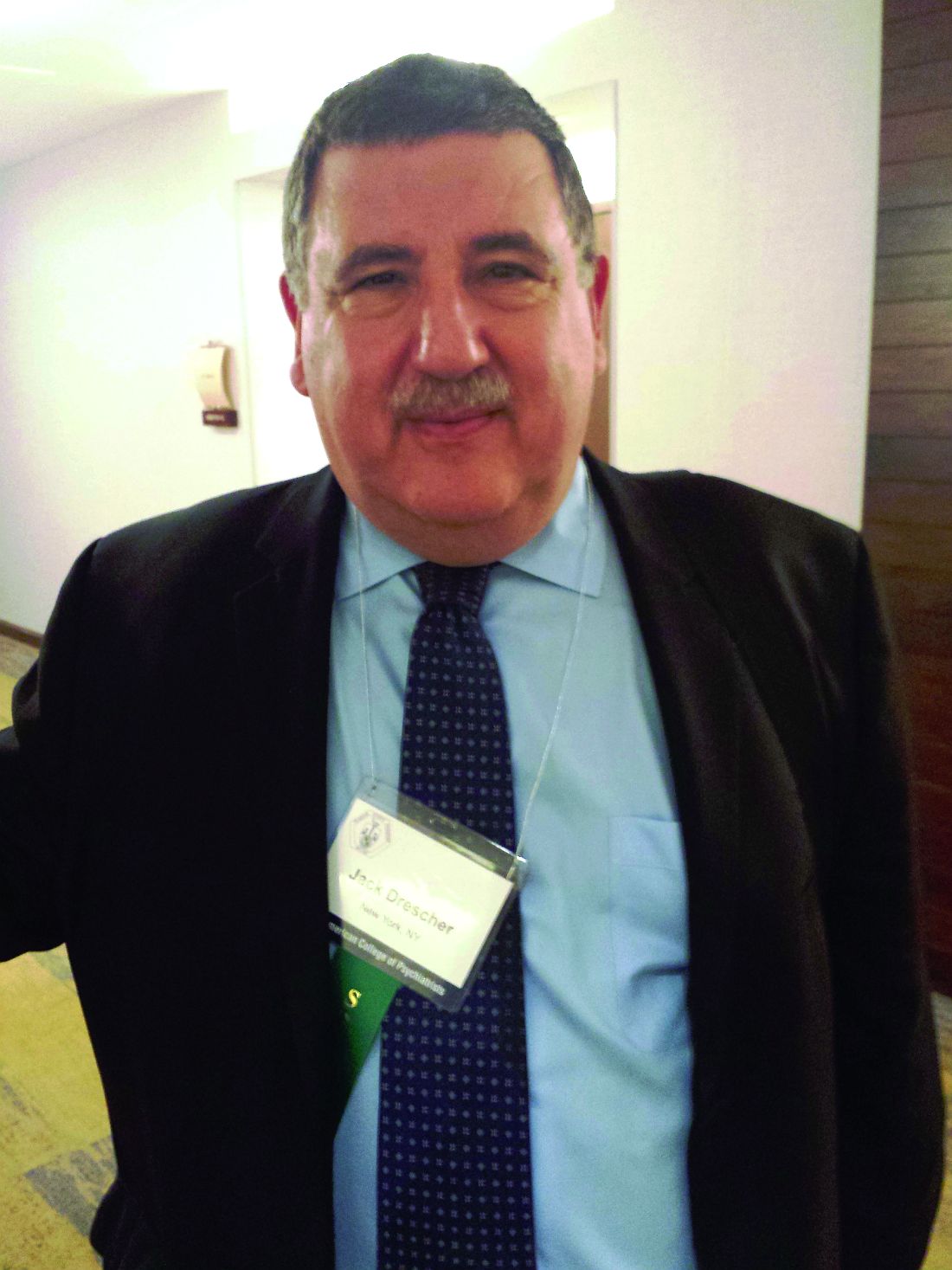
“There are some [clinicians] who believe they can predict who will persist and who will not. But they have not published, to my satisfaction, a way to show anyone else how to tell the difference,” said Jack Drescher, MD, a member of the World Health Organization’s (WHO’s) Working Group on the Classification of Sexual Disorders and Sexual Health and clinical professor of psychiatry and behavioral sciences at New York Medical College, Valhalla.
The WHO working group was tasked with categorizing sex and gender diagnoses in the forthcoming International Classification of Diseases (ICD-11). Dr. Drescher said that he and his fellow WHO working group members have identified at least three discrete transgender populations.
“There are persisters, desisters, and those who first develop gender dysphoria in adolescence and adulthood,” he said. “It would be a clinical mistake to think that there is a seamless transition from childhood gender dysphoria into adolescent and adult gender dysphoria.”
Dr. Drescher said that he believes the Dutch model for treating younger children diagnosed with what is interchangeably referred to as gender dysphoria or gender variance demonstrates the greatest sensitivity to how fluid the situation can be for many of these children. The absence of biomarkers for dysphoria that will persist into adulthood and the finding that a minority of prepubescent gender dysphoria diagnoses persist into adolescence inform the Dutch approach.
This approach, which originated at the VU University Medical Center Amsterdam, is based on 2 decades of research and practice. It assumes that it is better not to actively transition a child socially but to remain neutral to the way in which the child expresses gender identity. If children persist into late adolescence in this model, they are assisted in transitioning. If not, they are supported socially as they adjust to their natal gender. Puberty may sometimes have to be suppressed until the time one of the two paths has been decided.
“In my opinion, it is the most conservative approach,” Dr. Drescher said of the Dutch model. “They are the most cognizant of how much we don’t know, and they do a lot of good research.”
An approach originating at the Child and Adolescent Gender Center Clinic, which is affiliated with the University of California, San Francisco, supports a child socially into a cross-gendered role without medical or surgical intervention but also suppresses puberty. This method is based on the presumption of an adult transgender outcome, despite the absence of a way to predict results, said Dr. Drescher, who also cautions about the iatrogenic effects of such a presumption. “It takes a lot of work to socially transition a child in one direction. It would take a lot to transition back in the other direction, and there is no good empirical data as to whether this is entirely a benign process,” he said.
A third method originated in Toronto at the Centre for Addiction and Mental Health. This method actively discourages a child’s atypical gender interests and views transsexualism as an undesirable outcome that can be prevented, despite what Dr. Drescher said is a complete lack of evidence to either support or refute this claim. This method largely has been abandoned, in part since Ontario and five U.S. states and the District of Columbia have passed laws banning efforts to change a minor’s sexual orientation or gender identity. This method does have puberty suppression in children whose gender dysphoria appears to be persisting into adolescence in common with the other two.
Dr. Drescher said puberty suppression has helped decrease the levels of anxiety, depression, and suicidal ideation typically associated with this cohort. Postponing the development of secondary sexual characteristics gives those who ultimately will desist from their dysphoria more time to let it run its course. The Dutch first initiated this procedure 2 decades ago and have shown that any possible future side effects are outweighed by the psychosocial advantages it provides in the present.
The clinical view of gender dysphoria probably will get a jolt in 2018 upon publication of the ICD-11. In an interview, Dr. Drescher said that, if the condition is no longer categorized by the WHO as a mental disorder and is instead called “gender incongruence” in a chapter dedicated to gender and sexuality issues as currently planned, “it is likely the [American Psychiatric Association] will follow suit and remove gender dysphoria from the DSM. However, I don’t know how long that will take,” he said.
In his presentation, Dr. Drescher said that the causes for gender dysphoria remain unknown, as do the ways in which gender identity develops. It is also unclear how biological, psychosocial, and environmental factors affect gender dysphoria. What is clear, he said, is that “we have to rethink our developmental literature.”
Meanwhile, although gender dysphoria affects a relatively small percentage of the population – less than 1% of “nonreferred” children and adolescents, according to the DSM-5 – the number of prepubescent children presenting to gender clinics is on the rise. This increase might be driven more by social forces than by scientific ones. Dr. Drescher made an anecdotal observation during the presentation that more children are presenting to gender clinics already socially transitioned by their parents than there are children in the research literature on persisters and desisters.
Dr. Drescher recalled in the interview that, during the public comment period for the DSM-5, gender dysphoria elicited the third most responses, compared with other diagnoses, despite its rarity as a condition. “Interest in the subject far outweighs its prevalence.”
Gender-related glossary of terms
"There are so many moving parts to our understanding of gender," said Jack Drescher, MD, during a plenary session at the annual meeting of the American College of Psychiatrists. For that reason, "language is very important" when addressing children who might have questions about their gender identity, he said.
To help establish as much clarity as possible when discussing gender in the clinical setting, Dr. Drescher offered the following glossary of terms. These are not listed alphabetically but in a stepwise fashion aimed at leading to a clearer understanding of successive terms.
Sex: The biological attributes of being male or female. This includes sex chromosomes, gonads, sex hormones, and nonambiguous internal and external genitalia.
Gender: The public - and typically the legal - recognition of one's lived role as a boy, girl, man, woman or of other biological factors in combination with psychosocial factors that are seen as contributing to identity development.
Sexual orientation: A person's erotic response tendency or sexual attractions, either directed toward individuals of the same sex (homosexual), the other sex (heterosexual), or both sexes (bisexual).
Gender identity: Often an independent variable from sexual orientation, this refers to how an individual identifies as either male, female, or, in some cases, some other category.
Gender assignment: The natal presentation as either male or female. The historical terms are "biological male" or "biological female"; also occasionally known as "birth assigned male" or "birth assigned female."
Gender atypical: The somatic features or behaviors not statistically typical in individuals with the same assigned gender in a given society or era.
Gender nonconforming: Typically used as an alternative descriptive term for "gender atypical".
Gender dysphoria: The conflict between a person's assigned gender and that person's gender identity and expression; replaced "gender identity disorder" in the DSM-5.
Gender variant: Often used by those who are concerned the term "gender dysphoria" will unnecessarily pathologize a child.
Gender expression: How an individual demonstrates gender to others, including by way of dress, behavior, and appearance. Increasingly, the term is used in antidiscrimination documents.
Desister: Prepubescent children who present with gender dysphoria but who do not become transgender adults.
Persister: This refers to children who present with gender dysphoria and progress to a transgender adulthood.
Gender reassignment: An official - and often legal - change of gender by way of cross-sex endocrine therapy and/or gender reassignment surgery.
Transsexual: An individual who modifies the body via endocrine and/or surgical means to conform with gender identity either partially or completely.
Transwoman: A person, such as Caitlyn Jenner, who transitions from a male sex assignment to become female.
Transman: A person who transitions from a female sex assignment to become male.
Transgender: The "T" in the acronym LGBT; the popular - not scientific - inclusive term for those whose gender identity, gender expression, or behavior does not conform to that which is typically associated with the natal sex assignment.
Cisgender: From the Latin for "on the same side"; used in the transgender community to describe those whose gender identities align with their natal assignment.
Gender beliefs: Used to refer to the implicit, typically binary, cultural views on the "essential" qualities of men and women.
[email protected]
On Twitter @whitneymcknight
SCOTTSDALE, ARIZ. – The treatment of preadolescents who present with gender questions is often complicated by the absence of evidence-based data on who is most likely to remain gender dysphoric into adulthood and who is not, an expert said at the annual meeting of the American College of Psychiatrists.
“There are some [clinicians] who believe they can predict who will persist and who will not. But they have not published, to my satisfaction, a way to show anyone else how to tell the difference,” said Jack Drescher, MD, a member of the World Health Organization’s (WHO’s) Working Group on the Classification of Sexual Disorders and Sexual Health and clinical professor of psychiatry and behavioral sciences at New York Medical College, Valhalla.
The WHO working group was tasked with categorizing sex and gender diagnoses in the forthcoming International Classification of Diseases (ICD-11). Dr. Drescher said that he and his fellow WHO working group members have identified at least three discrete transgender populations.
“There are persisters, desisters, and those who first develop gender dysphoria in adolescence and adulthood,” he said. “It would be a clinical mistake to think that there is a seamless transition from childhood gender dysphoria into adolescent and adult gender dysphoria.”
Dr. Drescher said that he believes the Dutch model for treating younger children diagnosed with what is interchangeably referred to as gender dysphoria or gender variance demonstrates the greatest sensitivity to how fluid the situation can be for many of these children. The absence of biomarkers for dysphoria that will persist into adulthood and the finding that a minority of prepubescent gender dysphoria diagnoses persist into adolescence inform the Dutch approach.
This approach, which originated at the VU University Medical Center Amsterdam, is based on 2 decades of research and practice. It assumes that it is better not to actively transition a child socially but to remain neutral to the way in which the child expresses gender identity. If children persist into late adolescence in this model, they are assisted in transitioning. If not, they are supported socially as they adjust to their natal gender. Puberty may sometimes have to be suppressed until the time one of the two paths has been decided.
“In my opinion, it is the most conservative approach,” Dr. Drescher said of the Dutch model. “They are the most cognizant of how much we don’t know, and they do a lot of good research.”
An approach originating at the Child and Adolescent Gender Center Clinic, which is affiliated with the University of California, San Francisco, supports a child socially into a cross-gendered role without medical or surgical intervention but also suppresses puberty. This method is based on the presumption of an adult transgender outcome, despite the absence of a way to predict results, said Dr. Drescher, who also cautions about the iatrogenic effects of such a presumption. “It takes a lot of work to socially transition a child in one direction. It would take a lot to transition back in the other direction, and there is no good empirical data as to whether this is entirely a benign process,” he said.
A third method originated in Toronto at the Centre for Addiction and Mental Health. This method actively discourages a child’s atypical gender interests and views transsexualism as an undesirable outcome that can be prevented, despite what Dr. Drescher said is a complete lack of evidence to either support or refute this claim. This method largely has been abandoned, in part since Ontario and five U.S. states and the District of Columbia have passed laws banning efforts to change a minor’s sexual orientation or gender identity. This method does have puberty suppression in children whose gender dysphoria appears to be persisting into adolescence in common with the other two.
Dr. Drescher said puberty suppression has helped decrease the levels of anxiety, depression, and suicidal ideation typically associated with this cohort. Postponing the development of secondary sexual characteristics gives those who ultimately will desist from their dysphoria more time to let it run its course. The Dutch first initiated this procedure 2 decades ago and have shown that any possible future side effects are outweighed by the psychosocial advantages it provides in the present.
The clinical view of gender dysphoria probably will get a jolt in 2018 upon publication of the ICD-11. In an interview, Dr. Drescher said that, if the condition is no longer categorized by the WHO as a mental disorder and is instead called “gender incongruence” in a chapter dedicated to gender and sexuality issues as currently planned, “it is likely the [American Psychiatric Association] will follow suit and remove gender dysphoria from the DSM. However, I don’t know how long that will take,” he said.
In his presentation, Dr. Drescher said that the causes for gender dysphoria remain unknown, as do the ways in which gender identity develops. It is also unclear how biological, psychosocial, and environmental factors affect gender dysphoria. What is clear, he said, is that “we have to rethink our developmental literature.”
Meanwhile, although gender dysphoria affects a relatively small percentage of the population – less than 1% of “nonreferred” children and adolescents, according to the DSM-5 – the number of prepubescent children presenting to gender clinics is on the rise. This increase might be driven more by social forces than by scientific ones. Dr. Drescher made an anecdotal observation during the presentation that more children are presenting to gender clinics already socially transitioned by their parents than there are children in the research literature on persisters and desisters.
Dr. Drescher recalled in the interview that, during the public comment period for the DSM-5, gender dysphoria elicited the third most responses, compared with other diagnoses, despite its rarity as a condition. “Interest in the subject far outweighs its prevalence.”
Gender-related glossary of terms
"There are so many moving parts to our understanding of gender," said Jack Drescher, MD, during a plenary session at the annual meeting of the American College of Psychiatrists. For that reason, "language is very important" when addressing children who might have questions about their gender identity, he said.
To help establish as much clarity as possible when discussing gender in the clinical setting, Dr. Drescher offered the following glossary of terms. These are not listed alphabetically but in a stepwise fashion aimed at leading to a clearer understanding of successive terms.
Sex: The biological attributes of being male or female. This includes sex chromosomes, gonads, sex hormones, and nonambiguous internal and external genitalia.
Gender: The public - and typically the legal - recognition of one's lived role as a boy, girl, man, woman or of other biological factors in combination with psychosocial factors that are seen as contributing to identity development.
Sexual orientation: A person's erotic response tendency or sexual attractions, either directed toward individuals of the same sex (homosexual), the other sex (heterosexual), or both sexes (bisexual).
Gender identity: Often an independent variable from sexual orientation, this refers to how an individual identifies as either male, female, or, in some cases, some other category.
Gender assignment: The natal presentation as either male or female. The historical terms are "biological male" or "biological female"; also occasionally known as "birth assigned male" or "birth assigned female."
Gender atypical: The somatic features or behaviors not statistically typical in individuals with the same assigned gender in a given society or era.
Gender nonconforming: Typically used as an alternative descriptive term for "gender atypical".
Gender dysphoria: The conflict between a person's assigned gender and that person's gender identity and expression; replaced "gender identity disorder" in the DSM-5.
Gender variant: Often used by those who are concerned the term "gender dysphoria" will unnecessarily pathologize a child.
Gender expression: How an individual demonstrates gender to others, including by way of dress, behavior, and appearance. Increasingly, the term is used in antidiscrimination documents.
Desister: Prepubescent children who present with gender dysphoria but who do not become transgender adults.
Persister: This refers to children who present with gender dysphoria and progress to a transgender adulthood.
Gender reassignment: An official - and often legal - change of gender by way of cross-sex endocrine therapy and/or gender reassignment surgery.
Transsexual: An individual who modifies the body via endocrine and/or surgical means to conform with gender identity either partially or completely.
Transwoman: A person, such as Caitlyn Jenner, who transitions from a male sex assignment to become female.
Transman: A person who transitions from a female sex assignment to become male.
Transgender: The "T" in the acronym LGBT; the popular - not scientific - inclusive term for those whose gender identity, gender expression, or behavior does not conform to that which is typically associated with the natal sex assignment.
Cisgender: From the Latin for "on the same side"; used in the transgender community to describe those whose gender identities align with their natal assignment.
Gender beliefs: Used to refer to the implicit, typically binary, cultural views on the "essential" qualities of men and women.
[email protected]
On Twitter @whitneymcknight
EXPERT ANALYSIS AT THE AMERICAN COLLEGE OF PSYCHIATRISTS ANNUAL MEETING