User login
Study boosts surgical left atrial appendage occlusion
WASHINGTON – Surgical left atrial appendage occlusion in older patients with atrial fibrillation already undergoing cardiac surgery was associated with a 38% reduction in thromboembolism and a 15% lower risk of all-cause mortality during the subsequent year in a large observational study.
“Although randomized trial data are needed, our study demonstrates strong support for the benefits of closing the left atrial appendage at the time of cardiac surgery in patients with atrial fibrillation,” Daniel J. Friedman, MD, said at the annual meeting of the American College of Cardiology.

Thirty-seven percent of patients underwent surgical left atrial appendage occlusion (LAAO) at the time of their primary heart operation. They were slightly younger, more often female, in better cardiovascular health, and more likely to have their surgery at an academic medical center than were patients who didn’t have LAAO. Adjustment for all of these factors was made in the statistical analysis.
The primary outcome was rehospitalization for thromboembolism – that is, ischemic stroke, transient ischemic attack, or other systemic arterial embolism – within 1 year. This occurred in 1.6% of the LAAO group and 2.5% of the non-LAAO group, for an unadjusted 37% and an adjusted 38% relative risk reduction, reported Dr. Friedman, a cardiology research fellow at the Duke Clinical Research Institute in Durham, N.C.
Turning to prespecified secondary endpoints, he noted that the all-cause mortality rate at 1 year was 7.0% in the LAAO group and 10.8% in the comparison arm, for a significant adjusted 15% risk reduction in the closure group. The composite endpoint of thromboembolism, hemorrhagic stroke, or death occurred in 8.7% of the LAAO group compared with 13.5% of non-LAAO patients, representing an adjusted 30% reduction in risk.
In an exploratory analysis, Dr. Friedman and his coinvestigators determined that, in patients discharged without oral anticoagulation, LAAO was associated with a 71% reduction in risk of thromboembolism. In contrast, LAAO didn’t significantly affect thromboembolic risk in patients discharged on an oral anticoagulant.
The STS database did not include information on the methods or completeness of LAAO.
Discussant David J. Wilber, MD, urged care in attempting to translate the study findings into clinical practice.
“I must say, given the diversity of surgical occlusion techniques – everything from excision to internal sutures to external clipping and stapling – I’m really a bit surprised to see the relatively strong outcomes, with a very strong signal for reduction in thromboembolism and also an opportunity to decrease mortality. I guess I’d say I’d be a little bit cautious. I’d want to see data assessing the completeness of closure and its success long term before we take this to heart clinically,” said Dr. Wilber, professor of cardiology and pediatrics and codirector of the Cardiovascular Research Institute at Loyola University in Maywood, Ill.
Dr. Friedman shared Dr. Wilber’s reservations about applying these observational data to clinical practice.
“I think what we can say is that this study supports the concept that the left atrial appendage is important as a source of thromboembolism in atrial fibrillation, and that exclusion of the left atrial appendage can be a potentially viable treatment option for patients,” he said. “I think it’s going to be increasingly important to bring this up as a possible treatment option when we’re discussing cardiac surgery with our heart teams – and that means talking to the surgeon, the cardiologist who may be following the patient afterwards, and even the cardiothoracic anesthesiologist who’s going to be manning the transesophageal echo probe and may be able to give us some insight as to the quality of closure before the patient leaves the operating room.”
There is considerable surgical interest in LAAO for stroke prevention in AF in light of the success of the percutaneous Watchman device. However, many cardiothoracic surgeons have refrained from performing the brief occlusion procedure because of what up until now has been a lack of evidence as to safety and efficacy.
Asked if his study findings imply that oral anticoagulation can routinely be stopped after LAAO provided the surgeon believes the appendage has been successfully excluded from the circulation, Dr. Friedman replied that he does not.
Although the left atrial appendage has been implicated as the site of thrombus formation in 90% of thromboembolic events occurring in patients with AF, ligation of the appendage doesn’t address that other 10%. Dr. Friedman said he believes it’s appropriate to await the results of an ongoing prospective randomized trial of LAAO led by investigators at McMaster University in Hamilton, Ont.
Dr. Friedman reported having no financial conflicts regarding his study, funded by the Burroughs Welcome Fund and the Food and Drug Administration.
WASHINGTON – Surgical left atrial appendage occlusion in older patients with atrial fibrillation already undergoing cardiac surgery was associated with a 38% reduction in thromboembolism and a 15% lower risk of all-cause mortality during the subsequent year in a large observational study.
“Although randomized trial data are needed, our study demonstrates strong support for the benefits of closing the left atrial appendage at the time of cardiac surgery in patients with atrial fibrillation,” Daniel J. Friedman, MD, said at the annual meeting of the American College of Cardiology.
Thirty-seven percent of patients underwent surgical left atrial appendage occlusion (LAAO) at the time of their primary heart operation. They were slightly younger, more often female, in better cardiovascular health, and more likely to have their surgery at an academic medical center than were patients who didn’t have LAAO. Adjustment for all of these factors was made in the statistical analysis.
The primary outcome was rehospitalization for thromboembolism – that is, ischemic stroke, transient ischemic attack, or other systemic arterial embolism – within 1 year. This occurred in 1.6% of the LAAO group and 2.5% of the non-LAAO group, for an unadjusted 37% and an adjusted 38% relative risk reduction, reported Dr. Friedman, a cardiology research fellow at the Duke Clinical Research Institute in Durham, N.C.
Turning to prespecified secondary endpoints, he noted that the all-cause mortality rate at 1 year was 7.0% in the LAAO group and 10.8% in the comparison arm, for a significant adjusted 15% risk reduction in the closure group. The composite endpoint of thromboembolism, hemorrhagic stroke, or death occurred in 8.7% of the LAAO group compared with 13.5% of non-LAAO patients, representing an adjusted 30% reduction in risk.
In an exploratory analysis, Dr. Friedman and his coinvestigators determined that, in patients discharged without oral anticoagulation, LAAO was associated with a 71% reduction in risk of thromboembolism. In contrast, LAAO didn’t significantly affect thromboembolic risk in patients discharged on an oral anticoagulant.
The STS database did not include information on the methods or completeness of LAAO.
Discussant David J. Wilber, MD, urged care in attempting to translate the study findings into clinical practice.
“I must say, given the diversity of surgical occlusion techniques – everything from excision to internal sutures to external clipping and stapling – I’m really a bit surprised to see the relatively strong outcomes, with a very strong signal for reduction in thromboembolism and also an opportunity to decrease mortality. I guess I’d say I’d be a little bit cautious. I’d want to see data assessing the completeness of closure and its success long term before we take this to heart clinically,” said Dr. Wilber, professor of cardiology and pediatrics and codirector of the Cardiovascular Research Institute at Loyola University in Maywood, Ill.
Dr. Friedman shared Dr. Wilber’s reservations about applying these observational data to clinical practice.
“I think what we can say is that this study supports the concept that the left atrial appendage is important as a source of thromboembolism in atrial fibrillation, and that exclusion of the left atrial appendage can be a potentially viable treatment option for patients,” he said. “I think it’s going to be increasingly important to bring this up as a possible treatment option when we’re discussing cardiac surgery with our heart teams – and that means talking to the surgeon, the cardiologist who may be following the patient afterwards, and even the cardiothoracic anesthesiologist who’s going to be manning the transesophageal echo probe and may be able to give us some insight as to the quality of closure before the patient leaves the operating room.”
There is considerable surgical interest in LAAO for stroke prevention in AF in light of the success of the percutaneous Watchman device. However, many cardiothoracic surgeons have refrained from performing the brief occlusion procedure because of what up until now has been a lack of evidence as to safety and efficacy.
Asked if his study findings imply that oral anticoagulation can routinely be stopped after LAAO provided the surgeon believes the appendage has been successfully excluded from the circulation, Dr. Friedman replied that he does not.
Although the left atrial appendage has been implicated as the site of thrombus formation in 90% of thromboembolic events occurring in patients with AF, ligation of the appendage doesn’t address that other 10%. Dr. Friedman said he believes it’s appropriate to await the results of an ongoing prospective randomized trial of LAAO led by investigators at McMaster University in Hamilton, Ont.
Dr. Friedman reported having no financial conflicts regarding his study, funded by the Burroughs Welcome Fund and the Food and Drug Administration.
At ACC 17
Key clinical point:
Major finding: The ancillary surgical procedure was associated with a 38% reduction in thromboembolism and a 15% lower risk of all-cause mortality at 1 year, compared with no appendage closure.
Data source: A retrospective comparative effectiveness study using the Society of Thoracic Surgeons database of more than 10,000 Medicare recipients with atrial fibrillation who underwent cardiac surgery, 37% of whom underwent surgical left atrial appendage occlusion during their primary operation.
Disclosures: The presenter reported having no financial conflicts. The Burroughs Welcome Fund and the Food and Drug Administration funded the study.
Hospitalists seek tools for more efficient admissions
Moving patients safely and efficiently through the admission process is always a priority for hospitalists. Is there a way to optimize and standardize the process?
“In hopes of improving admission efficiency, while simultaneously increasing quality of care, we decided to use Lean/Six Sigma methodology to streamline our admission process,” says Escher Howard-Williams, MD, lead author of an abstract called “Standardizing the Admission Process Using Lean/Six Sigma One Piece Flow.”1
A basic tenet of the methodology is called “one piece flow” (OPF), the idea that standardized processes are more efficient and less prone to error when completed from start to finish without interruption. In the study, hospitalists committed to performing all patient admissions in OPF, focusing on one patient from initiation of chart review through exam, order entry and documentation, without interruption. Researchers then analyzed times, including time to call back to ED, time at initiation of chart review, time of evaluation of patient, time orders were placed, and time of sign-out note completed, before and after implementation of OPF. They found a substantial reduction in time of the admission process across all time points with OPF.
“When you are trying to improve quality of care in your institution, dissecting the overall work flow will allow you to discover areas that hinder the overall process,” Dr. Howard-Williams says. “Reframing your process to focus on providing excellent quality care will allow you to find workable solutions to improve the quality of care and efficiency in your practice. As part of this process, developing a team with an appropriate variety of members lays the foundation for success.”
Dr. Howard-Williams hopes that the study will inspire others to reflect on their own practices.
“If, during that reflection, they can identify areas that they would like to improve quality, we would encourage them to join us,” she says. “They will have the opportunity to build their personal work flow maps, find choke points and devise a plan for moving forward with new solutions.”
Reference
1. Howard-Williams E, Liles A, Stephens J, lanza-Kaduce K. Standardizing the admission process using Lean/Six Sigma One Piece Flow [abstract]. J Hosp Med. 2016;11(suppl 1). Available at: http://www.shmabstracts.com/abstract/standardizing-the-admission-process-using-lean-six-sigma-one-piece-flow/. Accessed March 7, 2017.
Using shock index in the ED to predict hospital admission and inpatient mortality
CLINICAL QUESTION: Can shock index (SI) in the ED predict the likelihood for hospital admission and inpatient mortality?
BACKGROUND: SI is defined as heart rate divided by systolic blood pressure. It is postulated to have an inverse relationship to cardiac output. SI has been studied as a prognostic metric of poor outcomes in patients with myocardial infarction, gastrointestinal hemorrhage, sepsis, and trauma. There are no large studies on SI in the general ED population.
SETTING: Academic tertiary care center.
SYNOPSIS: All ED patients over 18 years of age over a 12-month period were included in the study for a total of 58,633 charts. Charts were excluded if the patient presented in cardiac arrest, left prior to full evaluation in the ED, or had an incomplete or absent first set of vital signs. Likelihood ratio (LR) values of greater than 5 and 10 were considered moderate and large increases in the outcomes, respectively. Authors found SI greater than 1.2 had a positive LR of 11.69 for admission to the hospital and a positive LR of 5.82 for inpatient mortality.
This study identified potential thresholds for SI but did not validate them. Whether SI would be a useful tool for triage remains unanswered.
BOTTOM LINE: Initial SI greater than 1.2 at presentation to the ED was associated with increased likelihood of hospital admission and inpatient mortality.
CITATIONS: Balhara KS, Hsieh YH, Hamade B, et al. Clinical metrics in emergency medicine: the shock index and the probability of hospital admission and inpatient mortality. Emerg Med J. 2017 Feb;34(2):89-94.
Dr. Dietsche is a clinical instructor, Division of Hospital Medicine, University of Colorado School of Medicine, Aurora.
Gorsuch keeps cards close during nomination hearing
Throughout his marathon confirmation hearing, U.S. Supreme Court nominee Neil Gorsuch remained tight-lipped about how he might rule on major health care issues if confirmed to the country’s highest court, pledging to look at the facts of each case and rule according to the law.
The Senate Committee on the Judiciary wrapped up its nearly week-long questioning of Judge Gorsuch on March 23. Inquiries during the hearing ranged from Judge Gorsuch’s stance on abortion, to his support of religious freedom, to whether he would uphold President Trump’s controversial Executive Order on travel and immigration. Through it all, Judge Gorsuch, who presides over Denver’s 10th Circuit, kept his composure and refused to offer insight into which way he would lean when deciding such hot-button issues.

“If I were to start telling you which are my favorite [Supreme Court] precedents or which are my least favorite precedents, or if I viewed precedents in that fashion, I would be tipping my hand and suggesting to litigants that I’ve already made up my mind about their cases. That’s not a fair judge. I didn’t want that kind of judge when I was a lawyer, and I don’t want to be that kind of judge now.”
During the hearing, the case of Roe v. Wade was brought up repeatedly, and multiple senators from both parties questioned whether Judge Gorsuch would vote to overturn the right to an abortion. Sen. Lindsey Graham, (R-S.C.) discussed recent legislation he supports that would prohibit abortion if the probable postfertilization age of the fetus is 20 weeks or greater.
“We’re one of seven nations that allow wholesale, on demand, unlimited abortion at 20 weeks. I’d like to get out of that club,” Sen. Graham said during the hearing. “I’m just letting everybody know that if this legislation passes, it will be challenged before you and you will have to look at a new theory of how the state can protect the unborn ... Here’s what I think. You will read the briefs, look at the facts, and make a decision, am I fair to conclude that?”
“Senator, I can promise you no more than that, and I guarantee no less than that in every single case that comes before me,” Judge Gorsuch responded.
Sen. Dianne Feinstein (D-Calif.) queried whether Judge Gorsuch considered Roe to be “super precedent.”
“Senator, [the ruling] has been reaffirmed many times,” Judge Gorsuch responded, stressing that all Supreme Court precedent deserves respect and consideration when deciding new challenges.
The judge defended his 10th Circuit decision that found in favor of Hobby Lobby Stores after the company objected to the Affordable Care Act’s contraceptive coverage requirements based on religious grounds. In a separate case, Judge Gorsuch dissented from a ruling not to rehear a challenge by the Little Sisters of the Poor against certain contraceptive coverage provisions.

“Senator, our job there was to apply the statute as best we could understand its purpose as expressed in its text,” Judge Gorsuch said. “And I think every judge who faced that case – everyone – found it a hard case and did their level best and that’s all any judge can promise or guarantee. I respect all of my colleagues who addressed that case.”
The subject of religious freedom also was addressed during questions regarding President Trump’s Executive Order on travel and immigration and whether Judge Gorsuch would uphold the Executive Order if it came before the high court.
“President Trump promised a Muslim ban,” Sen. Patrick J. Leahy (D-Vt.) said during the hearing. “He still has on his website to this day that he’s called for a total and complete shutdown of Muslims entering the United States. And a Republican congressman recently said the best thing the president can do for his Muslim ban is to make sure he has Gorsuch on the Supreme Court.”
Judge Gorsuch called the unnamed congressman’s remark “silly,” adding that the congressman had “no idea” how Judge Gorsuch would rule in any case that comes before him. Sen. Leahy went on to ask Judge Gorsuch whether the president has the authority to block Jews from coming to the country or ban residents of Israel.
“We have a Constitution,” Judge Gorsuch replied. “And it does guarantee free exercise [of religion]. It also guarantees equal protection of the laws and a whole lot else besides, and the Supreme Court has held that due process rights extend even to undocumented persons in this country. I will apply the law faithfully and fearlessly and without regard to persons.”
A vote by the Senate Judiciary Committee is expected April 3 followed by a Senate floor vote later that week. Supreme Court justices require 60 votes for confirmation. Republicans control the Senate 52-48, so eight Democrats are needed to confirm Judge Gorsuch. Senate Minority Leader Charles E. Schumer (D-N.Y.) vowed on March 23 to oppose Judge Gorsuch and asked other Democrats to join him, setting up a potential filibuster against Judge Gorsuch’s confirmation.
[email protected]
On Twitter @legal_med
Throughout his marathon confirmation hearing, U.S. Supreme Court nominee Neil Gorsuch remained tight-lipped about how he might rule on major health care issues if confirmed to the country’s highest court, pledging to look at the facts of each case and rule according to the law.
The Senate Committee on the Judiciary wrapped up its nearly week-long questioning of Judge Gorsuch on March 23. Inquiries during the hearing ranged from Judge Gorsuch’s stance on abortion, to his support of religious freedom, to whether he would uphold President Trump’s controversial Executive Order on travel and immigration. Through it all, Judge Gorsuch, who presides over Denver’s 10th Circuit, kept his composure and refused to offer insight into which way he would lean when deciding such hot-button issues.
“If I were to start telling you which are my favorite [Supreme Court] precedents or which are my least favorite precedents, or if I viewed precedents in that fashion, I would be tipping my hand and suggesting to litigants that I’ve already made up my mind about their cases. That’s not a fair judge. I didn’t want that kind of judge when I was a lawyer, and I don’t want to be that kind of judge now.”
During the hearing, the case of Roe v. Wade was brought up repeatedly, and multiple senators from both parties questioned whether Judge Gorsuch would vote to overturn the right to an abortion. Sen. Lindsey Graham, (R-S.C.) discussed recent legislation he supports that would prohibit abortion if the probable postfertilization age of the fetus is 20 weeks or greater.
“We’re one of seven nations that allow wholesale, on demand, unlimited abortion at 20 weeks. I’d like to get out of that club,” Sen. Graham said during the hearing. “I’m just letting everybody know that if this legislation passes, it will be challenged before you and you will have to look at a new theory of how the state can protect the unborn ... Here’s what I think. You will read the briefs, look at the facts, and make a decision, am I fair to conclude that?”
“Senator, I can promise you no more than that, and I guarantee no less than that in every single case that comes before me,” Judge Gorsuch responded.
Sen. Dianne Feinstein (D-Calif.) queried whether Judge Gorsuch considered Roe to be “super precedent.”
“Senator, [the ruling] has been reaffirmed many times,” Judge Gorsuch responded, stressing that all Supreme Court precedent deserves respect and consideration when deciding new challenges.
The judge defended his 10th Circuit decision that found in favor of Hobby Lobby Stores after the company objected to the Affordable Care Act’s contraceptive coverage requirements based on religious grounds. In a separate case, Judge Gorsuch dissented from a ruling not to rehear a challenge by the Little Sisters of the Poor against certain contraceptive coverage provisions.
“Senator, our job there was to apply the statute as best we could understand its purpose as expressed in its text,” Judge Gorsuch said. “And I think every judge who faced that case – everyone – found it a hard case and did their level best and that’s all any judge can promise or guarantee. I respect all of my colleagues who addressed that case.”
The subject of religious freedom also was addressed during questions regarding President Trump’s Executive Order on travel and immigration and whether Judge Gorsuch would uphold the Executive Order if it came before the high court.
“President Trump promised a Muslim ban,” Sen. Patrick J. Leahy (D-Vt.) said during the hearing. “He still has on his website to this day that he’s called for a total and complete shutdown of Muslims entering the United States. And a Republican congressman recently said the best thing the president can do for his Muslim ban is to make sure he has Gorsuch on the Supreme Court.”
Judge Gorsuch called the unnamed congressman’s remark “silly,” adding that the congressman had “no idea” how Judge Gorsuch would rule in any case that comes before him. Sen. Leahy went on to ask Judge Gorsuch whether the president has the authority to block Jews from coming to the country or ban residents of Israel.
“We have a Constitution,” Judge Gorsuch replied. “And it does guarantee free exercise [of religion]. It also guarantees equal protection of the laws and a whole lot else besides, and the Supreme Court has held that due process rights extend even to undocumented persons in this country. I will apply the law faithfully and fearlessly and without regard to persons.”
A vote by the Senate Judiciary Committee is expected April 3 followed by a Senate floor vote later that week. Supreme Court justices require 60 votes for confirmation. Republicans control the Senate 52-48, so eight Democrats are needed to confirm Judge Gorsuch. Senate Minority Leader Charles E. Schumer (D-N.Y.) vowed on March 23 to oppose Judge Gorsuch and asked other Democrats to join him, setting up a potential filibuster against Judge Gorsuch’s confirmation.
[email protected]
On Twitter @legal_med
Enlisting social networks for better health outcomes
As a hospitalist, you typically have little, if any, contact with patients outside the hospital and, at most, you’ll only spend a couple of hours a year in front of any particular patient. The vast majority of the determinants of your patients’ health occur when you’re not there.
In a commentary in the New England Journal of Medicine entitled “Engineering Social Incentives for Health,” lead author David A. Asch, MD, MBA, addresses that issue.1
“The motivation for the piece is that the people who are in a position to influence a patient’s health are their friends and family, and yet so much of how we have structured health care is between a clinician and a patient,” he says. “We often fail to engage the people in patients’ everyday lives, who can be quite willing partners in improving health care. There are all sorts of things they can do to help patients with hard-to-control diabetes or ... heart failure, or anything that might have put them in the hospital in the first place.”
The column describes a ladder of social engineering strategies, from very simple to complex. One example on the simple end might be to help a patient remember a daily medication by having him place the medication bottle where his partner can see him taking – or not taking – it. (The alternative is that medications are taken in a private place, such in the bathroom, where no one might be watching over the routine to keep the patient on track.)
Moving up the ladder, a hospitalist might help set up a network of other patients with heart failure, so that they can help each other in a kind of peer mentorship.
“These peer-to-peer connections might require Web-based platforms or social support groups, so that kind of activity is a lot more complicated, but the general theme is: Can hospitalists think about ways to constructively engage the social networks that already surround patients, so they don’t need to invoke the health system to do it?”
It’s long been known that people with more social support do better: People who are married do better; people who have more friends do better. “Up until now, it’s just been an observation,” Dr. Asch says. “I think we’re at a point where we could begin to prescribe social support in the way we might prescribe a diuretic. I’d like to try it out at least. I think that’s the call to action.”
Reference
1. Asch D, Rosin R. Engineering social incentives for health. NEJM. 2016;375:2511-2513.
Effect of Plate in Close Proximity to Empty External-Fixation Pin Site on Long-Bone Torsional Strength
Take-Home Points
- The location of a bicortical defect in proximity to a tibia plate does not appear to affect the torsional stiffness or torsional failure strength of the bone.
- External fixator pin placement should be based on considerations other than the potential for creating a distal stress riser after definitive fracture management.
A stress riser in cortical bone may be considered any abrupt change in the contour or consistency of the hollow structure, such as a surface defect, that not only weakens the bone but concentrates stresses at that transition point.1 A cortical defect that is 20% of the bone diameter is associated with a 34% decrease in torsional strength, thus representing a “stress riser.”2 High-energy and complex tibia fractures are often provisionally stabilized with external fixation that gives the soft tissues time to recover before definitive fracture fixation. Pin diameter for a medium-size tibia external fixator typically is 5.0 mm, resulting in a 10-mm defect in bicortical placement. Therefore, any tibia with a diameter of <50 mm is at risk for a stress riser fracture.
Although it had been established that sizable cortical defects can decrease the torsional strength of long bone,2 the effect of a plate in close proximity to a defect secondary to an empty external-fixator pin site on torsional strength has not been determined. We conducted a study to evaluate this effect. The null hypothesis was there would be no difference in tibia torsional strength attributable to varying the proximity of a tibia midshaft plate to a 5.0-mm bicortical defect.
Methods
Forty fourth-generation, medium-size left composite tibias (Pacific Research Laboratories) were divided into 8 groups of 5 bones (Figure 1). 
Torsion testing to failure was performed for all specimens in a manner similar to that described by Gardner and colleagues.3 Impression molds for the composite tibia constructed from polymethylmethacrylate encased the superior and distal ends, leaving 25.5 cm of exposed midshaft. This allowed the composites to be rigidly clamped into a materials testing system (858 Mini-Bionix; MTS) equipped with a 100.0-Nm torsional load cell (Figure 2).
Results
Graphical results for torsional stiffness are presented in Figure 3. R2 for all stiffness calculations was >0.99. 


Discussion
Many tibia fractures require provisional stabilization with an external fixator that spans the knee, because of the high-energy nature of the injury or other, higher-priority polytrauma concerns. When the patient or injury is suitable for definitive fixation, the external fixator typically is removed in favor of internal fixation with a plate and screws. Depending on the nature and location of the fracture and the subsequent plate, the empty cortical pin-site defects, often lying at varying distances from the distal end of the plate, can potentially serve as stress risers for fracture.4
Other studies have evaluated long-bone cortical defects biomechanically1,2,4 and clinically,5-7 and multiple studies have been conducted on the effects of plates on long-bone strength for fracture stabilization.8-13 The present study evaluated the torsional strength of long bones in the presence of a bicortical defect and the proximity of the defect to a plate. There were no differences in stiffness or failure load between any of the groups of plated and unplated fourth-generation composite tibias tested to failure in torsion with varying distal bicortical defects. Hypothetically, one would expect the torsional stiffness of these specimens to increase with the mere addition of a metallic diaphyseal plate. However, this study demonstrated that the addition of a plate did not affect the torsional stiffness or strength of the tibias. Clinically, it is common practice to place external fixator pins as far as possible outside the planned incision site for definitive fracture fixation. Thus, we also hypothesized that the presence of a bicortical pin-site defect and its proximity to the plate would alter the torsional strength of the tibia specimens, and that the distal pin-site defect’s location farthest from the plate would exhibit greater strength, but this did not occur. Although other studies have shown that the presence of bicortical defects decreases the strength of long bones, we were unable to quantify this decrease because the 2 intact groups of composites, plated and unplated, survived failure testing.
This study had several limitations, first being the use of composite tibias as opposed to human cadaver bone. Although fourth-generation composite bone models have been validated as a suitable and accurate biomechanical substitute for cadaver specimens,14 anatomical variations in cadaver tibias may transfer forces differently through plates, screws, and distal pin sites. In order to test plated specimens against the unplated controls, we did not simulate a mid-shaft fracture in any of the tibias. The pin-site defects were intended to reflect the mechanical effects of bicortical defects immediately after pin removal and in the absence of any degree of bone healing. Finally, this study focused on pin-site defects that were distal to a midshaft plate and that may not represent the effects of bicortical pin-site defects proximal to the plate.
Given the results of this biomechanical study in composite tibias, varying the proximity of a bicortical defect to a plate does not affect the torsional stiffness or torsional failure strength of the bone. Placement of an intended bicortical defect should be based on considerations other than the potential for creating a distal stress riser after definitive fracture management.
Am J Orthop. 2017;46(2):E108-E111. Copyright Frontline Medical Communications Inc. 2017. All rights reserved.
1. Brooks DB, Burstein AH, Frankel VH. The biomechanics of torsional fractures. The stress concentration effect of a drill hole. J Bone Joint Surg Am. 1970;52(3):507-514.
2. Edgerton BC, An KN, Morrey BF. Torsional strength reduction due to cortical defects in bone. J Orthop Res. 1990;8(6):851-855.
3. Gardner MP, Chong AC, Pollock AG, Wooley PH. Mechanical evaluation of large-size fourth-generation composite femur and tibia models. Ann Biomed Eng. 2010;38(3):613-620.
4. Wysocki RW, Sheinkop MB, Virkus WW, Della Valle CJ. Femoral fracture through a previous pin site after computer-assisted total knee arthroplasty. J Arthroplasty. 2008;23(3):462-465.
5. Burstein AH, Currey J, Frankel VH, Heiple KG, Lunseth P, Vessely JC. Bone strength. The effect of screw holes. J Bone Joint Surg Am. 1972;54(6):1143-1156.
6. Clark CR, Morgan C, Sonstegard DA, Matthews LS. The effect of biopsy-hole shape and size on bone strength. J Bone Joint Surg Am. 1977;59(2):213-217.
7. Evans PE, Thomas WG. Tibial fracture through a traction-pin site. A report of two cases. J Bone Joint Surg Am. 1984;66(9):1475-1476.
8. Stoffel K, Dieter U, Stachowiak G, Gächter A, Kuster MS. Biomechanical testing of the LCP—how can stability in locked internal fixators be controlled? Injury. 2003;34(suppl 2):B11-B19.
9. Klaue K, Fengels I, Perren SM. Long-term effects of plate osteosynthesis: comparison of four different plates. Injury. 2000;31(suppl 2):B51-B62.
10. Uhthoff HK, Poitras P, Backman DS. Internal plate fixation of fractures: short history and recent developments. J Orthop Sci. 2006;11(2):118-126.
11. Takemoto RC, Sugi MT, Kummer F, Koval KJ, Egol KA. The effects of locked and unlocked neutralization plates on load bearing of fractures fixed with a lag screw. J Orthop Trauma. 2012;26(9):519-522.
12. Wagner M. General principles for the clinical use of the LCP. Injury. 2003;34(suppl 2):B31-B42.
13. Strauss EJ, Schwarzkopf R, Kummer F, Egol KA. The current status of locked plating: the good, the bad, and the ugly. J Orthop Trauma. 2008;22(7):479-486.
14. Elfar J, Menorca RM, Reed JD, Stanbury S. Composite bone models in orthopaedic surgery research and education. J Am Acad Orthop Surg. 2014;22(2):111-120.
Take-Home Points
- The location of a bicortical defect in proximity to a tibia plate does not appear to affect the torsional stiffness or torsional failure strength of the bone.
- External fixator pin placement should be based on considerations other than the potential for creating a distal stress riser after definitive fracture management.
A stress riser in cortical bone may be considered any abrupt change in the contour or consistency of the hollow structure, such as a surface defect, that not only weakens the bone but concentrates stresses at that transition point.1 A cortical defect that is 20% of the bone diameter is associated with a 34% decrease in torsional strength, thus representing a “stress riser.”2 High-energy and complex tibia fractures are often provisionally stabilized with external fixation that gives the soft tissues time to recover before definitive fracture fixation. Pin diameter for a medium-size tibia external fixator typically is 5.0 mm, resulting in a 10-mm defect in bicortical placement. Therefore, any tibia with a diameter of <50 mm is at risk for a stress riser fracture.
Although it had been established that sizable cortical defects can decrease the torsional strength of long bone,2 the effect of a plate in close proximity to a defect secondary to an empty external-fixator pin site on torsional strength has not been determined. We conducted a study to evaluate this effect. The null hypothesis was there would be no difference in tibia torsional strength attributable to varying the proximity of a tibia midshaft plate to a 5.0-mm bicortical defect.
Methods
Forty fourth-generation, medium-size left composite tibias (Pacific Research Laboratories) were divided into 8 groups of 5 bones (Figure 1).
Torsion testing to failure was performed for all specimens in a manner similar to that described by Gardner and colleagues.3 Impression molds for the composite tibia constructed from polymethylmethacrylate encased the superior and distal ends, leaving 25.5 cm of exposed midshaft. This allowed the composites to be rigidly clamped into a materials testing system (858 Mini-Bionix; MTS) equipped with a 100.0-Nm torsional load cell (Figure 2).
Results
Graphical results for torsional stiffness are presented in Figure 3. R2 for all stiffness calculations was >0.99.
Discussion
Many tibia fractures require provisional stabilization with an external fixator that spans the knee, because of the high-energy nature of the injury or other, higher-priority polytrauma concerns. When the patient or injury is suitable for definitive fixation, the external fixator typically is removed in favor of internal fixation with a plate and screws. Depending on the nature and location of the fracture and the subsequent plate, the empty cortical pin-site defects, often lying at varying distances from the distal end of the plate, can potentially serve as stress risers for fracture.4
Other studies have evaluated long-bone cortical defects biomechanically1,2,4 and clinically,5-7 and multiple studies have been conducted on the effects of plates on long-bone strength for fracture stabilization.8-13 The present study evaluated the torsional strength of long bones in the presence of a bicortical defect and the proximity of the defect to a plate. There were no differences in stiffness or failure load between any of the groups of plated and unplated fourth-generation composite tibias tested to failure in torsion with varying distal bicortical defects. Hypothetically, one would expect the torsional stiffness of these specimens to increase with the mere addition of a metallic diaphyseal plate. However, this study demonstrated that the addition of a plate did not affect the torsional stiffness or strength of the tibias. Clinically, it is common practice to place external fixator pins as far as possible outside the planned incision site for definitive fracture fixation. Thus, we also hypothesized that the presence of a bicortical pin-site defect and its proximity to the plate would alter the torsional strength of the tibia specimens, and that the distal pin-site defect’s location farthest from the plate would exhibit greater strength, but this did not occur. Although other studies have shown that the presence of bicortical defects decreases the strength of long bones, we were unable to quantify this decrease because the 2 intact groups of composites, plated and unplated, survived failure testing.
This study had several limitations, first being the use of composite tibias as opposed to human cadaver bone. Although fourth-generation composite bone models have been validated as a suitable and accurate biomechanical substitute for cadaver specimens,14 anatomical variations in cadaver tibias may transfer forces differently through plates, screws, and distal pin sites. In order to test plated specimens against the unplated controls, we did not simulate a mid-shaft fracture in any of the tibias. The pin-site defects were intended to reflect the mechanical effects of bicortical defects immediately after pin removal and in the absence of any degree of bone healing. Finally, this study focused on pin-site defects that were distal to a midshaft plate and that may not represent the effects of bicortical pin-site defects proximal to the plate.
Given the results of this biomechanical study in composite tibias, varying the proximity of a bicortical defect to a plate does not affect the torsional stiffness or torsional failure strength of the bone. Placement of an intended bicortical defect should be based on considerations other than the potential for creating a distal stress riser after definitive fracture management.
Am J Orthop. 2017;46(2):E108-E111. Copyright Frontline Medical Communications Inc. 2017. All rights reserved.
1. Brooks DB, Burstein AH, Frankel VH. The biomechanics of torsional fractures. The stress concentration effect of a drill hole. J Bone Joint Surg Am. 1970;52(3):507-514.
2. Edgerton BC, An KN, Morrey BF. Torsional strength reduction due to cortical defects in bone. J Orthop Res. 1990;8(6):851-855.
3. Gardner MP, Chong AC, Pollock AG, Wooley PH. Mechanical evaluation of large-size fourth-generation composite femur and tibia models. Ann Biomed Eng. 2010;38(3):613-620.
4. Wysocki RW, Sheinkop MB, Virkus WW, Della Valle CJ. Femoral fracture through a previous pin site after computer-assisted total knee arthroplasty. J Arthroplasty. 2008;23(3):462-465.
5. Burstein AH, Currey J, Frankel VH, Heiple KG, Lunseth P, Vessely JC. Bone strength. The effect of screw holes. J Bone Joint Surg Am. 1972;54(6):1143-1156.
6. Clark CR, Morgan C, Sonstegard DA, Matthews LS. The effect of biopsy-hole shape and size on bone strength. J Bone Joint Surg Am. 1977;59(2):213-217.
7. Evans PE, Thomas WG. Tibial fracture through a traction-pin site. A report of two cases. J Bone Joint Surg Am. 1984;66(9):1475-1476.
8. Stoffel K, Dieter U, Stachowiak G, Gächter A, Kuster MS. Biomechanical testing of the LCP—how can stability in locked internal fixators be controlled? Injury. 2003;34(suppl 2):B11-B19.
9. Klaue K, Fengels I, Perren SM. Long-term effects of plate osteosynthesis: comparison of four different plates. Injury. 2000;31(suppl 2):B51-B62.
10. Uhthoff HK, Poitras P, Backman DS. Internal plate fixation of fractures: short history and recent developments. J Orthop Sci. 2006;11(2):118-126.
11. Takemoto RC, Sugi MT, Kummer F, Koval KJ, Egol KA. The effects of locked and unlocked neutralization plates on load bearing of fractures fixed with a lag screw. J Orthop Trauma. 2012;26(9):519-522.
12. Wagner M. General principles for the clinical use of the LCP. Injury. 2003;34(suppl 2):B31-B42.
13. Strauss EJ, Schwarzkopf R, Kummer F, Egol KA. The current status of locked plating: the good, the bad, and the ugly. J Orthop Trauma. 2008;22(7):479-486.
14. Elfar J, Menorca RM, Reed JD, Stanbury S. Composite bone models in orthopaedic surgery research and education. J Am Acad Orthop Surg. 2014;22(2):111-120.
Onstep hernia repair decreased postop pain during sexual activity
The Onstep technique for inguinal hernia repair is associated with a lower incidence of postoperative pain during sexual activity than the Lichtenstein surgical technique, according to a paper published online in Surgery.
This study was a part of the Onli trial, the objective of which was to evaluate chronic pain and sexual dysfunction after inguinal hernia repair involving mesh fixation with sutures (Lichtenstein), compared with no mesh fixation (Onstep). The investigators reported the findings of a large study: 20.8% of inguinal repair patients experienced pain during sexual activity 1.4-1.7 years after their operation (Pain. 2006;122:258-63).
“The Onstep technique could be considered when surgeons and patients are discussing the optimal technique for repair of an inguinal hernia,” wrote Kristoffer Andresen, MD, of the University of Copenhagen, and his coauthors. “If the patient has pain during sexual activity as part of the complaint from the hernia, the Onstep technique seems to have a greater chance of removing this pain and alleviating the complaints.”
Among the 17 patients in the Onstep group who experienced postoperative pain during sexual activity, 8 had experienced preoperative pain during sexual activity and 7 had not. In the Lichtenstein group, 14 of the 30 patients who experienced postoperative pain had experienced preoperative pain, 14 had not. The remaining patients in both groups reported not having sexual activity before surgery or declined to answer.
The Lichtenstein technique was associated with new pain in 14 of the 70 patients (20%) while 7 of the 74 patients (9%) in the Onstep group reported new pain after surgery.
“For patients who experienced pain during sexual activity preoperatively, the Onstep technique removed the pain during sexual activity in the majority of patients while still resulting in few new cases,” the authors wrote.
Among the Lichtenstein group, 20 patients experienced pain from the surgical scar, compared with 7 patients in the Onstep group.
When asked about the degree of impairment of sexual function because of the postoperative pain, four patients in the Lichtenstein group said they had moderate to severe impairment, but none in the Onstep group reported that level of impairment.
Commenting on the possible mechanisms that might result in pain during sexual activity after hernia repair, the authors noted that mesh has been found to shrink from a mass around the vas deferens after a Lichtenstein repair. Previous studies have also found sutures around the iliohypogastric and the ilioinguinal nerves, which could also result in some pain.
“Because the mesh in the Onstep technique is not fixed with sutures, the risk of capturing nerves during the fixation of mesh is nonexistent,” they wrote. “There could, however, still be a risk of scar tissue and mesh shrinkage.”
PolySoft mesh (Bard Davol) was used for the Lichtenstein group and SoftMesh (Bard Davol) was used for the Onstep group.
In the 6 months’ follow-up period, two patients in the Lichtenstein group and one patient in the Onstep group experienced a recurrence of their hernia.
Three authors reported personal fees and travel grants from private industry – including Bard Medical – outside the submitted work. No other conflicts of interest were declared.
FROM SURGERY
Key clinical point: The Onstep technique for inguinal hernia repair is associated with a lower incidence of pain during sexual activity than the Lichtenstein technique.
Major finding: The Lichtenstein technique was associated with new pain in 14 of the 70 patients (20%) while 7 of the 74 patients (9%) in the Onstep group reported new pain after surgery.
Data source: A randomized trial in 259 patients undergoing inguinal hernia repair.
Disclosures: Three authors reported personal fees and travel grants from private industry – including Bard Medical – outside the submitted work. No other conflicts of interest were declared.
OpenNotes: Patient engagement with low physician hassle
ORLANDO – Early evidence suggests that OpenNotes is helping to engage patients and improve health outcomes while not creating undue burden for physicians.
A 2010 pilot project at Beth Israel Deaconess Medical Center in Boston, Geisinger Health System in Pennsylvania, and Harborview Medical Center in Seattle tested OpenNotes, a program that allows patients to see the entirety of a physician’s notes within their medical record and not just a summarized version.
Homer Chin, MD, of the department of medical informatics and outcomes research at Oregon Health & Science University, Portland, an associate with the OpenNotes Program, and physician champion for the Northwest OpenNotes Consortium, noted that a survey of the 105 primary care physicians participating in the pilot revealed they were apprehensive when they heard their visit notes would be completely available to patients.
Prior to the launch of the pilot, 24% of physicians expected significantly longer visits because of the availability of OpenNotes, 42% said they expected to spend more time addressing patient questions outside of visits, and 39% said they expected to spend more time writing/editing/dictating notes, Dr. Chin said at the annual meeting of the Healthcare Information and Management Systems Society.
Their concerns never quite materialized.
According to a survey of the participating physicians after the yearlong pilot, only 2% reported visits took significantly longer, 3% said they spent more time addressing patient questions outside of visits, and 11% said they spent more time writing/editing/dictating notes.
With regard to writing notes, “what we are finding is that most physicians are saying they are changing the way they write the note a little bit, but it is not taking more time,” Dr. Chin said. “They are just watching for certain terms and writing them in a different way, but it is not necessarily taking more time.”
More than 70% of patients who participated in the trial reported they are taking better care of themselves, more than 77% said they have a better understanding of their medical condition, more than 69% said they are better prepared for visits and more than 60% said they are more adherent to their prescription medication regimens.
Importantly, 85% said the availability of OpenNotes would affect their future choice of providers.
Dr. Chin also discussed the results of a survey of patients using OpenNotes in the Virginia Commonwealth University Health System in Richmond, noting that of roughly 420 respondents, 70% said their contact with their providers did not change, with nearly 20% saying they were contacting their provider less. Just over 40% said that reading their notes made them less worried about something health related, while a little more than 50% said there was no change. Nearly 85% said they thought seeing the notes helped them take better care of themselves. Nearly 90% of patients said that they understood some or all of their doctors’ notes.

It has been incredibly valuable “when the patients use the portal to prepare themselves for the visit with the provider,” Mr. Kravitz said. “They will review their case. They’ll look at past x-rays or reports, any kind of information, the results for laboratory and anything else, but they will message their provider. They are very heavy into messaging. It’s secure messaging within the portal and any type of questions, especially after they’ve had an appointment, they’ve thought of something they didn’t think about in the appointment, they have the opportunity to message back to the provider’s office.”
Geisinger saw 620,000 encounter reviews in OpenNotes out of 2 million patient visits in its last fiscal year, with virtually no complaints about the information in the OpenNotes.
Dr. Chin stressed that the implemented OpenNotes needs to be driven by physicians.
“I would emphasize that this has to be a clinician operational leader supported effort, and not an IT effort, so that when clinicians complain, you can point them to their department head, the chief medical officer, and not the IT people,” he said. “You’ve got to have good communication to providers. Our advice is to start with one department. You might do a pilot for a very short period of time, but there is enough evidence now to really implement it throughout the organization. We encourage people not to allow individual providers to opt out on their own, to make their own decision to opt out, to do it as an organizational effort.”
Michael Day, chief information officer of Ascension Health and Columbia St. Mary’s Health System of Milwaukee, offered the same advice.
“You’ve got to have leadership commitment up front,” Mr. Day said. “This really has to have good, strong physician leadership. The key executive in charge of all of this was the president of our medical group which drove a lot of the change with a lot of support from other areas.”
He noted that at his organization there was “a lot of physician grumbling” when OpenNotes was announced. However, there has been “relatively no impact. I think they’ve all agreed that this has made care better. We’ve also seen an actual improvement in the quality of the documentation.”
None of the presenters reported any conflicts of interest.
ORLANDO – Early evidence suggests that OpenNotes is helping to engage patients and improve health outcomes while not creating undue burden for physicians.
A 2010 pilot project at Beth Israel Deaconess Medical Center in Boston, Geisinger Health System in Pennsylvania, and Harborview Medical Center in Seattle tested OpenNotes, a program that allows patients to see the entirety of a physician’s notes within their medical record and not just a summarized version.
Homer Chin, MD, of the department of medical informatics and outcomes research at Oregon Health & Science University, Portland, an associate with the OpenNotes Program, and physician champion for the Northwest OpenNotes Consortium, noted that a survey of the 105 primary care physicians participating in the pilot revealed they were apprehensive when they heard their visit notes would be completely available to patients.
Prior to the launch of the pilot, 24% of physicians expected significantly longer visits because of the availability of OpenNotes, 42% said they expected to spend more time addressing patient questions outside of visits, and 39% said they expected to spend more time writing/editing/dictating notes, Dr. Chin said at the annual meeting of the Healthcare Information and Management Systems Society.
Their concerns never quite materialized.
According to a survey of the participating physicians after the yearlong pilot, only 2% reported visits took significantly longer, 3% said they spent more time addressing patient questions outside of visits, and 11% said they spent more time writing/editing/dictating notes.
With regard to writing notes, “what we are finding is that most physicians are saying they are changing the way they write the note a little bit, but it is not taking more time,” Dr. Chin said. “They are just watching for certain terms and writing them in a different way, but it is not necessarily taking more time.”
More than 70% of patients who participated in the trial reported they are taking better care of themselves, more than 77% said they have a better understanding of their medical condition, more than 69% said they are better prepared for visits and more than 60% said they are more adherent to their prescription medication regimens.
Importantly, 85% said the availability of OpenNotes would affect their future choice of providers.
Dr. Chin also discussed the results of a survey of patients using OpenNotes in the Virginia Commonwealth University Health System in Richmond, noting that of roughly 420 respondents, 70% said their contact with their providers did not change, with nearly 20% saying they were contacting their provider less. Just over 40% said that reading their notes made them less worried about something health related, while a little more than 50% said there was no change. Nearly 85% said they thought seeing the notes helped them take better care of themselves. Nearly 90% of patients said that they understood some or all of their doctors’ notes.
It has been incredibly valuable “when the patients use the portal to prepare themselves for the visit with the provider,” Mr. Kravitz said. “They will review their case. They’ll look at past x-rays or reports, any kind of information, the results for laboratory and anything else, but they will message their provider. They are very heavy into messaging. It’s secure messaging within the portal and any type of questions, especially after they’ve had an appointment, they’ve thought of something they didn’t think about in the appointment, they have the opportunity to message back to the provider’s office.”
Geisinger saw 620,000 encounter reviews in OpenNotes out of 2 million patient visits in its last fiscal year, with virtually no complaints about the information in the OpenNotes.
Dr. Chin stressed that the implemented OpenNotes needs to be driven by physicians.
“I would emphasize that this has to be a clinician operational leader supported effort, and not an IT effort, so that when clinicians complain, you can point them to their department head, the chief medical officer, and not the IT people,” he said. “You’ve got to have good communication to providers. Our advice is to start with one department. You might do a pilot for a very short period of time, but there is enough evidence now to really implement it throughout the organization. We encourage people not to allow individual providers to opt out on their own, to make their own decision to opt out, to do it as an organizational effort.”
Michael Day, chief information officer of Ascension Health and Columbia St. Mary’s Health System of Milwaukee, offered the same advice.
“You’ve got to have leadership commitment up front,” Mr. Day said. “This really has to have good, strong physician leadership. The key executive in charge of all of this was the president of our medical group which drove a lot of the change with a lot of support from other areas.”
He noted that at his organization there was “a lot of physician grumbling” when OpenNotes was announced. However, there has been “relatively no impact. I think they’ve all agreed that this has made care better. We’ve also seen an actual improvement in the quality of the documentation.”
None of the presenters reported any conflicts of interest.
AT HIMSS 2017
VIDEO: Pain and impaired QOL persist after open endometrial cancer surgery
NATIONAL HARBOR, MD. – Patient-reported outcomes from a prospective cohort study of minimally invasive versus open surgery for women with endometrial cancer showed that the disability from open surgery persisted for longer than had previously been recognized. Further, for a subset of patients, impairment in sexual functioning was significant, and persistent, regardless of the type of surgery.
At 3 weeks after surgery, patients who had open surgery had greater pain as measured by the Brief Pain Inventory (minimally important difference greater than 1, P = .0004). By 3 months post surgery, responses on the Functional Assessment of Cancer Therapy–General were still significantly lower for the open-surgery group, compared with the minimally invasive group (P = .0011).
Although patients’ pain and overall state of health were better at 3 weeks post surgery, regardless of whether women had open, laparoscopic, or robotic surgery, the reduced overall quality of life experienced by patients who had open surgery persisted.
“What was a bit different from other studies … is that we found that this is maintained even at 3 months, and it was clinically and statistically different,” Sarah Ferguson, MD, said in a video interview at the annual meeting of the Society of Gynecologic Oncology. “So that was really, I think, an interesting finding, that this doesn’t just impact the very short term. Three months is a fairly long time after a primary surgery, and [it’s] important for women to know this.”
The video associated with this article is no longer available on this site. Please view all of our videos on the MDedge YouTube channel
Patients in the eight-center study had histologically confirmed clinical stage I or II endometrial cancer. The open-surgery arm of the study involved 106 patients, and 414 had minimally invasive surgery (168 laparascopic, 246 robotic).
The robotic and laparoscopic arms showed no statistically significant differences for any patient-reported outcome, even after adjusting for potentially confounding variables, said Dr. Ferguson of Princess Margaret Cancer Centre at the University of Toronto. Accordingly, investigators compared both minimally invasive arms grouped together against open surgery.
Overall, about 80% of patients completed the quality-of-life questionnaires. The response rate for the sexual-functioning questionnaires, however, was much lower, ranging from about a quarter to a half of the participants.
When Dr. Ferguson and her colleagues examined the characteristics of the patients who did complete the sexual-functioning questionnaires, they found that these women were more likely to be younger, partnered, premenopausal and sexually active at the time of diagnosis. Both of the surgical groups “met the clinical cutoff for sexual dysfunction” on the Female Sexual Function Index questionnaire, she said.
For the sexual function questionnaires, differences between the open and minimally invasive groups were not significant at any time point throughout the 26 weeks that patients were studied. “Though it’s a small population, I think these results are important,” said Dr. Ferguson. “These variables may be helpful for us to target patients in our practice, or in future studies, who require intervention.”
Though the study was not randomized, Dr. Ferguson said that the baseline characteristics were similar between groups, and the investigators’ intention-to-treat analysis used a statistical model that adjusted for many potential confounding variables.
Dr. Ferguson reported having no conflicts of interest.
[email protected]
On Twitter @karioakes
AT THE ANNUAL MEETING ON WOMEN'S CANCER

Residual symptoms of schizophrenia: What are realistic treatment goals?
The course of chronic psychiatric conditions, such as schizophrenia, differs from chronic medical conditions, such as diabetes. Some patients with chronic psychiatric conditions achieve remission and become symptom-free, while others continue to have lingering signs of disease for life.
Residual symptoms of schizophrenia are not fully defined in the literature, which poses a challenge because they are central in the overall treatment of schizophrenia spectrum disorders.1 During this phase of schizophrenia, patients continue to have symptoms after psychosis has subsided. These patients might continue to have negative symptoms such as social and emotional withdrawal and low energy. Although frank psychotic behavior has disappeared, the patient might continue to hold strange beliefs. Pharmacotherapy is the primary treatment option for psychiatric conditions, but the psychosocial aspect may have greater importance when treating residual symptoms and patients with chronic psychiatric illness.2
A naturalistic study in Germany evaluated the occurrence and characteristics of residual symptoms in patients with schizophrenia.3 The authors used a Positive and Negative Syndrome Scale symptom severity score >1 for those purposes, which is possibly a stringent criterion to define residual symptoms. This multicenter study enrolled 399 individuals age 18 to 65 with a DSM-IV-TR diagnosis of schizophrenia, schizophreniform disorder, delusional disorder, or schizoaffective disorder.3 Of the 236 patients achieving remission at discharge, 94% had at least 1 residual symptom and 69% had at least 4 residual symptoms. Therefore, residual symptoms were highly prevalent in remitted patients. The most frequent residual symptoms were:
- blunted affect
- conceptual disorganization
- passive or apathetic social withdrawal
- emotional withdrawal
- lack of judgment and insight
- poor attention
- somatic concern
- difficulty with abstract thinking
- anxiety
- poor rapport.3
Of note, positive symptoms, such as delusions and hallucinatory behavior, were found in remitted patients at discharge (17% and 10%, respectively). The study concluded that the severity of residual symptoms was associated with relapse risk and had an overall negative impact on the outcome of patients with schizophrenia.3 The study noted that residual symptoms may be greater in number or volume than negative symptoms and questioned the origins of residual symptoms because most were present at baseline in more than two-third of patients.
Patients with residual symptoms of schizophrenia usually are older and therefore present specific management challenges for clinicians. Changes associated with aging, such as medical problems, cognitive deficits, and lack of social support, could create new care needs for this patient population. Although the biopsychosocial model used to treat chronic psychiatric conditions, especially schizophrenia, is preferred, older schizophrenia patients with residual symptoms often need more psychosocial interventions compared with young adults with schizophrenia.
Managing residual symptoms in schizophrenia
Few studies are devoted to pharmacological treatment of older adults with schizophrenia, likely because pharmacotherapy for older patients with schizophrenia can be challenging. Evidence-based treatment is based primarily on findings from younger patients who survived into later life. Clinicians often use the adage of geriatric psychiatry, “start low, go slow,” because older patients are susceptible to adverse effects associated with psychiatric medications, including cardiovascular, metabolic, anticholinergic, and extrapyramidal effects, orthostasis, sedation, falls, and neuroleptic malignant syndrome.
Older patients with schizophrenia are at an increased risk for extrapyramidal symptoms (EPS) and anticholinergic adverse effects, perhaps because of degeneration of dopaminergic and cholinergic neurons.4 Lowering the anticholinergic load by discontinuing or reducing the dosage of medications with anticholinergic properties, when possible, is a key principle when treating these patients. This tactic could help improve cognition and quality of life by decreasing the risk of other anticholinergic adverse effects, including delirium, constipation, urinary retention, and blurred vision.
Patients treated with typical antipsychotics are nearly twice as likely to develop tardive dyskinesia compared with those receiving atypical antipsychotics.5 Sedation, orthostatic hypotension, and anticholinergic effects can cause cognitive clouding, worsen cognitive impairment, and increase the risk of falls, especially in older patients.6 Clozapine and olanzapine have the strongest association with clinically significant weight gain and treatment-induced type 2 diabetes mellitus.7
The appropriate starting dosage of antipsychotics in older patients with schizophrenia is one-fourth of the starting adult dosage. Total daily maintenance dosages may be one-third to one-half of the adult dosage.6 Consensus guidelines for dosing atypical antipsychotics for older patients with schizophrenia are as shown in Table 1.8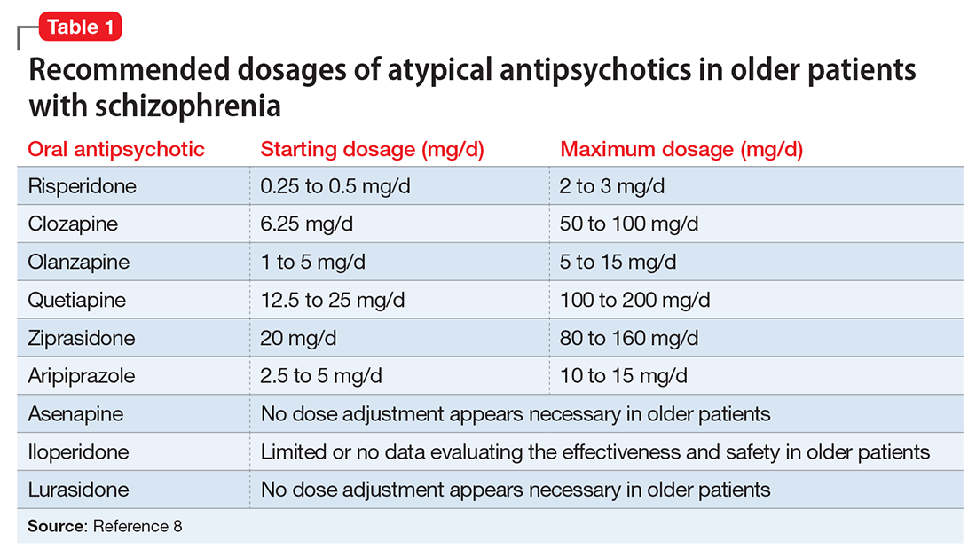
To ensure safety, patients should be regularly monitored with a complete blood count, comprehensive metabolic panel, lipid panel, hemoglobin A1C, electrocardiogram, orthostatic vital signs, Abnormal Involuntary Movement Scale, and weight check.7,9
When negative symptoms remain after a patient has achieved remission, it is important to evaluate whether the symptoms are related to adverse effects of medication (eg, parkinsonism syndrome), untreated depressive symptoms, or persistent positive symptoms, such as paranoia. Management of these symptoms consists of treating the cause, for example, using antipsychotics for primary positive symptoms, antidepressants for depression, anxiolytics for anxiety, and anti-parkinsonian agents or antipsychotic dosage reduction for EPS.
It is important to differentiate between negative symptoms of schizophrenia and depression in these patients. Negative symptoms of schizophrenia include affective flattening, alogia, avolition, and anhedonia. In depression, patients could have depressed mood, cognitive problems, sleep disturbances, and loss of appetite. Also, long-term symptoms are more consistent with negative symptomatology.
Keep in mind the potential for pharmacokinetic drug–drug interaction when using a combination of selective serotonin reuptake inhibitors (SSRIs), such as fluoxetine, paroxetine, and fluvoxamine (to treat negative/depressive symptoms), because all are significant inhibitors of cytochrome P450 enzymes and increase antipsychotic plasma level. The Expert Treatment Guidelines for Patients with Schizophrenia recommends SSRIs, followed by venlafaxine then bupropion to treat depressive symptoms after optimizing second-generation antipsychotics.9
Another point to consider when treating residual symptoms in patients with schizophrenia is to not discontinue antipsychotic medications. Relapse rates for these patients can occur up to 5 times higher than for those who continue treatment.10 A way to address this problem could be the use of depot antipsychotic medications, but there are no set recommendations for the use of long-acting injectable antipsychotics in older patients. These medications should be used with caution and at lowest effective dosages to offset potential adverse effects.
With the introduction of typical and atypical antipsychotics, the use of electroconvulsive therapy in older patients with schizophrenia has declined. In a 2009 meta-analysis of studies that included patients with refractory schizophrenia and repetitive transcranial magnetic stimulation (rTMS), results revealed a mixed effect size for controlled and uncontrolled studies. The authors stated the need for further controlled trials, assessing the efficacy of rTMS on negative and positive symptoms of schizophrenia.11
Psychotherapy and psychosocial interventions
Patients with schizophrenia who have persistent psychotic symptoms while receiving adequate pharmacotherapy should be offered adjunctive cognitive, behaviorally oriented psychotherapy to reduce symptom severity. Cognitive-behavioral therapy (CBT) has been shown to help reduce relapse rates, reduce psychotic symptoms, and improve patients’ mental state.12 Amotivation and lack of insight can be particularly troublesome, which CBT can help address.12 Psychoeducation can:
- empower patients to understand their illness
- help them cope with their disease
- be aware of symptom relapse
- seek help sooner rather than later.
Also, counseling and supportive therapy are recommended by the American Psychiatric Association guidelines. Providers should involve family and loved ones in this discussion, so that they can help collaborate with care and provide a supportive and non-judgmental environment.
Older patients with residual symptoms of schizophrenia are less likely to have completed their education, pursued a career, or developed long-lasting relationships. Family members who were their support system earlier in life, such as parents, often are unable to provide care for them by the time patients with schizophrenia become older. These patients also are less likely to get married or have children, meaning that they are more likely to live alone. The advent of the interdisciplinary team, integration of several therapeutic modalities, the provision of case managers, and assertive community treatment (ACT) teams has provided help with social support, relapses, and hospitalizations, for older patients with schizophrenia.13 Key elements of ACT include:
- a multidisciplinary team, including a medication prescriber
- a shared caseload among team members
- direct service provision by team members
- frequent patient contact
- low patient to staff ratios
- outreach to patients in the community.
Medical care
Patients with schizophrenia are at higher risk for several comorbid medical conditions, such as diabetes, coronary artery disease, and digestive and liver disorders, compared with individuals without schizophrenia. This risk is associated with numerous factors, including sedentary lifestyle, high rates of lifetime cigarette use (70% to 80% of schizophrenia outpatients age <67 smoke), poor self-management skills, frequent homelessness, and unhealthy diet.
Although substantial attention is devoted to the psychiatric and behavioral management of patients with schizophrenia, many barriers impede the detection and treatment of their medical conditions. Patients with schizophrenia could experience delays in diagnosing a medical disorder, leading to more acute comorbidities at the time of diagnosis and premature mortality. Studies have confirmed that cardiovascular diseases are the leading cause of premature death among psychiatric patients in the United States.14 Key risk factors include smoking, obesity, hypertension, dyslipidemia, diabetes, and lack of physical activity, all of which are more common among patients with schizophrenia compared with the general population.15 In addition, antipsychotics are associated with adverse metabolic effects.16
What are realistic treatment goals to manage residual symptoms in schizophrenia?
We believe that because remission in schizophrenia has been defined consensually, the bar for treatment expectations is set higher than it was 20 years ago. There can be patient-, family-, and system-related variables affecting the feasibility of treating residual symptoms. Providers who treat patients with schizophrenia should consider the following treatment goals:
- Prevent relapse and acute psychiatric hospitalization
- Use evidence-based strategies to minimize or monitor adverse effects
- Monitor compliance and consider use of depot antipsychotics combined with patients’ preference
- Facilitate ongoing safety assessment, including suicide risk
- Monitor negative and cognitive symptoms in addition to positive symptoms, using evidence-based management
- Encourage collaboration of care with family, caretakers, and other members of the treatment team
- Empower patients by providing psychoeducation and social skills training and assisting in their vocational rehabilitation
- Educate the patient and family about healthy lifestyle interventions and medical comorbidities common with schizophrenia
- Perform baseline screening and follow-up for early detection and treatment of medical comorbidities in patients with schizophrenia
- Improve functional status and quality of life.
In addition to meeting these treatment goals, a measurement-based method can be implemented to monitor improvement and status of the independent treatment domains. A collection of rating instruments can be found in Table 2.17-30
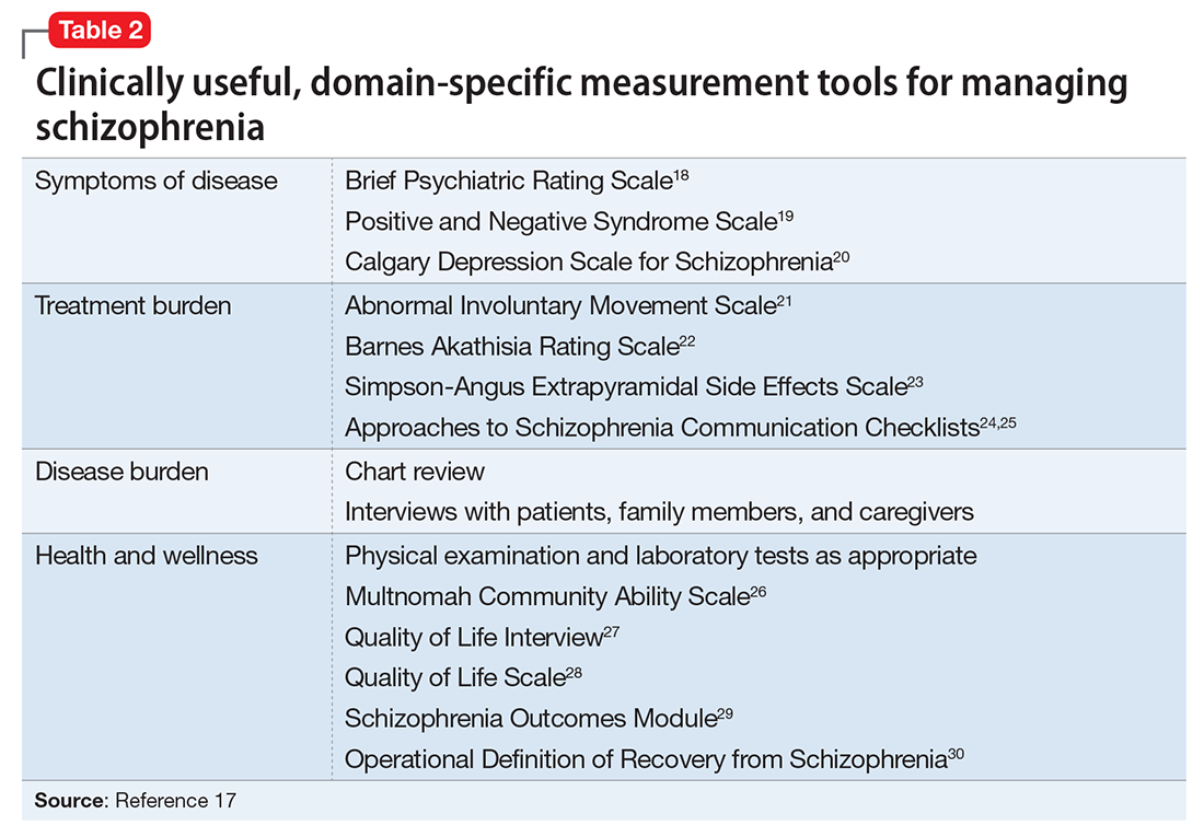
The clinical presentation of patients with residual symptoms of schizophrenia differs from that of other patients with schizophrenia. Our understanding of residual symptoms in schizophrenia has come a long way in the last decade; however, we are still far from pinning the complex nature of these symptoms, let alone their management. Given the risk of morbidity and disability, there clearly is a need for further investigation and investment of time and resources to support developing novel pharmacological treatment options to manage residual symptoms in patients with schizophrenia.
Because patients with residual symptoms of schizophrenia usually are older, psychiatrists should be responsible for implementing necessary screening assessments and should closely collaborate with primary care practitioners and other specialists, and when necessary, treat comorbid medical conditions. The importance of educating patients, their families, and the treatment team cannot be overlooked. Further, psychiatric treatment facilities should offer and promote healthy lifestyle interventions.
1. Kaiser S, Lyne J, Agartz I, et al. Individual negative symptoms and domains - relevance for assessment, pathomechanisms and treatment [published online July 21, 2016]. Schizophr Res. doi:10.1016/j.schres.2016.07.013.
2. Taylor M, Chaudhry I, Cross M, et al. Towards consensus in the long-term management of relapse prevention in schizophrenia. Hum Psychopharmacol. 2005;20(3):175-181.
3. Schennach R, Riedel M, Obermeier M, et al. What are residual symptoms in schizophrenia spectrum disorder? Clinical description and 1-year persistence within a naturalistic trial. Eur Arch Psychiatry Clin Neurosci. 2015;265(2):107-116.
4. Caligiuri MP, Jeste DV, Lacro JP. Antipsychotic-induced movement disorders in the elderly: epidemiology and treatment recommendations. Drugs Aging. 2000;17(5):363-384.
5. Dolder CR, Jeste DV. Incidence of tardive dyskinesia with typical versus atypical antipsychotics in very high risk patients. Biol Psychiatry. 2003;53(12):1142-1145.
6. Sable JA, Jeste DV. Antipsychotic treatment for late-life schizophrenia. Curr Psychiatry Rep. 2002;4(4):299-306.
7. Newcomer JW. Second-generation (atypical) antipsychotics and metabolic effects: a comprehensive literature review. CNS Drugs. 2005;19(suppl 1):1-93.
8. Khan AY, Redden W, Ovais M, et al. Current concepts in the diagnosis and treatment of schizophrenia in later life. Current Geriatric Reports. 2015;4(4):290-300.
9. Alexopoulos GS, Streim J, Carpenter D, et al; Expert Consensus Panel for Using Antipsychotic Drugs in Older Patients. Using antipsychotic agents in older patients. J Clin Psychiatry. 2004;65(suppl 2):5-99; discussion 100-102; quiz 103-104.
10. Robinson D, Woerner MG, Alvir JM, et al. Predictors of relapse following response from a first episode of schizophrenia or schizoaffective disorder. Arch Gen Psychiatry. 1999;56(3):241-247.
11. Freitas C, Fregni F, Pascual-Leone A. Meta-analysis of the effects of repetitive transcranial magnetic stimulation (rTMS) on negative and positive symptoms in schizophrenia. Schizophr Res. 2009;108(1-3):11-24.
12. Rector NA, Beck AT. Cognitive behavioral therapy for schizophrenia: an empirical review. J Nerv Ment Dis. 2012;200(10):832-839.
13. Stobbe J, Mulder NC, Roosenschoon BJ, et al. Assertive community treatment for elderly people with severe mental illness. BMC Psychiatry. 2010;10:84.
14. Hennekens CH, Hennekens AR, Hollar D, et al. Schizophrenia and increased risks of cardiovascular disease. Am Heart J. 2005;150(6):1115-1121.
15. Bushe CJ, Taylor M, Haukka J. Mortality in schizophrenia: a measurable clinical point. J Psychopharmacol. 2010;24(suppl 4):17-25.
16. Nasrallah HA, Meyer JM, Goff DC, et al. Low rates of treatment for hypertension, dyslipidemia, and diabetes in schizophrenia: data from the CATIE schizophrenia trial sample at baseline. Schizophr Res. 2006;86(1-3):15-22.
17. Nasrallah HA, Targum SD, Tandon R, et al. Defining and measuring clinical effectiveness in the treatment of schizophrenia. Psychiatr Serv. 2005;56(3):273-282.
18. Overall JE, Gorham DR. The Brief Psychiatric Rating Scale (BPRS): recent developments in ascertainment and scaling. Psychopharmacol Bull. 1988;24:97-99.
19. Kay SR, Fiszbein A, Opler LA. The Positive and Negative Syndrome Scale (PANSS) for schizophrenia. Schizophr Bull. 1987;13(2):261-276.
20. Addington D, Addington J, Schissel B. A depression rating scale for schizophrenics. Schizophr Res. 1990;3(4):247-251.
21. Guy W. ECDEU Assessment manual for psychopharmacology revised, 1976. Rockville, MD: US Department of Health, Education, and Welfare; Public Health Service; Alcohol, Drug Abuse, and Mental Health Administration; National Institute of Mental Health Psychopharmacology Research Branch, Division of Extramural Research Programs; 1976.
22. Barnes TR. A rating scale for drug-induced akathisia. Br J Psychiatry. 1989;154:672-676.
23. Simpson GM, Angus JWS. A rating scale for extrapyramidal side effects. Acta Psychiatr Scand. 1970;45(212):11-19.
24. Dott SG, Weiden P, Hopwood P, et al. An innovative approach to clinical communication in schizophrenia: the Approaches to Schizophrenia Communication checklists. CNS Spectr. 2001;6(4):333-338.
25. Dott SG, Knesevich J, Miller A, et al. Using the ASC program: a training guide. J Psychiatr Pract. 2001;7(1):64-68.
26. Barker S, Barron N, McFarland BH, et al. Multnomah Community Ability Scale: user’s manual. Portland, OR: Western Mental Health Research Center, Oregon Health Sciences University; 1994.
27. Lehman AF. A quality of life interview for the chronically mentally ill. Eval Program Plann. 1988;11(1):51-62.
28. Heinrichs DW, Hanlon TE, Carpenter WT Jr. The Quality of Life Scale: an instrument for rating the schizophrenic deficit syndrome. Schizophr Bull. 1984;10(3):388-398.
29. Becker M, Diamond R, Sainfort F. A new patient focused index for measuring quality of life in persons with severe and persistent mental illness. Qual Life Res. 1993;2(4):239-251.
30. Liberman RP, Kopelowicz A, Ventura J, et al. Operational criteria and factors related to recovery from schizophrenia. Int Rev Psychiatry. 2009;14(4):256-272.
The course of chronic psychiatric conditions, such as schizophrenia, differs from chronic medical conditions, such as diabetes. Some patients with chronic psychiatric conditions achieve remission and become symptom-free, while others continue to have lingering signs of disease for life.
Residual symptoms of schizophrenia are not fully defined in the literature, which poses a challenge because they are central in the overall treatment of schizophrenia spectrum disorders.1 During this phase of schizophrenia, patients continue to have symptoms after psychosis has subsided. These patients might continue to have negative symptoms such as social and emotional withdrawal and low energy. Although frank psychotic behavior has disappeared, the patient might continue to hold strange beliefs. Pharmacotherapy is the primary treatment option for psychiatric conditions, but the psychosocial aspect may have greater importance when treating residual symptoms and patients with chronic psychiatric illness.2
A naturalistic study in Germany evaluated the occurrence and characteristics of residual symptoms in patients with schizophrenia.3 The authors used a Positive and Negative Syndrome Scale symptom severity score >1 for those purposes, which is possibly a stringent criterion to define residual symptoms. This multicenter study enrolled 399 individuals age 18 to 65 with a DSM-IV-TR diagnosis of schizophrenia, schizophreniform disorder, delusional disorder, or schizoaffective disorder.3 Of the 236 patients achieving remission at discharge, 94% had at least 1 residual symptom and 69% had at least 4 residual symptoms. Therefore, residual symptoms were highly prevalent in remitted patients. The most frequent residual symptoms were:
- blunted affect
- conceptual disorganization
- passive or apathetic social withdrawal
- emotional withdrawal
- lack of judgment and insight
- poor attention
- somatic concern
- difficulty with abstract thinking
- anxiety
- poor rapport.3
Of note, positive symptoms, such as delusions and hallucinatory behavior, were found in remitted patients at discharge (17% and 10%, respectively). The study concluded that the severity of residual symptoms was associated with relapse risk and had an overall negative impact on the outcome of patients with schizophrenia.3 The study noted that residual symptoms may be greater in number or volume than negative symptoms and questioned the origins of residual symptoms because most were present at baseline in more than two-third of patients.
Patients with residual symptoms of schizophrenia usually are older and therefore present specific management challenges for clinicians. Changes associated with aging, such as medical problems, cognitive deficits, and lack of social support, could create new care needs for this patient population. Although the biopsychosocial model used to treat chronic psychiatric conditions, especially schizophrenia, is preferred, older schizophrenia patients with residual symptoms often need more psychosocial interventions compared with young adults with schizophrenia.
Managing residual symptoms in schizophrenia
Few studies are devoted to pharmacological treatment of older adults with schizophrenia, likely because pharmacotherapy for older patients with schizophrenia can be challenging. Evidence-based treatment is based primarily on findings from younger patients who survived into later life. Clinicians often use the adage of geriatric psychiatry, “start low, go slow,” because older patients are susceptible to adverse effects associated with psychiatric medications, including cardiovascular, metabolic, anticholinergic, and extrapyramidal effects, orthostasis, sedation, falls, and neuroleptic malignant syndrome.
Older patients with schizophrenia are at an increased risk for extrapyramidal symptoms (EPS) and anticholinergic adverse effects, perhaps because of degeneration of dopaminergic and cholinergic neurons.4 Lowering the anticholinergic load by discontinuing or reducing the dosage of medications with anticholinergic properties, when possible, is a key principle when treating these patients. This tactic could help improve cognition and quality of life by decreasing the risk of other anticholinergic adverse effects, including delirium, constipation, urinary retention, and blurred vision.
Patients treated with typical antipsychotics are nearly twice as likely to develop tardive dyskinesia compared with those receiving atypical antipsychotics.5 Sedation, orthostatic hypotension, and anticholinergic effects can cause cognitive clouding, worsen cognitive impairment, and increase the risk of falls, especially in older patients.6 Clozapine and olanzapine have the strongest association with clinically significant weight gain and treatment-induced type 2 diabetes mellitus.7
The appropriate starting dosage of antipsychotics in older patients with schizophrenia is one-fourth of the starting adult dosage. Total daily maintenance dosages may be one-third to one-half of the adult dosage.6 Consensus guidelines for dosing atypical antipsychotics for older patients with schizophrenia are as shown in Table 1.8
To ensure safety, patients should be regularly monitored with a complete blood count, comprehensive metabolic panel, lipid panel, hemoglobin A1C, electrocardiogram, orthostatic vital signs, Abnormal Involuntary Movement Scale, and weight check.7,9
When negative symptoms remain after a patient has achieved remission, it is important to evaluate whether the symptoms are related to adverse effects of medication (eg, parkinsonism syndrome), untreated depressive symptoms, or persistent positive symptoms, such as paranoia. Management of these symptoms consists of treating the cause, for example, using antipsychotics for primary positive symptoms, antidepressants for depression, anxiolytics for anxiety, and anti-parkinsonian agents or antipsychotic dosage reduction for EPS.
It is important to differentiate between negative symptoms of schizophrenia and depression in these patients. Negative symptoms of schizophrenia include affective flattening, alogia, avolition, and anhedonia. In depression, patients could have depressed mood, cognitive problems, sleep disturbances, and loss of appetite. Also, long-term symptoms are more consistent with negative symptomatology.
Keep in mind the potential for pharmacokinetic drug–drug interaction when using a combination of selective serotonin reuptake inhibitors (SSRIs), such as fluoxetine, paroxetine, and fluvoxamine (to treat negative/depressive symptoms), because all are significant inhibitors of cytochrome P450 enzymes and increase antipsychotic plasma level. The Expert Treatment Guidelines for Patients with Schizophrenia recommends SSRIs, followed by venlafaxine then bupropion to treat depressive symptoms after optimizing second-generation antipsychotics.9
Another point to consider when treating residual symptoms in patients with schizophrenia is to not discontinue antipsychotic medications. Relapse rates for these patients can occur up to 5 times higher than for those who continue treatment.10 A way to address this problem could be the use of depot antipsychotic medications, but there are no set recommendations for the use of long-acting injectable antipsychotics in older patients. These medications should be used with caution and at lowest effective dosages to offset potential adverse effects.
With the introduction of typical and atypical antipsychotics, the use of electroconvulsive therapy in older patients with schizophrenia has declined. In a 2009 meta-analysis of studies that included patients with refractory schizophrenia and repetitive transcranial magnetic stimulation (rTMS), results revealed a mixed effect size for controlled and uncontrolled studies. The authors stated the need for further controlled trials, assessing the efficacy of rTMS on negative and positive symptoms of schizophrenia.11
Psychotherapy and psychosocial interventions
Patients with schizophrenia who have persistent psychotic symptoms while receiving adequate pharmacotherapy should be offered adjunctive cognitive, behaviorally oriented psychotherapy to reduce symptom severity. Cognitive-behavioral therapy (CBT) has been shown to help reduce relapse rates, reduce psychotic symptoms, and improve patients’ mental state.12 Amotivation and lack of insight can be particularly troublesome, which CBT can help address.12 Psychoeducation can:
- empower patients to understand their illness
- help them cope with their disease
- be aware of symptom relapse
- seek help sooner rather than later.
Also, counseling and supportive therapy are recommended by the American Psychiatric Association guidelines. Providers should involve family and loved ones in this discussion, so that they can help collaborate with care and provide a supportive and non-judgmental environment.
Older patients with residual symptoms of schizophrenia are less likely to have completed their education, pursued a career, or developed long-lasting relationships. Family members who were their support system earlier in life, such as parents, often are unable to provide care for them by the time patients with schizophrenia become older. These patients also are less likely to get married or have children, meaning that they are more likely to live alone. The advent of the interdisciplinary team, integration of several therapeutic modalities, the provision of case managers, and assertive community treatment (ACT) teams has provided help with social support, relapses, and hospitalizations, for older patients with schizophrenia.13 Key elements of ACT include:
- a multidisciplinary team, including a medication prescriber
- a shared caseload among team members
- direct service provision by team members
- frequent patient contact
- low patient to staff ratios
- outreach to patients in the community.
Medical care
Patients with schizophrenia are at higher risk for several comorbid medical conditions, such as diabetes, coronary artery disease, and digestive and liver disorders, compared with individuals without schizophrenia. This risk is associated with numerous factors, including sedentary lifestyle, high rates of lifetime cigarette use (70% to 80% of schizophrenia outpatients age <67 smoke), poor self-management skills, frequent homelessness, and unhealthy diet.
Although substantial attention is devoted to the psychiatric and behavioral management of patients with schizophrenia, many barriers impede the detection and treatment of their medical conditions. Patients with schizophrenia could experience delays in diagnosing a medical disorder, leading to more acute comorbidities at the time of diagnosis and premature mortality. Studies have confirmed that cardiovascular diseases are the leading cause of premature death among psychiatric patients in the United States.14 Key risk factors include smoking, obesity, hypertension, dyslipidemia, diabetes, and lack of physical activity, all of which are more common among patients with schizophrenia compared with the general population.15 In addition, antipsychotics are associated with adverse metabolic effects.16
What are realistic treatment goals to manage residual symptoms in schizophrenia?
We believe that because remission in schizophrenia has been defined consensually, the bar for treatment expectations is set higher than it was 20 years ago. There can be patient-, family-, and system-related variables affecting the feasibility of treating residual symptoms. Providers who treat patients with schizophrenia should consider the following treatment goals:
- Prevent relapse and acute psychiatric hospitalization
- Use evidence-based strategies to minimize or monitor adverse effects
- Monitor compliance and consider use of depot antipsychotics combined with patients’ preference
- Facilitate ongoing safety assessment, including suicide risk
- Monitor negative and cognitive symptoms in addition to positive symptoms, using evidence-based management
- Encourage collaboration of care with family, caretakers, and other members of the treatment team
- Empower patients by providing psychoeducation and social skills training and assisting in their vocational rehabilitation
- Educate the patient and family about healthy lifestyle interventions and medical comorbidities common with schizophrenia
- Perform baseline screening and follow-up for early detection and treatment of medical comorbidities in patients with schizophrenia
- Improve functional status and quality of life.
In addition to meeting these treatment goals, a measurement-based method can be implemented to monitor improvement and status of the independent treatment domains. A collection of rating instruments can be found in Table 2.17-30
The clinical presentation of patients with residual symptoms of schizophrenia differs from that of other patients with schizophrenia. Our understanding of residual symptoms in schizophrenia has come a long way in the last decade; however, we are still far from pinning the complex nature of these symptoms, let alone their management. Given the risk of morbidity and disability, there clearly is a need for further investigation and investment of time and resources to support developing novel pharmacological treatment options to manage residual symptoms in patients with schizophrenia.
Because patients with residual symptoms of schizophrenia usually are older, psychiatrists should be responsible for implementing necessary screening assessments and should closely collaborate with primary care practitioners and other specialists, and when necessary, treat comorbid medical conditions. The importance of educating patients, their families, and the treatment team cannot be overlooked. Further, psychiatric treatment facilities should offer and promote healthy lifestyle interventions.
1. Kaiser S, Lyne J, Agartz I, et al. Individual negative symptoms and domains - relevance for assessment, pathomechanisms and treatment [published online July 21, 2016]. Schizophr Res. doi:10.1016/j.schres.2016.07.013.
2. Taylor M, Chaudhry I, Cross M, et al. Towards consensus in the long-term management of relapse prevention in schizophrenia. Hum Psychopharmacol. 2005;20(3):175-181.
3. Schennach R, Riedel M, Obermeier M, et al. What are residual symptoms in schizophrenia spectrum disorder? Clinical description and 1-year persistence within a naturalistic trial. Eur Arch Psychiatry Clin Neurosci. 2015;265(2):107-116.
4. Caligiuri MP, Jeste DV, Lacro JP. Antipsychotic-induced movement disorders in the elderly: epidemiology and treatment recommendations. Drugs Aging. 2000;17(5):363-384.
5. Dolder CR, Jeste DV. Incidence of tardive dyskinesia with typical versus atypical antipsychotics in very high risk patients. Biol Psychiatry. 2003;53(12):1142-1145.
6. Sable JA, Jeste DV. Antipsychotic treatment for late-life schizophrenia. Curr Psychiatry Rep. 2002;4(4):299-306.
7. Newcomer JW. Second-generation (atypical) antipsychotics and metabolic effects: a comprehensive literature review. CNS Drugs. 2005;19(suppl 1):1-93.
8. Khan AY, Redden W, Ovais M, et al. Current concepts in the diagnosis and treatment of schizophrenia in later life. Current Geriatric Reports. 2015;4(4):290-300.
9. Alexopoulos GS, Streim J, Carpenter D, et al; Expert Consensus Panel for Using Antipsychotic Drugs in Older Patients. Using antipsychotic agents in older patients. J Clin Psychiatry. 2004;65(suppl 2):5-99; discussion 100-102; quiz 103-104.
10. Robinson D, Woerner MG, Alvir JM, et al. Predictors of relapse following response from a first episode of schizophrenia or schizoaffective disorder. Arch Gen Psychiatry. 1999;56(3):241-247.
11. Freitas C, Fregni F, Pascual-Leone A. Meta-analysis of the effects of repetitive transcranial magnetic stimulation (rTMS) on negative and positive symptoms in schizophrenia. Schizophr Res. 2009;108(1-3):11-24.
12. Rector NA, Beck AT. Cognitive behavioral therapy for schizophrenia: an empirical review. J Nerv Ment Dis. 2012;200(10):832-839.
13. Stobbe J, Mulder NC, Roosenschoon BJ, et al. Assertive community treatment for elderly people with severe mental illness. BMC Psychiatry. 2010;10:84.
14. Hennekens CH, Hennekens AR, Hollar D, et al. Schizophrenia and increased risks of cardiovascular disease. Am Heart J. 2005;150(6):1115-1121.
15. Bushe CJ, Taylor M, Haukka J. Mortality in schizophrenia: a measurable clinical point. J Psychopharmacol. 2010;24(suppl 4):17-25.
16. Nasrallah HA, Meyer JM, Goff DC, et al. Low rates of treatment for hypertension, dyslipidemia, and diabetes in schizophrenia: data from the CATIE schizophrenia trial sample at baseline. Schizophr Res. 2006;86(1-3):15-22.
17. Nasrallah HA, Targum SD, Tandon R, et al. Defining and measuring clinical effectiveness in the treatment of schizophrenia. Psychiatr Serv. 2005;56(3):273-282.
18. Overall JE, Gorham DR. The Brief Psychiatric Rating Scale (BPRS): recent developments in ascertainment and scaling. Psychopharmacol Bull. 1988;24:97-99.
19. Kay SR, Fiszbein A, Opler LA. The Positive and Negative Syndrome Scale (PANSS) for schizophrenia. Schizophr Bull. 1987;13(2):261-276.
20. Addington D, Addington J, Schissel B. A depression rating scale for schizophrenics. Schizophr Res. 1990;3(4):247-251.
21. Guy W. ECDEU Assessment manual for psychopharmacology revised, 1976. Rockville, MD: US Department of Health, Education, and Welfare; Public Health Service; Alcohol, Drug Abuse, and Mental Health Administration; National Institute of Mental Health Psychopharmacology Research Branch, Division of Extramural Research Programs; 1976.
22. Barnes TR. A rating scale for drug-induced akathisia. Br J Psychiatry. 1989;154:672-676.
23. Simpson GM, Angus JWS. A rating scale for extrapyramidal side effects. Acta Psychiatr Scand. 1970;45(212):11-19.
24. Dott SG, Weiden P, Hopwood P, et al. An innovative approach to clinical communication in schizophrenia: the Approaches to Schizophrenia Communication checklists. CNS Spectr. 2001;6(4):333-338.
25. Dott SG, Knesevich J, Miller A, et al. Using the ASC program: a training guide. J Psychiatr Pract. 2001;7(1):64-68.
26. Barker S, Barron N, McFarland BH, et al. Multnomah Community Ability Scale: user’s manual. Portland, OR: Western Mental Health Research Center, Oregon Health Sciences University; 1994.
27. Lehman AF. A quality of life interview for the chronically mentally ill. Eval Program Plann. 1988;11(1):51-62.
28. Heinrichs DW, Hanlon TE, Carpenter WT Jr. The Quality of Life Scale: an instrument for rating the schizophrenic deficit syndrome. Schizophr Bull. 1984;10(3):388-398.
29. Becker M, Diamond R, Sainfort F. A new patient focused index for measuring quality of life in persons with severe and persistent mental illness. Qual Life Res. 1993;2(4):239-251.
30. Liberman RP, Kopelowicz A, Ventura J, et al. Operational criteria and factors related to recovery from schizophrenia. Int Rev Psychiatry. 2009;14(4):256-272.