User login
Most children with growth disorders benefit from growth hormone
ORLANDO – About 80% of children with growth disorders will attain their expected normal adult height if adequately treated with growth hormone, a large observational study has determined.
Somatropin recombinant human growth hormone seemed most effective for patients with growth hormone deficiency, idiopathic short stature, and for those who were born small for gestational age. Patients who had Turner syndrome or a homeobox-deficiency gene didn’t do quite as well, although most did still improve their final height.
The Genetics and Neuroendocrinology of Short Stature International Study (GENESIS) followed more than 22,000 children who were treated in 30 countries from 1999 to 2015.
All of the patients were treated with somatropin recombinant human growth hormone (Humatrope). Eli Lilly, which makes the medication, sponsored the study and presented the results during a poster session at the annual meeting of the Endocrine Society. Representatives at the poster declined to discuss results with the media during the open session.
“The results speak for themselves,” said Cheri Deal, MD, PhD, of the University of Montreal, Quebec, one of the presenting authors. “It’s an incredible story.”
The GENESIS cohort comprised more than 22,000 children who were treated for a variety of growth disorders at 827 international sites.
Subanalyses have been published, but this is the first release of the data on 5,076 patients with full follow-up. Most of the patients (61%) received the medication for growth hormone deficiency. Other indications were Turner syndrome (14%), idiopathic short stature (11%), short-stature homeobox-containing gene deficiency (3%), small for gestational age (5%), and chronic renal insufficiency (0.3%). Other diagnoses, plus unknown causes, made up the remainder of the diagnoses.
The investigators, led by Christopher J. Child, PhD, an Eli Lilly scientist, split the cohort into diagnostic groups and treatment subgroups: those who attained normal adult height, those who attained normal adult height with at least 4 years of treatment, and those who gained normal adult height who started treatment at younger than 10 years of age.
At baseline, those with growth hormone deficiency were a mean of 11 years old; their height was a mean of 2.4 standard deviations (SDS) below normal. They received a mean of 6 years of treatment; 86% achieved a normal adult height, with an overall gain of 1.39 SDS. Those who started somatropin before age 10 years made bigger gains. The final height gain was 1.78 SDS after a mean of 9.5 years of treatment.
At baseline, those with idiopathic short stature were a mean of 12 years old; their mean height was 2.3 SDS below normal. They received a mean of 4.7 years of treatment; 82% achieved normal height, with a gain of 1.1 SDS. Starting somatropin at a younger age (mean 8 years) improved outcomes slightly, with a final height gain of 1.6 SDS after 8 years of treatment.
Patients with Turner syndrome were a mean of 10 years when they started treatment; their mean height was 2.65 SDS below normal. They received a mean of 6.4 years of treatment; 66% achieved normal adult height, with a mean height gain of 0.95 SDS. Early treatment didn’t alter outcomes much in this group. After a mean of 8.5 years of treatment, 65% achieved normal height, with a height gain of 0.84 SDS.
Patients with a short-stature homeobox-containing gene deficiency were a mean of 11 years when they started treatment. At that time, their height was a mean of 2.36 SDS below normal. They were treated for a mean of 4.7 years; 76% achieved a normal adult height, with a mean gain of 0.86 SDS. Early treatment did improve outcomes a bit. After a mean treatment time of 7 years, 78% gained normal adult height, with a height gain of 0.84 SDS.
Children who were small for gestational age started treatment when they were a mean of 10.5 years. At that time, their mean height was 2.57 SDS below normal. They were treated for a mean of 5.4 years; 78% attained a normal adult height, with a mean gain of 1.1 SDS. Early treatment improved outcomes. After a mean treatment time of 7.8 years, 81% achieved normal adult height, with a mean gain of 1.41 SDS.
The less-robust gains seen in the children with genetically influenced growth disorders “wasn’t unexpected” and is in line with what has been observed in a number of clinical trials, the investigators said.
Eli Lilly manufactures the somatropin product used in the study, and it sponsored the study. Dr. Child is an employee of the company. Dr. Deal did not have any financial disclosures.
[email protected]
On Twitter @Alz_gal
AT ENDO 2017
Key clinical point:
Major finding: Overall, 80% of the treated children reached normal adult height.
Data source: The prospective observational cohort study comprised 5,076 children.
Disclosures: Eli Lilly manufactures the somatropin product used in the study, and it sponsored the study. Dr. Child is an employee of the company. Dr. Deal did not have any financial disclosures.
Michigan model embeds pharmacists into primary care
SAN DIEGO – Incorporating pharmacists into primary care offices has the potential to significantly impact patient care and save health care costs, according to Hae Mi Choe, PharmD.
Drug-related morbidity in the United States costs an estimated $290 billion annually, or about 13% of total health care expenditures, said Dr. Choe, associate dean at the University of Michigan College of Pharmacy and director of pharmacy innovations and partnerships at the University of Michigan Medical Group, Ann Arbor.

Dr. Choe described her experience helping to create a group practice model at the university health system for clinical pharmacists as part of the patient-centered medical home movement.
A total of 11 pharmacists are embedded in 14 primary care clinics in the University of Michigan Health System (now known as Michigan Medicine) to provide disease management and comprehensive medication review services. The pharmacists’ time at each site varies depending on patient volume, but typically ranges from 1 to 3 days per week.
They work to identify patients struggling to manage their diabetes, hypertension, or hyperlipidemia. “For patients who may not have reached their HbA1c goal, for instance, we may reach out to the physician and say, ‘Do you think Mrs. Jones would benefit from seeing a pharmacist?’ ” she said.
For comprehensive medication review, pharmacists schedule two visits. The first involves sitting down with the patient to review all of their medications, “trying to understand the patients’ perception of their medication, their preference in how they go about their treatment regimen, their understanding of the disease state, and identify potential barriers to treatment like medication cost,” Dr. Choe explained.
“Then we schedule the patient back in 2 weeks, after we’ve discussed issues with physicians and created a treatment plan,” she continued. “At that time, we discuss new treatment plan to improve efficacy, safety, and lower drug costs.”
Combined, both visits amount to 75-90 minutes of the pharmacist’s time.
“We try to provide comprehensive medication review at least once a year for complex patients,” she said. “It’s our version of an annual physical exam. One patient I saw was taking 32 medications prescribed by nine different specialists. She was taking her medication eight times per day.”
Another patient who came in for a comprehensive medication review transported his drugs in a suitcase. “He told me that he was about to go to Florida, and that the suitcase for his medications was bigger than the one for his clothes,” Dr. Choe said.
“You’d be surprised what patients take that you were not aware of,” she added. “Research shows that up to 50% of the time, patients do not take medications as prescribed. For antihypertensives, roughly one-quarter of patients don’t fill the prescription to begin with, and another one-quarter don’t take it as prescribed. That’s 50% right there.”
Pharmacists in the Michigan Medicine practice model have full access to the health system’s electronic medical record, including bidirectional communication with physicians.
“We also have population-level data, so if I wanted to drill down on clinic A and ask, who in clinic A is not achieving HbA1c, blood pressure, or other quality metrics, with a click of a button we can drill down to that population base,” she said. The pharmacists have been granted special clinical privileges, including the ability to initiate medication, adjust dosing, and discontinue therapy based on delegation protocols.
When Dr. Choe and her associates analyzed 2,674 interventions made during the first year of the program’s implementation, 50% involved increasing a dose, followed by adding a medication (20%), decreasing a dose (13%), deleting a medication (9%), and optimizing a medication regimen (8%). In addition, patients with a baseline HbA1c of greater than 7% experienced a 1% drop in their HbA1c level. Results were similar for patients whose baseline HbA1c level was greater than 8% and greater than 9% (drops of 1.4% and 1.9%, respectively).
The pharmacist program has expanded to Michigan Medicine chronic kidney disease clinics, outpatient psychiatric clinic, anticoagulation services, transitions of care services, palliative care services, transplant services, transplant clinics, oncology clinics, and telehealth services, Dr. Choe said.
For clinicians looking to embed pharmacists into their clinics, she recommended identifying key stakeholders with whom to collaborate.
“That could be a physician champion, and having a lead pharmacist who not only has the strong clinical ability but leadership skills to build a program,” she said. “You would also benefit from having someone in your team with a clinical operation background, and someone with quality and data expertise. Start small and aim for a bigger impact.”
Dr. Choe reported having no financial disclosures.
SAN DIEGO – Incorporating pharmacists into primary care offices has the potential to significantly impact patient care and save health care costs, according to Hae Mi Choe, PharmD.
Drug-related morbidity in the United States costs an estimated $290 billion annually, or about 13% of total health care expenditures, said Dr. Choe, associate dean at the University of Michigan College of Pharmacy and director of pharmacy innovations and partnerships at the University of Michigan Medical Group, Ann Arbor.
Dr. Choe described her experience helping to create a group practice model at the university health system for clinical pharmacists as part of the patient-centered medical home movement.
A total of 11 pharmacists are embedded in 14 primary care clinics in the University of Michigan Health System (now known as Michigan Medicine) to provide disease management and comprehensive medication review services. The pharmacists’ time at each site varies depending on patient volume, but typically ranges from 1 to 3 days per week.
They work to identify patients struggling to manage their diabetes, hypertension, or hyperlipidemia. “For patients who may not have reached their HbA1c goal, for instance, we may reach out to the physician and say, ‘Do you think Mrs. Jones would benefit from seeing a pharmacist?’ ” she said.
For comprehensive medication review, pharmacists schedule two visits. The first involves sitting down with the patient to review all of their medications, “trying to understand the patients’ perception of their medication, their preference in how they go about their treatment regimen, their understanding of the disease state, and identify potential barriers to treatment like medication cost,” Dr. Choe explained.
“Then we schedule the patient back in 2 weeks, after we’ve discussed issues with physicians and created a treatment plan,” she continued. “At that time, we discuss new treatment plan to improve efficacy, safety, and lower drug costs.”
Combined, both visits amount to 75-90 minutes of the pharmacist’s time.
“We try to provide comprehensive medication review at least once a year for complex patients,” she said. “It’s our version of an annual physical exam. One patient I saw was taking 32 medications prescribed by nine different specialists. She was taking her medication eight times per day.”
Another patient who came in for a comprehensive medication review transported his drugs in a suitcase. “He told me that he was about to go to Florida, and that the suitcase for his medications was bigger than the one for his clothes,” Dr. Choe said.
“You’d be surprised what patients take that you were not aware of,” she added. “Research shows that up to 50% of the time, patients do not take medications as prescribed. For antihypertensives, roughly one-quarter of patients don’t fill the prescription to begin with, and another one-quarter don’t take it as prescribed. That’s 50% right there.”
Pharmacists in the Michigan Medicine practice model have full access to the health system’s electronic medical record, including bidirectional communication with physicians.
“We also have population-level data, so if I wanted to drill down on clinic A and ask, who in clinic A is not achieving HbA1c, blood pressure, or other quality metrics, with a click of a button we can drill down to that population base,” she said. The pharmacists have been granted special clinical privileges, including the ability to initiate medication, adjust dosing, and discontinue therapy based on delegation protocols.
When Dr. Choe and her associates analyzed 2,674 interventions made during the first year of the program’s implementation, 50% involved increasing a dose, followed by adding a medication (20%), decreasing a dose (13%), deleting a medication (9%), and optimizing a medication regimen (8%). In addition, patients with a baseline HbA1c of greater than 7% experienced a 1% drop in their HbA1c level. Results were similar for patients whose baseline HbA1c level was greater than 8% and greater than 9% (drops of 1.4% and 1.9%, respectively).
The pharmacist program has expanded to Michigan Medicine chronic kidney disease clinics, outpatient psychiatric clinic, anticoagulation services, transitions of care services, palliative care services, transplant services, transplant clinics, oncology clinics, and telehealth services, Dr. Choe said.
For clinicians looking to embed pharmacists into their clinics, she recommended identifying key stakeholders with whom to collaborate.
“That could be a physician champion, and having a lead pharmacist who not only has the strong clinical ability but leadership skills to build a program,” she said. “You would also benefit from having someone in your team with a clinical operation background, and someone with quality and data expertise. Start small and aim for a bigger impact.”
Dr. Choe reported having no financial disclosures.
EXPERT ANALYSIS AT ACP INTERNAL MEDICINE
VIDEO: Careful TKI hiatus makes CML pregnancy possible
NEW YORK – The success that tyrosine kinase inhibitors have had in prolonging life and producing deep hematologic and molecular remissions in patients with chronic myeloid leukemia has led to an unexpected bonus for young women living with the disease: an opportunity to safely become pregnant and mother a child.
The approach is not yet routine and poses a level of risk to both the mother and fetus, especially because tyrosine kinase inhibitors (TKIs) are teratogenic. But with careful planning, close gestational monitoring, and with support from skilled obstetricians, the scenario of a successful pregnancy in women with chronic myeloid leukemia (CML) has now played out several dozen times at a handful of U.S. centers, Mrinal S. Patnaik, MD, said in a talk at the conference held by Imedex.
“We make it clear that this is experimental and is associated with risk, and we share the data [from case reports]; but if the woman wants to go forward,” a protocol now exists “to successfully get them to pregnancy,” said Dr. Patnaik, a hematologist oncologist at the Mayo Clinic in Rochester, Minn.
At Mayo alone, upwards of 20 women with CML have been successfully shepherded through pregnancy, he said in a video interview.
The prospect for a planned pregnancy is reserved for women with their CML well controlled for at least 2 years using a TKI, most often imatinib (Gleevec). In addition to being under complete hematologic control, the candidate patient must also show a deep molecular response, which means a blood level of the BRC-ABL tyrosine kinase that drives CML at least 4 or 4.5 logs (10,000-50,000-fold) below pretreatment levels or molecularly undetectable.
The patient then monitors her ovulatory cycle and stops her medication at the time of ovulation, attempts conception, and then monitors whether pregnancy has actually started. If it has, she needs to stay off her TKI regimen through at least the first 18 weeks of gestation, although an even longer drug holiday is preferred. If not, she resumes the medication and repeats the process later if she wants.
Once the women is pregnant and remains off her TKI regimen Dr. Patnaik and his associates closely follow the woman for signs of a molecular or hematologic relapse, although the latter are unusual. If a resurgence of CML stem cells occurs, the woman receives treatment with pegylated interferon-alpha, which is safe during pregnancy. When possible, TKI treatment remains on hold into the breast-feeding period.
During pregnancy and delivery, the patient requires careful and regular follow-up by a maternal-fetal medicine specialist and has an ongoing risk for high platelet counts causing placental blood clots, fetuses that are small for gestational age, preterm labor, premature rupture of membranes, and other complications.
“These are manageable with good obstetrical care,” Dr. Patnaik said. “We have developed a good system to work out the obstetrical complications.
“By and large we can be successful, but it requires a lot of monitoring and a lot of patient compliance with regular follow-ups,” he stressed.
In a video interview at the meeting, Dr. Patnaik discussed the approach he takes with his patients.
The video associated with this article is no longer available on this site. Please view all of our videos on the MDedge YouTube channel
[email protected]
On Twitter @mitchelzoler
EXPERT ANALYSIS FROM A MEETING ON HEMATOLOGIC MALIGNANCIES

Caution urged in extending dual antiplatelet therapy
SNOWMASS, COLO. – Think very carefully before extending the duration of dual antiplatelet therapy beyond 6 months in drug-eluting stent recipients with stable ischemic heart disease, Patrick T. O’Gara, MD, advised at the Annual Cardiovascular Conference at Snowmass.
Six months of dual antiplatelet therapy (DAPT) in this setting received a Class I recommendation in the 2016 American College of Cardiology/American Heart Association guideline focused update on DAPT duration (J Am Coll Cardiol. 2016 Sep 6;68[10]:1082-115). That’s a departure from previous guidelines, which recommended 12 months of DAPT. The shortened DAPT duration of 6 months is consistent with European Society of Cardiology recommendations.
In contrast, extending DAPT beyond the 6-month mark garnered a relatively weak Class IIb recommendation in the ACC/AHA focused update, meaning it “could be considered,” noted Dr. O’Gara, director of clinical cardiology at Brigham and Women’s Hospital, Boston, and professor of medicine at Harvard Medical School.
Considerable enthusiasm for extending DAPT well beyond 6 months after drug-eluting stent implantation has been generated in some quarters by the positive results of the PEGASUS TIMI 54 trial. But Dr. O’Gara and the other members of the guideline writing committee had reservations about the study, which together with other concerning evidence led to the weak Class IIb recommendation.
PEGASUS TIMI 54 included 21,162 patients with stable ischemic heart disease 1-3 years after a myocardial infarction who were randomized to low-dose aspirin plus either placebo or ticagrelor (Brilinta) at 60 mg or 90 mg b.i.d. and followed prospectively for a median of 33 months (N Engl J Med. 2015 May 7;372[19]:1791-800).
The primary efficacy endpoint, a composite of cardiovascular death, MI, or stroke, occurred in 9.0% of placebo-treated patients, compared with 7.8% of patients on either ticagrelor regimen, for a statistically significant 15% relative risk reduction in the DAPT group.
But there is more to the study than first meets the eye.
“I think what we as practitioners sometimes lose track of is that the investigators in this particular trial were very careful to enroll patients with stable ischemic heart disease who were at high risk of ischemic events over the next 3-5 years,” Dr. O’Gara noted. “These were patients who were generally older, patients with diabetes, chronic kidney disease, multivessel coronary disease, or who had had a second MI.”
Thus, the deck was stacked in favor of obtaining a result showing maximum efficacy. Yet, for every 10,000 patients treated with ticagrelor at 90 mg b.i.d., there were only 40 fewer cardiovascular events per year, compared with placebo. And that came at a cost of 41 more TIMI major bleeding events.
“That’s a wash at 90 mg,” the cardiologist said.
At 60 mg b.i.d. – the dose ultimately approved by the Food and Drug Administration – there were 42 fewer primary cardiovascular events per year per 10,000 treated patients, a benefit that came at the expense of 31 more TIMI major bleeding events.
“These are really razor thin margins, and I would encourage you to make a risk-benefit assessment of the trade-off between ischemia and bleeding in your decision-making,” Dr. O’Gara said.
The ACC/AHA guideline writing committee also took into account a meta-analysis of six randomized clinical trials totaling more than 33,000 high-risk patients post-MI who were assigned to more than 1 year of DAPT or aspirin alone. Extended DAPT brought a 22% reduction in the relative risk of major adverse cardiovascular events, but this was accompanied with a 73% increase in the risk of major bleeding (Eur Heart J. 2016 Jan 21;37[4]:390-9).
Turning to DAPT duration post-PCI in patients with an acute coronary syndrome, Dr. O’Gara noted that the 2016 ACC/AHA guideline focused update gave a Class I indication for 12 months of DAPT in recipients of a drug-eluting stent, but a weaker IIb recommendation for consideration of extending DAPT beyond that point – provided the patient was not at high bleeding risk and didn’t have significant bleeding during the first 12 months on DAPT.
“I think there’s a lot of individual and institutional variation with respect to this kind of decision-making, and I don’t think our guidelines are meant to be proscriptive, because our patients are quite nuanced,” the cardiologist observed.
The question physicians always have to ask in considering extended DAPT is, “How many ischemic events am I going to prevent at the expense of how many bleeding events?”
The investigators in the landmark DAPT study of extended therapy have analyzed their data in a fashion that has enabled them to develop a risk scoring system, known as the DAPT prediction rule, which is readily calculated based on factors including age, presence of diabetes, heart failure, and the size of the treated vessel.
For patients with a high DAPT score, assignment to an additional 18 months of DAPT after the initial 12 months of dual therapy was associated with a net 1.67% reduction in adverse events – both ischemic and bleeding – compared with the rate in patients who stopped DAPT at 12 months. For those with a low DAPT score, extended dual antiplatelet therapy resulted in a 1.03% net increase in adverse events (JAMA. 2016 Apr 26;315[16]:1735-49).
“I should warn you that the discriminatory power of this particular score is relatively modest,” Dr. O’Gara noted. “The C-statistic is not higher than about 0.7. But I do think that the DAPT score meets the sniff test biologically and clinically. It’s a real good first step. I do think this particular score needs to be validated externally in other populations going forward.”
Dr. O’Gara reported having no financial conflicts of interest.
EXPERT ANALYSIS FROM THE CARDIOVASCULAR CONFERENCE AT SNOWMASS
Adolescents with an incarcerated parent at higher risk of mental health problems
Having or having had a parent in jail appears to be a risk factor for adolescent mental health problems, a Minnesota study found.
In a study of 122,180 children from the 8th, 9th, and 11th grades drawn from the 2013 Minnesota Student Survey, parental incarceration was strongly associated with higher rates of mental health problems such as internalizing, self-injury, suicidal ideation, and suicidal attempts. Children of currently incarcerated parents were two and a half to four times as likely to experience these mental health problems, and children of formerly incarcerated parents were roughly twice as likely to experience them, compared with children whose parents had not been jailed, said Laurel Davis, PhD, and Rebecca J. Shlafer, PhD, MPH, of the department of pediatrics at the University of Minnesota, Minneapolis.
For youth with currently incarcerated parents, almost half reported moderate symptoms of internalizing, and more than one-third reported at least one purposeful self-injury. However, only 30% reported any type of treatment for a mental health concern, the researchers said.
Parental closeness was protective against negative mental health outcomes, they noted. “Children of incarcerated parents are a vulnerable population and they should continue to be the targets of intervention efforts aiming to buttress their personal and social resources, including strong relationships with caregivers.”
Studies suggest that an estimated 1.9 million U.S. children have a parent in a state or federal prison, and millions more have a parent in county jails.
In Minnesota alone, 2,500 young people who had a parent in jail or whose parent had been in jail reported attempting suicide, and more than 5,000 children of incarcerated or formerly incarcerated parents had reported purposely having injured themselves in the last year. “This represents an enormous challenge to our systems of care,” said Dr. Davis and Dr. Shlafer. “Identifying and intervening with these youth should be a priority with governments and social service organizations.”
Read more at the Journal of Adolescence (2017;54:120-34).
FROM THE JOURNAL OF ADOLESCENCE
How stress controls hemoglobin levels in blood

Researchers say they have discovered a new mechanism through which globin genes are expressed.
Their discovery, described in Cell Research, indicates that cellular stress is needed for the production of hemoglobin.
“Surprisingly, we have revealed an entirely new mechanism through which hemoglobin gene expression is regulated by stress,” said study author Raymond Kaempfer, PhD, of the Hebrew University of Jerusalem in Israel.
“An intracellular signal, essential for coping with stress, is absolutely necessary to allow for hemoglobin production. That stress signal is activated by the hemoglobin gene itself. Although we have long known that this signal strongly inhibits protein synthesis in general, during hemoglobin gene expression, it first plays its indispensable, positive role before being turned off promptly to allow for massive hemoglobin formation needed for breathing.”
To produce a globin protein molecule, the DNA of the globin gene is first transcribed into a long RNA molecule from which internal segments must be spliced out to generate the RNA template for protein synthesis in the red cell.
The researchers found that, for each of the adult and fetal globin genes, the splicing of its RNA is strictly controlled by an intracellular stress signal.
The signal, which has been known for a long time, involves an enzyme called PKR. This enzyme remains silent unless it is activated by a specific RNA structure thought to occur only in RNA made by viruses.
What the researchers discovered is that the long RNAs transcribed from the globin genes each contain a short intrinsic RNA element that is capable of strongly activating PKR.
Unless the PKR enzyme is activated in this manner, the long RNA cannot be spliced to form the mature RNA template for globin protein synthesis.
Once activated, PKR will place a phosphate onto a key initiation factor needed for the synthesis of all proteins, called eIF2-alpha. That, in turn, leads to inactivation of eIF2-alpha, resulting in a block in protein synthesis. This process is essential for coping with stress.
The researchers discovered that, once activated, PKR must phosphorylate eIF2-alpha, and that phosphorylated eIF2-alpha is essential to form the machinery needed to splice globin RNA.
In the splicing process, removal of an internal RNA segment causes the mature RNA product to refold such that it no longer will activate PKR. This allows for unimpeded synthesis on this RNA of the essential globin protein chains at maximal rates, which allows for effective oxygen breathing.
In other words, the ability to activate PKR remains transient, serving solely to enable splicing.
Thus, the researchers have demonstrated a novel, positive role for PKR activation and eIF2-alpha phosphorylation in human globin RNA splicing, in contrast to the long-standing negative role of this intracellular stress response in protein synthesis.
The realization that stress is essential may have implications for how we understand hemoglobin expression.
“What this boils down to is that, even at the cellular level, stress and the ability to mount a stress response are essential to our survival,” Dr Kaempfer said. “We have long known this in relation to other biological processes, and now we see that it is at play even for the tiny molecules that carry oxygen in our blood.” ![]()
Lifetime headache, suicide attempts may be linked in older patients
, a study of 1,965 community-dwelling participants aged 65 years and older suggests.
The results hold “even after adjusting for various confounding variables such as depression,” reported Raffaella Calati, PhD, of Inserm U1061 – Hôpital La Colombière in Montpellier, France, and her associates.
The participants were screened at a neurology hospital. A special questionnaire was developed to assess nonmigrainous and migraine headache. In addition, the participants were examined using the Mini-International Neuropsychiatry Interview, which can be used to identify suicidal ideation, suicide attempts, and Axis I psychiatric disorders.
The association between lifetime suicidal attempts and headache proved significant (odds ratio, 1.92; 95% confidence interval, 1.17-3.15). Of the 17% of people who reported migraine, 29% of them reported lifetime suicide attempts, and, of the 10% of people who reported nonmigrainous headaches, 17% reported lifetime suicide attempts. Subjects who reported lifetime suicide attempts shared several characteristics (Eur Psychiatry. 2017;41:132-9).
“The main finding in this cohort of elderly subjects from the general population was the association between lifetime [suicide attempts] and lifetime headache,” Dr. Calati and her associates wrote. “Interestingly, the association remained significant when controlling for other variables such as gender, living alone, tobacco and alcohol consumption, and depressive/manic, hypomanic, and anxiety disorders.”
The findings could lead to early detection of patients at risk of suicide attempts and help guide the choice of treatment, they said. “In headache subjects the use of drugs associated with a suicidal risk warning, such as antiepileptic drugs (used in headache), should be carefully monitored with closer follow-ups evaluating suicidal risk.”
[email protected]
On Twitter @ginalhenderson
FROM EUROPEAN PSYCHIATRY
Gotta catch ’em all: Is Pokémon Go an intervention for schizophrenia?
San Diego – A 22-year-old Minnesota patient with schizophrenia tried to “catch ’em all” during last year’s Pokémon Go craze, and he ended up landing something even more important: motivation to get outside and meet people.
That’s the word from clinicians who report that the game dramatically transformed the young man’s life, coaxing him to leave his house, chat with other players, and even stop worrying so much about his movement disorder.
Could Pokémon Go become a treatment for people with mental illness who need motivation to leave their homes? It’s not clear, and the decline of the Pokémon Go phenomenon may make it difficult for researchers to find out, at least until another version sweeps the nation.
The Minnesota clinicians want to study the idea; they also want to know why it seems – based on a tiny sample – that patients with schizophrenia may have trouble tolerating the “augmented reality” built into the game.

Dr. Elmaghraby and his coauthor Suzanne Geier Jasberg, MD, an attending psychiatrist with PrairieCare Medical Group in Minneapolis, described their findings in a poster at the International Congress on Schizophrenia Research.
The Pokémon Go game appeared in the summer of 2016, and during that time, many of the young patients attending a first-episode psychosis clinic began talking about playing it, Dr. Elmaghraby said in an interview.
“They have the negative symptoms – they’re socially isolated, withdrawing from friends and families, and don’t engage with society,” she said. But the game requires users to travel around the real world in search of Pokémon characters.
“People who hadn’t left the house in many days were getting multiple steps per day by going out in the environment and engaging with other people,” she said.
The poster focuses on the 22-year-old male, who had the disorganized type of schizophrenia with auditory and visual hallucinations. He also had residual dyskinetic movements related to a previous stint on risperidone.
His thought processes were improving, but he’d had trouble leaving the house for 6 months. Then, the game coaxed him into a new phase.
“He demonstrated remarkable improvement in his negative symptoms, most notably motivation,” the clinicians wrote. “The game seemed to have a unique ability to motivate this young person to engage more robustly in social interactions.”
They also noticed that several patients, including the young man, engaged in a peculiar behavior: They turned off the “augmented reality” in the game.
Normally, Pokémon Go players keep the augmented reality feature on, allowing them to see Pokémon characters as if they’re actually nearby. Smartphone screens create the illusion by blending their live camera view of the world with images of the characters. (Think about how Dick Van Dyke appears to dance with animated penguins in “Mary Poppins,” and you’ll get the idea.)
In this augmented reality, your smartphone screen may makes it appear as if a Pokémon character is on top of the coffee cup at your desk, said report coauthor Dr. Jasberg. This feature adds to the immediacy and fun of the game.
But players can turn off this feature, eliminating the view of the world through the smartphone camera. Instead of appearing as if they’re nearby in the real world, the characters simply show up on a green screen, Dr. Jasberg said. (Players still have to go places to find them.)
The patients couldn’t explain why they preferred to turn off the feature, which is easily done, Dr. Elmaghraby said. However, they indicated that it’s not in order to preserve battery life, she said.
Dr. Elmaghraby speculates that their choices may have something to do with their underlying sensory processing dysfunction.
The clinicians hope to study how the brains of patients with schizophrenia work when they play the game with the augmented reality turned off and on. And they’re intrigued by how such games as Pokémon Go might encourage people to move and become socially engaged.
There’s been fairly little published research into the effects of the Pokémon Go craze, possibly because it erupted so recently. Several studies have examined its effects on exercise, with one analysis of college students suggesting that it especially boosted activity levels in the formerly sedentary (Int J Health Geogr. 2017 Feb 22;16[1]:8). Another study of young adults found that the increase of activity in players was moderate and vanished after 6 weeks (BMJ. 2016 Dec 13;355:i6270).
For now, Dr. Jasberg encouraged clinicians to be aware of Pokémon Go and understand that it’s a low-risk intervention. The clinicians didn’t notice any negative impacts, although it’s possible that parents may have gotten zinged by a distinctly modern phenomenon – overtaxed smartphone data plans.
The authors reported no relevant disclosures.
San Diego – A 22-year-old Minnesota patient with schizophrenia tried to “catch ’em all” during last year’s Pokémon Go craze, and he ended up landing something even more important: motivation to get outside and meet people.
That’s the word from clinicians who report that the game dramatically transformed the young man’s life, coaxing him to leave his house, chat with other players, and even stop worrying so much about his movement disorder.
Could Pokémon Go become a treatment for people with mental illness who need motivation to leave their homes? It’s not clear, and the decline of the Pokémon Go phenomenon may make it difficult for researchers to find out, at least until another version sweeps the nation.
The Minnesota clinicians want to study the idea; they also want to know why it seems – based on a tiny sample – that patients with schizophrenia may have trouble tolerating the “augmented reality” built into the game.
Dr. Elmaghraby and his coauthor Suzanne Geier Jasberg, MD, an attending psychiatrist with PrairieCare Medical Group in Minneapolis, described their findings in a poster at the International Congress on Schizophrenia Research.
The Pokémon Go game appeared in the summer of 2016, and during that time, many of the young patients attending a first-episode psychosis clinic began talking about playing it, Dr. Elmaghraby said in an interview.
“They have the negative symptoms – they’re socially isolated, withdrawing from friends and families, and don’t engage with society,” she said. But the game requires users to travel around the real world in search of Pokémon characters.
“People who hadn’t left the house in many days were getting multiple steps per day by going out in the environment and engaging with other people,” she said.
The poster focuses on the 22-year-old male, who had the disorganized type of schizophrenia with auditory and visual hallucinations. He also had residual dyskinetic movements related to a previous stint on risperidone.
His thought processes were improving, but he’d had trouble leaving the house for 6 months. Then, the game coaxed him into a new phase.
“He demonstrated remarkable improvement in his negative symptoms, most notably motivation,” the clinicians wrote. “The game seemed to have a unique ability to motivate this young person to engage more robustly in social interactions.”
They also noticed that several patients, including the young man, engaged in a peculiar behavior: They turned off the “augmented reality” in the game.
Normally, Pokémon Go players keep the augmented reality feature on, allowing them to see Pokémon characters as if they’re actually nearby. Smartphone screens create the illusion by blending their live camera view of the world with images of the characters. (Think about how Dick Van Dyke appears to dance with animated penguins in “Mary Poppins,” and you’ll get the idea.)
In this augmented reality, your smartphone screen may makes it appear as if a Pokémon character is on top of the coffee cup at your desk, said report coauthor Dr. Jasberg. This feature adds to the immediacy and fun of the game.
But players can turn off this feature, eliminating the view of the world through the smartphone camera. Instead of appearing as if they’re nearby in the real world, the characters simply show up on a green screen, Dr. Jasberg said. (Players still have to go places to find them.)
The patients couldn’t explain why they preferred to turn off the feature, which is easily done, Dr. Elmaghraby said. However, they indicated that it’s not in order to preserve battery life, she said.
Dr. Elmaghraby speculates that their choices may have something to do with their underlying sensory processing dysfunction.
The clinicians hope to study how the brains of patients with schizophrenia work when they play the game with the augmented reality turned off and on. And they’re intrigued by how such games as Pokémon Go might encourage people to move and become socially engaged.
There’s been fairly little published research into the effects of the Pokémon Go craze, possibly because it erupted so recently. Several studies have examined its effects on exercise, with one analysis of college students suggesting that it especially boosted activity levels in the formerly sedentary (Int J Health Geogr. 2017 Feb 22;16[1]:8). Another study of young adults found that the increase of activity in players was moderate and vanished after 6 weeks (BMJ. 2016 Dec 13;355:i6270).
For now, Dr. Jasberg encouraged clinicians to be aware of Pokémon Go and understand that it’s a low-risk intervention. The clinicians didn’t notice any negative impacts, although it’s possible that parents may have gotten zinged by a distinctly modern phenomenon – overtaxed smartphone data plans.
The authors reported no relevant disclosures.
AT THE ICSR BIENNIAL MEETING
Coronary flow reserve reveals hidden cardiovascular risk
SNOWMASS, COLO. – Mounting evidence attests to the value of noninvasive measurement of coronary flow reserve as a means of classifying cardiovascular risk in patients with stable coronary artery disease (CAD) more accurately than is possible via coronary angiography or measurement of fractional flow reserve, Marcelo F. Di Carli, MD, reported at the Annual Cardiovascular Conference at Snowmass.
“We use CFR [coronary flow reserve] as a way to exclude coronary disease. It’s a good practical measure of multivessel ischemic CAD. When the CFR is normal, you can with high confidence exclude the possibility of high-risk CAD,” according to Dr. Di Carli, executive director of the cardiovascular imaging program and chief of the division of nuclear medicine and molecular imaging at Brigham and Women’s Hospital, Boston.
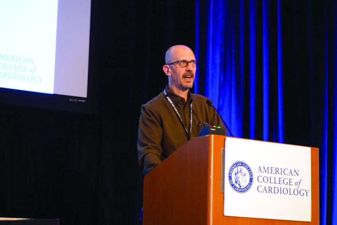
Most recently, he and his coinvestigators utilized CFR to provide new insight into the paradox that women have a higher cardiovascular disease death rate than men, even though their prevalence of obstructive CAD is lower.
Their NIH-sponsored study included 329 consecutive patients with a left ventricular ejection fraction greater than 40% – 43% of them women – who underwent coronary angiography several days after noninvasive assessment of CFR via myocardial perfusion positron emission tomography. The women had a lower burden of angiographic CAD and a lower pretest clinical risk score than the men. Nevertheless, during a median of 3 years of follow-up, the women had an adjusted twofold greater risk of the composite endpoint of cardiovascular death, nonfatal MI, or heart failure.
This excess cardiovascular risk in women was independently associated with a very low CFR, defined as less than 1.6. Dr. Di Carli and his coinvestigators calculated that this impaired CFR mediated 40% of the excess risk in women. Thus, a low CFR represents a novel hidden biologic risk for ischemic heart disease (Circulation. 2017 Feb 7;135[6]:566-77).
CFR is defined as the ratio of absolute coronary flow or myocardial perfusion between drug-induced hyperemia and rest. It can be quantified noninvasively using positron emission tomography or MRI.
CFR integrates into a single measure the three components of CAD: the focal stenosis, the diffuse atherosclerotic plaque typically present to a varying degree throughout a target vessel, and microvascular dysfunction.
CFR is a measure of coronary physiology, as is invasive fractional flow reserve (FFR). However, FFR measures only the severity of stenosis and extent of diffuse disease; it doesn’t assess microvascular dysfunction. This is a limitation because it means FFR can give false-negative readings in patients without significant obstructive coronary disease who have severe microvascular dysfunction.
As for angiography, Dr. Di Carli continued, it’s now evident that this purely anatomic assessment is of limited value as a marker of clinical risk and is inadequate to guide management decisions in the setting of stable CAD. After all, angiographically guided revascularization has not reduced cardiovascular events in clinical trials comparing it with optimal medical therapy, as in the COURAGE and BARI-2D trials.
“It’s clear that there’s been a paradigm shift in how we manage patients with stable CAD. For many years the coronary angiogram was the cornerstone of what we did: how we understand the symptoms, the patient’s risk, and ultimately how we proceed with treatment. But there is no benefit in basing treatment solely on what the lesions look like anatomically. That’s why we’ve turned to functional testing of coronary physiology,” he said.
CFR has opened a window on the importance of microvascular dysfunction, which is present in about half of patients with stable CAD and has been shown to predict cardiovascular risk independent of whether or not severe obstructive disease is present.
In an earlier study, Dr. Di Carli and coworkers demonstrated that quantification of CFR enhances stratification for risk of cardiac death among diabetes patients (Circulation. 2012 Oct 9;126[15]:1858-68). The study included 2,783 patients, of whom 1,172 were diabetic, who underwent measurement of CFR and were subsequently followed for a median of 1.4 years, during which 137 cardiac deaths occurred.
Diabetes patients without known CAD who had a low CFR had a high cardiac death rate of 2.8%/year, similar to the 2.0%/year rate in nondiabetic patients with a history of acute MI or revascularization. On the other hand, diabetes patients with a normal CFR and without known CAD had a cardiac mortality rate of only 0.3%/year, comparable to the 0.5% rate in nondiabetics without known CAD who had preserved systolic function and a normal stress perfusion study.
In the future, CFR may aid in decision making as to whether an individual with stable CAD is best treated by percutaneous coronary intervention, surgical revascularization, or guideline-directed medical therapy. For example, if CFR indicates the presence of an isolated severe focal stenosis, and this is confirmed by angiography and FFR, PCI may be the best option, while diffuse disease as demonstrated by CFR may be better treated surgically or using optimal medical therapy. But this needs to be established in prospective clinical trials, added Dr. Di Carli.
He reported having no financial conflicts regarding his presentation.
SNOWMASS, COLO. – Mounting evidence attests to the value of noninvasive measurement of coronary flow reserve as a means of classifying cardiovascular risk in patients with stable coronary artery disease (CAD) more accurately than is possible via coronary angiography or measurement of fractional flow reserve, Marcelo F. Di Carli, MD, reported at the Annual Cardiovascular Conference at Snowmass.
“We use CFR [coronary flow reserve] as a way to exclude coronary disease. It’s a good practical measure of multivessel ischemic CAD. When the CFR is normal, you can with high confidence exclude the possibility of high-risk CAD,” according to Dr. Di Carli, executive director of the cardiovascular imaging program and chief of the division of nuclear medicine and molecular imaging at Brigham and Women’s Hospital, Boston.
Most recently, he and his coinvestigators utilized CFR to provide new insight into the paradox that women have a higher cardiovascular disease death rate than men, even though their prevalence of obstructive CAD is lower.
Their NIH-sponsored study included 329 consecutive patients with a left ventricular ejection fraction greater than 40% – 43% of them women – who underwent coronary angiography several days after noninvasive assessment of CFR via myocardial perfusion positron emission tomography. The women had a lower burden of angiographic CAD and a lower pretest clinical risk score than the men. Nevertheless, during a median of 3 years of follow-up, the women had an adjusted twofold greater risk of the composite endpoint of cardiovascular death, nonfatal MI, or heart failure.
This excess cardiovascular risk in women was independently associated with a very low CFR, defined as less than 1.6. Dr. Di Carli and his coinvestigators calculated that this impaired CFR mediated 40% of the excess risk in women. Thus, a low CFR represents a novel hidden biologic risk for ischemic heart disease (Circulation. 2017 Feb 7;135[6]:566-77).
CFR is defined as the ratio of absolute coronary flow or myocardial perfusion between drug-induced hyperemia and rest. It can be quantified noninvasively using positron emission tomography or MRI.
CFR integrates into a single measure the three components of CAD: the focal stenosis, the diffuse atherosclerotic plaque typically present to a varying degree throughout a target vessel, and microvascular dysfunction.
CFR is a measure of coronary physiology, as is invasive fractional flow reserve (FFR). However, FFR measures only the severity of stenosis and extent of diffuse disease; it doesn’t assess microvascular dysfunction. This is a limitation because it means FFR can give false-negative readings in patients without significant obstructive coronary disease who have severe microvascular dysfunction.
As for angiography, Dr. Di Carli continued, it’s now evident that this purely anatomic assessment is of limited value as a marker of clinical risk and is inadequate to guide management decisions in the setting of stable CAD. After all, angiographically guided revascularization has not reduced cardiovascular events in clinical trials comparing it with optimal medical therapy, as in the COURAGE and BARI-2D trials.
“It’s clear that there’s been a paradigm shift in how we manage patients with stable CAD. For many years the coronary angiogram was the cornerstone of what we did: how we understand the symptoms, the patient’s risk, and ultimately how we proceed with treatment. But there is no benefit in basing treatment solely on what the lesions look like anatomically. That’s why we’ve turned to functional testing of coronary physiology,” he said.
CFR has opened a window on the importance of microvascular dysfunction, which is present in about half of patients with stable CAD and has been shown to predict cardiovascular risk independent of whether or not severe obstructive disease is present.
In an earlier study, Dr. Di Carli and coworkers demonstrated that quantification of CFR enhances stratification for risk of cardiac death among diabetes patients (Circulation. 2012 Oct 9;126[15]:1858-68). The study included 2,783 patients, of whom 1,172 were diabetic, who underwent measurement of CFR and were subsequently followed for a median of 1.4 years, during which 137 cardiac deaths occurred.
Diabetes patients without known CAD who had a low CFR had a high cardiac death rate of 2.8%/year, similar to the 2.0%/year rate in nondiabetic patients with a history of acute MI or revascularization. On the other hand, diabetes patients with a normal CFR and without known CAD had a cardiac mortality rate of only 0.3%/year, comparable to the 0.5% rate in nondiabetics without known CAD who had preserved systolic function and a normal stress perfusion study.
In the future, CFR may aid in decision making as to whether an individual with stable CAD is best treated by percutaneous coronary intervention, surgical revascularization, or guideline-directed medical therapy. For example, if CFR indicates the presence of an isolated severe focal stenosis, and this is confirmed by angiography and FFR, PCI may be the best option, while diffuse disease as demonstrated by CFR may be better treated surgically or using optimal medical therapy. But this needs to be established in prospective clinical trials, added Dr. Di Carli.
He reported having no financial conflicts regarding his presentation.
EXPERT ANALYSIS FROM THE CARDIOVASCULAR CONFERENCE AT SNOWMASS
Bundled maternal HIV, well-baby visits boost ART adherence
SEATTLE – When new moms can get their well-baby visits and HIV care together in the same office, they have better antiretroviral adherence, better viral suppression, and breast-feed longer, according to a randomized trial of 472 new moms with HIV in Cape Town, South Africa.
“It’s a simple and highly effective strategy for promoting maternal postpartum engagement” in HIV care, said lead investigator Landon Myer, MD, professor and head of epidemiology and biostatistics at the University of Cape Town.

Antiretroviral treatment management is often a routine part of prenatal care, but care splits after birth, with moms generally sent to an adult HIV clinic and babies in follow-up care at the pediatrician’s office. It’s a logistics problem for many, and women tend to prioritize the care of their infants over their own HIV.
“There’s a big push [globally] to identify interventions that can enhance women’s antiretroviral therapy (ART) adherence post partum,” Dr. Myer said.
The investigators had a hunch that bundling care would help. They randomized 234 women to centers with combined HIV and pediatric care within a week of birth and 238 to the usual split care approach. In the latter group, the mothers were referred to adult HIV services soon after delivery.
At 12 months, 77% of the women in the integrated-care group had viral loads below 50 copies/mL, versus 56% of women in the split care group. Women in the integrated group breastfed for about 9 months, versus 3 months in the control group. The findings were statistically significant.
“We were surprised by how big the differences were,” Dr. Myer said at the Conference on Retroviruses & Opportunistic Infections in partnership with the International Antiviral Society.
Mother-to-child transmission was low, at about 0.55%, and did not differ by arm. Vaccination rates, vitamin use, and other infant outcomes were also similar in both groups. Just a few women in each arm dropped out before the 12-month, postpartum visit.
The mothers were a median of 28 years old, and all had started ART during pregnancy at a median of 21 weeks gestation, with a median pre-ART T-cell count of 354 cells/microL. Three-quarters had viral suppression below 50 copies/mL at randomization. About a quarter were giving birth for the first time. Mothers in the bundled-care group were referred back to adult HIV services at the end of breastfeeding.
Dr. Myer had no disclosures. The work was funded by the National Institutes of Health.
SEATTLE – When new moms can get their well-baby visits and HIV care together in the same office, they have better antiretroviral adherence, better viral suppression, and breast-feed longer, according to a randomized trial of 472 new moms with HIV in Cape Town, South Africa.
“It’s a simple and highly effective strategy for promoting maternal postpartum engagement” in HIV care, said lead investigator Landon Myer, MD, professor and head of epidemiology and biostatistics at the University of Cape Town.
Antiretroviral treatment management is often a routine part of prenatal care, but care splits after birth, with moms generally sent to an adult HIV clinic and babies in follow-up care at the pediatrician’s office. It’s a logistics problem for many, and women tend to prioritize the care of their infants over their own HIV.
“There’s a big push [globally] to identify interventions that can enhance women’s antiretroviral therapy (ART) adherence post partum,” Dr. Myer said.
The investigators had a hunch that bundling care would help. They randomized 234 women to centers with combined HIV and pediatric care within a week of birth and 238 to the usual split care approach. In the latter group, the mothers were referred to adult HIV services soon after delivery.
At 12 months, 77% of the women in the integrated-care group had viral loads below 50 copies/mL, versus 56% of women in the split care group. Women in the integrated group breastfed for about 9 months, versus 3 months in the control group. The findings were statistically significant.
“We were surprised by how big the differences were,” Dr. Myer said at the Conference on Retroviruses & Opportunistic Infections in partnership with the International Antiviral Society.
Mother-to-child transmission was low, at about 0.55%, and did not differ by arm. Vaccination rates, vitamin use, and other infant outcomes were also similar in both groups. Just a few women in each arm dropped out before the 12-month, postpartum visit.
The mothers were a median of 28 years old, and all had started ART during pregnancy at a median of 21 weeks gestation, with a median pre-ART T-cell count of 354 cells/microL. Three-quarters had viral suppression below 50 copies/mL at randomization. About a quarter were giving birth for the first time. Mothers in the bundled-care group were referred back to adult HIV services at the end of breastfeeding.
Dr. Myer had no disclosures. The work was funded by the National Institutes of Health.
AT CROI
Key clinical point:
Major finding: At 12 months, 77% of the women in the integrated-care group had viral loads below 50 copies/mL, versus 56% of women in the control arm. Women in the integrated group breastfed for about 9 months, versus 3 months in the split-care group.
Data source: A randomized trial of 472 new moms with HIV and their babies in Cape Town, South Africa.
Disclosures: Dr. Myer had no disclosures. The work was funded by the National Institutes of Health.
