Coronary heart disease remains the leading cause of death of US adults aged 35 years and older due to the downstream consequences of arterial occlusion and ensuing myocardial ischemia.1,2 Among patients with coronary heart disease, 30% to 33% will present with ST-segment elevation myocardial infarction (STEMI).3-5
For STEMI patients, access to a facility with percutaneous coronary intervention (PCI) capabilities is critical. Door-to-balloon (D2B) time is used as a performance measure to assess the quality of care that STEMI patients are receiving. Data show that decreasing D2B times can improve outcomes of STEMI patients, with lower D2B time associated with lower mortality rates.5 To obtain and maintain the lowest D2B times possible, health care systems must adopt best practice models to continually improve patient outcomes.
The current best practice for patients with an acute STEMI involves bypassing non-PCI centers and transporting patients directly to PCI-enabled centers—as long as the patient’s total emergency medical services (EMS) contact-to-balloon time remains low. In a study by Le May et al,6 patients directly transported to a PCI-enabled center had a mortality rate of 5% within 180 days compared to 11.5% for those who were first seen at a center without PCI capabilities.
Longer EMS transport time to PCI centers, however, requires earlier cardiac catheterization laboratory (CCL) activation to prevent delays in care. Emergency physicians (EPs) must work with local EMS, PCI and non-PCI centers, and interventional cardiologists to establish protocols to expedite STEMI patient care. Previous studies, however, have shown that paramedics are able to diagnose STEMI patients during transport and can activate the catheterization team from the field, with beneficial patient outcomes.7-11 Prior to July 2011, all patients with a suspected MI who were transported to our institution via EMS were not diagnosed with STEMI until evaluation by an EP. Because the CCL had no in-house staff after hours, the interventional cardiology team required 30 minutes to respond and prepare. In 2011, our EMS, in conjunction with county hospitals, EPs, and cardiologists, implemented an initiative that empowered paramedics to activate STEMI alerts from the field. This provided advanced notification to the interventional cardiology laboratory team to initiate preparation for patients in need of immediate PCI.
Since the best practice for STEMI patients involves early diagnosis and immediate activation of the cardiac catheterization team, we sought to improve the outcomes for STEMI patients by utilizing prehospital care initiatives to lower D2B times. Other health care systemshave utilized EPs to receive the transmitted electrocardiogram (ECG) and to initiate CCL team activation. We found that having both the EP and cardiologist review the ECG delayed the activation process when compared to direct activation by EMS.Our initiatives to improve time to CCL activation included the following:
(1) Training and enabling paramedics to diagnose a STEMI on the ECG;
(2) Bypassing non-PCI centers for PCI centers during STEMI patient transport;
(3) Allowing paramedics to activate the CCL at our institution prior to transmitting an ECG and prior to patient arrival at the hospital; and
(4) Bypassing the ED to go directly to the CCL when the CCL was ready to receive the patient. (When the CCL was not ready, patients were transported to the ED. After reviewing the transmitted ECG, the EPs could cancel the STEMI alert if indicated.)
By empowering EMS to diagnose and activate the CCL, we also measured the rate of unnecessary activations, which was defined as patients who did not present with chest pain and an ECG showing ≥1 mm ST segment elevation in two contiguous limb leads, or ≥2 mm ST segment elevation in two contiguous precordial leads.
Causes of unnecessary activation by EMS included left ventricular hypertrophy, left bundle branch block, early repolarization, non-specific ST segment changes, pericarditis, and ventricular paced rhythms. We recorded and analyzed D2B times and the unnecessary activation rates, and continuously used these data for quality improvement. Our goal was to decrease D2B times and unnecessary activation rates, while maintaining these improvements over time.
Our ED has 93,000 patient visits annually, and our hospital serves as the only PCI center within a county encompassing 1,255 square miles. Our hospital EMS used 12 Lifepak monitors to help interpret patients’ ECG. To study the impact of our initiative, data were collected each year from 2011 to 2015. The 2011 data were recorded before EMS began to activate the CCL and served as the baseline for the D2B times. These data were then compared to postintervention D2B data from 2012 to 2015.
In addition to collecting unnecessary activation rates by EMS paramedics from 2012 to 2015, we continuously used the unnecessary activation data to identify why these activations were occurring and to devise a strategy to improve rates. Emergency physicians have a leadership role regarding EMS training and oversight, ongoing paramedic education, and the quality improvement process.
A Reduction in D2B Time
Our baseline D2B time prior to the intervention was 52.5 minutes. After our intervention, average D2B times for 2012, 2013, 2014, and 2015 were 38.2, 33.5, 39, and 37.25 minutes, respectively. The institution’s baseline rate of unnecessary activation prior to intervention in 2011 was 19.1%. After intervention, the EMS unnecessary activation rates for 2012, 2013, 2014, and 2015 were 30%, 18%, 15%, and 19% respectively, averaging 20.5% over 4 years, or 17.3% for 2013 through 2015.
Emergency medical services averaged approximately 164 STEMI activations each year. We decreased D2B times for patients by over 15 minutes from 2011 compared to 2012 through 2015. These results were sustained, with attention to metrics, over four years. Furthermore, the proportion of STEMI patients receiving immediate PCI within the recommended 90 minutes was 100% of patients in 2014 and 2015, reflecting consistent improvement over 2012 and 2013.
We also demonstrated an overall reduction in the unnecessary CCL activation rate by EMS over the time of our intervention. Our analysis of unnecessary activation causes and EMS education led to improvement in the unnecessary activation rate from 2012-2015, and these rates have been sustained over time. The rate of unnecessary activation by EMS in 2012 to 2015 was 20.5%.
Conclusion
Lower D2B time for STEMI patients is associated with lower mortality following PCI for patients with acute MI, fewer complications, and shorter length of stay in the hospital. We successfully lowered D2B time after empowering EMS to activate our interventional cardiology laboratory prior to arrival at the hospital for patients with acute STEMI, coupled with the ability to bypass the ED for patients with acute STEMI presentations. With rigorous attention to metrics and ongoing aggressive medic education, we were able to achieve consistent, sustained D2B times under 40 minutes over the course of four consecutive years. We present our experience as a potential model of a multifaceted intervention for other systems to consider replicating.
References
1. Fosbol EL, Granger CB, Jollis JG, et al. The impact of a statewide pre-hospital STEMI strategy to bypass hospitals without percutaneous coronary intervention capability on treatment times. Circulation. 2013;127(5):604-612. doi:10.1161/CIRCULATIONAHA.112.118463. 2. Mozaffarian D, Benjamin EJ, Go AS, et al; American Heart Association Statistics Committee; Stroke Statistics Subcommittee. Executive summary: heart disease and stroke statistics—2016 update: a report from the American Heart Association. Circulation. 2016;133(4):447-454. doi:10.1161/CIR.0000000000000366. 3. Hasdai D, Behar S, Wallentin L, et al. A prospective survey of the characteristics, treatments and outcomes of patients with acute coronary syndromes in Europe and the Mediterranean basin; the Euro Heart Survey of Acute Coronary Syndromes (Euro Heart Survey ACS). Eur Heart J. 2002;23(15):1190-1201. 4. Fox KA, Goodman SG, Klein W, et al. Management of acute coronary syndromes. Variations in practice and outcome; findings from the Global Registry of Acute Coronary Events (GRACE). Eur Heart J. 2002;23(15)1177-1189. 5. McNamara RL, Wang Y, Herrin J, et al. Effect of door-to-balloon time on mortality in patients with ST-segment elevation myocardial infarction. J Am Coll Cardiol. 2006;47(11):2180-2186. doi:10.1016/j.jacc.2005.12.072. 6. Le May MR, Wells GA, So DY, et al. Reduction in mortality as a result of direct transport from the field to a receiving center for primary percutaneous coronary intervention. J Am Coll Cardiol.2012;60(14):1223-1230. doi:10.1016/j.jacc.2012.07.008. 7. Camp-Rogers T, Dante S, Kontos MC, Roberts CS, Kreisa L, Kurz MC. The impact of prehospital activation of the cardiac catheterization team on time to treatment for patients presenting with ST-segment-elevation myocardial infarction. Am J Emerg Med. 2011;29(9):1117-1124. doi:10.1016/j.ajem.2010.08.005. 8. Bates ER, Jacobs AK. Time to treatment in patients with STEMI. N Engl J Med. 2013;369(10):889-892. doi:10.1056/NEJMp1308772. 9. Franco E, Mateos A, Acebal C, et al. Prehospital activation of cardiac catheterization teams in ST-segment elevation myocardial infarction. Rev Port Cardiol. 2014;33(9):545-553. doi:10.1016/j.repc.2014.03.007. 10. Hammond BB. Four steps to reducing door-to-balloon time. J Emerg Nurs. 2010;36(3):217-220. doi:10.1016/j.jen.2009.05.019. 11. Mumma BE, Kontos MC, Peng SA, Diercks DB. Association between prehospital electrocardiogram use and patient home distance from the percutaneous coronary intervention center on total reperfusion time in ST-segment-elevation myocardial infarction patients: a retrospective analysis from the national cardiovascular data registry. Am Heart J. 2014;167(6):915-920. doi:10.1016/j.ahj.2014.03.014.
Our institution lowered door-to-balloon times by empowering EMS to activate the cardiology laboratory prior to arrival at the hospital.
Our institution lowered door-to-balloon times by empowering EMS to activate the cardiology laboratory prior to arrival at the hospital.
Coronary heart disease remains the leading cause of death of US adults aged 35 years and older due to the downstream consequences of arterial occlusion and ensuing myocardial ischemia.1,2 Among patients with coronary heart disease, 30% to 33% will present with ST-segment elevation myocardial infarction (STEMI).3-5
For STEMI patients, access to a facility with percutaneous coronary intervention (PCI) capabilities is critical. Door-to-balloon (D2B) time is used as a performance measure to assess the quality of care that STEMI patients are receiving. Data show that decreasing D2B times can improve outcomes of STEMI patients, with lower D2B time associated with lower mortality rates.5 To obtain and maintain the lowest D2B times possible, health care systems must adopt best practice models to continually improve patient outcomes.
The current best practice for patients with an acute STEMI involves bypassing non-PCI centers and transporting patients directly to PCI-enabled centers—as long as the patient’s total emergency medical services (EMS) contact-to-balloon time remains low. In a study by Le May et al,6 patients directly transported to a PCI-enabled center had a mortality rate of 5% within 180 days compared to 11.5% for those who were first seen at a center without PCI capabilities.
Longer EMS transport time to PCI centers, however, requires earlier cardiac catheterization laboratory (CCL) activation to prevent delays in care. Emergency physicians (EPs) must work with local EMS, PCI and non-PCI centers, and interventional cardiologists to establish protocols to expedite STEMI patient care. Previous studies, however, have shown that paramedics are able to diagnose STEMI patients during transport and can activate the catheterization team from the field, with beneficial patient outcomes.7-11 Prior to July 2011, all patients with a suspected MI who were transported to our institution via EMS were not diagnosed with STEMI until evaluation by an EP. Because the CCL had no in-house staff after hours, the interventional cardiology team required 30 minutes to respond and prepare. In 2011, our EMS, in conjunction with county hospitals, EPs, and cardiologists, implemented an initiative that empowered paramedics to activate STEMI alerts from the field. This provided advanced notification to the interventional cardiology laboratory team to initiate preparation for patients in need of immediate PCI.
Since the best practice for STEMI patients involves early diagnosis and immediate activation of the cardiac catheterization team, we sought to improve the outcomes for STEMI patients by utilizing prehospital care initiatives to lower D2B times. Other health care systemshave utilized EPs to receive the transmitted electrocardiogram (ECG) and to initiate CCL team activation. We found that having both the EP and cardiologist review the ECG delayed the activation process when compared to direct activation by EMS.Our initiatives to improve time to CCL activation included the following:
(1) Training and enabling paramedics to diagnose a STEMI on the ECG;
(2) Bypassing non-PCI centers for PCI centers during STEMI patient transport;
(3) Allowing paramedics to activate the CCL at our institution prior to transmitting an ECG and prior to patient arrival at the hospital; and
(4) Bypassing the ED to go directly to the CCL when the CCL was ready to receive the patient. (When the CCL was not ready, patients were transported to the ED. After reviewing the transmitted ECG, the EPs could cancel the STEMI alert if indicated.)
By empowering EMS to diagnose and activate the CCL, we also measured the rate of unnecessary activations, which was defined as patients who did not present with chest pain and an ECG showing ≥1 mm ST segment elevation in two contiguous limb leads, or ≥2 mm ST segment elevation in two contiguous precordial leads.
Causes of unnecessary activation by EMS included left ventricular hypertrophy, left bundle branch block, early repolarization, non-specific ST segment changes, pericarditis, and ventricular paced rhythms. We recorded and analyzed D2B times and the unnecessary activation rates, and continuously used these data for quality improvement. Our goal was to decrease D2B times and unnecessary activation rates, while maintaining these improvements over time.
Our ED has 93,000 patient visits annually, and our hospital serves as the only PCI center within a county encompassing 1,255 square miles. Our hospital EMS used 12 Lifepak monitors to help interpret patients’ ECG. To study the impact of our initiative, data were collected each year from 2011 to 2015. The 2011 data were recorded before EMS began to activate the CCL and served as the baseline for the D2B times. These data were then compared to postintervention D2B data from 2012 to 2015.
In addition to collecting unnecessary activation rates by EMS paramedics from 2012 to 2015, we continuously used the unnecessary activation data to identify why these activations were occurring and to devise a strategy to improve rates. Emergency physicians have a leadership role regarding EMS training and oversight, ongoing paramedic education, and the quality improvement process.
A Reduction in D2B Time
Our baseline D2B time prior to the intervention was 52.5 minutes. After our intervention, average D2B times for 2012, 2013, 2014, and 2015 were 38.2, 33.5, 39, and 37.25 minutes, respectively. The institution’s baseline rate of unnecessary activation prior to intervention in 2011 was 19.1%. After intervention, the EMS unnecessary activation rates for 2012, 2013, 2014, and 2015 were 30%, 18%, 15%, and 19% respectively, averaging 20.5% over 4 years, or 17.3% for 2013 through 2015.
Emergency medical services averaged approximately 164 STEMI activations each year. We decreased D2B times for patients by over 15 minutes from 2011 compared to 2012 through 2015. These results were sustained, with attention to metrics, over four years. Furthermore, the proportion of STEMI patients receiving immediate PCI within the recommended 90 minutes was 100% of patients in 2014 and 2015, reflecting consistent improvement over 2012 and 2013.
We also demonstrated an overall reduction in the unnecessary CCL activation rate by EMS over the time of our intervention. Our analysis of unnecessary activation causes and EMS education led to improvement in the unnecessary activation rate from 2012-2015, and these rates have been sustained over time. The rate of unnecessary activation by EMS in 2012 to 2015 was 20.5%.
Conclusion
Lower D2B time for STEMI patients is associated with lower mortality following PCI for patients with acute MI, fewer complications, and shorter length of stay in the hospital. We successfully lowered D2B time after empowering EMS to activate our interventional cardiology laboratory prior to arrival at the hospital for patients with acute STEMI, coupled with the ability to bypass the ED for patients with acute STEMI presentations. With rigorous attention to metrics and ongoing aggressive medic education, we were able to achieve consistent, sustained D2B times under 40 minutes over the course of four consecutive years. We present our experience as a potential model of a multifaceted intervention for other systems to consider replicating.
References
1. Fosbol EL, Granger CB, Jollis JG, et al. The impact of a statewide pre-hospital STEMI strategy to bypass hospitals without percutaneous coronary intervention capability on treatment times. Circulation. 2013;127(5):604-612. doi:10.1161/CIRCULATIONAHA.112.118463. 2. Mozaffarian D, Benjamin EJ, Go AS, et al; American Heart Association Statistics Committee; Stroke Statistics Subcommittee. Executive summary: heart disease and stroke statistics—2016 update: a report from the American Heart Association. Circulation. 2016;133(4):447-454. doi:10.1161/CIR.0000000000000366. 3. Hasdai D, Behar S, Wallentin L, et al. A prospective survey of the characteristics, treatments and outcomes of patients with acute coronary syndromes in Europe and the Mediterranean basin; the Euro Heart Survey of Acute Coronary Syndromes (Euro Heart Survey ACS). Eur Heart J. 2002;23(15):1190-1201. 4. Fox KA, Goodman SG, Klein W, et al. Management of acute coronary syndromes. Variations in practice and outcome; findings from the Global Registry of Acute Coronary Events (GRACE). Eur Heart J. 2002;23(15)1177-1189. 5. McNamara RL, Wang Y, Herrin J, et al. Effect of door-to-balloon time on mortality in patients with ST-segment elevation myocardial infarction. J Am Coll Cardiol. 2006;47(11):2180-2186. doi:10.1016/j.jacc.2005.12.072. 6. Le May MR, Wells GA, So DY, et al. Reduction in mortality as a result of direct transport from the field to a receiving center for primary percutaneous coronary intervention. J Am Coll Cardiol.2012;60(14):1223-1230. doi:10.1016/j.jacc.2012.07.008. 7. Camp-Rogers T, Dante S, Kontos MC, Roberts CS, Kreisa L, Kurz MC. The impact of prehospital activation of the cardiac catheterization team on time to treatment for patients presenting with ST-segment-elevation myocardial infarction. Am J Emerg Med. 2011;29(9):1117-1124. doi:10.1016/j.ajem.2010.08.005. 8. Bates ER, Jacobs AK. Time to treatment in patients with STEMI. N Engl J Med. 2013;369(10):889-892. doi:10.1056/NEJMp1308772. 9. Franco E, Mateos A, Acebal C, et al. Prehospital activation of cardiac catheterization teams in ST-segment elevation myocardial infarction. Rev Port Cardiol. 2014;33(9):545-553. doi:10.1016/j.repc.2014.03.007. 10. Hammond BB. Four steps to reducing door-to-balloon time. J Emerg Nurs. 2010;36(3):217-220. doi:10.1016/j.jen.2009.05.019. 11. Mumma BE, Kontos MC, Peng SA, Diercks DB. Association between prehospital electrocardiogram use and patient home distance from the percutaneous coronary intervention center on total reperfusion time in ST-segment-elevation myocardial infarction patients: a retrospective analysis from the national cardiovascular data registry. Am Heart J. 2014;167(6):915-920. doi:10.1016/j.ahj.2014.03.014.
A 29-year-old man presented to the ED with a 3-day history of constant left-sided low back pain that radiated to his left buttock and groin. The patient stated the pain worsened with movement, making it difficult for him to walk. He reported lifting heavy boxes at work, but denied any trauma. The patient also denied recent fevers, chills, chest pain, dyspnea, abdominal pain, urinary or fecal incontinence, weakness, numbness, or saddle anesthesia. Regarding his medical history, he had an appendectomy as a child, but reported no other surgeries or medical issues. His social history was significant for narcotic and inhalant use and daily tobacco use. The patient also reported taking heroin intravenously (IV) 6 months prior.
Vital signs at presentation were: heart rate (HR), 92 beats/min; respiratory rate, 15 breaths/min; blood pressure, 118/80 mm Hg; and temperature, 98.2°F. Oxygen saturation was 98% on room air.
The patient was a well-developed young man in no apparent distress. Dermatological examination showed bilateral track marks in the antecubital fossa. The musculoskeletal (MSK) examination demonstrated left gluteal tenderness to palpation and decreased active and passive range of motion of the left hip, especially with internal rotation and flexion. He had no midline tenderness, and the lower extremities had normal pulses and no motor or sensory deficits.
The patient’s pain improved with IV fluids, diazepam, and ketorolac, and he was able to ambulate with assistance. He was clinically diagnosed with sciatica, and discharged home with prescriptions for diazepam and ibuprofen. He was also instructed to follow-up with an orthopedist within 7 days from discharge.
The patient returned to the ED the following day with similar complaints of unabating left-sided pain and difficulty ambulating. His vital signs were notable for an elevated HR of 106 beats/min. Physical examination findings were unchanged from his presentation the previous day, and an X-ray of the lumbar spine showed no abnormalities.
After receiving IV analgesics, the patient’s pain improved and his tachycardia resolved. He was discharged home with instructions to continue taking diazepam, and was also given prescriptions for prednisone and oxycodone/acetaminophen. He was instructed to follow-up with an orthopedist within 24 hours.
Over the next 9 days, the patient was seen twice by an orthopedist, who ordered imaging of the lumbar spine, including a repeat X-ray and contrast-enhanced magnetic resonance imaging (MRI), both of which were unremarkable. The patient completed the prescribed course of diclofenac, oxycodone/acetaminophen, and prednisone, but experienced only minimal pain relief.The orthopedist prescribed the diclofenac to supplement the medication regimen that he was already on.
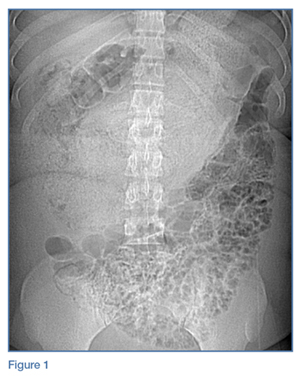
At the second follow-up visit, the orthopedist ordered an MRI of the patient’s left hip, which demonstrated inflammation of the left sacroiliac joint (SIJ) with effusion, and a 1-cm by 1-cm collection adjacent to the left psoas muscle; these findings were concerning for septic arthritis (Figure). Based on the MRI study, a computed tomography (CT)-guided arthrocentesis of the left SIJ was performed by an interventional radiologist.
Figure Following the arthrocentesis, the orthopedist referred the patient to the ED. At this presentation,the emergency physician (EP) ordered blood cultures, blood work, urinalysis, and a urinary toxicology screen, and started the patient on IV ceftriaxone and vancomycin. The laboratory studies were significant for the following elevated inflammatory markers: erythrocyte sedimentation rate (ESR), 19 mm/h; C-reactive protein (CRP), 2.45 mg/L; white blood cell count (WBC), 13.6 K/uL with normal differential; and lactate level, 2.6 mg/dL. The toxicology screen was positive for opioids. The basic metabolic panel, chest X-ray, and urinalysis were all unremarkable. An electrocardiogram showed sinus tachycardia.
The patient was admitted to the hospital, and infectious disease services was contacted. While awaiting transport to the inpatient floor, the patient admitted to IV drug use 4 weeks prior to his initial presentation—not the 6 months he initially reported at the first ED visit.
The blood cultures grew Candida parapsilosis, and culture from the SIJ arthrocentesis grew Pseudomonas aeruginosa. The infectious disease physician switched the patient’s antibiotic therapy to IV cefepime and fluconazole. The patient also was seen by an orthopedist, who determined that no surgical intervention was required.
Follow-up laboratory studies showed inflammatory markers peaking at the following levels: ESR, 36 mm/h; CRP, 4.84 mg/L; and WBC, 32.1 K/uL with 90% neutrophils. These markers normalized throughout his hospital stay. The patient was also tested for hepatitis and human immunodeficiency virus, both of which were negative. A transesophageal echocardiogram showed no obvious masses or vegetations.
The patient had an uncomplicated hospital course, and was discharged home on hospital day 6 with a 4-week prescription of oral fluconazole and levofloxacin, and instructed to follow-up with both infectious disease and the orthopedist. To address his history of IV drug use, he also was given follow-up with pain management.
One month later, the patient returned a fourth time to the ED for evaluation of bilateral lower extremity pain and swelling. He stated that he had been mostly bed-bound at home since his discharge from the hospital due to continued pain with weight-bearing.
The patient’s vital signs were normal. The EP ordered a duplex ultrasound study, which showed extensive bilateral lower extremity deep vein thrombosis. He was started on subcutaneous therapeutic enoxaparin and admitted to the inpatient hospital. During admission,a left lower lobe pulmonary artery embolism was found on chest CT angiography, though he had no cardiac or respiratory symptoms. He was discharged home with a 3-month prescription for oral rivaroxaban.
At a 4-month follow-up visit, the patient reported minimal residual disability after completing the course of treatment. During the follow-up, the patient denied using IV heroin; he was referred to a pain management specialist, who placed the patient on methadone.
Discussion
Infectious sacroiliitis (ISI) is a rare form of infectious arthritis affecting the SIJ, with an incidence of 1 to 2 reported cases per year.1 The literature on ISI currently consists only of case reports and case series. This infection is often diagnosed after the disease has progressed, with a mean time to diagnosis of 43.3 days.2
Infectious arthritis of any joint has a prevalence of 2 to 10 per 100,000 people. In 50% of cases, the knee is the joint most commonly affected, followed by the hip, shoulder, and elbow.3 Regardless of location, infectious arthritis is associated with significant morbidity and mortality due to sepsis and irreversible loss of joint function.4
Risk factors for ISI include IV drug use, pregnancy, trauma, endocarditis, and immunosuppression.1 The decision to initiate the workup for ISI can be difficult to make because the condition may present without signs of an infectious etiology, such as toxic appearance, inflammatory changes surrounding the joint, or even fever—only 41% of affected patients in one case series were febrile.2 The workup is often time-consuming, invasive, and expensive.
Although delayed diagnosis and treatment of septic arthritis is associated with significant adverse effects, there is unfortunately no consensus to guide the workup for ISI. As opposed to Kocher’s criteria for the differentiation of septic hip arthritis from transient synovitis in pediatric patients or well-known red-flags for further evaluation of low back pain, physicians are left without much guidance when considering laboratory workup or imaging decisions to evaluate for ISI.
Sacroiliac Joint
As previously noted, the SIJ is not commonly affected by infection. It is a diarthrodial, L-shaped joint comprised of the posterior ilium and sacrum, and is a near-rigid structure with very limited movement that provides stability to the axial skeleton.5 The SIJ is often overlooked as a secondary cause of low back pain in younger patients with rheumatologic conditions (eg, ankylosing spondylitis, Reiter syndrome), pregnancy-associated ligamentous laxity, and osteoarthritis in elderly patients. In one study, 88.2% of sacroiliitis cases were inflammatory, 8.8% infectious, and 2.9% degenerative.6
Signs and Symptoms
As our case illustrates, ISI often presents with nonspecific symptoms and physical findings.7 Patients typically present with fever, painful manipulation of the SIJ, and unilateral lumbo-gluteal pain.2 The components of the history and physical examination suspicious for an infectious etiology include the subacute presentation; unresolved pain despite treatment; tenderness to palpation; decreased range of motion; and recent IV drug use, which increases the risk of infectious disease due to unsterile practices and direct inoculation of pathogens into the bloodstream8 and a further predilection into the axial skeleton. 9 It is important to obtain an accurate social history; however, patients may not be forthright about disclosing sensitive information such as sexual history and illicit drug use.
Physical Assessment
The SIJ is best appreciated in the seated patient by palpating one fingerbreadth medial to the posterior superior iliac spine as he or she slowly bends forward.10 Tenderness elicited while in this position is suggestive of SIJ inflammation. The area of tenderness may be lower than anticipated and lateral to the gluteal cleft, as synovial fluid is typically relegated to the lower half of the joint.
Several adjunctive physical examination maneuvers, such as the Gaenslen test and Flexion Abduction External Rotation test (FABER test or Patrick’s test) can isolate SIJ pathology or dysfunction. The Gaenslen test is performed by asking the patient to lie supine and flex the affected hip and knee, with the lumbar spine flat against the examination table. Hyperextending the contralateral thigh downward will reproduce pain in the affected SIJ.
The FABER test is a simple but less specific examination technique to assess joint pain in the hip, lumbar, and sacroiliac joints.11 In this assessment, the clinician flexes the patient’s affected knee to 90°, externally rotates the hip, and applies downward pressure on the knee. Pain reproduced in the affected SI region is sensitive for joint inflammation.
Laboratory and Imaging Studies
Laboratory studies typically show inconsistent and nonspecific findings, such as the elevated ESR and CRP levels seen in our patient.2,12 Imaging studies to assess the SIJ for signs of infection are therefore essential for confirming infection.
Magnetic resonance imaging is the preferred imaging modality to assess for ISI, since it has the highest sensitivity in visualizing joint effusion and bone marrow edema compared to other modalities. Computed tomography, however, can be helpful in visualizing associated abscesses and guiding arthrocentesis.12 Plain X-ray may not demonstrate early changes in bone.13 The confirmatory study for ISI is synovial fluid analysis and culture.7
Treatment
Infectious sacroiliitis secondary to P aeruginosa, a gram-negative bacillus, is difficult to treat because of the glycocalyx and slime production that protects the pathogen from antibiotics, the development of multiple-antimicrobial resistance, and poor drug penetration into bones and abscesses.14 Antibiotic treatment should cover Staphylococcus aureus and may be broadened to cover gram-negative bacilli. The recommended duration of treatment is at least a 2-week course of IV antibiotics, followed by a 6-week course of oral antibiotics.2 Therapy also includes pain control and surgical intervention for abscesses, osteomyelitis, and refractory cases.7
Complications
Complications and long-term sequelae are common in ISI, often due to late diagnosis of the condition.Our case illustrates the delayed diagnosis of Pseudomonas ISI with candidemia in a young man with a history of IV drug use presenting with atraumatic low back pain. His clinical course was complicated by a thromboembolic event, likely secondary to immobility and a hypercoagulable state from infection and inflammation.15 Infectious sacroiliitis secondary to P aeruginosa is most commonly seen in patients with immunosuppression, hospitalization, and IV drug use.2
Summary
Infectious sacroiliitis remains a diagnostic challenge for physicians due to its rare incidence and nonspecific clinical manifestations. Our case illustrates the importance of maintaining a high level of clinical suspicion for infectious arthritis in young patients presenting with common MSK complaints in the presence of infectious risk factors. Emergency physicians should consider red flags, abnormal vital signs, and patient recidivism when deciding on the most appropriate workup.
References
1. Mancarella L, De Santis M, Magarelli N, Ierardi AM, Bonomo L, Ferraccioli G. Septic sacroiliitis: an uncommon septic arthritis. Clin Exp Rheumatol. 2009;27(6):1004-1008. 2. Hermet M, Minichiello E, Flipo RM, et al. Infectious sacroiliitis: a retrospective, multicentre study of 39 adults. BMC Infect Dis. 2012;12:305.doi:10.1186/1471-2334-12-305. 3. Abelson A. Septic Arthritis. Cleveland Clinic. http://www.clevelandclinicmeded.com/medicalpubs/diseasemanagement/rheumatology/septic-arthritis. Published August 2010. Accessed October 28, 2016. 4. Goldenberg DL. Septic arthritis. Lancet. 1998;351(9097):197-202. doi:10.1016/S0140-6736(97)09522-6. 5. Vleeming A, Schuenke MD, Masi AT, Carreiro JE, Danneels L, Willard FH. The sacroiliac joint: an overview of its anatomy, function and potential clinical implications. J Anat. 2012;221(6):537-567. doi:10.1111/j.1469-7580.2012.01564.x. 6. Owlia MB, Danesh-Ardakani M. Frequency of sacroiliitis among patients with low back pain. Electron Physician. 2016;8(3):2094-2100. doi:10.19082/2094. 7. Zimmermann B 3rd, Mikolich DJ, Lally EV. Septic sacroiliitis. Semin Arthritis Rheum. 1996;26(3):592-604. 8. Brtalik D, Pariyadath M. A case report of infectious sacroiliitis in an adult presenting to the emergency department with inability to walk. J Emerg Med. 2017:52(3)e65-e68. doi:10.1016/j.jemermed.2016.10.022. 9. Ferraro K, Cohen MA. Acute septic sacroiliitis in an injection drug user. Am J Emerg Med. 2004;22(1):60-61. 10. Safran M, Botser IB. Hip anatomy and biomechanics. In: Miller MD, Thompson SR, eds. DeLee & Drez’s Orthopaedic Sports Medicine. Vol 2. 4th ed. Philadelphia, PA: Elsevier Saunders; 2015:917-932.e1. 11. LeBlond RF, Brown DD, Suneja M, Szot JF. The spine, pelvic, and extremities. In: LeBlond RF, Brown DD, Suneja M, Szot JF. eds. DeGowin’s Diagnostic Examination. 10th ed. New York, NY: McGraw-Hill; 2015:508-576. 12. Scott KR, Rising KL, Conlon LW. Infectious sacroiliitis. J Emerg Med. 2014;47(3):83-84. doi:10.1016/j.jemermed.2014.05.001. 13. Cinar M, Sanal HT, Yilmaz S, et al. Radiological followup of the evolution of inflammatory process in sacroiliac joint with magnetic resonance imaging: a case with pyogenic sacroiliitis. Case Rep Rheumatol. 2012;2012:509136. doi:10.1155/2012/509136. 14. Calza L, Manfredi R, Marinacci G, Fortunato L, Chiodo F. Community-acquired Pseudomonas aeruginosa sacro-iliitis in a previously healthy patient. J Med Microbiol. 2002;51(7):620-622. 15. Levi M, Keller TT, van Gorp E, ten Cate H. Infection and inflammation and the coagulation system. Cardiovasc Res. 2003;60(1):26-39.
A 29-year-old man presented for evaluation of unabating left-sided low back pain that radiated to his left buttock and groin.
A 29-year-old man presented for evaluation of unabating left-sided low back pain that radiated to his left buttock and groin.
Case
A 29-year-old man presented to the ED with a 3-day history of constant left-sided low back pain that radiated to his left buttock and groin. The patient stated the pain worsened with movement, making it difficult for him to walk. He reported lifting heavy boxes at work, but denied any trauma. The patient also denied recent fevers, chills, chest pain, dyspnea, abdominal pain, urinary or fecal incontinence, weakness, numbness, or saddle anesthesia. Regarding his medical history, he had an appendectomy as a child, but reported no other surgeries or medical issues. His social history was significant for narcotic and inhalant use and daily tobacco use. The patient also reported taking heroin intravenously (IV) 6 months prior.
Vital signs at presentation were: heart rate (HR), 92 beats/min; respiratory rate, 15 breaths/min; blood pressure, 118/80 mm Hg; and temperature, 98.2°F. Oxygen saturation was 98% on room air.
The patient was a well-developed young man in no apparent distress. Dermatological examination showed bilateral track marks in the antecubital fossa. The musculoskeletal (MSK) examination demonstrated left gluteal tenderness to palpation and decreased active and passive range of motion of the left hip, especially with internal rotation and flexion. He had no midline tenderness, and the lower extremities had normal pulses and no motor or sensory deficits.
The patient’s pain improved with IV fluids, diazepam, and ketorolac, and he was able to ambulate with assistance. He was clinically diagnosed with sciatica, and discharged home with prescriptions for diazepam and ibuprofen. He was also instructed to follow-up with an orthopedist within 7 days from discharge.
The patient returned to the ED the following day with similar complaints of unabating left-sided pain and difficulty ambulating. His vital signs were notable for an elevated HR of 106 beats/min. Physical examination findings were unchanged from his presentation the previous day, and an X-ray of the lumbar spine showed no abnormalities.
After receiving IV analgesics, the patient’s pain improved and his tachycardia resolved. He was discharged home with instructions to continue taking diazepam, and was also given prescriptions for prednisone and oxycodone/acetaminophen. He was instructed to follow-up with an orthopedist within 24 hours.
Over the next 9 days, the patient was seen twice by an orthopedist, who ordered imaging of the lumbar spine, including a repeat X-ray and contrast-enhanced magnetic resonance imaging (MRI), both of which were unremarkable. The patient completed the prescribed course of diclofenac, oxycodone/acetaminophen, and prednisone, but experienced only minimal pain relief.The orthopedist prescribed the diclofenac to supplement the medication regimen that he was already on.
At the second follow-up visit, the orthopedist ordered an MRI of the patient’s left hip, which demonstrated inflammation of the left sacroiliac joint (SIJ) with effusion, and a 1-cm by 1-cm collection adjacent to the left psoas muscle; these findings were concerning for septic arthritis (Figure). Based on the MRI study, a computed tomography (CT)-guided arthrocentesis of the left SIJ was performed by an interventional radiologist.
Figure Following the arthrocentesis, the orthopedist referred the patient to the ED. At this presentation,the emergency physician (EP) ordered blood cultures, blood work, urinalysis, and a urinary toxicology screen, and started the patient on IV ceftriaxone and vancomycin. The laboratory studies were significant for the following elevated inflammatory markers: erythrocyte sedimentation rate (ESR), 19 mm/h; C-reactive protein (CRP), 2.45 mg/L; white blood cell count (WBC), 13.6 K/uL with normal differential; and lactate level, 2.6 mg/dL. The toxicology screen was positive for opioids. The basic metabolic panel, chest X-ray, and urinalysis were all unremarkable. An electrocardiogram showed sinus tachycardia.
The patient was admitted to the hospital, and infectious disease services was contacted. While awaiting transport to the inpatient floor, the patient admitted to IV drug use 4 weeks prior to his initial presentation—not the 6 months he initially reported at the first ED visit.
The blood cultures grew Candida parapsilosis, and culture from the SIJ arthrocentesis grew Pseudomonas aeruginosa. The infectious disease physician switched the patient’s antibiotic therapy to IV cefepime and fluconazole. The patient also was seen by an orthopedist, who determined that no surgical intervention was required.
Follow-up laboratory studies showed inflammatory markers peaking at the following levels: ESR, 36 mm/h; CRP, 4.84 mg/L; and WBC, 32.1 K/uL with 90% neutrophils. These markers normalized throughout his hospital stay. The patient was also tested for hepatitis and human immunodeficiency virus, both of which were negative. A transesophageal echocardiogram showed no obvious masses or vegetations.
The patient had an uncomplicated hospital course, and was discharged home on hospital day 6 with a 4-week prescription of oral fluconazole and levofloxacin, and instructed to follow-up with both infectious disease and the orthopedist. To address his history of IV drug use, he also was given follow-up with pain management.
One month later, the patient returned a fourth time to the ED for evaluation of bilateral lower extremity pain and swelling. He stated that he had been mostly bed-bound at home since his discharge from the hospital due to continued pain with weight-bearing.
The patient’s vital signs were normal. The EP ordered a duplex ultrasound study, which showed extensive bilateral lower extremity deep vein thrombosis. He was started on subcutaneous therapeutic enoxaparin and admitted to the inpatient hospital. During admission,a left lower lobe pulmonary artery embolism was found on chest CT angiography, though he had no cardiac or respiratory symptoms. He was discharged home with a 3-month prescription for oral rivaroxaban.
At a 4-month follow-up visit, the patient reported minimal residual disability after completing the course of treatment. During the follow-up, the patient denied using IV heroin; he was referred to a pain management specialist, who placed the patient on methadone.
Discussion
Infectious sacroiliitis (ISI) is a rare form of infectious arthritis affecting the SIJ, with an incidence of 1 to 2 reported cases per year.1 The literature on ISI currently consists only of case reports and case series. This infection is often diagnosed after the disease has progressed, with a mean time to diagnosis of 43.3 days.2
Infectious arthritis of any joint has a prevalence of 2 to 10 per 100,000 people. In 50% of cases, the knee is the joint most commonly affected, followed by the hip, shoulder, and elbow.3 Regardless of location, infectious arthritis is associated with significant morbidity and mortality due to sepsis and irreversible loss of joint function.4
Risk factors for ISI include IV drug use, pregnancy, trauma, endocarditis, and immunosuppression.1 The decision to initiate the workup for ISI can be difficult to make because the condition may present without signs of an infectious etiology, such as toxic appearance, inflammatory changes surrounding the joint, or even fever—only 41% of affected patients in one case series were febrile.2 The workup is often time-consuming, invasive, and expensive.
Although delayed diagnosis and treatment of septic arthritis is associated with significant adverse effects, there is unfortunately no consensus to guide the workup for ISI. As opposed to Kocher’s criteria for the differentiation of septic hip arthritis from transient synovitis in pediatric patients or well-known red-flags for further evaluation of low back pain, physicians are left without much guidance when considering laboratory workup or imaging decisions to evaluate for ISI.
Sacroiliac Joint
As previously noted, the SIJ is not commonly affected by infection. It is a diarthrodial, L-shaped joint comprised of the posterior ilium and sacrum, and is a near-rigid structure with very limited movement that provides stability to the axial skeleton.5 The SIJ is often overlooked as a secondary cause of low back pain in younger patients with rheumatologic conditions (eg, ankylosing spondylitis, Reiter syndrome), pregnancy-associated ligamentous laxity, and osteoarthritis in elderly patients. In one study, 88.2% of sacroiliitis cases were inflammatory, 8.8% infectious, and 2.9% degenerative.6
Signs and Symptoms
As our case illustrates, ISI often presents with nonspecific symptoms and physical findings.7 Patients typically present with fever, painful manipulation of the SIJ, and unilateral lumbo-gluteal pain.2 The components of the history and physical examination suspicious for an infectious etiology include the subacute presentation; unresolved pain despite treatment; tenderness to palpation; decreased range of motion; and recent IV drug use, which increases the risk of infectious disease due to unsterile practices and direct inoculation of pathogens into the bloodstream8 and a further predilection into the axial skeleton. 9 It is important to obtain an accurate social history; however, patients may not be forthright about disclosing sensitive information such as sexual history and illicit drug use.
Physical Assessment
The SIJ is best appreciated in the seated patient by palpating one fingerbreadth medial to the posterior superior iliac spine as he or she slowly bends forward.10 Tenderness elicited while in this position is suggestive of SIJ inflammation. The area of tenderness may be lower than anticipated and lateral to the gluteal cleft, as synovial fluid is typically relegated to the lower half of the joint.
Several adjunctive physical examination maneuvers, such as the Gaenslen test and Flexion Abduction External Rotation test (FABER test or Patrick’s test) can isolate SIJ pathology or dysfunction. The Gaenslen test is performed by asking the patient to lie supine and flex the affected hip and knee, with the lumbar spine flat against the examination table. Hyperextending the contralateral thigh downward will reproduce pain in the affected SIJ.
The FABER test is a simple but less specific examination technique to assess joint pain in the hip, lumbar, and sacroiliac joints.11 In this assessment, the clinician flexes the patient’s affected knee to 90°, externally rotates the hip, and applies downward pressure on the knee. Pain reproduced in the affected SI region is sensitive for joint inflammation.
Laboratory and Imaging Studies
Laboratory studies typically show inconsistent and nonspecific findings, such as the elevated ESR and CRP levels seen in our patient.2,12 Imaging studies to assess the SIJ for signs of infection are therefore essential for confirming infection.
Magnetic resonance imaging is the preferred imaging modality to assess for ISI, since it has the highest sensitivity in visualizing joint effusion and bone marrow edema compared to other modalities. Computed tomography, however, can be helpful in visualizing associated abscesses and guiding arthrocentesis.12 Plain X-ray may not demonstrate early changes in bone.13 The confirmatory study for ISI is synovial fluid analysis and culture.7
Treatment
Infectious sacroiliitis secondary to P aeruginosa, a gram-negative bacillus, is difficult to treat because of the glycocalyx and slime production that protects the pathogen from antibiotics, the development of multiple-antimicrobial resistance, and poor drug penetration into bones and abscesses.14 Antibiotic treatment should cover Staphylococcus aureus and may be broadened to cover gram-negative bacilli. The recommended duration of treatment is at least a 2-week course of IV antibiotics, followed by a 6-week course of oral antibiotics.2 Therapy also includes pain control and surgical intervention for abscesses, osteomyelitis, and refractory cases.7
Complications
Complications and long-term sequelae are common in ISI, often due to late diagnosis of the condition.Our case illustrates the delayed diagnosis of Pseudomonas ISI with candidemia in a young man with a history of IV drug use presenting with atraumatic low back pain. His clinical course was complicated by a thromboembolic event, likely secondary to immobility and a hypercoagulable state from infection and inflammation.15 Infectious sacroiliitis secondary to P aeruginosa is most commonly seen in patients with immunosuppression, hospitalization, and IV drug use.2
Summary
Infectious sacroiliitis remains a diagnostic challenge for physicians due to its rare incidence and nonspecific clinical manifestations. Our case illustrates the importance of maintaining a high level of clinical suspicion for infectious arthritis in young patients presenting with common MSK complaints in the presence of infectious risk factors. Emergency physicians should consider red flags, abnormal vital signs, and patient recidivism when deciding on the most appropriate workup.
References
1. Mancarella L, De Santis M, Magarelli N, Ierardi AM, Bonomo L, Ferraccioli G. Septic sacroiliitis: an uncommon septic arthritis. Clin Exp Rheumatol. 2009;27(6):1004-1008. 2. Hermet M, Minichiello E, Flipo RM, et al. Infectious sacroiliitis: a retrospective, multicentre study of 39 adults. BMC Infect Dis. 2012;12:305.doi:10.1186/1471-2334-12-305. 3. Abelson A. Septic Arthritis. Cleveland Clinic. http://www.clevelandclinicmeded.com/medicalpubs/diseasemanagement/rheumatology/septic-arthritis. Published August 2010. Accessed October 28, 2016. 4. Goldenberg DL. Septic arthritis. Lancet. 1998;351(9097):197-202. doi:10.1016/S0140-6736(97)09522-6. 5. Vleeming A, Schuenke MD, Masi AT, Carreiro JE, Danneels L, Willard FH. The sacroiliac joint: an overview of its anatomy, function and potential clinical implications. J Anat. 2012;221(6):537-567. doi:10.1111/j.1469-7580.2012.01564.x. 6. Owlia MB, Danesh-Ardakani M. Frequency of sacroiliitis among patients with low back pain. Electron Physician. 2016;8(3):2094-2100. doi:10.19082/2094. 7. Zimmermann B 3rd, Mikolich DJ, Lally EV. Septic sacroiliitis. Semin Arthritis Rheum. 1996;26(3):592-604. 8. Brtalik D, Pariyadath M. A case report of infectious sacroiliitis in an adult presenting to the emergency department with inability to walk. J Emerg Med. 2017:52(3)e65-e68. doi:10.1016/j.jemermed.2016.10.022. 9. Ferraro K, Cohen MA. Acute septic sacroiliitis in an injection drug user. Am J Emerg Med. 2004;22(1):60-61. 10. Safran M, Botser IB. Hip anatomy and biomechanics. In: Miller MD, Thompson SR, eds. DeLee & Drez’s Orthopaedic Sports Medicine. Vol 2. 4th ed. Philadelphia, PA: Elsevier Saunders; 2015:917-932.e1. 11. LeBlond RF, Brown DD, Suneja M, Szot JF. The spine, pelvic, and extremities. In: LeBlond RF, Brown DD, Suneja M, Szot JF. eds. DeGowin’s Diagnostic Examination. 10th ed. New York, NY: McGraw-Hill; 2015:508-576. 12. Scott KR, Rising KL, Conlon LW. Infectious sacroiliitis. J Emerg Med. 2014;47(3):83-84. doi:10.1016/j.jemermed.2014.05.001. 13. Cinar M, Sanal HT, Yilmaz S, et al. Radiological followup of the evolution of inflammatory process in sacroiliac joint with magnetic resonance imaging: a case with pyogenic sacroiliitis. Case Rep Rheumatol. 2012;2012:509136. doi:10.1155/2012/509136. 14. Calza L, Manfredi R, Marinacci G, Fortunato L, Chiodo F. Community-acquired Pseudomonas aeruginosa sacro-iliitis in a previously healthy patient. J Med Microbiol. 2002;51(7):620-622. 15. Levi M, Keller TT, van Gorp E, ten Cate H. Infection and inflammation and the coagulation system. Cardiovasc Res. 2003;60(1):26-39.
A 61-year-old woman presented to the ED for evaluation of left-side facial pain following a fall. The patient stated that she lost her balance as she was getting out of her car and fell to the ground, striking her left face and head. She denied any loss of consciousness, and complained of primarily left periorbital pain and swelling. She also denied neck or extremity pain, and was ambulatory after the fall. Her medical history was significant for hypertension and gastroesophageal reflux disease, for which she took medications. She admitted to a modest use of alcohol but denied tobacco use.
On physical examination, the patient’s vital signs were: blood pressure, 148/92 mm Hg; heart rate, 104 beats/min; respiratory rate, 18 breaths/min; and temperature, 98.8oF. Oxygen saturation was 98% on room air. Examination of the head and face revealed marked left periorbital bruising and swelling, and abrasions to the left forehead and anterior temporal area. The left eye was swollen shut. The right pupil was round and reactive to light, with intact extraocular muscle movement. The patient was tender to palpation around the left periorbital area, but not on any other areas of her face or cranium. The neck was nontender in the midline posteriorly, and the patient’s neurological examination was normal. Examination of the lungs, heart, and abdomen were likewise normal. No measurement of visual acuity was obtained.
The emergency physician (EP) ordered a computed tomography (CT) scan of the head and face without contrast. Because the patient could not remember the date of her last tetanus shot, a tetanus immunization was administered. The EP made several attempts to open the patient’s left eye to examine the pupil and anterior chamber, but was unable to do so because of the marked swelling and the patient’s discomfort.
Radiology services reported that the CT scan of the head was normal, while the CT scan of the face revealed a left orbital floor fracture. The patient was discharged home with instructions to place ice on the areas of swelling and to avoid blowing her nose. She was also given a prescription for hydrocodone/acetaminophen and instructed to follow-up with an ophthalmologist in 1 week.
Unfortunately, the patient suffered permanent and complete loss of sight in the left eye. She sued the hospital and the EP for failure to perform a complete physical examination and consult with an ophthalmologist to determine the extent of her injuries. In addition, an overread of the CT scan of the face revealed entrapment of the left inferior rectus muscle, which the original radiologist did not include in his report. The jury returned a defense verdict.
Discussion
This case is unfortunate because the critical injury, entrapment of the inferior rectus muscle, was missed by two physicians—the EP and the radiologist. While this injury can sometimes be detected on CT, most clinicians agree that orbital muscle entrapment is a clinical diagnosis. The most significant omission in this case is that the EP neither examined the affected eye nor tested the extraocular muscles. If the EP had done so, then in all likelihood this injury would have been identified and ophthalmology services would have been consulted.
Visual acuity should be considered a sixth vital sign in patients who present with an eye injury. This test can be performed using a wall, pocket, or mobile-app Snellen chart.1 If the patient is unable to perform an eye examination, the EP should assess for light and color perception.1 A complete loss of vision implies injury to the optic nerve or globe.1
When possible, it is best to attempt to examine the eyes prior to the onset of significant eyelid swelling. In the presence of significant swelling, lid retractors (eg, paper clip retractors) can be used to allow proper examination of the eye. The pupil, sclera, anterior chamber, and eye movement should all be assessed. Limited vertical movement of the globe, vertical diplopia, and pain in the inferior orbit on attempted vertical movement are consistent with entrapment of the inferior rectus muscle.2 The presence of enophthalmos (posterior displacement of the globe within the orbit) and globe ptosis (downward displacement of the globe within the orbit) should be noted because these often indicate a significant fracture.2
The majority of orbital floor fractures do not require surgical repair. Most are followed for 5 to 10 days to allow swelling and orbital hemorrhage to subside.2 Prednisone (1 mg/kg/d for 7 days) can decrease edema and may limit the risk of diplopia from inferior rectus muscle contractions and fibrosis. However, the presence of tight entrapment of the inferior rectus muscle, or CT scan demonstration of the inferior rectus muscle within the maxillary sinus, is an indication for immediate surgical intervention.2
As physicians, it is imperative that we thoroughly examine the area of primary complaint which, as this case demonstrates, is not always easy.
References
1. Walker RA, Adhikari S. Eye emergencies. In: Tintinalli JE, Stapczynski JS, Ma OJ, Yealy DM, Meckler GD, Cline DM, eds. Tintinalli’s Emergency Medicine: A Comprehensive Study Guide. 8th ed. New York, NY: McGraw Hill; 2016:1543-1569.
A 61-year-old woman presented to the ED for evaluation of left-side facial pain following a fall. The patient stated that she lost her balance as she was getting out of her car and fell to the ground, striking her left face and head. She denied any loss of consciousness, and complained of primarily left periorbital pain and swelling. She also denied neck or extremity pain, and was ambulatory after the fall. Her medical history was significant for hypertension and gastroesophageal reflux disease, for which she took medications. She admitted to a modest use of alcohol but denied tobacco use.
On physical examination, the patient’s vital signs were: blood pressure, 148/92 mm Hg; heart rate, 104 beats/min; respiratory rate, 18 breaths/min; and temperature, 98.8oF. Oxygen saturation was 98% on room air. Examination of the head and face revealed marked left periorbital bruising and swelling, and abrasions to the left forehead and anterior temporal area. The left eye was swollen shut. The right pupil was round and reactive to light, with intact extraocular muscle movement. The patient was tender to palpation around the left periorbital area, but not on any other areas of her face or cranium. The neck was nontender in the midline posteriorly, and the patient’s neurological examination was normal. Examination of the lungs, heart, and abdomen were likewise normal. No measurement of visual acuity was obtained.
The emergency physician (EP) ordered a computed tomography (CT) scan of the head and face without contrast. Because the patient could not remember the date of her last tetanus shot, a tetanus immunization was administered. The EP made several attempts to open the patient’s left eye to examine the pupil and anterior chamber, but was unable to do so because of the marked swelling and the patient’s discomfort.
Radiology services reported that the CT scan of the head was normal, while the CT scan of the face revealed a left orbital floor fracture. The patient was discharged home with instructions to place ice on the areas of swelling and to avoid blowing her nose. She was also given a prescription for hydrocodone/acetaminophen and instructed to follow-up with an ophthalmologist in 1 week.
Unfortunately, the patient suffered permanent and complete loss of sight in the left eye. She sued the hospital and the EP for failure to perform a complete physical examination and consult with an ophthalmologist to determine the extent of her injuries. In addition, an overread of the CT scan of the face revealed entrapment of the left inferior rectus muscle, which the original radiologist did not include in his report. The jury returned a defense verdict.
Discussion
This case is unfortunate because the critical injury, entrapment of the inferior rectus muscle, was missed by two physicians—the EP and the radiologist. While this injury can sometimes be detected on CT, most clinicians agree that orbital muscle entrapment is a clinical diagnosis. The most significant omission in this case is that the EP neither examined the affected eye nor tested the extraocular muscles. If the EP had done so, then in all likelihood this injury would have been identified and ophthalmology services would have been consulted.
Visual acuity should be considered a sixth vital sign in patients who present with an eye injury. This test can be performed using a wall, pocket, or mobile-app Snellen chart.1 If the patient is unable to perform an eye examination, the EP should assess for light and color perception.1 A complete loss of vision implies injury to the optic nerve or globe.1
When possible, it is best to attempt to examine the eyes prior to the onset of significant eyelid swelling. In the presence of significant swelling, lid retractors (eg, paper clip retractors) can be used to allow proper examination of the eye. The pupil, sclera, anterior chamber, and eye movement should all be assessed. Limited vertical movement of the globe, vertical diplopia, and pain in the inferior orbit on attempted vertical movement are consistent with entrapment of the inferior rectus muscle.2 The presence of enophthalmos (posterior displacement of the globe within the orbit) and globe ptosis (downward displacement of the globe within the orbit) should be noted because these often indicate a significant fracture.2
The majority of orbital floor fractures do not require surgical repair. Most are followed for 5 to 10 days to allow swelling and orbital hemorrhage to subside.2 Prednisone (1 mg/kg/d for 7 days) can decrease edema and may limit the risk of diplopia from inferior rectus muscle contractions and fibrosis. However, the presence of tight entrapment of the inferior rectus muscle, or CT scan demonstration of the inferior rectus muscle within the maxillary sinus, is an indication for immediate surgical intervention.2
As physicians, it is imperative that we thoroughly examine the area of primary complaint which, as this case demonstrates, is not always easy.
Case
A 61-year-old woman presented to the ED for evaluation of left-side facial pain following a fall. The patient stated that she lost her balance as she was getting out of her car and fell to the ground, striking her left face and head. She denied any loss of consciousness, and complained of primarily left periorbital pain and swelling. She also denied neck or extremity pain, and was ambulatory after the fall. Her medical history was significant for hypertension and gastroesophageal reflux disease, for which she took medications. She admitted to a modest use of alcohol but denied tobacco use.
On physical examination, the patient’s vital signs were: blood pressure, 148/92 mm Hg; heart rate, 104 beats/min; respiratory rate, 18 breaths/min; and temperature, 98.8oF. Oxygen saturation was 98% on room air. Examination of the head and face revealed marked left periorbital bruising and swelling, and abrasions to the left forehead and anterior temporal area. The left eye was swollen shut. The right pupil was round and reactive to light, with intact extraocular muscle movement. The patient was tender to palpation around the left periorbital area, but not on any other areas of her face or cranium. The neck was nontender in the midline posteriorly, and the patient’s neurological examination was normal. Examination of the lungs, heart, and abdomen were likewise normal. No measurement of visual acuity was obtained.
The emergency physician (EP) ordered a computed tomography (CT) scan of the head and face without contrast. Because the patient could not remember the date of her last tetanus shot, a tetanus immunization was administered. The EP made several attempts to open the patient’s left eye to examine the pupil and anterior chamber, but was unable to do so because of the marked swelling and the patient’s discomfort.
Radiology services reported that the CT scan of the head was normal, while the CT scan of the face revealed a left orbital floor fracture. The patient was discharged home with instructions to place ice on the areas of swelling and to avoid blowing her nose. She was also given a prescription for hydrocodone/acetaminophen and instructed to follow-up with an ophthalmologist in 1 week.
Unfortunately, the patient suffered permanent and complete loss of sight in the left eye. She sued the hospital and the EP for failure to perform a complete physical examination and consult with an ophthalmologist to determine the extent of her injuries. In addition, an overread of the CT scan of the face revealed entrapment of the left inferior rectus muscle, which the original radiologist did not include in his report. The jury returned a defense verdict.
Discussion
This case is unfortunate because the critical injury, entrapment of the inferior rectus muscle, was missed by two physicians—the EP and the radiologist. While this injury can sometimes be detected on CT, most clinicians agree that orbital muscle entrapment is a clinical diagnosis. The most significant omission in this case is that the EP neither examined the affected eye nor tested the extraocular muscles. If the EP had done so, then in all likelihood this injury would have been identified and ophthalmology services would have been consulted.
Visual acuity should be considered a sixth vital sign in patients who present with an eye injury. This test can be performed using a wall, pocket, or mobile-app Snellen chart.1 If the patient is unable to perform an eye examination, the EP should assess for light and color perception.1 A complete loss of vision implies injury to the optic nerve or globe.1
When possible, it is best to attempt to examine the eyes prior to the onset of significant eyelid swelling. In the presence of significant swelling, lid retractors (eg, paper clip retractors) can be used to allow proper examination of the eye. The pupil, sclera, anterior chamber, and eye movement should all be assessed. Limited vertical movement of the globe, vertical diplopia, and pain in the inferior orbit on attempted vertical movement are consistent with entrapment of the inferior rectus muscle.2 The presence of enophthalmos (posterior displacement of the globe within the orbit) and globe ptosis (downward displacement of the globe within the orbit) should be noted because these often indicate a significant fracture.2
The majority of orbital floor fractures do not require surgical repair. Most are followed for 5 to 10 days to allow swelling and orbital hemorrhage to subside.2 Prednisone (1 mg/kg/d for 7 days) can decrease edema and may limit the risk of diplopia from inferior rectus muscle contractions and fibrosis. However, the presence of tight entrapment of the inferior rectus muscle, or CT scan demonstration of the inferior rectus muscle within the maxillary sinus, is an indication for immediate surgical intervention.2
As physicians, it is imperative that we thoroughly examine the area of primary complaint which, as this case demonstrates, is not always easy.
References
1. Walker RA, Adhikari S. Eye emergencies. In: Tintinalli JE, Stapczynski JS, Ma OJ, Yealy DM, Meckler GD, Cline DM, eds. Tintinalli’s Emergency Medicine: A Comprehensive Study Guide. 8th ed. New York, NY: McGraw Hill; 2016:1543-1569.
A 20-year-old woman with no significant medical history presented to the ED with a several-month history of worsening abdominal pain. She reported that although she previously had been evaluated at multiple EDs, no cause of her abdominal pain had been identified. The patient further noted that the pain had significantly increased the day of this presentation.
Figure 1
Physical examination revealed guarding and rebound tenderness in the midabdomen. Computed tomography (CT) studies of the abdomen and pelvis were performed; representative scout and axial images of the upper abdomen are shown above (Figures 1 and 2).
Figure 2
What is the suspected diagnosis?
Answer
The scout image of the abdomen revealed a distended stomach (white arrows, Figure 3), which displaced multiple loops of small bowel into the lower abdomen. The axial image through the upper abdomen showed air and solid material within the distended stomach (white arrows, Figure 4). Multiple foci of extraluminal (free) air were seen anteriorly (white asterisks, Figure 4). A coronal reformat of the CT better demonstrated the distended stomach filled with debris (white arrows, Figure 5), extraluminal air (white asterisk, Figure 5), and pneumatosis (air within the walls of multiple small bowel loops; red arrows, Figure 5).
These findings indicated a bowel obstruction and perforation due to the presence of a gastric bezoar. Upon further questioning, the patient admitted to a stress-related habit of eating her own hair (trichophagia) over the past 3 to 4 months.
Figure 3
Bezoars
Gastric bezoars are aggregates of nondigestible material that collect within the gastrointestinal system, usually fruit/vegetable matter (phytobezoars) or hair (trichobezoars). Phytobezoars are most common in patients with a history of reduced gastric motility and/or prior gastric surgery. Trichobezoars, similar to the one seen in this case, typically occur in young women and/or patients with psychiatric illness.1
Gastric bezoars are typically located in the gastric body but may extend into the small bowel and cause bowel obstruction. Trichobezoars that extend into the small bowel are referred to as “Rapunzel syndrome” (based on the fairy tale of the princess with long hair).
Figure 4
Clinical Presentation
Patients with gastric bezoars often present to the ED with nonspecific complaints of abdominal pain, including early satiety, weight loss, signs of anemia, abdominal pain, bloating, and symptoms of small bowel obstruction (SBO).2 Obtaining a thorough history is important to identify trichophagia, as only a small percentage of patients have evidence of alopecia on examination.
Figure 5
Workup
The workup for patients with gastric bezoars typically involves multiple imaging modalities. While abdominal radiography may demonstrate distention of the stomach, these findings are often nonspecific, and the characteristic feature of a mass with a diffusely mottled appearance is visualized in less than 20% of cases.
Computed tomography is the test of choice for detecting a bezoar, with a reported sensitivity of 97%.3 This modality is also useful for assessing the size of a bezoar and evaluating for complications such as SBO, perforation (free-air), or pneumatosis—all of which were revealed on this patient’s CT studies.
Treatment
The treatment for patients with large or obstructing gastric bezoars is surgical resection; both open and laparoscopic techniques have been described in the literature.2,4 The patient in this case was admitted to the hospital, where she underwent surgical removal of the bezoar. She was discharged home on hospital day 6 with outpatient psychiatric follow-up.
References
1. Guniganti P, Bradenham CH, Raptis C, Menias CO, Mellnick VM. Radiographics. 2015;35(7):1909-1921. doi:10.1148/rg.2015150062. 2. Fallon SC, Slater BJ, Larimer EL, Brandt ML, Lopez ME. The surgical management of Rapunzel syndrome: a case series and literature review. J Pediatr Surg. 2013;48(4):830-834. doi:10.1016/j.jpedsurg.2012.07.046. 3. Ripollés T, García-Aguayo J, Martínez MJ, Gil P. Gastrointestinal Bezoars: Sonographic and CT Characteristics. AJR Am J Roentgenol. 2001;177(1):65-69. doi:10.2214/ajr.177.1.1770065. 4. Flaherty DC, Aguilar F, Pradhan B, Grewal H. Rapunzel syndrome due to ingested hair extensions: Surgical and psychiatric considerations. Int J Surg Case Rep. 2015;17:155-157. doi:10.1016/j.ijscr.2015.11.009.
An otherwise healthy 20-year-old woman presented for evaluation of severe chronic abdominal pain.
An otherwise healthy 20-year-old woman presented for evaluation of severe chronic abdominal pain.
A 20-year-old woman with no significant medical history presented to the ED with a several-month history of worsening abdominal pain. She reported that although she previously had been evaluated at multiple EDs, no cause of her abdominal pain had been identified. The patient further noted that the pain had significantly increased the day of this presentation.
Figure 1
Physical examination revealed guarding and rebound tenderness in the midabdomen. Computed tomography (CT) studies of the abdomen and pelvis were performed; representative scout and axial images of the upper abdomen are shown above (Figures 1 and 2).
Figure 2
What is the suspected diagnosis?
Answer
The scout image of the abdomen revealed a distended stomach (white arrows, Figure 3), which displaced multiple loops of small bowel into the lower abdomen. The axial image through the upper abdomen showed air and solid material within the distended stomach (white arrows, Figure 4). Multiple foci of extraluminal (free) air were seen anteriorly (white asterisks, Figure 4). A coronal reformat of the CT better demonstrated the distended stomach filled with debris (white arrows, Figure 5), extraluminal air (white asterisk, Figure 5), and pneumatosis (air within the walls of multiple small bowel loops; red arrows, Figure 5).
These findings indicated a bowel obstruction and perforation due to the presence of a gastric bezoar. Upon further questioning, the patient admitted to a stress-related habit of eating her own hair (trichophagia) over the past 3 to 4 months.
Figure 3
Bezoars
Gastric bezoars are aggregates of nondigestible material that collect within the gastrointestinal system, usually fruit/vegetable matter (phytobezoars) or hair (trichobezoars). Phytobezoars are most common in patients with a history of reduced gastric motility and/or prior gastric surgery. Trichobezoars, similar to the one seen in this case, typically occur in young women and/or patients with psychiatric illness.1
Gastric bezoars are typically located in the gastric body but may extend into the small bowel and cause bowel obstruction. Trichobezoars that extend into the small bowel are referred to as “Rapunzel syndrome” (based on the fairy tale of the princess with long hair).
Figure 4
Clinical Presentation
Patients with gastric bezoars often present to the ED with nonspecific complaints of abdominal pain, including early satiety, weight loss, signs of anemia, abdominal pain, bloating, and symptoms of small bowel obstruction (SBO).2 Obtaining a thorough history is important to identify trichophagia, as only a small percentage of patients have evidence of alopecia on examination.
Figure 5
Workup
The workup for patients with gastric bezoars typically involves multiple imaging modalities. While abdominal radiography may demonstrate distention of the stomach, these findings are often nonspecific, and the characteristic feature of a mass with a diffusely mottled appearance is visualized in less than 20% of cases.
Computed tomography is the test of choice for detecting a bezoar, with a reported sensitivity of 97%.3 This modality is also useful for assessing the size of a bezoar and evaluating for complications such as SBO, perforation (free-air), or pneumatosis—all of which were revealed on this patient’s CT studies.
Treatment
The treatment for patients with large or obstructing gastric bezoars is surgical resection; both open and laparoscopic techniques have been described in the literature.2,4 The patient in this case was admitted to the hospital, where she underwent surgical removal of the bezoar. She was discharged home on hospital day 6 with outpatient psychiatric follow-up.
References
1. Guniganti P, Bradenham CH, Raptis C, Menias CO, Mellnick VM. Radiographics. 2015;35(7):1909-1921. doi:10.1148/rg.2015150062. 2. Fallon SC, Slater BJ, Larimer EL, Brandt ML, Lopez ME. The surgical management of Rapunzel syndrome: a case series and literature review. J Pediatr Surg. 2013;48(4):830-834. doi:10.1016/j.jpedsurg.2012.07.046. 3. Ripollés T, García-Aguayo J, Martínez MJ, Gil P. Gastrointestinal Bezoars: Sonographic and CT Characteristics. AJR Am J Roentgenol. 2001;177(1):65-69. doi:10.2214/ajr.177.1.1770065. 4. Flaherty DC, Aguilar F, Pradhan B, Grewal H. Rapunzel syndrome due to ingested hair extensions: Surgical and psychiatric considerations. Int J Surg Case Rep. 2015;17:155-157. doi:10.1016/j.ijscr.2015.11.009.
First EDition: Novel Blood Collection System May Reduce Contamination Rates, more
BY JEFF BAUER
Use of a blood collection system that diverts and sequesters the initial 1.5 to 2 mL of blood was associated with a significant decrease in blood culture contamination compared to standard practice, according to an open-label trial conducted at a single ED. The results were published online in the journal Clinical Infectious Diseases.
An estimated 0.6% to 6% of blood cultures are contaminated. Some blood cultures may become contaminated by skin fragments colonized with bacteria that are dislodged during venipuncture. Such false-positive results lead to increased costs and harm associated with unnecessary additional testing and treatment.
Researchers at the University of Nebraska Medical Center evaluated a novel sterile blood collection system, the SteriPath initial specimen diversion device (ISDD), to determine if it could reduce contamination rates by diverting and excluding the initial portion of collected blood. Investigators evaluated 1,808 blood cultures from 904 adult ED patients at an urban 689-bed university hospital. The patients’ mean age was 59 years, and 55% were male. For each patient, the first 20-mL blood sample was obtained using a standard procedure in which blood was drawn into a syringe and then injected into blood culture vials. A second 20-mL sample was obtained using the ISDD; the initial 1.5 to 2 mL of blood was diverted into a holding chamber, and the rest of the sample was directed into the blood culture vials. A culture was determined to be contaminated if one or more of several skin-residing organisms, including coagulase-negative staphylococci, Propionibacterium species, Micrococcus species, viridans group streptococci, Corynebacterium species, or Bacillus species, was recovered from only one of the paired cultures.
Compared to standard practice, use of the ISDD was associated with a significant reduction in blood culture contamination. Overall, two of the 904 samples (0.22%) collected with the ISDD were contaminated, compared to 16 of the 904 samples (1.78%) collected via standard practice (P = .001). Sensitivity was not affected by use of the ISDD; true septicemia was observed in 65 of 904 samples (7.2%) collected via ISDD and 69 of 904 samples (7.6%) collected via standard procedure (P = .41).
Rupp ME, Cavalieri RJ, Marolf C, Lyden E. Reduction in blood culture contamination through use of initial specimen diversion device. Clin Infect Dis. 2017 Apr 3. [Epub ahead of print]. doi:10.1093/cid/cix304.
FDA: Fluoroquinolone Use Not Linked to Retinal Detachment, Aortic Problems
LUCAS FRANKI
FRONTLINE MEDICAL NEWS
The Food and Drug Administration (FDA) has found no evidence of a link between fluoroquinolone antibiotic use and retinal detachment or aortic aneurysm and dissection, according to a new Drug Safety Communication update on potential serious, disabling adverse effects of oral and injectable fluoroquinolones.
Fluoroquinolones are used to treat acute bacterial sinusitis, acute bacterial exacerbation of chronic bronchitis, and uncomplicated urinary tract infections.
In a Safety Communication published May 12, 2016, the FDA noted that serious adverse effects were possible from fluoroquinolone usage and that fluoroquinolones should be prescribed only when no other treatment options are possible. Serious adverse effects associated with fluoroquinolone use include hallucination, depression, suicidal thoughts, tendinitis and tendon rupture, a pins-and-needles feeling in the arms and legs, joint pain and swelling, skin rash, and severe diarrhea.
After reviewing patient cases and study findings, the FDA said the evidence did not support an association between fluoroquinolone use and potential retinal or aortic dangers, according to its May 10, 2017, Drug Safety Communication update.
“We will continue to assess safety issues with fluoroquinolones, and will update the public if additional actions are needed,” the FDA said in a statement.
US Food and Drug Administration. FDA Drug Safety Communication: FDA updates warnings for oral and injectable fluoroquinolone antibiotics due to disabling side effects. May 10, 2017. https://www.fda.gov/Drugs/DrugSafety/ucm511530.htm. Accessed May 25, 2017.
Intravenous tPA Increases Risk of Mortality in Children With Acute Ischemic Stroke
SHARON WORCESTER
FRONTLINE MEDICAL NEWS
Intravenous thrombolysis with tissue plasminogen activator (tPA) is associated with adverse outcomes, including an increased risk of death, in children with acute ischemic stroke, based on a review of cases from the 2006-2010 Nationwide Inpatient Survey.
Of 20,587 patients ages 0 to 17 years who were included in the survey, 198 received an intervention, including tPA in 169 patients, intra-arterial thrombectomy (IAT) in five patients, and both tPA and IAT in 24 patients. The overall mortality rate was 7.8%, but in those who received tPA, it was 13.8%, compared with 7.7% in those who did not, Kathryn Ess, MD, of Rush University Medical Center, Chicago, reported at the annual meeting of the American Academy of Neurology. No deaths occurred in those who underwent only IAT, said Dr Ess.
Other outcomes were also worse for those who received tPA. For example, untreated patients were more likely to be discharged home than were tPA-treated patients (67.8% vs 47.5%), and intracerebral hemorrhage was more common in treated vs untreated patients (10.1% vs 3.8%). Costs for treated patients averaged $200,346 vs $123,015 for untreated patients.
Children included in the review had a mean age of 6 years, 43.9% were girls, and 47.7% were white. Treated patients were older (10 years vs 5.9 years), and comorbidities included Moyamoya disease in 12.4% of patients, cardiac valvular disease in 6.6%, and sickle cell disease in 6.5%. Those who received tPA had a higher prevalence of procoagulable conditions (15.2% vs 2%). Of note, the higher prevalence of intracerebral hemorrhage in treated patients was not explained by Moyamoya or sickle cell disease, as patients with those comorbidities were less likely than those without those conditions to receive treatment, Dr Ess said.
Though limited by the retrospective study design, small numbers of treated patients, a lack of data on stroke severity or functional outcomes, and the inclusion of data from years before newer thrombectomy devices became available, the findings highlight concerns about the safety and efficacy of tPA in children with ischemic stroke, she said, noting that few studies have looked at the utility of tPA with or without IAT in the pediatric population.
“Studies of the efficacy of ischemic stroke treatment in adults can’t necessarily be extrapolated to children,” she said, adding that this is especially true given the difference in etiologies of pediatric acute ischemic stroke.
Indeed, the findings underscore “the age-old adage that children are not just little adults,” said Andrew Southerland, MD, of the University of Virginia, Charlottesville, who was the discussant for the session. “We need prospective clinical trials in children,” he said.
HCV Seroconversion Rate 0.1% After Occupational Exposure
BIANCA NOGRADY
FRONTLINE MEDICAL NEWS
An analysis of 13 years of accidental occupational exposures to hepatitis C virus (HCV)-contaminated fluids or instruments has revealed a seroconversion rate of just 0.1%, significantly lower than that previously reported in the literature. This finding is from a longitudinal analysis of data from a prospectively maintained database of 1,361 occupational injuries involving HCV-positive source that occurred between 2002 and 2015 conducted by Francesco M. Egro, MD, and his colleagues from the University of Pittsburgh Medical Center. Results were published online in the American Journal of Infection Control.
The two incidents of seroconversion occurred in patients who were exposed to blood from an HCV-positive patient via percutaneous injuries to the thumb from a hollow-bore needle, representing an overall seroconversion rate of 0.1%. In both cases, the source patients whose blood was involved were not coinfected with hepatitis B virus or human immunodeficiency virus.
Researchers also conducted a review of literature on needlestick injuries and occupational exposure to HCV-infected blood and fluids; from this review, they calculated an overall seroconversion rate average of 0.7%, with an average rate of 0.8% for percutaneous exposures. The review did not include mucomembranous exposure, as there were not enough data.
In this study, 65% of exposures were caused by percutaneous injuries and 34% were caused by mucocutaneous injuries; the cause of the remaining 1% was uncertain.
The hand was the most common site of injury (63%), followed by the face and neck (28%), and the arm, foot, leg, or trunk (4%). There was no record of the anatomical location of the injury in 5% of cases.
In nearly three-quarters of cases, blood was the source of exposure, while blood-containing saliva accounted for 3% of cases. The remaining 24% of cases were linked to other fluids, such as peritoneal fluid, tracheal secretions, amniotic fluid, bloody irrigation fluid, and blood-containing feces.
“The risk of transmission after exposure to HCV-positive patients’ fluids or tissues other than blood is expected to be low, but has not been formally quantified,” the authors wrote. “Although there have been reports of HCV seroconversion after human bites and after punching a HCV-positive individual in the teeth, percutaneous exposures to the blood of a HCV-positive source remain the most common cause of occupational HCV transmission.”
While the rate of seroconversion was low, the authors encouraged prompt reporting, testing, and follow-up of exposed individuals.
Egro FM, Nwaiwu CA, Smith S, Harper JD, Spiess AM. Seroconversion rates among health care workers exposed to hepatitis C virus-contaminated body fluids: The University of Pittsburgh 13-year experience. Am J Infect Control. 2017 Apr 24. [Epub ahead of print]. doi:10.1016/j.ajic.2017.03.011.
BY JEFF BAUER
Use of a blood collection system that diverts and sequesters the initial 1.5 to 2 mL of blood was associated with a significant decrease in blood culture contamination compared to standard practice, according to an open-label trial conducted at a single ED. The results were published online in the journal Clinical Infectious Diseases.
An estimated 0.6% to 6% of blood cultures are contaminated. Some blood cultures may become contaminated by skin fragments colonized with bacteria that are dislodged during venipuncture. Such false-positive results lead to increased costs and harm associated with unnecessary additional testing and treatment.
Researchers at the University of Nebraska Medical Center evaluated a novel sterile blood collection system, the SteriPath initial specimen diversion device (ISDD), to determine if it could reduce contamination rates by diverting and excluding the initial portion of collected blood. Investigators evaluated 1,808 blood cultures from 904 adult ED patients at an urban 689-bed university hospital. The patients’ mean age was 59 years, and 55% were male. For each patient, the first 20-mL blood sample was obtained using a standard procedure in which blood was drawn into a syringe and then injected into blood culture vials. A second 20-mL sample was obtained using the ISDD; the initial 1.5 to 2 mL of blood was diverted into a holding chamber, and the rest of the sample was directed into the blood culture vials. A culture was determined to be contaminated if one or more of several skin-residing organisms, including coagulase-negative staphylococci, Propionibacterium species, Micrococcus species, viridans group streptococci, Corynebacterium species, or Bacillus species, was recovered from only one of the paired cultures.
Compared to standard practice, use of the ISDD was associated with a significant reduction in blood culture contamination. Overall, two of the 904 samples (0.22%) collected with the ISDD were contaminated, compared to 16 of the 904 samples (1.78%) collected via standard practice (P = .001). Sensitivity was not affected by use of the ISDD; true septicemia was observed in 65 of 904 samples (7.2%) collected via ISDD and 69 of 904 samples (7.6%) collected via standard procedure (P = .41).
Rupp ME, Cavalieri RJ, Marolf C, Lyden E. Reduction in blood culture contamination through use of initial specimen diversion device. Clin Infect Dis. 2017 Apr 3. [Epub ahead of print]. doi:10.1093/cid/cix304.
FDA: Fluoroquinolone Use Not Linked to Retinal Detachment, Aortic Problems
LUCAS FRANKI
FRONTLINE MEDICAL NEWS
The Food and Drug Administration (FDA) has found no evidence of a link between fluoroquinolone antibiotic use and retinal detachment or aortic aneurysm and dissection, according to a new Drug Safety Communication update on potential serious, disabling adverse effects of oral and injectable fluoroquinolones.
Fluoroquinolones are used to treat acute bacterial sinusitis, acute bacterial exacerbation of chronic bronchitis, and uncomplicated urinary tract infections.
In a Safety Communication published May 12, 2016, the FDA noted that serious adverse effects were possible from fluoroquinolone usage and that fluoroquinolones should be prescribed only when no other treatment options are possible. Serious adverse effects associated with fluoroquinolone use include hallucination, depression, suicidal thoughts, tendinitis and tendon rupture, a pins-and-needles feeling in the arms and legs, joint pain and swelling, skin rash, and severe diarrhea.
After reviewing patient cases and study findings, the FDA said the evidence did not support an association between fluoroquinolone use and potential retinal or aortic dangers, according to its May 10, 2017, Drug Safety Communication update.
“We will continue to assess safety issues with fluoroquinolones, and will update the public if additional actions are needed,” the FDA said in a statement.
US Food and Drug Administration. FDA Drug Safety Communication: FDA updates warnings for oral and injectable fluoroquinolone antibiotics due to disabling side effects. May 10, 2017. https://www.fda.gov/Drugs/DrugSafety/ucm511530.htm. Accessed May 25, 2017.
Intravenous tPA Increases Risk of Mortality in Children With Acute Ischemic Stroke
SHARON WORCESTER
FRONTLINE MEDICAL NEWS
Intravenous thrombolysis with tissue plasminogen activator (tPA) is associated with adverse outcomes, including an increased risk of death, in children with acute ischemic stroke, based on a review of cases from the 2006-2010 Nationwide Inpatient Survey.
Of 20,587 patients ages 0 to 17 years who were included in the survey, 198 received an intervention, including tPA in 169 patients, intra-arterial thrombectomy (IAT) in five patients, and both tPA and IAT in 24 patients. The overall mortality rate was 7.8%, but in those who received tPA, it was 13.8%, compared with 7.7% in those who did not, Kathryn Ess, MD, of Rush University Medical Center, Chicago, reported at the annual meeting of the American Academy of Neurology. No deaths occurred in those who underwent only IAT, said Dr Ess.
Other outcomes were also worse for those who received tPA. For example, untreated patients were more likely to be discharged home than were tPA-treated patients (67.8% vs 47.5%), and intracerebral hemorrhage was more common in treated vs untreated patients (10.1% vs 3.8%). Costs for treated patients averaged $200,346 vs $123,015 for untreated patients.
Children included in the review had a mean age of 6 years, 43.9% were girls, and 47.7% were white. Treated patients were older (10 years vs 5.9 years), and comorbidities included Moyamoya disease in 12.4% of patients, cardiac valvular disease in 6.6%, and sickle cell disease in 6.5%. Those who received tPA had a higher prevalence of procoagulable conditions (15.2% vs 2%). Of note, the higher prevalence of intracerebral hemorrhage in treated patients was not explained by Moyamoya or sickle cell disease, as patients with those comorbidities were less likely than those without those conditions to receive treatment, Dr Ess said.
Though limited by the retrospective study design, small numbers of treated patients, a lack of data on stroke severity or functional outcomes, and the inclusion of data from years before newer thrombectomy devices became available, the findings highlight concerns about the safety and efficacy of tPA in children with ischemic stroke, she said, noting that few studies have looked at the utility of tPA with or without IAT in the pediatric population.
“Studies of the efficacy of ischemic stroke treatment in adults can’t necessarily be extrapolated to children,” she said, adding that this is especially true given the difference in etiologies of pediatric acute ischemic stroke.
Indeed, the findings underscore “the age-old adage that children are not just little adults,” said Andrew Southerland, MD, of the University of Virginia, Charlottesville, who was the discussant for the session. “We need prospective clinical trials in children,” he said.
HCV Seroconversion Rate 0.1% After Occupational Exposure
BIANCA NOGRADY
FRONTLINE MEDICAL NEWS
An analysis of 13 years of accidental occupational exposures to hepatitis C virus (HCV)-contaminated fluids or instruments has revealed a seroconversion rate of just 0.1%, significantly lower than that previously reported in the literature. This finding is from a longitudinal analysis of data from a prospectively maintained database of 1,361 occupational injuries involving HCV-positive source that occurred between 2002 and 2015 conducted by Francesco M. Egro, MD, and his colleagues from the University of Pittsburgh Medical Center. Results were published online in the American Journal of Infection Control.
The two incidents of seroconversion occurred in patients who were exposed to blood from an HCV-positive patient via percutaneous injuries to the thumb from a hollow-bore needle, representing an overall seroconversion rate of 0.1%. In both cases, the source patients whose blood was involved were not coinfected with hepatitis B virus or human immunodeficiency virus.
Researchers also conducted a review of literature on needlestick injuries and occupational exposure to HCV-infected blood and fluids; from this review, they calculated an overall seroconversion rate average of 0.7%, with an average rate of 0.8% for percutaneous exposures. The review did not include mucomembranous exposure, as there were not enough data.
In this study, 65% of exposures were caused by percutaneous injuries and 34% were caused by mucocutaneous injuries; the cause of the remaining 1% was uncertain.
The hand was the most common site of injury (63%), followed by the face and neck (28%), and the arm, foot, leg, or trunk (4%). There was no record of the anatomical location of the injury in 5% of cases.
In nearly three-quarters of cases, blood was the source of exposure, while blood-containing saliva accounted for 3% of cases. The remaining 24% of cases were linked to other fluids, such as peritoneal fluid, tracheal secretions, amniotic fluid, bloody irrigation fluid, and blood-containing feces.
“The risk of transmission after exposure to HCV-positive patients’ fluids or tissues other than blood is expected to be low, but has not been formally quantified,” the authors wrote. “Although there have been reports of HCV seroconversion after human bites and after punching a HCV-positive individual in the teeth, percutaneous exposures to the blood of a HCV-positive source remain the most common cause of occupational HCV transmission.”
While the rate of seroconversion was low, the authors encouraged prompt reporting, testing, and follow-up of exposed individuals.
Egro FM, Nwaiwu CA, Smith S, Harper JD, Spiess AM. Seroconversion rates among health care workers exposed to hepatitis C virus-contaminated body fluids: The University of Pittsburgh 13-year experience. Am J Infect Control. 2017 Apr 24. [Epub ahead of print]. doi:10.1016/j.ajic.2017.03.011.
Dr. Lipetskaia is Assistant Professor, Department of Obstetrics and Gynecology, University of Rochester School of Medicine and Dentistry, Rochester, New York.
Dr. Doyle is Assistant Professor, Department of Obstetrics and Gynecology, University of Rochester School of Medicine and Dentistry, Rochester, New York.
Dr. Wood is Research Associate Professor, Departments of Obstetrics and Gynecology, Urology, and Neuroscience, University of Rochester School of Medicine and Dentistry, Rochester, New York.
Dr. Lipetskaia reports no financial relationships relevant to this video. Dr. Doyle reports that she receives grant or research support from the University of Rochester and has other financial relationships with ENDOGLOW, LLC. Dr. Wood reports that he receives grant or research support from IMAGEN, Inc, and the University of Rochester, and is a consultant to ENDOGLOW, LLC, and IMAGEN, Inc.
Author and Disclosure Information
Dr. Lipetskaia is Assistant Professor, Department of Obstetrics and Gynecology, University of Rochester School of Medicine and Dentistry, Rochester, New York.
Dr. Doyle is Assistant Professor, Department of Obstetrics and Gynecology, University of Rochester School of Medicine and Dentistry, Rochester, New York.
Dr. Wood is Research Associate Professor, Departments of Obstetrics and Gynecology, Urology, and Neuroscience, University of Rochester School of Medicine and Dentistry, Rochester, New York.
Dr. Lipetskaia reports no financial relationships relevant to this video. Dr. Doyle reports that she receives grant or research support from the University of Rochester and has other financial relationships with ENDOGLOW, LLC. Dr. Wood reports that he receives grant or research support from IMAGEN, Inc, and the University of Rochester, and is a consultant to ENDOGLOW, LLC, and IMAGEN, Inc.
The video associated with this article is no longer available on this site. Please view all of our videos on the MDedge YouTube channel
Visit the Society of Gynecologic Surgeons online: sgsonline.org
SAN DIEGO – Psychiatrists may encounter refugee patients from war-torn countries in virtually every part of the United States with complex mental health needs, including high rates of posttraumatic stress disorder, chronic pain, and somatic symptoms, according to two presenters at the annual meeting of the American Psychiatric Association.
Over the past decade, refugees from Middle Eastern counties – particularly Iraq, Syria, and Afghanistan – have increased fourfold as a percentage of all refugees in the United States, while those from Sub-Saharan Africa continue to make up a large share. Despite heated political wrangling, the U.S. Department of State recently increased limits on the number of refugees that can be accepted. California, Texas, New York, Michigan, Ohio, and Washington are the states resettling the most new arrivals.
Refugees with trauma exposure have high rates of posttraumatic stress disorder, chronic pain, and somatic symptoms. In addition, recent research suggests, these refugees may have poorly understood stressors related to migration and adjustment that also may be significant contributors to mental illness risk. Despite this, refugees generally have less access to mental health care than does the general population.
The presenters shared their perspectives on refugee mental health with findings that could inform the timing and nature of interventions in these potentially vulnerable populations.
Cynthia L. Arfken, PhD, of Wayne State University in Detroit, presented results from an ongoing cohort study of Syrian families presenting to a primary care clinic as part of their State Department–mandated health check upon resettlement. Arash Javanbakht, MD, also of the university, led the research.
The investigators recruited families at a primary care clinic in southeastern Michigan, where refugees receive health assessments within the first month of arrival in the United States.
The researchers consecutively enrolled and evaluated 297 individuals, including 59 children aged 6 and older (mean age, 11.3) from Syria. These families represented 95% of refugees seen at the clinic during the study period, from June to December 2016.
The researchers also collected hair and saliva samples from consenting families for a separate study looking at biomarkers and mental health outcomes.
Adults were screened for PTSD using the PTSD checklist for adults, and children for anxiety using the Screen for Child Anxiety Related Emotional Disorders, or SCARED, measure. Psychiatric nurses and bilingual health care workers helped the team obtain consent and conduct assessments.
The researchers found that 61% of the children had a probable anxiety diagnosis, and nearly 85% had probable separation anxiety. Higher child anxiety scores were associated with higher PTSD scores in mothers (P = .05).
Dr. Arfken said in an interview that she and her team were “shocked” at the high prevalence of probable anxiety disorders in the cohort, in part because they’d conducted an earlier study enrolling adult Iraqi refugees and “found hardly any psychiatric symptoms at all.”
The high levels of anxiety seen among the Syrian refugees may be related to the severity of the ongoing conflict, Dr. Arfken said. The children’s results were sufficiently jarring to the team that “we changed our whole plan,” she said, “to concentrate on following up both the children who showed distress and those who did not.” They also attempted some nonmedical interventions, such as dance and mindfulness groups.
Also at the conference, Christopher Morrow, MD, of the University of Maryland in Baltimore, presented findings from a case study that illuminates some of the potential mental health risks for resettled refugees.
Dr. Morrow described a 31-year-old man from Afghanistan who had worked for the U.S. Special Forces in Afghanistan as a translator and subsequently entered the United States as a refugee. About a year later he was admitted to an inpatient psychiatric unit after a violent suicide attempt and was treated for depression.
The researchers noted that the patient had no previous history of depression or other mental illness prior to arriving in the United States. “His symptoms developed over the course of the first year of resettlement,” Dr. Morrow said in an interview.
This patient, Dr. Morrow said, was single and was not religious, leaving him not inclined to join a mosque or other Islamic community group. He was placed in an unskilled work assignment, despite his well-developed skills as a translator. Over the course of a year, he became increasingly isolated and “decompensated to the point where there was a really violent suicide attempt.
“We think that some kind of programmed follow-up – be it a community resource or through primary care – could have helped stabilize him before he got to a point of real hopelessness,” Dr. Morrow said.
Dr. Morrow and his colleagues proposed two interventions as adjustments to current health policy for refugees: adding universal mental health screening to each refugee’s health check in the first month after arrival, and scheduling follow-up later in the resettlement process.
“If there is active follow-up, a way that you could check in with these individuals as they’re acclimating, that’s probably the point where you could intervene best,” he said.
Dr. Morrow and Dr. Arfken disclosed no conflicts of interest related to their research.
Children often present for evaluation of a melanocytic lesion that is new, evolving, or worrisome to parents and caregivers.
“In the pediatric health care system, malignant melanoma is considered an especially heinous crime. In San Diego, dedicated pediatric providers who identify this disease are members of an elite squad known as the mole patrol unit.” Dr. Sheila Fallon Friedlander opened her presentation on pediatric moles with this tongue-in-cheek statement, adapted from the popular TV show “Law and Order SVU,” at a pediatric dermatology meeting sponsored by Rady Children’s Hospital–San Diego and University of California, San Diego.
Dr. Sheila Fallon FriedlanderChildhood and adolescent melanoma is rare, but the incidence in the United States has been steadily increasing over the past 35 years. A multicenter, retrospective review conducted by Wong et al., using the National Cancer Institute Surveillance, Epidemiology, and End Results (SEER) database between 1973 and 2009, detected 1,317 cases of melanoma for an incidence rate of 6 (95% confidence interval, 5.7-6.3), and revealed an average increase in adolescent melanoma of 2% per year. The greatest incidence occurred in girls aged 15-19 years, and individuals living in geographic locations with low ultraviolet-B exposure, intermittently exposed to intense UV rays (Pediatrics. 2013 May;131[5]:846-54).
Only 104 cases were diagnosed in children aged less than 10 years, and the melanoma incidence in this age group was relatively unchanging from 1973 to 2009. Dr. Friedlander further emphasized, “Pediatric melanoma is extremely uncommon in patients less than 10 years of age, but more likely to be atypical.”
She continued by describing a group of surgical oncologists at MD Anderson Cancer Center in Houston, who conducted a retrospective review of children with cutaneous melanoma between 1988 and 2007 included in the SEER database, to determine the influence of age on disease presentation. Preadolescents younger than age 10 years were more ethnically diverse (nonwhite), more frequently presented with nontruncal primary melanocytic lesions, and increasingly were diagnosed with advanced disease, compared with their adolescent counterparts (J Pediatr Surg. 2013 Nov;48[11]:2207-13).
The National Cancer Institute
Cordoro et al. conducted a similar large retrospective cohort study of children given the diagnosis of melanoma from 1984 to 2009 at the University of California, San Francisco (J Am Acad Dermatol. 2013 Jun;68[6] 913-25). Discovering that 60% of 70 children did not present with classic ABCDE findings (asymmetry, border, color, diameter, evolving), this group suggested additional ABCD detection criteria (amelanosis, bleeding, bumps, color uniformity, variable diameter, and de novo development) to facilitate earlier diagnosis and treatment of pediatric melanoma.
Congenital melanocytic nevi (CMN) may have increased risk for malignant potential, and can be challenging for pediatric providers to manage. Among all CMN, the increase in melanoma risk is estimated as less than 1%. The risk for malignant melanoma is further increased in individuals with large or giant CMN (greater than 20 cm diameter adult size), with an absolute risk of approximately 2%-5%. The number of satellite nevi also is considered in risk stratification. The presence of greater than 20 satellite nevi is associated with a greater than fivefold risk of neurocutaneous melanosis. There is no documented association between an increased quantity of satellite nevi and malignant melanoma.
“One particularly challenging pigmented lesion identified among pediatric patients is a Spitz nevus,” according to Dr. Friedlander. This lesion presents with greater cytologic atypia than other benign congenital and acquired nevi, and often clinically mimics malignant melanoma if identified in adults. There also exists a subset of atypical Spitz nevi, consisting of lesions with greater cytologic atypia than benign Spitz nevi. A retrospective review at Massachusetts General Hospital, Boston, of 157 cases of Spitz-type melanocytic lesions identified between 1987 and 2002 revealed increased melanoma risk, minimal mortality, and moderate risk of regional lymph node metastasis (Arch Dermatol. 2011;147[10]:1173-9).
“Classic pediatric Spitz nevi with typical clinical features and history may be managed conservatively with clinical monitoring alone, but those with concerning features such as bleeding, asymmetry, or ulceration should be excised with clear margins,” Dr. Friedlander emphasized. She discouraged sentinel lymph node biopsy, however, given the positive outcomes of 24 patients at Boston Children’s Hospital with atypical Spitz nevi treated with excision alone, published by Cerrato et al. (Pediatr Dermatol. 2011 Dec 30;29[4]:448-53).
“In light of the rising incidence of pediatric melanoma, we need to identify high-risk patients, educate about mole surveillance, and encourage sun protection,” Dr. Friedlander stressed. Children with phenotype of Fitzpatrick I (fair skin, blonde or red hair, and blue eye color) are at highest risk, as are those with a high density of freckles who burn easily and tan poorly. Further risk factors highlighted include excessive sun exposure, indoor tanning, use of phototoxic medications, immunosuppression, and genetics. The first and best line of defense against harmful ultraviolet radiation is covering up (clothing with a tight weave, wet suits, and hats).
The American Academy of Pediatrics encourages staying in the shade when possible, and limiting sun exposure during the peak sun intensity hours, between 10 a.m. and 4 p.m. When physical protection is not possible, the American Academy of Dermatology endorses the application of water resistant, broad spectrum SPF of greater than 30 at least every 2 hours.
In the absence of a typical presentation, combining direct microscopy plus nail clipping histopathology – two diagnostic tests with different sensitivities and specificities – raises the likelihood of correctly diagnosing onychomycosis, according to a report published in Mycoses.
It is often difficult to diagnose nail diseases based solely on clinical features, and laboratory techniques for diagnosing onychomycoses in particular “remain a challenge,” said Fernanda G. Lavorato, MD, of Universidade do Estado do Rio de Janeiro, and her associates.
Isolating filamentous fungi in cultures is considered the preferred method for diagnosing the disorder, but this method lacks sensitivity and is not always accessible, since some dermatologic centers don’t have a mycology laboratory with personnel trained in sample collection and fungal processing.
copyright Manuel-F-O/Thinkstock
The investigators assessed the performance of cultures and two inexpensive and readily available techniques, direct microscopy and nail clipping for histopathological examination, at identifying toenail onychomycoses caused by dermatophyte and nondermatophyte molds. Their study sample comprised 212 adults who presented for diagnosis and treatment of toenail lesions to a single center during a 2-year period. Each patient had at least one lesion 2 mm wide and 3-5 mm long on the affected toenail.
The mean patient age was 58.8 years (range, 27-86 years). Most study participants (77.8%) had more than 1 affected nail. Many (29.7%) also had symptoms or signs of cutaneous lesions on the palm, sole, or interdigital region.
Direct microscopy was the most sensitive diagnostic test, correctly identifying 100% of the 122 cases of onychomycosis. In contrast, cultures identified only 34.4% of cases. This low sensitivity for culture testing was expected, and was “likely due to the rapid growth of fungi and bacteria comprising the local microbiota, which often prevents the growth of pathogenic fungi, particularly of slow-growing dermatophytes,” Dr. Lavorato and her associates said (Mycoses. 2017 May 15. doi:10.1111/myc.12633).
Histopathology of nail clippings was the most specific diagnostic test, correctly identifying 77% of cases. “Nail clipping histopathologic analysis complements the [microscopic] examination, particularly in cases of strong clinical suspicion but repeatedly negative mycological tests,” the investigators noted.
Direct microscopy showed greater accuracy with nondermatophytes, while nail clipping showed greater accuracy for dermatophytes, they added.
In this study, Trichophyton rubrum and T. mentagrophytes were the most frequently isolated dermatophytes, found in 70% and 23% of cases, respectively. Neoscytalidium dimidatum and Fusarium species were the most frequently isolated nondermatophytes, found in 44% and 28% of cases, respectively. In addition, Candida yeasts were isolated in samples from 14% of patients, and bacterial colonies were isolated in 70%.
The Mycology Laboratory at Pedro Ernesto University Hospital supported the study. Dr. Lavorato and her associates reported having no relevant financial disclosures.
Key clinical point: In the absence of a typical clinical presentation, combining direct microscopy plus nail clipping histopathology – two diagnostic tests with different sensitivities and specificities – raises the likelihood of correctly diagnosing onychomycosis.
Major finding: Direct microscopy was the most sensitive diagnostic test, correctly identifying 100% of the 122 cases of onychomycosis, while histopathology of nail clippings was the most specific diagnostic test, correctly identifying 77% of cases.
Data source: A single-center prospective cross-sectional study involving 212 adults suspected of having onychomycosis during a 2-year period.
Disclosures: The Mycology Laboratory at Pedro Ernesto University Hospital supported the study. Dr. Lavorato and her associates reported having no relevant financial disclosures.
For the May-June issue of the Journal of Community and Supportive Oncology, the Editor in Chief, Dr David Henry, discusses an editorial by Kevin Knopf, a JCSO editor, about drawing on modern portfolio theory to improve cancer care. Side effects come under scrutiny this issue, with a How We Do It article on prehabilitation for lymphedema in head and neck cancer patients, a Review article that examines pancreatitis associated with newer classes of antineoplastic therapies, and a research article that looks at prescriber adherence to antiemetic guidelines with trifluridine-tipiracil. In other research articles, investigators report on physician attitudes and prevalence of molecular testing in lung cancer; a comprehensive assessment of cancer survivors’ concerns to inform program development; and perceived financial hardship among patients with advanced cancer. Two Case Reports address the treatment of Kaposi sarcoma in patients with AIDS, and a third describes a rare case of hypoglycemia induced by a classic gastrointestinal stromal tumor. Finally, Dr Henry summarizes an in-depth interview on cardiotoxicity, which he did with his colleague, Dr Joseph Carver.
For the May-June issue of the Journal of Community and Supportive Oncology, the Editor in Chief, Dr David Henry, discusses an editorial by Kevin Knopf, a JCSO editor, about drawing on modern portfolio theory to improve cancer care. Side effects come under scrutiny this issue, with a How We Do It article on prehabilitation for lymphedema in head and neck cancer patients, a Review article that examines pancreatitis associated with newer classes of antineoplastic therapies, and a research article that looks at prescriber adherence to antiemetic guidelines with trifluridine-tipiracil. In other research articles, investigators report on physician attitudes and prevalence of molecular testing in lung cancer; a comprehensive assessment of cancer survivors’ concerns to inform program development; and perceived financial hardship among patients with advanced cancer. Two Case Reports address the treatment of Kaposi sarcoma in patients with AIDS, and a third describes a rare case of hypoglycemia induced by a classic gastrointestinal stromal tumor. Finally, Dr Henry summarizes an in-depth interview on cardiotoxicity, which he did with his colleague, Dr Joseph Carver.
Listen to the podcast below.
For the May-June issue of the Journal of Community and Supportive Oncology, the Editor in Chief, Dr David Henry, discusses an editorial by Kevin Knopf, a JCSO editor, about drawing on modern portfolio theory to improve cancer care. Side effects come under scrutiny this issue, with a How We Do It article on prehabilitation for lymphedema in head and neck cancer patients, a Review article that examines pancreatitis associated with newer classes of antineoplastic therapies, and a research article that looks at prescriber adherence to antiemetic guidelines with trifluridine-tipiracil. In other research articles, investigators report on physician attitudes and prevalence of molecular testing in lung cancer; a comprehensive assessment of cancer survivors’ concerns to inform program development; and perceived financial hardship among patients with advanced cancer. Two Case Reports address the treatment of Kaposi sarcoma in patients with AIDS, and a third describes a rare case of hypoglycemia induced by a classic gastrointestinal stromal tumor. Finally, Dr Henry summarizes an in-depth interview on cardiotoxicity, which he did with his colleague, Dr Joseph Carver.