User login
Azacitidine alone comparable to AZA combos for most MDS patients

A 3-arm phase 2 study of azacitidine alone or in combination with lenalidomide or vorinostat in patients with higher-risk myelodysplastic syndromes (MDS) or chronic myelomonocytic leukemia (CMML) has shown the combination therapies to have similar overall response rates (ORR) to azacitidine monotherapy. Based on these findings, investigators did not choose either combination arm for phase 3 testing of overall survival.
However, patients with CMML treated with the azacitidine-lenalidomide combination had twice the ORR compared with azacitidine monotherapy, they reported.
And patients with certain mutations, such as DNMT3A, BCOR, and NRAS, had higher overall response rates, although only those with the DNMT3A mutation were significant.
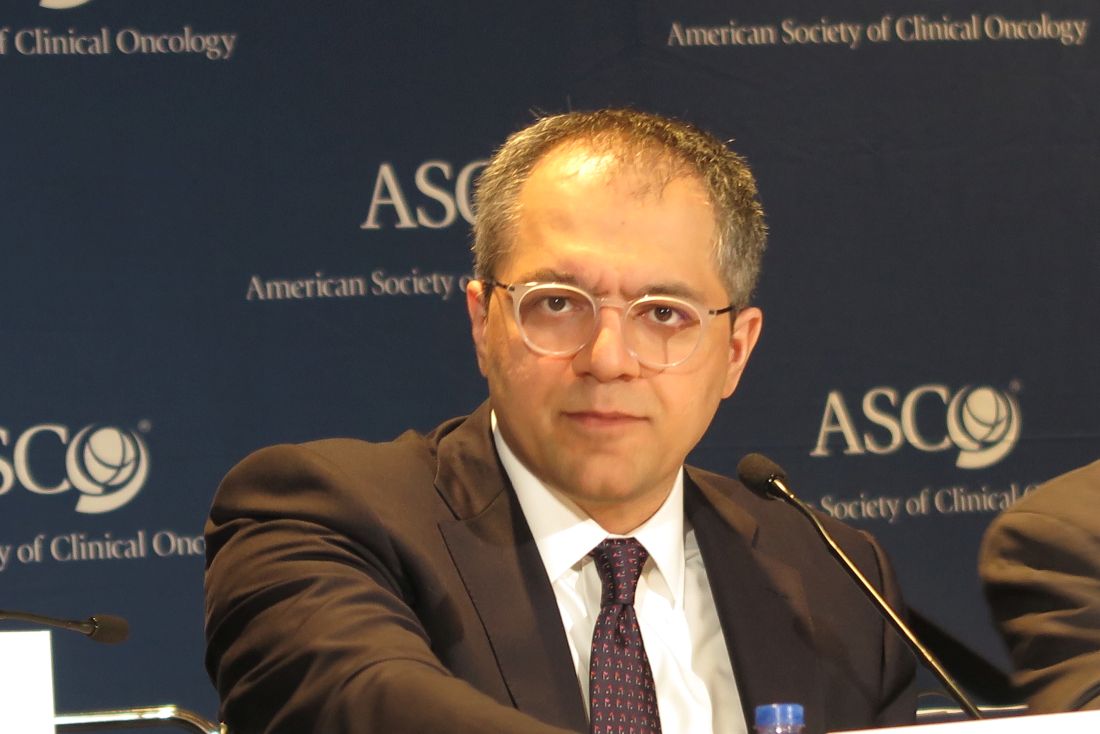
Mikkael A. Sekeres, MD, of the Cleveland Clinic in Cleveland, Ohio, and colleagues reported these findings in the Journal of Clinical Oncology on behalf of the North American Intergroup Study SWOG S117.
Doses of azacitidine were the same for monotherapy and combination arms: 75 mg/m2/day intravenously or subcutaneously on days 1 to 7 of a 28-day cycle.
Patients in the lenalidomide arm received 10 mg/day orally of that drug on days 1 to 21, and patients in the vorinostat arm received 300 mg twice daily orally on days 3 to 9.
Patient characteristics
Patients had MDS of IPSS Intermediate-2 or higher or bone marrow blasts 5% or greater. Patients with CMML had fewer than 20% blasts.
The investigators randomized 277 patients to receive either azacitidine alone (n=92), azacitidine plus lenalidomide (n=93), or azacitidine plus vorinostat (n=92).
Patients were a median age of 70 years (range, 28 to 93). Eighty-five patients (31%) were female, 53 (19%) had CMML, and 18 (6%) had treatment-related MDS. More than half the patients were transfusion-dependent at baseline.
Baseline characteristics were similar across the 3 arms. The investigators noted that the baseline characteristics were also similar across the 90 centers participating in the study, whether they were an MDS Center of Excellence or a high-volume center.
Adverse events
For the most part, therapy-related adverse events were similar across the arms.
Rates of grade 3 or higher febrile neutropenia and infection and infestations were similar for all 3 cohorts: 89% for azaciditine monotherapy, 91% for the lenalidomide combination, and 91% for the vorinostat combination.
However, the vorinostat arm had more grade 3 or higher gastrointestinal toxicities (14 patients, 15%) compared with the monotherapy arm (4 patients, 4%), P=0.02.
And patients receiving lenalidomide experienced more grade 3 or higher rash (14 patients, 16%) compared with patients receiving monotherapy (3 patients, 3%), P=0.005.
Patients in the combination arms stopped therapy at significantly higher rates than the monotherapy arm. Eight percent of patients receiving monotherapy stopped treatment compared with 20% in the lenalidomide arm and 21% in the vorinostat arm.
Patients in the combination arms also had more dose modifications not specified in the protocol than those in the monotherapy arm. Twenty-four percent receiving azacitidine monotherapy had non-protocol defined dose modifications, compared with 43% in the lenalidomide arm and 42% in the vorinostat arm.
Responses
The ORR for the entire study population was 38%.
Patients in the monotherapy arm had an ORR of 38%, those in the lenalidomide arm, 49%, and those in the vorinostate arm, 27%. Neither arm achieved significance compared with the monotherapy arm.
Patients who were treatment-naïve in the lenalidomide arm had a somewhat improved ORR compared with monotherapy, P=0.08.
The median duration of response for all cohorts was 15 months: 10 months for monotherapy, 14 months for lenalidomide, and 18 months for vorinostat.
Patients who were able to remain on therapy for 6 months or more in the lenalidomide arm achieved a higher ORR of 87% compared with monotherapy (62%, P=0.01). However, there was no difference in response duration with longer therapy.
The median overall survival (OS) was 17 months for all patients, 15 months for patients in the monotherapy group, 19 months for those in the lenalidomide arm, and 17 months for those in the vorinostat group.
CMML patients had similar OS across treatment arms, with the median not yet reached for patients in the monotherapy arm.
Subgroup responses
Patients with CMML in the lenalidomide arm had a significantly higher ORR than CMML patients in the monotherapy arm, 68% and 28%, respectively (P=0.02).
Median duration of response for CMML patients was 19 months, with no differences between the arms.
The investigators observed no differences in ORR for therapy-related MDS, IPSS subgroups, transfusion-dependent patients, or allogeneic transplant rates.
However, they noted ORR was better for patients with chromosome 5 abnormality regardless of treatment arm than for those without the abnormality (odds ratio, 2.17, P=0.008).
One hundred thirteen patients had mutational data available. They had a median number of 2 mutations (range, 0 to 7), with the most common being ASXL1 (n = 31), TET2 (n = 26), SRSF2 (n = 23), TP53 (n = 22), RUNX1 (n = 21), and U2AF1 (n = 19).
Patients with DNMT3A mutation had a significantly higher ORR than for patients without mutations, 67% and 34%, respectively P=0.025).
Patients with BCOR and NRAS mutations had numerically higher, but non-significant, ORR than non-mutated patients. Patients with BCOR mutation had a 57% ORR compared with 34% for non-mutated patients (P=0.23). Patients with NRAS mutation had a 60% ORR compared with 36% for non-mutated patients (P=0.28).
Patients with mutations in TET2 (P = .046) and TP53 (P = .003) had a worse response duration than those without mutations.
Response duration was significantly better with fewer mutations. For 2 or more mutations, the hazard ration was 6.86 versus no mutations (P=0.01).
The investigators believed under-dosing may have compromised response and survival in the combination arms. They suggested that studies focused on the subgroups that seemed to benefit from the combinations should be conducted. ![]()
A 3-arm phase 2 study of azacitidine alone or in combination with lenalidomide or vorinostat in patients with higher-risk myelodysplastic syndromes (MDS) or chronic myelomonocytic leukemia (CMML) has shown the combination therapies to have similar overall response rates (ORR) to azacitidine monotherapy. Based on these findings, investigators did not choose either combination arm for phase 3 testing of overall survival.
However, patients with CMML treated with the azacitidine-lenalidomide combination had twice the ORR compared with azacitidine monotherapy, they reported.
And patients with certain mutations, such as DNMT3A, BCOR, and NRAS, had higher overall response rates, although only those with the DNMT3A mutation were significant.
Mikkael A. Sekeres, MD, of the Cleveland Clinic in Cleveland, Ohio, and colleagues reported these findings in the Journal of Clinical Oncology on behalf of the North American Intergroup Study SWOG S117.
Doses of azacitidine were the same for monotherapy and combination arms: 75 mg/m2/day intravenously or subcutaneously on days 1 to 7 of a 28-day cycle.
Patients in the lenalidomide arm received 10 mg/day orally of that drug on days 1 to 21, and patients in the vorinostat arm received 300 mg twice daily orally on days 3 to 9.
Patient characteristics
Patients had MDS of IPSS Intermediate-2 or higher or bone marrow blasts 5% or greater. Patients with CMML had fewer than 20% blasts.
The investigators randomized 277 patients to receive either azacitidine alone (n=92), azacitidine plus lenalidomide (n=93), or azacitidine plus vorinostat (n=92).
Patients were a median age of 70 years (range, 28 to 93). Eighty-five patients (31%) were female, 53 (19%) had CMML, and 18 (6%) had treatment-related MDS. More than half the patients were transfusion-dependent at baseline.
Baseline characteristics were similar across the 3 arms. The investigators noted that the baseline characteristics were also similar across the 90 centers participating in the study, whether they were an MDS Center of Excellence or a high-volume center.
Adverse events
For the most part, therapy-related adverse events were similar across the arms.
Rates of grade 3 or higher febrile neutropenia and infection and infestations were similar for all 3 cohorts: 89% for azaciditine monotherapy, 91% for the lenalidomide combination, and 91% for the vorinostat combination.
However, the vorinostat arm had more grade 3 or higher gastrointestinal toxicities (14 patients, 15%) compared with the monotherapy arm (4 patients, 4%), P=0.02.
And patients receiving lenalidomide experienced more grade 3 or higher rash (14 patients, 16%) compared with patients receiving monotherapy (3 patients, 3%), P=0.005.
Patients in the combination arms stopped therapy at significantly higher rates than the monotherapy arm. Eight percent of patients receiving monotherapy stopped treatment compared with 20% in the lenalidomide arm and 21% in the vorinostat arm.
Patients in the combination arms also had more dose modifications not specified in the protocol than those in the monotherapy arm. Twenty-four percent receiving azacitidine monotherapy had non-protocol defined dose modifications, compared with 43% in the lenalidomide arm and 42% in the vorinostat arm.
Responses
The ORR for the entire study population was 38%.
Patients in the monotherapy arm had an ORR of 38%, those in the lenalidomide arm, 49%, and those in the vorinostate arm, 27%. Neither arm achieved significance compared with the monotherapy arm.
Patients who were treatment-naïve in the lenalidomide arm had a somewhat improved ORR compared with monotherapy, P=0.08.
The median duration of response for all cohorts was 15 months: 10 months for monotherapy, 14 months for lenalidomide, and 18 months for vorinostat.
Patients who were able to remain on therapy for 6 months or more in the lenalidomide arm achieved a higher ORR of 87% compared with monotherapy (62%, P=0.01). However, there was no difference in response duration with longer therapy.
The median overall survival (OS) was 17 months for all patients, 15 months for patients in the monotherapy group, 19 months for those in the lenalidomide arm, and 17 months for those in the vorinostat group.
CMML patients had similar OS across treatment arms, with the median not yet reached for patients in the monotherapy arm.
Subgroup responses
Patients with CMML in the lenalidomide arm had a significantly higher ORR than CMML patients in the monotherapy arm, 68% and 28%, respectively (P=0.02).
Median duration of response for CMML patients was 19 months, with no differences between the arms.
The investigators observed no differences in ORR for therapy-related MDS, IPSS subgroups, transfusion-dependent patients, or allogeneic transplant rates.
However, they noted ORR was better for patients with chromosome 5 abnormality regardless of treatment arm than for those without the abnormality (odds ratio, 2.17, P=0.008).
One hundred thirteen patients had mutational data available. They had a median number of 2 mutations (range, 0 to 7), with the most common being ASXL1 (n = 31), TET2 (n = 26), SRSF2 (n = 23), TP53 (n = 22), RUNX1 (n = 21), and U2AF1 (n = 19).
Patients with DNMT3A mutation had a significantly higher ORR than for patients without mutations, 67% and 34%, respectively P=0.025).
Patients with BCOR and NRAS mutations had numerically higher, but non-significant, ORR than non-mutated patients. Patients with BCOR mutation had a 57% ORR compared with 34% for non-mutated patients (P=0.23). Patients with NRAS mutation had a 60% ORR compared with 36% for non-mutated patients (P=0.28).
Patients with mutations in TET2 (P = .046) and TP53 (P = .003) had a worse response duration than those without mutations.
Response duration was significantly better with fewer mutations. For 2 or more mutations, the hazard ration was 6.86 versus no mutations (P=0.01).
The investigators believed under-dosing may have compromised response and survival in the combination arms. They suggested that studies focused on the subgroups that seemed to benefit from the combinations should be conducted. ![]()
Study contradicts AAP recommendations on infant room-sharing
Infants who slept in their own rooms by age 9 months slept significantly longer and better than those who continued sharing a room with their parents as recommended by the American Academy of Pediatrics, the authors of a prospective study of 279 mother-infant dyads reported June 5.
The findings “raise questions about the well-intended AAP recommendation that room-sharing should ideally occur for all infants until their first birthday,” wrote Ian M. Paul, MD, of Penn State University, Hershey, and his associates (Pediatrics. 2017 Jun 5. doi: 10.1542/peds.2017-0122). “Perhaps our most troubling finding was that room-sharing was associated with overnight transitions to bed-sharing, which is strongly discouraged by the AAP.”

Insufficient sleep leads to excess weight gain during infancy and sleep problems later in childhood and has negative implications for parents. “The desire to optimize infant sleep duration and consolidation, however, must be balanced with safe infant sleep,” the researchers emphasized. About 3,500 infants die annually of SIDS and other sleep-related deaths, about 90% of which occur before age 6 months. Currently, however, the AAP’s updated 2016 recommendations advise that infants sleep on a separate surface in their parents’ room for at least the first 6 months and ideally for 1 year (Pediatrics. 2016; 138:e20162938).
To examine relationships among where, how well, and how long infants slept, the researchers analyzed Brief Infant Sleep Questionnaires collected from first-time mothers of term singletons as part of the prospective, single-center Intervention Nurses Start Infants Growing on Healthy Trajectories (INSIGHT) study.
For 4-month-olds, average reported sleep duration was similar whether they slept alone or in the parental bedroom. Solo sleepers, however, had better sleep consolidation, averaging 46 more minutes of sleep at the longest stretch, compared with room sharers (P = .02). By age 9 months, infants who had slept alone by age 4 months averaged 40 more minutes of nightly sleep than room-sharers and 26 more minutes than infants who began sleeping alone after 4 months of age (P = .008). Furthermore, the average longest sleep span of early solo sleepers was 100 minutes more than that of room-sharers and 45 minutes more than that of infants who began sleeping alone between ages 4 and 9 months (P = .01).
At age 30 months, infants who had slept alone by age 9 months averaged 45 more minutes of nightly sleep than those who had shared a room (P = .004). Room-sharing at 4 months also was tied to a two-fold greater odds of having pillows, blankets, or other unsafe objects on the sleep surface, the researchers said. Together, the findings support revising the AAP recommendation until evidence conclusively supports it, they concluded.
The study’s funders included Penn State University, Hershey; Penn State Children’s Hospital; the U.S. Department of Agriculture; the Penn State Clinical and Translational Science Institute, and the National Institutes of Health/National Center for Advancing Translational Sciences. The investigators reported having no conflicts of interest.
The study by Dr. Paul and his associates is an important contribution to the literature, wrote Rachel Y. Moon, MD, and Fern R. Hauck, MD, in an accompanying editorial (Pediatrics. 2017 Jun 5. doi: 10.1542/peds.2017-1323). However, they said, more research is needed.
For example, it is unclear whether sleep consolidation in young infants is preferable from a physiologic perspective. “The ability to arouse is critical physiologically, and a leading hypothesis is that failure to arouse makes an infant vulnerable to SIDS,” they wrote.
Dr. Moon and Dr. Hauck also noted that the AAP’s recommendation on room-sharing without bed-sharing is based on studies conducted in England, New Zealand, and Scotland showing that room-sharing lowers the risk of SIDS, compared with solitary sleeping.
“More recent, unpublished data from the New Zealand Sudden and Unexplained Death in Infancy study show similar protection from room-sharing, with an adjusted odds ratio of 0.36 (95% confidence interval, 0.19-0.71) for room-sharing infants compared with solitary-sleeping infants (E. Mitchell, MBBS, personal communication, 2016),” they wrote. “Because none of these studies stratified the risk by infant age in months, it is difficult to determine the optimal endpoint for room-sharing.
“We strongly support more research, both about the physiology of infant sleep and arousal for room-sharing infants and about the consequences of room-sharing on parental and child sleep.”
Dr. Moon and Dr. Hauck are with the University of Virginia, Charlottesville. They had no relevant disclosures.
Infants who slept in their own rooms by age 9 months slept significantly longer and better than those who continued sharing a room with their parents as recommended by the American Academy of Pediatrics, the authors of a prospective study of 279 mother-infant dyads reported June 5.
The findings “raise questions about the well-intended AAP recommendation that room-sharing should ideally occur for all infants until their first birthday,” wrote Ian M. Paul, MD, of Penn State University, Hershey, and his associates (Pediatrics. 2017 Jun 5. doi: 10.1542/peds.2017-0122). “Perhaps our most troubling finding was that room-sharing was associated with overnight transitions to bed-sharing, which is strongly discouraged by the AAP.”
Insufficient sleep leads to excess weight gain during infancy and sleep problems later in childhood and has negative implications for parents. “The desire to optimize infant sleep duration and consolidation, however, must be balanced with safe infant sleep,” the researchers emphasized. About 3,500 infants die annually of SIDS and other sleep-related deaths, about 90% of which occur before age 6 months. Currently, however, the AAP’s updated 2016 recommendations advise that infants sleep on a separate surface in their parents’ room for at least the first 6 months and ideally for 1 year (Pediatrics. 2016; 138:e20162938).
To examine relationships among where, how well, and how long infants slept, the researchers analyzed Brief Infant Sleep Questionnaires collected from first-time mothers of term singletons as part of the prospective, single-center Intervention Nurses Start Infants Growing on Healthy Trajectories (INSIGHT) study.
For 4-month-olds, average reported sleep duration was similar whether they slept alone or in the parental bedroom. Solo sleepers, however, had better sleep consolidation, averaging 46 more minutes of sleep at the longest stretch, compared with room sharers (P = .02). By age 9 months, infants who had slept alone by age 4 months averaged 40 more minutes of nightly sleep than room-sharers and 26 more minutes than infants who began sleeping alone after 4 months of age (P = .008). Furthermore, the average longest sleep span of early solo sleepers was 100 minutes more than that of room-sharers and 45 minutes more than that of infants who began sleeping alone between ages 4 and 9 months (P = .01).
At age 30 months, infants who had slept alone by age 9 months averaged 45 more minutes of nightly sleep than those who had shared a room (P = .004). Room-sharing at 4 months also was tied to a two-fold greater odds of having pillows, blankets, or other unsafe objects on the sleep surface, the researchers said. Together, the findings support revising the AAP recommendation until evidence conclusively supports it, they concluded.
The study’s funders included Penn State University, Hershey; Penn State Children’s Hospital; the U.S. Department of Agriculture; the Penn State Clinical and Translational Science Institute, and the National Institutes of Health/National Center for Advancing Translational Sciences. The investigators reported having no conflicts of interest.
FROM PEDIATRICS
Key clinical point: Infants who slept in their own room by age 4 months slept significantly longer and better than those who continued sharing a room with their parents as recommended by the American Academy of Pediatrics.
Major finding: By age 9 months, infants who had slept alone by age 4 months averaged 40 more minutes of nightly sleep than room sharers and 26 more minutes than infants who began sleeping alone after 4 months of age (P = .008). Furthermore, the average longest sleep span of early solo sleepers was 100 minutes more than that of room-sharers and 45 minutes more than that of infants who began sleeping alone between ages 4 and 9 months (P = .01).
Data source: Secondary analyses of 279 mother-infant dyads from the single-center, prospective Intervention Nurses Start Infants Growing on Healthy Trajectories (INSIGHT) study.
Disclosures: The study funders included Penn State University, Hershey; Penn State Children’s Hospital; the U.S. Department of Agriculture; the Penn State Clinical and Translational Science Institute, and the National Institutes of Health/National Center for Advancing Translational Sciences. The investigators reported having no conflicts of interest.
The Professional Doctorate: What Are We Waiting for?
The increasingly complex health care system in the United States relies heavily on quality improvement, interprofessional collaboration, patient outcomes, health policy legislation, and advocacy. While important, most of these factors are outside the scope of the traditional master’s-level education program—necessitating the development of methods to help advanced practice providers, including NPs and PAs, obtain additional skills. The solution of choice, for many professions, has been the introduction of the “professional doctorate” as a complementary alternative to the typical research-focused doctoral program, such as the PhD.
Traditional PhD curricula prepare individuals to perform research that is typically specialized and confined to their field of study.1 While this research does produce new knowledge, it usually remains in the realm of academia and often does not address any specific “real-world” problem.2 But to be recognized, compensated, and identified as a full professional in modern societ
Analysis by Taylor and Maxwell and by Lee, Green, and Brennan has shown that, rather than theory, the workplace demands the application of knowledge geared toward daily professional duties.3,4 They envisioned a doctorate-prepared practitioner who had less skill in pure research but who would be able to apply theory to everyday problems in the workplace.4,5 Rather than devalue the contributions of classical PhD training, this model proposed the creation of a hybrid curriculum that would prepare individuals to use “applied research.”3 As the professional doctorate gained acceptance, it matured from the “first-generation” concept (which was quite similar to the PhD in structure) to “second-generation,” which is more focused on discipline and workplace realities.3,5 In general, these professional doctorates can be earned in less time than a PhD and do not require original research.
Over the past two decades, more than 500 unique professional practice doctorate programs have emerged across the US, in fields ranging from nursing to bioethics. One of the most prominent is the Doctor of Nursing Practice (DNP), designed for RNs seeking a post-professional degree in nursing. In 2004, following three years of research by a task force, the American Association of Colleges of Nursing (AACN) endorsed the DNP, with the goal that it would become the minimum educational standard for advanced practice nurses by 2015.6 According to the AACN, there are 289 DNP programs in the US, with an additional 128 in development.6
The PA profession has lagged behind not only our NP colleagues, but also many other health professions, in the adoption of a discipline-specific, doctoral-level degree. Our counterparts in audiology, physical therapy, occupational therapy, and athletic training have been part of the exponential growth in second-generation health care doctorates.7 While these programs may differ in concept, they share several similarities: They do not require original research; they include a clinical component; and they promote knowledge in the context of the workplace.5-8
In the past five years, PAs have started considering (or debating, depending on your perspective) a professional/clinical doctorate as the next step in our post-professional journey. It’s about time, when you consider that 16.8% of newly certified PAs intend to pursue additional education or clinical training, according to a recent report from the National Commission on Certification of Physician Assistants.9
There are already a few doctoral programs for PAs. Among the earliest clinically focused doctorate programs was the US Army/Air Force-Baylor DScPAS-EM program, designed to educate military PAs at the doctoral level upon completion of an 18-month emergency medicine residency.10 Lincoln Memorial University has a Doctor of Medical Science (DMS) program, comprised of one year of online advanced clinical medicine coursework and one year of online coursework focused on primary care, hospital medicine, emergency medicine, or education.11 And Lynchburg College in Virginia has just launched a post-professional doctoral program for PAs; this DMS program includes a clinical fellowship, as well as coursework in leadership training, health care management and law, organizational behavior, disaster medicine, and global health.12
While not strictly created for PAs, the Doctor of Health Science programs at Nova Southeastern University and A.T. Still University have been educating PAs at the doctoral level for more than 10 years.13,14 Later this year, A.T. Still University plans to introduce a post-professional Doctor of Physician Assistant Studies that will provide a pathway for PAs wishing to become leaders and scholar-practitioners, develop core leadership abilities, and/or enter PA education without the location-specific requirement of a clinical or academic residency.
When the push for professional practice doctorates started, pundits claimed they were just an attempt at a “cash grab” by universities looking to bolster their rosters (and their coffers). But advocates have long argued that these degrees provide practitioners with the knowledge and training required to offer advanced services in increasingly complex social and technologic environments.7 No less than The Institute of Medicine, The Joint Commission, and the Robert Wood Johnson Foundation have called for the reinvention of education programs to equip today’s health professionals
Why? First and foremost, to ensure quality patient outcomes. Beyond that, better prepared clinicians can help to address provider shortages. Those with doctorates can also serve as faculty, educating the next generation of health care providers. And practically speaking, for those seeking advanced education, holding a doctorate will create opportunities for increased decision-making and upward mobility in the workplace.
There is no question that our current health care environment is driven by the regulations and costs of the Affordable Care Act, as well as quality management systems and strategies. NPs and PAs are in a unique position to cost-effectively direct the care of, and advocate for, diverse patient populations. NPs and PAs who recognize this opportunity to serve need doctoral-level training tailored to this milieu.
Do you agree? Share your thoughts on professional doctorates with me at [email protected].
1. Carnegie Project on the Education Doctorate. Founding literature. www.cpedinitiative.org/?. Accessed May 8, 2017.
2. Costley C, Lester S. Work-based doctorates: professional extension at the highest levels. Studies in Higher Education. 2012;37(3):257-269.
3. Taylor N, Maxwell T. Enhancing the relevance of a professional doctorate: the case of the doctor of education degree at the University of New England. Asia-Pacific J Coop Edu. 2004;5(1):60-69.
4. Lee A, Green B, Brennan M. Organisational knowledge, professional practice and the professional doctorate at work. In: Garrick J, Rhodes C (eds). Research and Knowledge at Work. Perspectives, Case-studies and Innovative Strategies. London: Routledge; 2000.
5. Maxwell T. From first to second generation professional doctorate. Studies in Higher Education. 2003;28(3):279-291.
6. American Association of Colleges of Nursing. DNP fact sheet. www.aacn.nche.edu/media-relations/fact-sheets/dnp. Accessed May 8, 2017.
7. Zusman A. Degrees of change: how new kinds of professional doctorates are changing higher education institutions. Research and Occasional Paper Series. 2013;8(13):1-20.
8. Kumar S, Dawson K. Exploring the impact of a professional practice education doctorate in educational environments. Studies in Continuing Education. 2013;35(2):165-178.
9. National Commission on Certification of Physician Assistants. 2014 statistical profile of recently certified physician assistants: an annual report of the NCCPA. www.nccpa.net/Uploads/docs/RecentlyCertifiedReport2014.pdf. Accessed May 8, 2017.
10. Baylor University. Army-Baylor emergency medicine physician assistant (EMPA) program. www.baylor.edu/graduate/pa/index.php?id=936090. Accessed May 8, 2017.
11. Lincoln Memorial University. Doctor of medical science announcement. www.lmunet.edu/academics/schools/debusk-college-of-osteopathic-medicine/dms. Accessed May 8, 2017.
12. Lynchburg College. Doctor of medical science. www.lynchburg.edu/graduate/physician-assistant-medicine/doctor-of-medical-science/. Accessed May 8, 2017.
13. Nova Southeastern University. Doctor of health science program. http://healthsciences.nova.edu/healthsciences/dhs/. Accessed May 8, 2017.
14. A.T. Still University. About the college of graduate health studies – online. www.atsu.edu/college-of-graduate-health-studies. Accessed May 8, 2017.

The increasingly complex health care system in the United States relies heavily on quality improvement, interprofessional collaboration, patient outcomes, health policy legislation, and advocacy. While important, most of these factors are outside the scope of the traditional master’s-level education program—necessitating the development of methods to help advanced practice providers, including NPs and PAs, obtain additional skills. The solution of choice, for many professions, has been the introduction of the “professional doctorate” as a complementary alternative to the typical research-focused doctoral program, such as the PhD.
Traditional PhD curricula prepare individuals to perform research that is typically specialized and confined to their field of study.1 While this research does produce new knowledge, it usually remains in the realm of academia and often does not address any specific “real-world” problem.2 But to be recognized, compensated, and identified as a full professional in modern societ
Analysis by Taylor and Maxwell and by Lee, Green, and Brennan has shown that, rather than theory, the workplace demands the application of knowledge geared toward daily professional duties.3,4 They envisioned a doctorate-prepared practitioner who had less skill in pure research but who would be able to apply theory to everyday problems in the workplace.4,5 Rather than devalue the contributions of classical PhD training, this model proposed the creation of a hybrid curriculum that would prepare individuals to use “applied research.”3 As the professional doctorate gained acceptance, it matured from the “first-generation” concept (which was quite similar to the PhD in structure) to “second-generation,” which is more focused on discipline and workplace realities.3,5 In general, these professional doctorates can be earned in less time than a PhD and do not require original research.
Over the past two decades, more than 500 unique professional practice doctorate programs have emerged across the US, in fields ranging from nursing to bioethics. One of the most prominent is the Doctor of Nursing Practice (DNP), designed for RNs seeking a post-professional degree in nursing. In 2004, following three years of research by a task force, the American Association of Colleges of Nursing (AACN) endorsed the DNP, with the goal that it would become the minimum educational standard for advanced practice nurses by 2015.6 According to the AACN, there are 289 DNP programs in the US, with an additional 128 in development.6
The PA profession has lagged behind not only our NP colleagues, but also many other health professions, in the adoption of a discipline-specific, doctoral-level degree. Our counterparts in audiology, physical therapy, occupational therapy, and athletic training have been part of the exponential growth in second-generation health care doctorates.7 While these programs may differ in concept, they share several similarities: They do not require original research; they include a clinical component; and they promote knowledge in the context of the workplace.5-8
In the past five years, PAs have started considering (or debating, depending on your perspective) a professional/clinical doctorate as the next step in our post-professional journey. It’s about time, when you consider that 16.8% of newly certified PAs intend to pursue additional education or clinical training, according to a recent report from the National Commission on Certification of Physician Assistants.9
There are already a few doctoral programs for PAs. Among the earliest clinically focused doctorate programs was the US Army/Air Force-Baylor DScPAS-EM program, designed to educate military PAs at the doctoral level upon completion of an 18-month emergency medicine residency.10 Lincoln Memorial University has a Doctor of Medical Science (DMS) program, comprised of one year of online advanced clinical medicine coursework and one year of online coursework focused on primary care, hospital medicine, emergency medicine, or education.11 And Lynchburg College in Virginia has just launched a post-professional doctoral program for PAs; this DMS program includes a clinical fellowship, as well as coursework in leadership training, health care management and law, organizational behavior, disaster medicine, and global health.12
While not strictly created for PAs, the Doctor of Health Science programs at Nova Southeastern University and A.T. Still University have been educating PAs at the doctoral level for more than 10 years.13,14 Later this year, A.T. Still University plans to introduce a post-professional Doctor of Physician Assistant Studies that will provide a pathway for PAs wishing to become leaders and scholar-practitioners, develop core leadership abilities, and/or enter PA education without the location-specific requirement of a clinical or academic residency.
When the push for professional practice doctorates started, pundits claimed they were just an attempt at a “cash grab” by universities looking to bolster their rosters (and their coffers). But advocates have long argued that these degrees provide practitioners with the knowledge and training required to offer advanced services in increasingly complex social and technologic environments.7 No less than The Institute of Medicine, The Joint Commission, and the Robert Wood Johnson Foundation have called for the reinvention of education programs to equip today’s health professionals
Why? First and foremost, to ensure quality patient outcomes. Beyond that, better prepared clinicians can help to address provider shortages. Those with doctorates can also serve as faculty, educating the next generation of health care providers. And practically speaking, for those seeking advanced education, holding a doctorate will create opportunities for increased decision-making and upward mobility in the workplace.
There is no question that our current health care environment is driven by the regulations and costs of the Affordable Care Act, as well as quality management systems and strategies. NPs and PAs are in a unique position to cost-effectively direct the care of, and advocate for, diverse patient populations. NPs and PAs who recognize this opportunity to serve need doctoral-level training tailored to this milieu.
Do you agree? Share your thoughts on professional doctorates with me at [email protected].
1. Carnegie Project on the Education Doctorate. Founding literature. www.cpedinitiative.org/?. Accessed May 8, 2017.
2. Costley C, Lester S. Work-based doctorates: professional extension at the highest levels. Studies in Higher Education. 2012;37(3):257-269.
3. Taylor N, Maxwell T. Enhancing the relevance of a professional doctorate: the case of the doctor of education degree at the University of New England. Asia-Pacific J Coop Edu. 2004;5(1):60-69.
4. Lee A, Green B, Brennan M. Organisational knowledge, professional practice and the professional doctorate at work. In: Garrick J, Rhodes C (eds). Research and Knowledge at Work. Perspectives, Case-studies and Innovative Strategies. London: Routledge; 2000.
5. Maxwell T. From first to second generation professional doctorate. Studies in Higher Education. 2003;28(3):279-291.
6. American Association of Colleges of Nursing. DNP fact sheet. www.aacn.nche.edu/media-relations/fact-sheets/dnp. Accessed May 8, 2017.
7. Zusman A. Degrees of change: how new kinds of professional doctorates are changing higher education institutions. Research and Occasional Paper Series. 2013;8(13):1-20.
8. Kumar S, Dawson K. Exploring the impact of a professional practice education doctorate in educational environments. Studies in Continuing Education. 2013;35(2):165-178.
9. National Commission on Certification of Physician Assistants. 2014 statistical profile of recently certified physician assistants: an annual report of the NCCPA. www.nccpa.net/Uploads/docs/RecentlyCertifiedReport2014.pdf. Accessed May 8, 2017.
10. Baylor University. Army-Baylor emergency medicine physician assistant (EMPA) program. www.baylor.edu/graduate/pa/index.php?id=936090. Accessed May 8, 2017.
11. Lincoln Memorial University. Doctor of medical science announcement. www.lmunet.edu/academics/schools/debusk-college-of-osteopathic-medicine/dms. Accessed May 8, 2017.
12. Lynchburg College. Doctor of medical science. www.lynchburg.edu/graduate/physician-assistant-medicine/doctor-of-medical-science/. Accessed May 8, 2017.
13. Nova Southeastern University. Doctor of health science program. http://healthsciences.nova.edu/healthsciences/dhs/. Accessed May 8, 2017.
14. A.T. Still University. About the college of graduate health studies – online. www.atsu.edu/college-of-graduate-health-studies. Accessed May 8, 2017.
Test goes wide and deep to detect free tumor DNA in blood
CHICAGO – An experimental liquid biopsy method plumbs the depths of the genome to detect a broad array of mutations in blood samples that closely correspond to variants in tumors.
The high-intensity method for detecting circulating free DNA (cfDNA) in plasma detected at least one significant genetic variant in samples from 89% of patients with advanced breast, lung, and prostate cancers.
If validated in further studies, the technique could inform more accurate drug selection in patients with refractory disease and eventually may form the basis for plasma-based assays to detect early-stage cancers, said Pedram Razavi, MD, PhD, of Memorial Sloan Kettering Cancer Center in New York.
“This novel, high-intensity sequencing assay incorporates an unprecedented combination of depth and breadth of coverage compared to previous assays. High levels of concordance for variants between plasma and tissue provide strong evidence for high rates of tumor DNA detection in plasma,” he said at a press briefing at the annual meeting of the American Society of Clinical Oncology.
The technique also provides important insights into tumor biology, “including first exploration of mutational signatures in the plasma,” he added.
Deep scanning
The high-intensity sequencing method scans for 508 genes in a 2 million base-pair swath of genome, performing 60,000 repeat reads on each genome region to improve the assay’s sensitivity, and deep sequencing allows for detection of rare “needle-in-a-haystack” variants.
Dr. Razavi and his colleagues examined prospectively collected blood and tissues from 161 patients, 124 of whom had samples sufficient for concordance studies.
The samples were collected within 6 weeks of a de novo cancer diagnosis or evidence of disease progression, before the initiation of therapy.
Both cfDNA and genomic data from white blood cells (WBCs) of each patient were sequenced with the aforementioned 508-gene panel covering a broad range of known cancer variants and mutations.
Tumor tissues were sequenced with MSK-IMPACT, a 410 gene assay, with blinding of results in regard to plasma and WBC sequencing. Variants were classified as clonal or subclonal based on tumor sequencing in breast cancer and non–small cell lung cancer (NSCLC).
As noted before, 89% of patients had at least one genetic variant detected in both the tumor and in plasma, including 97% of patients with metastatic breast cancer, 85% of those with NSCLC, and 84% of patients with metastatic prostate cancer.
The investigators identified 864 clonal or subclonal variants in tissue samples from all three of these cancers, and 627 of the variants also were found in plasma.
In addition, 76% of clinically actionable somatic mutations identified in tumors also were found in plasma, which suggests that the high-intensity sequencing technique may be able to identify tumor heterogeneity that is not always evident in single tumor biopsies, Dr. Razavi said.

‘A clear advance’
“The work by Dr. Razavi and colleagues is a clear advance in the field because it surveys for the first time a much broader panel of genes – 508 genes in this case – and it does it with much deeper sequencing, which means it can detect much rarer alterations,” commented ASCO expert John Heymach, MD, PhD, from the University of Texas MD Anderson Cancer Center in Houston.
The study “helps illuminate a path toward a day when we will be using circulating tumor DNA assays for early detection of cancer, and not just for selecting certain therapies,” he added.
“We’re a long way from utilizing liquid biopsy for detecting cancers; already though, we’re seeing some utility of circulating tumor DNA in the domain of identifying novel alterations as a means of segmentation clinical trials,” commented ASCO expert Sumanta Kumar Pal, MD, from City of Hope in Duarte, Calif.

Dr. Heymach and Dr. Pal were not involved in the study, but were invited discussants at the briefing.
The study was funded in part by GRAIL. Dr. Razavi reported institutional research funding from the company. Multiple coauthors are employees of the company.
CHICAGO – An experimental liquid biopsy method plumbs the depths of the genome to detect a broad array of mutations in blood samples that closely correspond to variants in tumors.
The high-intensity method for detecting circulating free DNA (cfDNA) in plasma detected at least one significant genetic variant in samples from 89% of patients with advanced breast, lung, and prostate cancers.
If validated in further studies, the technique could inform more accurate drug selection in patients with refractory disease and eventually may form the basis for plasma-based assays to detect early-stage cancers, said Pedram Razavi, MD, PhD, of Memorial Sloan Kettering Cancer Center in New York.
“This novel, high-intensity sequencing assay incorporates an unprecedented combination of depth and breadth of coverage compared to previous assays. High levels of concordance for variants between plasma and tissue provide strong evidence for high rates of tumor DNA detection in plasma,” he said at a press briefing at the annual meeting of the American Society of Clinical Oncology.
The technique also provides important insights into tumor biology, “including first exploration of mutational signatures in the plasma,” he added.
Deep scanning
The high-intensity sequencing method scans for 508 genes in a 2 million base-pair swath of genome, performing 60,000 repeat reads on each genome region to improve the assay’s sensitivity, and deep sequencing allows for detection of rare “needle-in-a-haystack” variants.
Dr. Razavi and his colleagues examined prospectively collected blood and tissues from 161 patients, 124 of whom had samples sufficient for concordance studies.
The samples were collected within 6 weeks of a de novo cancer diagnosis or evidence of disease progression, before the initiation of therapy.
Both cfDNA and genomic data from white blood cells (WBCs) of each patient were sequenced with the aforementioned 508-gene panel covering a broad range of known cancer variants and mutations.
Tumor tissues were sequenced with MSK-IMPACT, a 410 gene assay, with blinding of results in regard to plasma and WBC sequencing. Variants were classified as clonal or subclonal based on tumor sequencing in breast cancer and non–small cell lung cancer (NSCLC).
As noted before, 89% of patients had at least one genetic variant detected in both the tumor and in plasma, including 97% of patients with metastatic breast cancer, 85% of those with NSCLC, and 84% of patients with metastatic prostate cancer.
The investigators identified 864 clonal or subclonal variants in tissue samples from all three of these cancers, and 627 of the variants also were found in plasma.
In addition, 76% of clinically actionable somatic mutations identified in tumors also were found in plasma, which suggests that the high-intensity sequencing technique may be able to identify tumor heterogeneity that is not always evident in single tumor biopsies, Dr. Razavi said.
‘A clear advance’
“The work by Dr. Razavi and colleagues is a clear advance in the field because it surveys for the first time a much broader panel of genes – 508 genes in this case – and it does it with much deeper sequencing, which means it can detect much rarer alterations,” commented ASCO expert John Heymach, MD, PhD, from the University of Texas MD Anderson Cancer Center in Houston.
The study “helps illuminate a path toward a day when we will be using circulating tumor DNA assays for early detection of cancer, and not just for selecting certain therapies,” he added.
“We’re a long way from utilizing liquid biopsy for detecting cancers; already though, we’re seeing some utility of circulating tumor DNA in the domain of identifying novel alterations as a means of segmentation clinical trials,” commented ASCO expert Sumanta Kumar Pal, MD, from City of Hope in Duarte, Calif.
Dr. Heymach and Dr. Pal were not involved in the study, but were invited discussants at the briefing.
The study was funded in part by GRAIL. Dr. Razavi reported institutional research funding from the company. Multiple coauthors are employees of the company.
AT ASCO 2017
Key clinical point: High-intensity sequencing of plasma samples appears capable of detecting actionable tumor mutations in a large proportion of samples.
Major finding: In 89% of patients with advanced cancers, genetic variants were identified in both tumor samples and circulating free DNA testing.
Data source: Prospective study of tissue and plasma samples from 124 patients with non–small cell lung cancer or metastatic breast and prostate cancers.
Disclosures: The study was funded in part by GRAIL. Dr. Razavi reported institutional research funding from the company. Multiple coauthors are employees of the company.
Cost, value of new cancer treatments rarely correlate
New high-priced cancer treatments rarely demonstrate value, compared with the prices being charged for them.
Researchers examined clinical trial data for new treatments using scoring frameworks developed by both the American Society of Clinical Oncology (ASCO) and the European Society for Medical Oncology (ESMO) that assess the value of cancer therapies by taking into consideration survival gains in relation to toxicity and quality. Findings were simultaneously published online June 2 in The Lancet Oncology (2017 June 2. doi: 10.1016/S1470-2045(17)30415-1) and presented at the annual meeting of the American Society of Clinical Oncology.
One of the findings from the research quantified the magnitude of how much benefit a new treatment offers patients.
“We were able to compare that across different treatments and we were able to calculate the drug cost per month of treatment with each new cancer treatment,” Christopher Booth, MD, of Queen’s University Cancer Research Institute, Kingston, Ont., and one of the report’s authors, said in an interview.
“We looked to see whether there is a relationship between cost and benefit. In most elements of our economy, if something is of higher value and better quality, we often pay more for it as opposed to something that offers less quality or less value,” he continued. “We found that that relationship does not hold true in the world of cancer drugs. In fact, if anything, we saw an inverse association, meaning that the drugs that are the most expensive actually have the smallest benefit for patients. That is probably the most important finding of the work.”
Dr. Booth suggested that this has to do with the fact that value is never a factor in pricing.
“In very general terms, the way that a new drug is priced is effectively the highest price the market will bear,” he said. “There really isn’t an approach currently to price drugs based on the return that they offer to patients and society. There is a conversation under way within the oncology community about whether we need to be shifting that conversation to something perhaps called value-based pricing where treatments that offer greater benefit to patients are priced higher than treatments that offer negligible benefit.”
He also noted that “there is not a lot of incentive for the research community or pharmaceutical companies to identify drugs that have larger and larger benefits, being that the financial return on their investment does not appear to be related to how much benefit the drug offers. If we shifted that conversation, it would hopefully, at least in some way, drive the research community and push us to find treatments that have large or meaningful benefits to patients instead of some of these treatments that are very expensive with important side effects that really offer very small improvements in patient outcomes.”
Dr. Booth continued: “I think it’s inevitable and I think what these score cards are doing, and even though they are not perfect, are getting the conversation into the mainstream and for the first time allowing oncologists and researchers and policy makers to start some kind of comparative analyses looking at one treatment compared to the other in the same disease setting as offering a greater or lesser benefit to patients,” which is an important conversation as health care systems are faced with stretching their limited resources to help the populations they serve.
The other key finding of the report was the lack of correlation between the scores yielded by the ASCO Value Framework and the ESMO Magnitude of Clinical Benefit Scale.
“We looked at a number of randomized trials of new cancer treatments and we scored them using the European approach and the American approach and found that there is actually fairly little agreement between the two systems,” Dr. Booth said.
The cause, he suggested, was tied to differences in methodology between the two systems, though he noted that both frameworks are evolving and “I suspect there will be convergence over time.”
FROM ASCO 2017
VIDEO: Olaparib improves outlook in women with BRCA-related HER2-negative MBC
CHICAGO – Compared with single-agent chemotherapy of the oncologist’s choice, the PARP inhibitor olaparib reduced the risk of progression or death by 42% in women with BRCA-related HER2-negative metastatic breast cancer, the OlympiAD trialists reported in a plenary session at the annual meeting of the American Society of Clinical Oncology.
Lead author Mark E. Robson, MD, of Memorial Sloan Kettering Cancer Center, New York, discussed findings of the randomized phase III trial, as well as strategies for building on the trial’s success and what the future holds for this class of agents in breast cancer.
Dr. Robson disclosed that he has a consulting or advisory role with McKesson and AstraZeneca; receives travel, accommodations, and/or expenses from AstraZeneca; receives honoraria from AstraZeneca; and receives research funding (institutional) from AstraZeneca, Abbvie, Myriad Genetics, Biomarin, Medivation, and Tesaro. The trial was funded by AstraZeneca.
The video associated with this article is no longer available on this site. Please view all of our videos on the MDedge YouTube channel
AT ASCO 2017

VIDEO: Abiraterone improves survival in high-risk prostate cancer
CHICAGO – Adding abiraterone acetate plus prednisone to androgen deprivation therapy in hormone-naive high-risk prostate cancer patients significantly improves overall survival, according to “practice-changing” results from the phase III LATITUDE trial and the multiarm, multistage STAMPEDE trial.
In the double-blind, randomized, placebo-controlled LATITUDE trial, 597 patients with newly diagnosed high-risk metastatic prostate cancer who received 1,000 mg of abiraterone acetate daily plus 5 mg of prednisone daily in addition to androgen deprivation therapy experienced a 38% reduction in the risk of death and a 53% reduction in the risk of radiographic progression or death at a median follow-up of 30.4 months, compared with 602 such patients who received placebo and androgen deprivation, Karim Fizazi, MD, PhD, of the Institut Gustave Roussy, Villejuif, France, reported at the annual meeting of the American Society of Clinical Oncology.
Significant improvements also were seen in the treatment vs. placebo group for all secondary endpoints, including time to prostate-specific-antigen progression (hazard ratio, 0.30), time to pain progression (HR, 0.70), time to next symptomatic skeletal event (HR, 0.70), time to chemotherapy (HR, 0.44), and time to subsequent prostate cancer therapy (HR, 0.42), Dr. Fizazi said.
He discussed the findings, including side effects, and ongoing and future studies, in a video interview.
The video associated with this article is no longer available on this site. Please view all of our videos on the MDedge YouTube channel
“Based on these findings, I believe that using abiraterone up front, together with androgen deprivation therapy, will become the next standard of care for these men,” he said.
Similarly, in the STAMPEDE trial, abiraterone acetate plus prednisone was associated with significantly improved overall survival in 960 high-risk prostate cancer patients starting hormone therapy, compared with 957 such patients receiving placebo and standard-of-care hormone therapy, according to Nicholas D. James, BSc MBBS, PhD, of the Institute of Cancer and Genomic Sciences, University of Birmingham, England.
“Overall, we’ve got about a 30% improvement in survival times for the upfront use of abiraterone. For our metastatic patients, we haven’t reached the median survival yet for the abiraterone patients, but it’s around 3.5 years median survival in the control arm, and we’re projecting that’s going to go up to 6.5 or 7 years in the abiraterone patients,” he said in a video interview, adding that he and his colleagues think this will be one of the biggest survival gains ever reported in adults in such a setting.
The video associated with this article is no longer available on this site. Please view all of our videos on the MDedge YouTube channel
Dr. James also discussed findings with respect to secondary endpoints, including a 70% improvement in failure-free survival in the treatment vs. placebo group – an improvement that he called an “absolutely spectacularly big gain” – and skeletal-related events, which decreased by 55% in the treatment group.
Both investigators discussed ongoing and recently completed studies that could shed additional light on the use of abiraterone in prostate cancer patients, including plans to look at the effects of giving both abiraterone and docetaxel together.
“My prediction will be that we will be giving both treatments in due course,” Dr. James said, adding, “I think these findings will certainly be practice changing.”
AT ASCO 2017
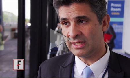
Interventions ease cancer-related stress, depression
CHICAGO – Clinicians have gotten much better in recent decades at helping patients cope with physical side effects from cancer care, but mental health interventions aimed at easing stress and fear are often absent or, at best, an afterthought.
As three new studies show, however, early psychological interventions can both improve the quality of life for patients with cancer and enhance clinical outcomes.
For example, an online stress management program dubbed STREAM, developed jointly by oncologists and psychologists, was associated with significantly better quality of life and lower levels of distress for patients 2 months after a new cancer diagnosis, compared with controls who had not yet used it, reported Viviane Hess, MD, a medical oncologist at the University Hospital of Basel (Switzerland).
“From randomized controlled trials, we know that cognitive behavioral therapies are effective in mediating distress. However, the majority of cancer patients lack psychological support, due to both barriers on the patients’ side and lack of resources on the providers’ side,” she said at a briefing at the annual meeting of the American Society of Clinical Oncology.
Other studies presented in the briefing show that psychological interventions can significantly reduce patient anxieties about the possibility of cancer recurrence and help relieve depressive symptoms for at least 6 months among patients with advanced cancers.
STREAM
Dr. Hess and her colleagues conducted a prospective, wait-list controlled trial for patients who had started therapy for a newly diagnosed cancer within the past 12 weeks. The patients were randomized to either immediate use of an online psychological intervention or to a delayed start.
The investigators chose a web-based intervention because a majority of patients with cancer already use the Internet for information, and because Internet interventions can overcome barriers of time and distance, with results comparable to those seen with face-to-face interventions, Dr. Hess said.
The intervention consisted of eight weekly sessions based on approaches used in cognitive behavioral therapy, with focus on bodily reactions to stress, cognitive stress reduction, feelings, and social interactions. Each of the eight sessions focused on a single topic. Patients were given written and audio information about each topic and then were asked to perform exercises and complete questionnaires.
For this “minimal contact” intervention, psychologists based in Basel reviewed each patient’s progress weekly and gave written feedback personalized for that patient via a secure online portal.
In all, 129 patients were randomized (65 to the intervention and 64 controls). The majority were women treated with curative intent for early-stage breast cancer. Patients newly diagnosed with lung, ovarian, and gastrointestinal cancers, as well as melanoma and lymphoma, also were included.
Patients in both the active intervention and control groups were assessed at baseline and at 2 months with three quality-of-life measures: the FACIT-F (Functional Assessment of Chronic Illness Therapy Fatigue subscale), DT (Distress Thermometer), and HADS (Hospital Anxiety and Depression Scale).
At 2 months, the mean FACIT-F score for patients in both the intervention and control groups increased, indicating an improved quality of life, but the change was significantly greater for patients in the intervention group (mean 8.59 points vs. controls; P = .0032). There was also a significant decrease in the DT scale from 6 points at baseline to 4 points at 2 months among patients randomized to the intervention, but no change among controls. There were no significant differences in either anxiety or depression between the groups, however.

“Most oncologists, particularly when we’re meeting patients for the first time and starting on curative intent therapy, it is assumed there is a level of distress … but we assume that that level of distress will fall naturally as we start them on therapy,” commented ASCO expert Don S. Dizon, MD, from the Massachusetts General Hospital Cancer Center in Boston, who was not involved in the study.
“What Dr. Hess is showing is that not only do they have distress, but it’s significant distress,” he said, adding that the study shows that online tools can be effective at providing support, and that the ease of access can help patients who might otherwise never meet with a mental health professional.
Recurrence fears assuaged
In a separate study, investigators from Australia reported results of a randomized phase II trial showing that an intervention called Conquer Fear was effective at lowering fear of recurrence among patients with successfully treated stage I-III breast, colorectal, or melanoma cancers who reported a high level of fear of recurrence.
“Fear of cancer recurrence is associated with poorer quality of life, greater distress, lack of planning for the future, avoidance or excessive use of screening, and greater health care utilization,” said Jane MacNeil Beith, MD, PhD, a medical oncologist at the University of Sydney.
Dr. Beith and her colleagues randomly assigned 222 patients to either the Conquer Fear intervention or relaxation training (control group).
Patients assigned to the control group received five 1-hour individual, face-to-face relaxation sessions over 10 weeks. The sessions included muscle relaxation, meditative relaxation, and visualization and quick relaxation techniques taught by trained study instructors. Patients in each group received instructions for home-based relaxation techniques.
The Conquer Fear intervention is intended to help patients with values clarification, detached mindfulness (focusing on the moment), attention training, meta-cognitive therapy (worry or not worry), and a behavioral contract to ensure appropriate follow-up.
They measured fear of recurrence using the validated 42-item Fear of Cancer Recurrence Inventory (FCRI), which scores fear on a scale of 0-168, with higher scores indicating higher levels of fear, Dr. Beith said.
The mean baseline FCRI scores were 82.7 for patients assigned to the intervention and 85.7 in controls.
Participants in both groups had improvement of all outcomes, but patients in the active intervention group had significantly lower FCRI total scores at the end of the intervention and at 3 and 6 months of follow-up. The severity of fear of recurrence was also lower among patients in the intervention group immediately afterward and at 3 months, but not at 6 months, she reported.
“I applaud Dr. Beith for her trial, particularly because we as oncologists have only about 15 minutes with our patients, and the assessments for our patients, even in follow-up, for the most part concentrate on cancer-related outcomes, because that’s what our patients want to know. They want to know if they’re fine, or they want to know, is their cancer back,” Dr. Dizon commented.
The study demonstrates fear of recurrence is real, but that there are also interventions that can help patients reduce those fears, he said.
CALM
In a third study, Gary Rodin, MD, from the department of supportive care at Princess Margaret Cancer Centre in Toronto, and his colleagues reported that a brief psychological intervention with the acronym CALM (for Managing Cancer and Living Meaningfully) was associated with a “clinically meaningful” reduction in depressive symptoms in patients with advanced cancer.

Patients in CALM receive three to six individual sessions delivered over 3-6 months. The program helps patients cope with symptom management and improve communications with health care providers, understand changes in themselves and their relations with others, find a personal sense of meaning and purpose, and learn to live with thoughts of the future and mortality, Dr. Rodin explained.
In a clinical trial, 52% of the 151 patients randomized to the intervention had clinically important reductions in depressive symptoms at 3 months, compared with 33% of controls assigned to usual care. At 6 months, the respective reductions in depressive symptoms were 64% vs. 35%.
The intervention also was effective at preventing depression among 137 patients without depressive symptoms at baseline. In this group, 13% of patients who received the CALM intervention had depressive symptoms at 3 months, compared with 30% of controls.
In addition, patients who received CALM support at both 3 and 6 months said they were better prepared for the end of life, reported that they had greater opportunity to talk about their concerns for the future, were less frightened, and said they had greater ability to express and manage their feelings. At 6 months these effects were strengthened, and the CALM group also felt more able to understand their cancer experience, deal with changes in relationships as a result of cancer, explore ways of communicating with their health care team and family, and clarify their values and beliefs.
“The intervention addresses both the practical problems that people face, and there are many – How do I manage my pain? My symptoms? – but also the profound problems and issues that people face – What is the meaning of my life? – when having a terminal diagnosis, for instance,” he said.
The investigators are currently establishing a global network to train health professionals in the delivery of CALM, and to evaluate the program’s effectiveness in different clinical settings and cultures, he said.
The STREAM study was institutionally supported. Dr. Hess disclosed travel and accommodations from CSL Behring. The Conquer Fear study was funded by Cancer Australia, beyondBlue, and the Australian National Breast Cancer Foundation. Dr. Beith reported no relevant disclosures. The CALM study was supported by the Canadian Institute of Health Research. Dr. Rodin reported no relevant disclosures.
CHICAGO – Clinicians have gotten much better in recent decades at helping patients cope with physical side effects from cancer care, but mental health interventions aimed at easing stress and fear are often absent or, at best, an afterthought.
As three new studies show, however, early psychological interventions can both improve the quality of life for patients with cancer and enhance clinical outcomes.
For example, an online stress management program dubbed STREAM, developed jointly by oncologists and psychologists, was associated with significantly better quality of life and lower levels of distress for patients 2 months after a new cancer diagnosis, compared with controls who had not yet used it, reported Viviane Hess, MD, a medical oncologist at the University Hospital of Basel (Switzerland).
“From randomized controlled trials, we know that cognitive behavioral therapies are effective in mediating distress. However, the majority of cancer patients lack psychological support, due to both barriers on the patients’ side and lack of resources on the providers’ side,” she said at a briefing at the annual meeting of the American Society of Clinical Oncology.
Other studies presented in the briefing show that psychological interventions can significantly reduce patient anxieties about the possibility of cancer recurrence and help relieve depressive symptoms for at least 6 months among patients with advanced cancers.
STREAM
Dr. Hess and her colleagues conducted a prospective, wait-list controlled trial for patients who had started therapy for a newly diagnosed cancer within the past 12 weeks. The patients were randomized to either immediate use of an online psychological intervention or to a delayed start.
The investigators chose a web-based intervention because a majority of patients with cancer already use the Internet for information, and because Internet interventions can overcome barriers of time and distance, with results comparable to those seen with face-to-face interventions, Dr. Hess said.
The intervention consisted of eight weekly sessions based on approaches used in cognitive behavioral therapy, with focus on bodily reactions to stress, cognitive stress reduction, feelings, and social interactions. Each of the eight sessions focused on a single topic. Patients were given written and audio information about each topic and then were asked to perform exercises and complete questionnaires.
For this “minimal contact” intervention, psychologists based in Basel reviewed each patient’s progress weekly and gave written feedback personalized for that patient via a secure online portal.
In all, 129 patients were randomized (65 to the intervention and 64 controls). The majority were women treated with curative intent for early-stage breast cancer. Patients newly diagnosed with lung, ovarian, and gastrointestinal cancers, as well as melanoma and lymphoma, also were included.
Patients in both the active intervention and control groups were assessed at baseline and at 2 months with three quality-of-life measures: the FACIT-F (Functional Assessment of Chronic Illness Therapy Fatigue subscale), DT (Distress Thermometer), and HADS (Hospital Anxiety and Depression Scale).
At 2 months, the mean FACIT-F score for patients in both the intervention and control groups increased, indicating an improved quality of life, but the change was significantly greater for patients in the intervention group (mean 8.59 points vs. controls; P = .0032). There was also a significant decrease in the DT scale from 6 points at baseline to 4 points at 2 months among patients randomized to the intervention, but no change among controls. There were no significant differences in either anxiety or depression between the groups, however.
“Most oncologists, particularly when we’re meeting patients for the first time and starting on curative intent therapy, it is assumed there is a level of distress … but we assume that that level of distress will fall naturally as we start them on therapy,” commented ASCO expert Don S. Dizon, MD, from the Massachusetts General Hospital Cancer Center in Boston, who was not involved in the study.
“What Dr. Hess is showing is that not only do they have distress, but it’s significant distress,” he said, adding that the study shows that online tools can be effective at providing support, and that the ease of access can help patients who might otherwise never meet with a mental health professional.
Recurrence fears assuaged
In a separate study, investigators from Australia reported results of a randomized phase II trial showing that an intervention called Conquer Fear was effective at lowering fear of recurrence among patients with successfully treated stage I-III breast, colorectal, or melanoma cancers who reported a high level of fear of recurrence.
“Fear of cancer recurrence is associated with poorer quality of life, greater distress, lack of planning for the future, avoidance or excessive use of screening, and greater health care utilization,” said Jane MacNeil Beith, MD, PhD, a medical oncologist at the University of Sydney.
Dr. Beith and her colleagues randomly assigned 222 patients to either the Conquer Fear intervention or relaxation training (control group).
Patients assigned to the control group received five 1-hour individual, face-to-face relaxation sessions over 10 weeks. The sessions included muscle relaxation, meditative relaxation, and visualization and quick relaxation techniques taught by trained study instructors. Patients in each group received instructions for home-based relaxation techniques.
The Conquer Fear intervention is intended to help patients with values clarification, detached mindfulness (focusing on the moment), attention training, meta-cognitive therapy (worry or not worry), and a behavioral contract to ensure appropriate follow-up.
They measured fear of recurrence using the validated 42-item Fear of Cancer Recurrence Inventory (FCRI), which scores fear on a scale of 0-168, with higher scores indicating higher levels of fear, Dr. Beith said.
The mean baseline FCRI scores were 82.7 for patients assigned to the intervention and 85.7 in controls.
Participants in both groups had improvement of all outcomes, but patients in the active intervention group had significantly lower FCRI total scores at the end of the intervention and at 3 and 6 months of follow-up. The severity of fear of recurrence was also lower among patients in the intervention group immediately afterward and at 3 months, but not at 6 months, she reported.
“I applaud Dr. Beith for her trial, particularly because we as oncologists have only about 15 minutes with our patients, and the assessments for our patients, even in follow-up, for the most part concentrate on cancer-related outcomes, because that’s what our patients want to know. They want to know if they’re fine, or they want to know, is their cancer back,” Dr. Dizon commented.
The study demonstrates fear of recurrence is real, but that there are also interventions that can help patients reduce those fears, he said.
CALM
In a third study, Gary Rodin, MD, from the department of supportive care at Princess Margaret Cancer Centre in Toronto, and his colleagues reported that a brief psychological intervention with the acronym CALM (for Managing Cancer and Living Meaningfully) was associated with a “clinically meaningful” reduction in depressive symptoms in patients with advanced cancer.
Patients in CALM receive three to six individual sessions delivered over 3-6 months. The program helps patients cope with symptom management and improve communications with health care providers, understand changes in themselves and their relations with others, find a personal sense of meaning and purpose, and learn to live with thoughts of the future and mortality, Dr. Rodin explained.
In a clinical trial, 52% of the 151 patients randomized to the intervention had clinically important reductions in depressive symptoms at 3 months, compared with 33% of controls assigned to usual care. At 6 months, the respective reductions in depressive symptoms were 64% vs. 35%.
The intervention also was effective at preventing depression among 137 patients without depressive symptoms at baseline. In this group, 13% of patients who received the CALM intervention had depressive symptoms at 3 months, compared with 30% of controls.
In addition, patients who received CALM support at both 3 and 6 months said they were better prepared for the end of life, reported that they had greater opportunity to talk about their concerns for the future, were less frightened, and said they had greater ability to express and manage their feelings. At 6 months these effects were strengthened, and the CALM group also felt more able to understand their cancer experience, deal with changes in relationships as a result of cancer, explore ways of communicating with their health care team and family, and clarify their values and beliefs.
“The intervention addresses both the practical problems that people face, and there are many – How do I manage my pain? My symptoms? – but also the profound problems and issues that people face – What is the meaning of my life? – when having a terminal diagnosis, for instance,” he said.
The investigators are currently establishing a global network to train health professionals in the delivery of CALM, and to evaluate the program’s effectiveness in different clinical settings and cultures, he said.
The STREAM study was institutionally supported. Dr. Hess disclosed travel and accommodations from CSL Behring. The Conquer Fear study was funded by Cancer Australia, beyondBlue, and the Australian National Breast Cancer Foundation. Dr. Beith reported no relevant disclosures. The CALM study was supported by the Canadian Institute of Health Research. Dr. Rodin reported no relevant disclosures.
AT ASCO 2017
Key clinical point: Oncologists and patients may focus on only the clinical aspects of care while overlooking cancer-associated distress, anxiety, or fear.
Major finding: Each of three psychological interventions was effective at reducing mental health symptoms in patients with cancer.
Data source: Randomized trials of interventions aimed at relieving mental health symptoms associated with a new cancer diagnoses, fear of cancer recurrence, and advanced or metastatic disease.
Disclosures: The STREAM study was institutionally supported. Dr. Hess disclosed travel and accommodations from CSL Behring. The Conquer Fear study was funded by Cancer Australia, beyondBlue, and the Australian National Breast Cancer Foundation. Dr. Beith reported no relevant disclosures. The CALM study was supported by the Canadian Institute of Health Research. Dr. Rodin reported no relevant disclosures.
VIDEO: TRK inhibitor shows 76% ORR across diverse cancers harboring TRK fusions
CHICAGO – An integrated analysis of three trials has shown that larotrectinib, an oral selective inhibitor of tropomyosin receptor kinase (TRK), has durable efficacy across diverse adult and pediatric cancers harboring TRK fusions, netting an impressive 76% overall response rate.
Lead study author David Hyman, MD, chief of early drug development at Memorial Sloan Kettering Cancer Center in New York, discussed highlights of the analysis, larotrectinib’s regulatory status, and implications for TRK fusion testing in clinical care at the annual meeting of the American Society of Clinical Oncology.
Dr. Hyman disclosed that he has a consulting or advisory role with Atara Biotherapeutics, Chugai Pharma, and CytomX Therapeutics, and that he receives research funding from AstraZeneca and Puma Biotechnology. The study was funded by Loxo Oncology.
The video associated with this article is no longer available on this site. Please view all of our videos on the MDedge YouTube channel
AT ASCO 2017
Severe health conditions decrease among childhood cancer survivors

CHICAGO—The 15-year cumulative incidence of severe health conditions for survivors of childhood cancer has decreased over the past 30 years, from 12.7% for those diagnosed in the 1970s to 10.1% and 8.9% for those diagnosed in the 1980s and 1990s, respectively. And the decreases were greatest for patients with Wilms’ tumor and Hodgkin lymphoma (HL), followed by patients with astrocytoma, non-Hodgkin lymphoma (NHL), and acute lymphoblastic leukemia (ALL).
Investigators of the Childhood Cancer Survivor Study (CCSS) undertook a retrospective cohort analysis of children aged 0 – 14 years diagnosed with cancer between 1970 and 1999. Their goal was to determine whether cancer therapy modifications have maintained cure rates while decreasing the risk of late effects of therapy.
Todd M. Gibson, PhD, of St Jude Children’s Research Hospital in Memphis, Tennessee, presented the findings at the 2017 annual meeting of the American Society for Clinical Oncology (ASCO) as abstract LBA10500.
Researchers analyzed data from 23,600 childhood cancer survivors in the CCSS who were alive 5 years after diagnosis. The patients had leukemia, lymphoma, CNS malignancies, Wilms tumor, neuroblastoma, or soft-tissue/bone sarcoma.
Dr Gibson noted that while 83% of children with a malignancy achieve a 5-year survival, more than half develop at least one severe, disabling, life-threatening health condition by age 50.
The survivors were a median age at last follow-up of 28 years (range, 5-63) and the median time since diagnosis was 21 years (range, 5-43).
The investigators found significant decreases in severe health conditions in 6 diagnostic groups:
- Wilms tumor, decreased from 13% to 5% (P<0.0001)
- HL, decreased from 18% to 11% (P<0.0001)
- Astrocytoma, decreased from 15% to 9% (P=0.004)
- NHL, decreased from 10% to 6% (P=0.04)
- ALL, decreased from 9% to 7% (P=0.002)
- Ewings sarcoma, decreased from 19% to 10% (P=0.01)
They found no reductions in subsequent severe health conditions among survivors of neuroblastoma, acute myeloid leukemia (AML), soft tissue sarcoma, or osteosarcoma.
The investigators believe the decreases were driven mainly by a reduced incidence of endocrine conditions, subsequent malignant neoplasms, gastrointestinal and neurological conditions, but not cardiac or pulmonary conditions.
They also analyzed the reduction in treatment intensities by decade for different diseases and found they correlated with the reduced incidence of serious chronic health conditions by 15 years after diagnosis.
The National Institutes of Health funded the study.