User login
Rash in HIV-positive man

Taking into consideration the patient’s 2 predisposing risk factors (AIDS and dementia), the FP diagnosed seborrheic dermatitis. The FP also noted seborrheic dermatitis on the scalp. The scalp is frequently involved in cases of seborrheic dermatitis on the face.
Seborrheic dermatitis is a common, chronic, relapsing dermatitis affecting sebum-rich areas of the body. Its presentation may vary from mild erythema to greasy scale, and rarely, to erythroderma. Patients with seborrheic dermatitis may be colonized with certain species of lipophilic yeast of the genus Malassezia (also known as Pityrosporum). Malassezia, however, is considered normal skin flora because it is also found in unaffected people.
Some evidence suggests that an overgrowth of Malassezia may produce different irritants or metabolites on affected skin, leading to inflammation. It’s postulated that an overgrowth of Malassezia leads to the inflammatory dermatitis in seborrhea.
The goals of treatment are to diminish the fungal overgrowth and treat the inflammatory response. Thus, the mainstay of treatment is ongoing topical antifungals in shampoos and other preparations, along with a topical steroid to treat the inflammation.
The FP prescribed 2% ketoconazole shampoo to be used twice weekly and suggested that a selenium-containing shampoo be used on the other days of the week, as well as a 2.5% hydrocortisone cream to be applied twice daily to the face until the dermatitis resolved. The FP also prescribed a topical antifungal, cyclopirox 1% cream, to be applied twice daily on an ongoing basis to keep the Malassezia under control. At a one-month follow-up, the patient’s skin and scalp were cleared.
Photos and text for Photo Rounds Friday courtesy of Richard P. Usatine, MD. This case was adapted from: Hancock M, Bae Y, Usatine R. Seborrheic dermatitis. In: Usatine R, Smith M, Mayeaux EJ, et al, eds. Color Atlas of Family Medicine. 2nd ed. New York, NY: McGraw-Hill; 2013: 871-877.
To learn more about the Color Atlas of Family Medicine, see: www.amazon.com/Color-Family-Medicine-Richard-Usatine/dp/0071769641/
You can now get the second edition of the Color Atlas of Family Medicine as an app by clicking on this link: usatinemedia.com

Daily 150-mg aspirin dose slashes preterm preeclampsia risk
While low-dose aspirin has been shown for decades to decrease the risk of preterm preeclampsia, yet to be established are the optimal dose and time of day to administer aspirin; how preeclampsia risk is best assessed; and at what gestational week it is best to start aspirin.
New findings from a randomized, double-blind, placebo-controlled trial show that a daily 150-mg dose of aspirin, taken at night, significantly reduced incidence of preterm preeclampsia in women identified as being at high risk in the first trimester (N Engl J Med. 2017 June 28. doi: 10.1056/NEJMoa1704559).
After 156 women dropped out of the study or were lost to follow-up, investigators had results from 798 women in the aspirin group and 822 in the placebo group, with about 80% of subjects having taken aspirin or placebo as directed. The study’s primary outcome was delivery with preeclampsia before 37 weeks, seen in 13 women (1.6%) in the aspirin group, compared with 35 (4.3%) in the placebo group (odds ratio 0.38; 95% confidence interval, 0.20 to 0.74; P = .004).
Other decisions incorporated into the study design, including those about aspirin dosage (higher than the currently recommended 60-80 mg for this population), taking aspirin at night, and the gestational age at which to start aspirin, were based on results from prior trials, studies, and meta-analyses, the researchers said.
Screening at 11-13 weeks’ gestation identifies less than 40% of term preeclampsia, studies have shown, and aspirin did not reduce term preeclampsia in this study, the investigators said.
The European Union Seventh Framework Program and the Fetal Medicine Foundation (UK) sponsored the study. None of the investigators had any relevant financial disclosures.
FROM NEW ENGLAND JOURNAL OF MEDICINE
Key clinical point:
Major finding: Among patients taking aspirin, 1.6% developed preterm preeclampsia, compared with 4.3% of those taking placebo (odds ratio 0.38; 95% confidence interval, 0.20 to 0.74; P = .004).
Data source: A randomized, international, multicenter trial enrolling nearly 1,800 women identified through screening as being at high risk of preeclampsia.
Disclosures: The European Union Seventh Framework Program and the Fetal Medicine Foundation (UK) sponsored the study. None of the investigators had any relevant financial disclosures.
Atypical Femoral Fracture Due to Daily Ibandronate
As more people are surviving breast cancer, more will be given bisphosphonate (BP) for bone metastases—and may be at risk for treatment adverse effects, such as atypical femoral fracture (AFF). Clinicians from Royal Victoria Hospital, Belfast, United Kingdom, describe what they learned in the first reported case of AFF caused by daily ibandronate.
The patient, a 55-year-old woman, had been diagnosed and treated for breast cancer. Twelve years later, she began having hip and lower back pain and was diagnosed with bony metastatic spread. She was started on hormone-suppressing therapy as well as daily ibandronic acid. Four years later, she began having new lower limb and groin pain. Radiography, bone scans and other tests indicated further metastatic spread to her left femur.
While she was waiting for scheduled radiotherapy, she fell, fracturing her left femur. Radiographs revealed AFF, presumed secondary to her BP therapy rather than metastasis. She underwent intramedullary nail fixation.
Evidence suggests that AFFs are stress fractures, the clinicians say. Repetitive loading on bone can lead to micro cracks, which are even more vulnerable to stress when BPs suppress the normal bone repair process.
Atypical femoral fracture is rare. Most cases have been patients receiving intravenous BP; to the clinicians’ knowledge this is the first report of AFF with an oral BP prescribed for bone metastatic breast cancer. But incidence of AFF grows with prolonged treatment, and the risk is raised with oral dosing, which is much higher in cancer cases, the clinicians note, compared with osteoporosis.
Because patients often have prodromal pain before an overt break, the clinicians suggest asking all oncology patients on BP about pain in thigh, hip, or groin. Bone scans and magnetic resonance imaging may reveal an imminent fracture. The clinicians caution that AFFs can be bilateral, so the opposite side also should be imaged.
Source:
Espey R, Grimes S, Heyburn G, Kealey WD. BMJ Case Rep. 2017;2017. pii: bcr-2016-217489.
doi: 10.1136/bcr-2016-217489.
First Cancer Treatment Based on Biomarkers Is Approved
In an “important first,” the FDA has fast-tracked a cancer treatment based on a biomarker rather than on the location of the tumor.
Pembrolizumab (Keytruda) has already been approved for certain patients with metastatic melanoma, metastatic non-small cell lung cancer, and other cancers. With the new approval, pembrolizumab is now indicated for adult and pediatric patients with unresectable or metastatic solid tumors that have a biomarker known as microsatellite instability-high (MSI-H) or mismatch repair deficient (dMMR). Microsatellite instability-high and dMMR tumors have abnormalities that affect the proper repair of DNA inside the cell. Pembrolizumab blocks a cellular pathway (PD-1/PD-L1—proteins found in immune cells and some cancer cells), which may help the immune system fight the cancer cells.
Tumors with the biomarkers are most commonly found in colorectal, endometrial, and gastrointestinal cancers. About 5% of patients with metastatic colorectal cancer have MSI-H or dMMR tumors. Less commonly, they are found in cancers in the breast, prostate, bladder, thyroid gland, and other locations.
In 5 clinical trials, of 149 patients treated with pembrolizumab for 15 cancer types, 40% had a complete or partial response. For most of those patients, the response lasted ≥ 6 months.
Oral combo produces deep, durable responses in MM

An all-oral treatment regimen can produce deep and durable responses in patients with newly diagnosed multiple myeloma (MM), according to a speaker at the 22nd Congress of the European Hematology Association (EHA).
In a phase 1/2 study, patients with newly diagnosed MM received treatment with ixazomib, lenalidomide, and dexamethasone for up to 12 cycles, followed by maintenance with ixazomib alone.
The overall response rate was 88% for the entire cohort and 80% among patients who did not proceed to transplant.
The median progression-free survival was 35.4 months in the entire cohort and 29.4 months in patients who did not proceed to transplant.
Most patients did not complete all 12 cycles of induction, but 38% proceeded to maintenance, and 12% remain on study treatment at a median follow-up of 56 months.
Shaji Kumar, MD, of the Mayo Clinic in Rochester, Minnesota, presented these results at the EHA Congress as abstract S408. The research was sponsored by Millennium Pharmaceuticals, Inc., a subsidiary of Takeda Pharmaceutical Company Limited.
Patients
The trial included 65 patients. Their median age was 66 (range, 34-86), and 55% were male. Eight percent of patients had high-risk cytogenetics.
ECOG performance status was 0 for 43% of patients, 1 for 52%, and 2 for 5%. ISS stage was 1 for 43% of patients, 2 for 43%, and 3 for 14%.
Treatment
Patients received ixazomib (1.68 - 3.95 mg/m2 in phase 1 and 4.0 mg in phase 2 on days 1, 8, and 15) plus lenalidomide (25 mg on days 1-21), and dexamethasone (40 mg on days 1, 8, 15, and 22) for up to twelve, 28-day cycles. Patients could then receive ixazomib maintenance until disease progression or unacceptable toxicity.
Twenty-three patients (35%) discontinued induction to undergo hematopoietic stem cell transplant (HSCT).
Thirty-seven patients discontinued study treatment for other reasons—17 of them during induction. Twenty-one patients discontinued due to progressive disease, 9 due to adverse events (AEs), 5 due to patient withdrawal, 1 due to unsatisfactory response, and 1 due to other reasons.
Twenty-five patients completed induction and went on to receive ixazomib maintenance. Five patients remain on study treatment.
Safety
Seventy-eight percent of patients had a grade 3 or higher AE, 46% had a serious AE, 57% had an AE leading to dose reduction, and 15% had an AE leading to discontinuation of any of the study drugs. There were 2 on-study deaths.
The most common grade 3 or higher AEs were neutropenia, thrombocytopenia, diarrhea, back pain, vomiting, nausea, peripheral neuropathy, and rashes/eruptions/exanthems.
Efficacy
The overall response rate was 88% for the entire cohort and 80% among patients who did not go on to receive HSCT.
The complete response (CR) rates were 23% and 32%, respectively. Thirty four percent and 32%, respectively, had a very good partial response (VGPR). And 30% and 17%, respectively, had a partial response (PR).
Thirty-two percent of patients who received maintenance (n=8) had an improvement in their response during the maintenance phase. Two patients went from a VGPR to a stringent CR, 3 went from a VGPR to a CR, 2 went from a VGPR to a near CR, and 1 went from a PR to a VGPR.
Sixteen patients were evaluated for minimal residual disease (MRD). Of the patients with a stringent CR/CR, 89% (8/9) were MRD-negative.
The median progression-free survival was 35.4 months in the entire cohort and 29.4 months in patients who did not proceed to HSCT. The 4-year overall survival rate was 84% and 82%, respectively.
“Long-term follow-up, a median follow-up of 56 months, with the combination of ixazomib, lenalidomide, and dexamethasone demonstrates that this is an efficacious regimen for newly diagnosed myeloma patients, with excellent tolerability, and something that can be maintained for a long period of time,” Dr Kumar said. “These results form the basis for an ongoing phase 3 program.” ![]()
Mapping the genomic landscape of T-ALL

Researchers say they have uncovered new details of the genomic landscape of T-lineage acute lymphoblastic leukemia (T-ALL).
The team believes their findings will aid the development of drugs to target newly discovered mutations and enable researchers to engineer better mouse models to probe the leukemia’s aberrant biological machinery.
Charles Mullighan, MD, MBBS, of St. Jude Children’s Research Hospital in Memphis, Tennessee, and his colleagues conducted this research and reported their findings in Nature Genetics.
“This first comprehensive and systematic analysis in a large group of patients revealed many new mutations that are biologically significant as well as new drug targets that could be clinically important,” Dr Mullighan said.
“Leukemias typically arise from multiple genetic changes that work together. Most previous studies have not had the breadth of genomic data in enough patients to identify the constellations of mutations and recognize their associations.”
Dr Mullighan and his colleagues sequenced the genomes of 264 children and young adults with T-ALL. This revealed 106 driver genes, half of which had not been identified in childhood T-ALL.
“We went into this study knowing that we didn’t know the full genomic landscape of T-ALL,” said Stephen Hunger, MD, of the Children’s Hospital of Philadelphia in Pennsylvania.
“But we were surprised that over half of the new targets and mutations were previously unrecognized. It was particularly unexpected and very striking that some mutations were exclusively found in some subtypes of T-ALL but not others.”
The researchers’ analysis confirmed that T-ALL was driven by mutations in known signaling pathways, including JAK-STAT, Ras, and PTEN-PI3K. However, the study also revealed new mutations in those known pathways.
In addition, the researchers identified cases in which the same T-ALL subtype had mutations in different pathways triggered by the same founding mutation.
“We believe this finding suggests we can target such subtypes with an inhibitor drug for one of the pathways, and it’s likely to be effective,” Dr Mullighan said.
He and his colleagues also believe the mutations uncovered in this study will enable researchers to create mouse models that more accurately reflect human T-ALL.
“We now have a launching pad, if you will, to design mouse models that include multiple genetic mutations to more faithfully reflect the leukemias we see in humans,” Dr Mullighan said.
Data from this study are available to researchers through the St. Jude PeCan data portal and the TARGET Data Matrix. ![]()
FDA puts pembrolizumab trials on clinical hold

The US Food and Drug Administration (FDA) has placed clinical holds on 3 trials testing combination therapy with pembrolizumab (Keytruda) in patients with multiple myeloma (MM)—KEYNOTE-183, KEYNOTE-185, and KEYNOTE-023.
In 2 of these trials—KEYNOTE-183 and -185—there were more deaths among patients receiving pembrolizumab than among patients receiving comparator treatments.
KEYNOTE-183 is a phase 3 study of pomalidomide and low-dose dexamethasone with or without pembrolizumab in refractory or relapsed and refractory MM.
KEYNOTE-185 is a phase 3 study of lenalidomide and low-dose dexamethasone with or without pembrolizumab in newly diagnosed and treatment-naïve MM.
The excess deaths in the pembrolizumab arms of KEYNOTE-183 and -185 led to a pause in enrollment for both trials, which was announced last month.
Now, the FDA says available data suggest the risks of pembrolizumab plus pomalidomide or lenalidomide outweigh any potential benefit for MM patients.
Therefore, all patients enrolled in KEYNOTE-183 and -185 will discontinue investigational treatment with pembrolizumab.
The same applies to patients in the pembrolizumab/lenalidomide/dexamethasone cohort of KEYNOTE-023.
KEYNOTE-023 is a phase 1 trial of pembrolizumab in combination with backbone treatments. Cohort 1 of this trial was designed to evaluate pembrolizumab in combination with lenalidomide and dexamethasone in MM patients who received prior treatment with an immunomodulatory agent (lenalidomide, pomalidomide, or thalidomide).
Pembrolizumab is a humanized monoclonal antibody that blocks interaction between the programmed cell death protein 1 (PD-1) and its receptor ligands, PD-L1 and PD-L2.
Pembrolizumab is FDA-approved to treat classical Hodgkin lymphoma, melanoma, non-small cell lung cancer, head and neck cancer, urothelial carcinoma, and microsatellite instability-high solid tumors. ![]()
What’s Her Dry-agnosis?
ANSWER
The correct answer is eczema/atopic dermatitis (choice “c”).
Patients with eczema have a low threshold for itching, so they scratch, often making the condition appear far worse than it really is. In such cases, it’s typical for the problem to be mistaken for impetigo (choice “a”) or yeast infection (choice “d”). The latter, much like psoriasis (choice “b”), is quite rare in the perioral area. Furthermore, the patient had no indicative signs of psoriasis.
DISCUSSION
Eczema is one of several manifestations of atopic dermatitis, a syndrome that affects more than 20% of newborns in this country. These children have extraordinarily dry, thin skin that overreacts to wetting and drying, as well as scratching. Certain areas are especially prone to these changes and consequently develop scaling and itching.
The perioral area is one, in large part because it is kept moist by food, drink, nasal secretions, and saliva (due to habitual lip licking). The scaly rash becomes inflamed; on people with skin of color, this frequently manifests with hyperpigmentation.
When picked enough, this type of rash can become impetiginized—that is, superficially infected with staph or strep. But in this patient’s case, antibiotics were of no practical use.
A topical steroid ointment (hydrocortisone 2.5%) was used, and the patient was urged to stop picking (or licking!) and to use moisturizers (eg, petroleum jelly). The family was educated about the problem and its origins, and the parents were reassured of the self-limiting nature of postinflammatory hyperpigmentation (a major source of concern).
In this case, the key to making the correct diagnosis was the significance of the personal and family history of atopy—and an appre
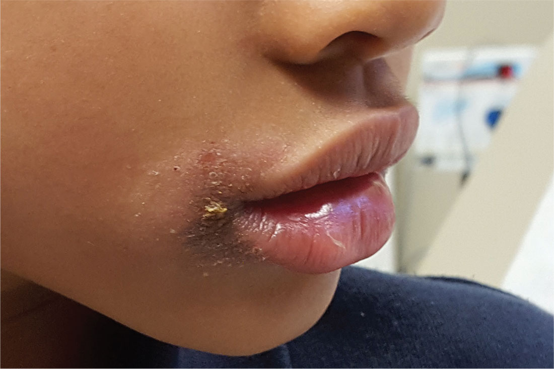
The persistent rash around this 5-year-old African-American girl’s mouth is causing a great deal of concern for her parents, who request referral to dermatology after four months of attempted treatment. Oral and topical antibiotics, as well as antifungal products (nystatin and fluconazole), have been used to no good effect.
The patient is often seen licking her lips, which dries and irritates them. Fine crusting surrounds her mouth, particularly the left lateral oral commissure. The skin in the affected areas is darker than the rest.
Elsewhere, her type IV skin is quite dry, with focal areas of scaling on the arms and antecubital region. No rash is seen on extensor areas, nor are there any changes in her nails.
The child is markedly atopic, as are her siblings, who are present for the exam. The patient and her siblings are all congested, breathing through their mouths (“allergies,” according to their parents).

Loop ileostomy tops colectomy for IBD rescue
SEATTLE – Diverting loop ileostomies save patients with inflammatory bowel disease (IBD) with severe colitis from rushed total abdominal colectomies, buying time for patient optimization before surgery, and perhaps even saving colons, according to a report from the University of California, Los Angeles.
Urgent colectomy is the standard of care, but it’s a big operation when patients aren’t doing well. Immunosuppression, malnutrition, and other problems lead to high rates of complications.
In 2013, UCLA physicians decided to try rescue diverting loop ileostomies (DLIs), a relatively quick, minimally invasive option to temporarily divert the fecal stream, instead. The idea is to give the colon a chance to heal and the patient another shot at medical management and recovery before definitive surgery. There’s even a chance of colon salvage.
The approach has been working well at UCLA. Investigators previously reported good results for their first eight patients. They presented updated results for the series – now up to 34 patients – at the annual meeting of the American Society of Colon and Rectal Surgeons.
So far, DLI allowed 91% of patients (31/34) to avoid urgent total colectomies. It’s “a safe alternative. Patients undergoing DLI have acceptably low complication rates and most are afforded time for medical and nutritional optimization prior to proceeding with their definitive surgical care,” said presenter Tara Russell, MD, a UCLA surgery resident.
“Currently, [almost every] patient presenting with acute colitis who we aren’t able to get to the point of discharge with medical optimization” is now offered rescue DLI at the university, and patients have been eager for a chance at avoiding total colectomy. The only patients who are not offered DLI are those with, for instance, fulminant toxic megacolon, Dr. Russell said.
The DLI approach failed in just 2 of the 18 ulcerative colitis patients and 1 of the 16 Crohn’s patients in the series. All three went on to emergent total colectomies 11-53 days after the procedure.
The majority of DLI patients tolerated oral intake by postop day 1, and the median time to resuming a regular diet was 2 days. Most people were discharged within a day or 2 of diversion, and a few took longer to achieve medical rescue. Almost 90% had an improvement in nutritional status, and over 80% went on to elective laparoscopic definitive procedures or colon salvage.
Two patients had postop wound infections, “but there were no other complications” with DLI, Dr. Russell said.
All DLIs were performed with a single-incision laparoscopic approach and took an average of about a half hour. Most of the diversions were in the right lower abdominal quadrant.
The mean age of the patients was 36 years, with a range of 16-81 years. Just over half were men. Of 21 patients who met systemic inflammatory response syndrome criteria at the time of operation, 13 (62%) resolved within 24 hours of DLI.
AT ASCRS 2017
Key clinical point:
Major finding: DLI allowed 91% of patients (31/34) to avoid urgent total colectomies.
Data source: A report on 34 patients with severe, acute inflammatory bowel disease colitis.
Disclosures: The presenter had no disclosures.
Coaching ‘No’
In a recent column entitled “To the limit,” I tried to make the case that the negative consequences of permissive parenting are numerous enough to warrant the attention of primary care pediatricians and family physicians. The evidence linking atypical sensory adaptation, behavior difficulties, sleep deprivation, and obesity to a permissive parenting style is just beginning to appear in the literature, but the numbers are in sync with the anecdotal observations of many experienced pediatricians like me. In that previous column,
First, let me make it clear that I don’t consider parenting style to be a topic that needs to occur on the checklist of every patient at every health maintenance visit. You already are overburdened with the demands of experts who have lobbied to have their favorite hot button issues included in your 15 minutes of face-to-face time with your young patients.
We also must accept our limited role as advisors. There are many ways to skin a cat and to raise a child. Homogeneity is not our goal. We must respect the cultural and philosophical differences that exist in our society. However, in my opinion, the unhealthy consequences of permissive parenting deserve a sensitive attempt at education and some gentle anticipatory guidance ... hopefully without an aroma of condescension.
The opportunities for our input begin in the first few months of life when parents are faced with the difficult questions of whether it is safe and appropriate to allow their infant to cry himself to sleep and whether a mom must allow her infant to use her breast as a pacifier. With the transition to solid food comes the challenge of how to manage the inevitable rejection of new tastes, colors, and textures. Of course, most parents find these issues challenging, but to what degree a parent can internalize your reassurance and advice is a good reflection on where he or she sits on the permissive to authoritarian spectrum of parenting.
With an infant’s rapidly advancing motor skills comes the question of when, where, and how to create boundaries to keep the child safe ... and to protect the environment from the surprisingly destructive power of an inquisitive toddler. Here the permissive parent will be continually challenged when he or she finds that simply saying “No” or “Don’t” doesn’t always work ... to some extent because, up to this point, the child has never encountered a situation in which s/he hasn’t gotten what s/he wants.

This is not an issue in which we should allow ourselves to get bogged down in circuitous philosophical arguments. We must keep our advice practical and focused on issues of safety and health. I have found that a significant number of permissive parents can learn the difficult skill of saying “No” to their children. It takes time, but it is time well spent.
Dr. Wilkoff practiced primary care pediatrics in Brunswick, Maine for nearly 40 years. He has authored several books on behavioral pediatrics, including “How to Say No to Your Toddler.” Email him at [email protected].
In a recent column entitled “To the limit,” I tried to make the case that the negative consequences of permissive parenting are numerous enough to warrant the attention of primary care pediatricians and family physicians. The evidence linking atypical sensory adaptation, behavior difficulties, sleep deprivation, and obesity to a permissive parenting style is just beginning to appear in the literature, but the numbers are in sync with the anecdotal observations of many experienced pediatricians like me. In that previous column,
First, let me make it clear that I don’t consider parenting style to be a topic that needs to occur on the checklist of every patient at every health maintenance visit. You already are overburdened with the demands of experts who have lobbied to have their favorite hot button issues included in your 15 minutes of face-to-face time with your young patients.
We also must accept our limited role as advisors. There are many ways to skin a cat and to raise a child. Homogeneity is not our goal. We must respect the cultural and philosophical differences that exist in our society. However, in my opinion, the unhealthy consequences of permissive parenting deserve a sensitive attempt at education and some gentle anticipatory guidance ... hopefully without an aroma of condescension.
The opportunities for our input begin in the first few months of life when parents are faced with the difficult questions of whether it is safe and appropriate to allow their infant to cry himself to sleep and whether a mom must allow her infant to use her breast as a pacifier. With the transition to solid food comes the challenge of how to manage the inevitable rejection of new tastes, colors, and textures. Of course, most parents find these issues challenging, but to what degree a parent can internalize your reassurance and advice is a good reflection on where he or she sits on the permissive to authoritarian spectrum of parenting.
With an infant’s rapidly advancing motor skills comes the question of when, where, and how to create boundaries to keep the child safe ... and to protect the environment from the surprisingly destructive power of an inquisitive toddler. Here the permissive parent will be continually challenged when he or she finds that simply saying “No” or “Don’t” doesn’t always work ... to some extent because, up to this point, the child has never encountered a situation in which s/he hasn’t gotten what s/he wants.
This is not an issue in which we should allow ourselves to get bogged down in circuitous philosophical arguments. We must keep our advice practical and focused on issues of safety and health. I have found that a significant number of permissive parents can learn the difficult skill of saying “No” to their children. It takes time, but it is time well spent.
Dr. Wilkoff practiced primary care pediatrics in Brunswick, Maine for nearly 40 years. He has authored several books on behavioral pediatrics, including “How to Say No to Your Toddler.” Email him at [email protected].