User login
How to prioritize CVD reduction in type 2 diabetes
LOS ANGELES – In the opinion of Mikhail N. Kosiborod, MD, the paradigm of treating patients with type 2 diabetes should shift from a narrow focus on hemoglobin A1c control to a broader strategy of reducing cardiovascular risk.
“We already know that the number one killer of patients with diabetes is cardiovascular disease, and we already know that lowering HbA1c as a general strategy does not substantially lower the risk of most important CVD events,” Dr. Kosiborod, a cardiologist at Saint Luke’s Mid-America Heart Institute, Kansas City, Mo., said at the World Congress on Insulin Resistance, Diabetes & Cardiovascular Disease.
Physicians know that some medications lower the risk of cardiovascular events – including cardiovascular death – substantially, and other drugs don’t. “The bottom line is that we are not talking about ignoring HbA1c, but it’s how you get there that’s important – how you do it and in whom,” Dr. Kosiborod explained.
He pointed to a meta-analysis of four large diabetes trials involving 27,049 participants and 2,370 major vascular events (Diabetologia. 2009 Nov;52[11]:2288-98). It found that the general strategy of targeting more-intensive glucose lowering modestly reduced nonfatal myocardial infarction and increased major hypoglycemia over 4.4 years in people with type 2 diabetes – yet there was no difference in the effect of intensive glucose control on cardiovascular death or hospitalization for heart failure.
“Some point to the benefit of glucose control on the risk of nonfatal myocardial infarction, but that’s a modest benefit,” he said. “It’s observed beyond the randomization phase of clinical trials and takes many years to see it. It’s a large, very long-term investment for a modest reduction in MI risk, with no benefit in death or heart failure. So, when you test intensive glucose control as a general strategy, it has not been successful in reducing cardiovascular complications of type 2 diabetes.”
However, there is now evidence that specific classes of medications, such as sodium-glucose co-transporter-2 (SGLT2) inhibitors and glucagon-like peptide-1 (GLP-1) receptor agonists, initially developed for glucose lowering in type 2 diabetes, can significantly reduce cardiovascular risk within a relatively short time frame.
In EMPA-REG (Empagliflozin Cardiovascular Outcome Event Trial in Type 2 Diabetes Mellitus Patients), the first trial to demonstrate such benefits, all patients had established CVD, compared with 67% of patients in CANVAS (Canagliflozin Cardiovascular Assessment Study), a second RCT program to report cardiovascular outcomes with SGLT2 inhibitors. In the meantime, about 15%-20% of patients in real-world clinical practice have established CVD.
This led Dr. Kosiborod and his associates to launch CVD-REAL (Comparative Effectiveness of Cardiovascular Outcomes in New Users of SGLT2 Inhibitors), a real-world comparative effectiveness study that evaluated hospitalization for heart failure and total mortality among new users of SGLT2 inhibitors, compared with other glucose-lowering drugs.
In all, 154,528 patients in six countries were initiated on an SGLT2 inhibitor, and 154,528 were initiated on other glucose-lowering drugs (Circulation. 2017 May 18. doi: 10/1161/circulationaha.117.029190). The greatest exposure time was observed from canagliflozin (53%) followed by dapagliflozin (42%) and empagliflozin (5%).
The pooled analysis showed that initiation of SGLT2 inhibitors was associated with a significantly lower risk of heart failure events, compared with other glucose-lowering drugs (risk ratio, 0.61; P less than .001). The researchers observed an overall 39% lower risk of heart failure hospitalization, 51% reduction in total death, and 46% reduction in the composite of heart failure hospitalization or death.
“There was no heterogeneity across countries, despite the fact that the health care systems were very different and the prescribing patterns were very different,” he said.
Dr. Kosiborod, who is also professor of medicine at the University of Missouri-Kansas City, noted that 13% of patients from CVD-REAL had established CVD, while 87% did not. When comparing the results within these two key subgroups, “what’s striking is the difference in event rates, stratified by treatment allocation,” he said of the unpublished data.
“If you look at the composite outcome of heart failure or death, you see an almost seven-fold difference in annualized event rates – about 7% per year in patients with established CVD, compared with about 1% per year in the primary prevention cohort,” he explained. “But the relative risk reduction associated with SGLT2 inhibitors versus other glucose-lowering drugs is identical across both patient groups. That’s a good lesson in epidemiology: You can have patients with dramatically different absolute risks, dramatically different absolute risk reductions, and therefore dramatically different numbers needed to treat, but identical relative risk reductions.”
Dr. Kosiborod also pointed out that heart failure is emerging as one of the most important outcomes in trials patents with type 2 diabetes.
“That’s because people with diabetes who develop heart failure have very poor outcomes,” he said. “Among elderly patients with type 2 diabetes who develop new heart failure, there’s less than 25% survival at 5 years. That’s the reason, I think, that if you really want to impact survival and complication rates in people with diabetes, preventing and treating heart failure is one of the surest ways of doing so.
“You shouldn’t just think of the patient in front of you as someone who has an A1c of 7%, 8%, or 9%,” he cautioned. “You should also start thinking of where the patient is on the spectrum of cardiovascular disease, all the way from CVD risk factors only to symptomatic heart failure.”
Some evidence already exists to help clinicians make treatment decisions based on where the patients fall on that spectrum, he continued.
For example, clinical trials have demonstrated that in patients with established atherosclerotic cardiovascular disease, GLP-1 receptor agonists and SGLT2 inhibitors can reduce the risk of cardiovascular events, including, in some cases, cardiovascular death.
“We don’t have a lot of data demonstrating benefit for patients with recent acute coronary syndrome,” he said. “Some compounds have proven to be neutral, but none has been proven to save lives in this patient group.
“Now, we also have data for people with prior stroke that pioglitazone may be beneficial in managing those patients to prevent recurrent stroke and MI, based on the recent IRIS Trial, provided they don’t have heart failure at baseline,” Dr. Kosiborod added. “We don’t have definitive data yet in people with established heart failure, but those studies are ongoing.”
Dr. Kosiborod disclosed that he is a consultant for Amgen, AstraZeneca, Boehringer Ingelheim, Eisai, Glytec, GSK, Intarcia, Merck (Diabetes), Novartis, Novo Nordisk, Sanofi, and ZS Pharma. He has also received research grants from AstraZeneca and Boehringer Ingelheim.
EXPERT ANALYSIS FROM WCIRDC 2017
Heart attacks bring 12 weeks of higher stroke risk
LOS ANGELES – , based on a sample of Medicare beneficiaries.
The period of elevated stroke risk following an MI extends beyond the 30-day window that has traditionally been considered the interval of highest risk, Alexander E. Merkler, MD, said at the International Stroke Conference, sponsored by the American Heart Association.
Beyond 12 weeks after MI discharge, the stroke incidence showed no significant difference compared with people without a recent MI history, said Dr. Merkler, a neurologist at Weill Cornell Medicine in New York.
He calculated these statistically significant elevated risk rates after adjusting for demographic measures, stroke risk factors, and the comorbidities included in the Charlson Comorbidity Index.
These increased stroke rates were independent of periprocedural strokes that might have happened during MI interventions, as the analysis excluded MI patients with a history of a stroke either before or during their MI hospitalization.
To run this analysis, Dr. Merkler and his associates used data collected in a 5% sample of Medicare beneficiaries who were at least 66 years old during 2008-2015. Among these 1.7 million people were 46,182 who were hospitalized for an MI.
Several factors associated with an acute MI likely contribute to an elevated stroke risk, including stasis in the heart and generation of microthrombi, and a possibly systemic proinflammatory state, Dr. Merkler suggested.
Dr. Merkler had no disclosures.
SOURCE: Merkler AE et al., International Stroke Conference abstract 172 (Stroke. 2018 Jan; 49[Suppl 1]:A172).
REPORTING FROM ISC 2018
Key clinical point: A patient’s stroke risk is elevated for 12 weeks following a myocardial infarction.
Major finding: The stroke rate was 2.7-fold, 2.0-fold, and 1.6-fold above background at 4, 8, and 12 weeks after an MI.
Study details: A review of 1.7 million Medicare beneficiaries during 2008-2015.
Disclosures: Dr. Merkler had no disclosures.
Source: Merkler AE et al., International Stroke Conference abstract 172 (Stroke. 2018 Jan;49[Suppl 1]:A172).
Pembrolizumab plus SBRT shows promise for advanced solid tumors
SAN FRANCISCO – Pembrolizumab immunotherapy with multi-site stereotactic body radiotherapy (SBRT) appears to be a safe and effective treatment in patients with advanced solid tumors, according to findings from a phase 1 study.
Of 79 patients with metastatic solid tumors who progressed on standard treatment and who were enrolled in the study, 68 underwent multi-site SBRT, received at least one cycle of pembrolizumab (Keytruda), and had imaging follow-up. The overall objective response rate in those 68 patients was 13.2%, Jeffrey Lemons, MD, reported at the ASCO-SITC Clinical Immuno-Oncology Symposium.
When responses in the non-irradiated lesions (out-of-field responses) were measured based on a 30% reduction in any single lesion, the rate was 26.9%. But when defined by a 30% reduction in aggregate diameter of the non-irradiated measurable lesions, the rate was 13.5%, he said. While both approaches for measuring response are acceptable, Dr. Lemons noted, it’s important to be sure which one is being used in a given study.
Overall, 73 patients received both SBRT and pembrolizumab (5 had no imaging follow-up). They had a mean age of 62 years and a median of five prior therapies. Cancer types included ovarian/fallopian tube cancer (12.3%), non–small cell lung cancer (9.6%), breast cancer (8.2%), cholangiocarcinoma (8.2%), endometrial cancer (8.2%), colorectal cancer (6.8%), head and neck cancer (5.5%), and other tumors, each with less than 5% accrual (41.2%).
The number of sites treated with SBRT was two in 94.5% of patients, three in 4.1%, and four in 1.3%; 151 lesions in total were treated.
The premise for combining pembrolizumab and SBRT is that response to anti-programmed cell death-1 (PD1) therapy seems to correspond with interferon-gamma signaling, and that SBRT can stimulate innate and adaptive immunity to potentially augment immunotherapy, Dr. Lemons explained. In addition, anti-PD1 treatment outcomes are improved with lower disease burden.
Multi-site radiation is an emerging paradigm for eradicating metastatic disease, he said.
Patients included in the study had metastatic solid tumors and had progressed on standard treatment. They had measurable disease by RECIST, and metastases amenable to SBRT with 0.25 cc to 65 cc of viable tumor.
Tumors larger than 65 cc were partially targeted with radiotherapy. Radiation doses were adapted from recently completed and ongoing National Cancer Institute trials and ranged from 30-50 Gy (3-5 fractions) based on anatomic location.
Pembrolizumab was initiated within 7 days of the final SBRT treatment.
Dose-limiting toxicities, all grade 3, occurred in six patients during a median follow-up of 5.5 months, and included pneumonitis in three patients, hepatic failure in one patient, and colitis in two patients, but there were no radiation dose reductions, Dr. Lemons said.
“This is the first and largest prospective trial to determine the safety of this combination,” he explained. “There was some intriguing clinical activity ... and we feel that this justifies further randomized studies
The University of Chicago sponsored the study. Dr. Lemons reported having no disclosures.
SOURCE: Lemons J et al., ASCO-SITC abstract #20.
REPORTING FROM THE CLINICAL IMMUNO-ONCOLOGY SYMPOSIUM
Key clinical point: Pembrolizumab plus multi-site SBRT appears safe and effective for advanced solid tumors.
Major finding: The overall objective response rate was 13.2%.
Study details: A phase 1 study of 79 patients.
Disclosures: The University of Chicago sponsored the study. Dr. Lemons reported having no disclosures
Source: Lemons J et al. ASCO-SITC abstract #20.
Trial seeks improved regimens for pregnant women with HIV
A new phase 3 trial will compare the safety and efficacy of the current first-line antiretroviral regimen for pregnant women with HIV to that of two other regimens, each of which include the newer antiretroviral drug dolutegravir (DTG).
The World Health Organization recommends efavirenz /emtricitabine/tenofovir disoproxil fumarate (EFV/FTC/TDF) for pregnant women who have HIV and live in low-resource settings, but the regimen is not well tolerated.
The new phase 3 trial, known as IMPAACT 2010 or VESTED (Virologic Efficacy and Safety of Antiretroviral Therapy Combinations with TAF/TDF, EFV, and DTG), will compare the recommended regimen with DTG plus emtricitabine/tenofovir alafenamide (FTC/TAF) and DTG plus emtricitabine/tenofovir disoproxil fumarate (FTC/TDF) in hopes of finding a better alternative.
The trial has sites open in Zimbabwe and the United States, but more sites are expected to open over the coming months in Botswana, Brazil, Haiti, India, Malawi, South Africa, Tanzania, Thailand, Uganda, the United States, and Zimbabwe, according to a statement from the U.S. National Institutes of Health.
The study is receiving funding in part from the Eunice Kennedy Shriver National Institute of Child Health and Human Development, as well as from the National Institute of Allergy and Infectious Diseases. The drugs used in the study have been provided by Gilead Sciences, Mylan, and ViiV Healthcare. ViiV is also covering nonparticipant costs for the International Maternal Pediatric Adolescent AIDS Clinical Trials (IMPAACT) network, which is conducting the study.
Cerebrospinal tract may help decide mild stroke treatment
LOS ANGELES – In acute ischemic stroke patients with small perfusion lesions of less than 15 mL, involvement of the corticospinal tract (CST) may help guide the decision whether to treat with alteplase.
Specifically, patients with hypoperfusion in the CST, but without CST infarction, were more likely to have a modified Rankin Scale score (mRS) of 0-2 at 90 days after treatment with alteplase. Patients with no CST involvement, or with a CST infarct, had worse outcomes with treatment.
The better outcomes “might indicate that hypoperfused CST tissue at baseline could be salvaged by timely reperfusion,” said Min Lou, MD, PhD, vice chair of neurology at Zhejiang University, Hangzhou, China, during her presentation of the study at the International Stroke Conference sponsored by the American Heart Association.
Previous studies have left physicians uncertain what to do with patients presenting with mild stroke, especially strokes that are judged to be potentially disabling despite a low National Institutes of Health Stroke Scale (NIHSS) score. These patients have a lower risk of hemorrhagic conversion, estimated at 0%-2%.
A meta-analysis showed that alteplase treatment in patients with a baseline NIHSS score of 0-4 was associated with a 48% increase in the chance of an excellent outcome. But another study showed that, among patients with a perfusion lesion smaller than 15 mL, treatment with alteplase was less likely than no treatment at all to confer an excellent outcome.
“The challenge is to identify what kind of minor stroke patient would benefit from thrombolysis therapy,” said Dr. Lou.
The researchers analyzed data from the International Stroke Perfusion Imaging Registry (INSPIRE) database, selecting 412 patients with an acute perfusion lesion less than 15 mL who had undergone computed tomography perfusion or magnetic resonance perfusion imaging within 4.5 hours of symptom onset, had had the scan repeated at 24 hours, and who were followed up at 90 days.
Among the patients, 248 were treated with alteplase, and 164 were not. The alteplase-treated group had a mean NIHSS score of 5.0, compared with 4.0 in the untreated group (P = .001). CST hypoperfusion was more common in the treated group (32.3% versus 15.9%, P less than .001), as was CST infarction (14.9% versus 5.5%, P = .004) and 24-hour intracerebral hemorrhage (ICH) (9.3% versus 0.6%, P less than .001).
The researchers divided subjects into three groups: no CST lesion (306 patients), CST hypoperfusion with no CST infarct (60), and CST infarct (46). The 24-hour ICH frequency varied between the groups, with the highest frequency in the CST infarct group (15.2%), followed by the CST hypoperfusion with no infarct group (10.0%), and the no CST lesion group (3.6%, P = .002).
At 90 days, higher frequency of an mRS score of 0-1 was only associated with alteplase treatment in the CST hypoperfusion with no infarct group (76.7% versus 47.1%, P = .035).
The reverse was true in the other groups: No treatment was associated with better odds of an mRS score of 0-1 in the group with no CST lesion (87.7% versus 79.8%, P = .067) and particularly in the group with CST infarct (88.9% versus 32.4%, P = .006).
The study is limited by the fact that it is a retrospective analysis, the investigators cautioned.
The National Natural Science Foundation of China funded the study. Dr. Lou reported having no financial disclosures.
AT ISC 2018
Key clinical point:
Major finding: 76.7% of patients with cerebrospinal tract hypoperfusion but no infarct achieved a modified Rankin Scale score of 0-1, compared with 47.1% of untreated patients.
Data source: A retrospective analysis of 412 patients drawn from the International Stroke Perfusion Imaging Registry.
Disclosures: The National Natural Science Foundation of China funded the study. Dr. Lou reported having no financial disclosures.
Elderly at highest CV risk get short-statined
ANAHEIM, CALIF. – Adults older than age 75 years with known atherosclerotic cardiovascular disease are significantly less likely than younger patients to receive a high-intensity statin for secondary prevention, even though they actually tolerate statin therapy better, Michael G. Nanna, MD, said at the American Heart Association scientific sessions.
This was among the eye-opening findings from his analysis of data from the PALM (Patient and Provider Assessment of Lipid Management) Registry, a national registry that provides a snapshot of how cardiologists, primary care physicians, and endocrinologists in real-world community practice care for their patients with known atherosclerotic cardiovascular disease (ASCVD) or at high risk for it.
The impetus for this study was the dearth of information about what’s going on in everyday clinical practice in terms of statin utilization and side effects in the elderly since release of the 2013 American College of Cardiology and American Heart Association cholesterol guidelines. Those guidelines highlighted the lack of randomized clinical trial data to support the use of statins in patients over age 75, who had typically been excluded from participation in the major studies.
The guidelines recommended moderate-intensity statin therapy for secondary prevention in the elderly, and didn’t take a firm position regarding statins for primary prevention in older patients.
What’s happening in community practice
For primary prevention in the elderly, physicians appear to be extrapolating from their practice patterns in younger at-risk patients. Sixty-three percent of patients younger than age 75 at high risk for ASCVD were on a statin for primary prevention, as were an equal percentage of older patients. Moreover, 10.2% of older patients were on a high-intensity statin for primary prevention, a rate not significantly different from the 12.3% in younger at-risk patients.
Statin therapy for secondary prevention in the elderly was a different story. Older patients were significantly less likely to receive any statin for secondary prevention. And they were much less likely to get a high-intensity statin, by a margin of 23.5% to 36.2%.
Indeed, in a multivariate regression analysis adjusted for patient demographics, diabetes, smoking, heart failure, body mass index, insurance type, income, and whether a patient saw a cardiologist, older patients with ASCVD were 42% less likely to receive a high-intensity statin than patients younger than age 75.
“It’s interesting that older patients who have ASCVD are actually the group at highest risk of events, yet they’re the least likely to receive a high-intensity statin,” Dr. Nanna observed in an interview.
Of note, older patients were significantly less likely to report any side effect on a statin, by a margin of 41.3% to 46.6%. They were also markedly less likely to report myalgias, by a margin of 23.3% to 33.3%.
“One of the reasons why folks have shied away from treating older patients with statins, and especially with high-intensity statins, is the theoretical risk of more side effects and drug interactions. We didn’t see that,” Dr. Nanna said.
What’s next
“My dream is that studies like this will motivate folks to fund a randomized clinical trial looking at high-intensity statins in older adults,” Dr. Nanna said. “I think there are funding challenges because both rosuvastatin and atorvastatin are generic at this point. But I think it needs to be done.”
Rumor has it, he added, that the first randomized trial of statin therapy in the elderly will be in the primary prevention setting. “That’s an area where we’re all essentially operating in an evidence-free zone,” Dr. Nanna said.
Regeneron and Sanofi fund the PALM Registry. Dr. Nanna reported having no relevant financial conflicts of interest.
REPORTING FROM THE AHA SCIENTIFIC SESSIONS
Key clinical point:
Major finding: Patients over age 75 with known cardiovascular disease were 42% less likely to receive a high-intensity statin for secondary prevention.
Study details: This was an analysis of more than 7,700 patients in the observational PALM Registry conducted in 138 U.S. community cardiology, primary care, and endocrinology practices.
Disclosures: Regeneron and Sanofi fund the PALM Registry. The presenter reported having no financial conflicts of interest.
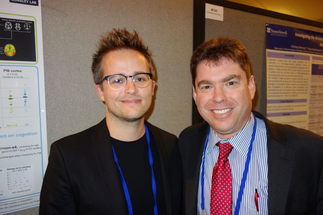
APOE4 may drive tau deposition in Alzheimer’s
SAN DIEGO – The apolipoprotein E e4 allele is well known for its association with amyloid deposition in Alzheimer’s disease, but now it also appears to help drive the other key pathological process in the disease: deposition of hyperphosphorylated tau protein.
That’s according to the authors of a PET neuroimaging study presented at the annual meeting of the American Neurological Association.
The finding suggests a pathophysiologic mechanism for Alzheimer’s disease cases that predominantly affect memory.
The team then compared the results with uptake of the radiotracers in 71 cognitively normal control subjects who were a mean age of 79 years, 23 of whom (32%) were APOE4 carriers.
APOE4 was associated with higher cortical amyloid in the controls, but not tau deposition. Although AV1451 uptake in the temporal lobe was increased in e4 carriers, the effect was not statistically significant after controlling for PiB uptake.
“We saw an APOE4 effect on amyloid but not on tau in normal people,” said senior investigator Gil Rabinovici, MD, a neurologist and professor of memory and aging at the University of California, San Francisco.
All the AD patients had PiB uptake, with no difference in uptake between e4 carriers and noncarriers. However, APOE4 carriers had higher AV1451 uptake in their anterior medial temporal lobes (MTL), a difference that remained unchanged after controlling for PiB.
Carriers of the e4 allele who had AV1451 uptake in their MTLs had a harder time than other AD patients on a test in which they were asked to recall a series of words after a 10-minute break (California Verbal Learning Test), but they did not perform worse on other cognitive measures.
“In cognitively normal individuals, the effect of APOE4 on tau pathology seems to be mediated by the effect of e4 on Ab [amyloid-beta] deposition,” the investigators noted. “However, when assessing amyloid-positive symptomatic AD patients, APOE4 was associated with increased AV1451 binding in the MTL.
“This suggests that, in addition to its effect on Ab pathology, APOE4 might influence the topographical distribution of tau pathology and, potentially, the cognitive symptoms in patients,” the researchers concluded.
“We are very interested in heterogeneity in Alzheimer’s disease – age of onset, rate of progression, which brain areas are affected,” Dr. Rabinovici said. “This study is beginning to dig into some of the factors that might explain that; APOE4 is one potential modifier. It may have a direct effect on tau phosphorylation, and it changes where tau is located in the setting of Alzheimer’s disease.”
The finding of increased MTL tau in APOE4-positive Alzheimer’s patients helps explain why patients who carry the allele tend to have more memory problems, he said, while AD patients who don’t carry the allele tend to have more of a cortical-predominant presentation, with more visual-spatial and language problems.
“I think it’s very likely that APOE4-related disease may be driven by a different mechanism than APOE4-negative disease,” Dr. Rabinovici said. “In the future, APOE4-positive or APOE4-negative might be used to stratify therapy and the measurements used for disease progression. APOE4 itself may be an interesting therapeutic target because it has downstream effects on amyloid and tau.”
Meanwhile, there’s just not a lot of tau pathology in cognitively normal people, which is likely why the effect didn’t show up in the controls, noted lead investigator Renaud La Joie, PhD, a neuroimaging researcher at UCSF.
The work was funded in part by the National Institutes of Health. Avid Pharmaceuticals provided the AV1451. Dr. Rabinovici is an advisor for Genentech, Merck, and Roche, and has research support from Avid Radiopharmaceuticals and Eli Lilly, among others.
SOURCE: La Joie R, et al. Abstract M183, American Neurological Association 2017 annual meeting.
REPORTING FROM ANA 2017
Key clinical point:
Major finding: APOE4 carriers had higher AV1451-uptake in their anterior medial temporal lobes, a difference that remained unchanged after controlling for Pittsburgh compound B.
Study details: An analysis of radiotracer PET imaging in 67 Alzheimer’s disease patients and 71 controls.
Disclosures: The work was funded in part by the National Institutes of Health. Avid Pharmaceuticals provided the AV1451. The senior investigator is an advisor for Genentech, Merck, and Roche, and has research support from Avid Radiopharmaceuticals and Eli Lilly, among others.
Source: La Joie R, et al. Abstract M183, American Neurological Association 2017 annual meeting.
FDA approves irritable bowel syndrome treatment
(IBS-C).
Plecanatide had previously been approved to treat adults with chronic idiopathic constipation (CIC).
Plecanatide met both of its primary endpoints, with reductions in abdominal pain in both studies, compared with placebo (30.2% vs. 17.8% in study 1, P < .001; 21.5% vs. 14.2% in study 2, P = .009).
Plecanatide is the only prescription, once-daily medication that treats both CIC and IBS-C in adults. The drug should be available in the first quarter of 2018.
Birth cohort affected 2015-2016 flu vaccine effectiveness
The influenza vaccine introduced in 2009 showed reduced effectiveness during the 2015-2016 influenza season, but only in adults born between 1958 and 1979, according to an analysis published online in the Journal of Infectious Diseases.
Using the Influenza Vaccine Effectiveness Network, researchers analyzed data from 2,115 patients with medically attended acute respiratory illness who tested positive for A(H1N1)pdm09 influenza virus, and 14,696 patients who tested negative for the influenza virus, from 2010-2011 to 2015-2016 (excluding the 2014-2015 influenza season).
Overall, 48% of the influenza virus–negative patients and 28% of the virus-positive patients had received at least one dose of the seasonal inactivated influenza vaccine more than 2 weeks before they fell ill.
However, the vaccine, which was based on the A/California/07/2009 strain of the A(H1N1)pdm09 virus, was only 47% effective during the 2015-2016 season, compared with 61% effectiveness during the 2010-2011 season through to the 2013-2014 season.
When researchers looked at vaccine effectiveness by birth cohort, they found that one particular cohort – individuals born between 1958 and 1979 – showed a significantly reduced vaccine effectiveness (22%) during the 2015-2016 season. By comparison, vaccine effectiveness in this cohort was 61% during the 2010-2013 seasons, and 56% during the 2013-2014 season.
When this birth cohort was excluded from analysis of the 2015-2016 season, the overall vaccine effectiveness for that season was 61%.
While the vaccine was based on an early reference strain of A(H1N1)pdm09, the virus itself later acquired mutations in the hemagglutinin gene, leading to the emergence of new genetic clades, including 6B, which dominated in the 2013-2014 influenza season, and 6B.1, which dominated in 2015-2016.
“Limited serologic data suggest that some adults born during 1958-1979 (age range in 2015-2016, 36-57 years) have decreased antibody titers against A(H1N1)pdm09 group 6B and 6B.1 viruses,” wrote Brendan Flannery, PhD, from the Centers for Disease Control and Prevention, and his coauthors.
They suggested that individuals in this cohort may have been immunologically primed with A/USSR/90/1977-like viruses, which were the first group of A(H1N1) viruses that this cohort would have been exposed to. A(H1N1) strains didn’t circulate between 1958 and 1977. Vaccination with A(H1N1)pdm09 viruses may have induced antibodies against shared antigenic components found on early versions of A(H1N1)pdm09.
If these shared antigenic epitopes were then altered in the later 6B and 6B.1 viruses, that might account for decreased antibody titers in this age group.
“Replacement of the A/California/07/2009(H1N1)pdm09 vaccine reference strain with A/Michigan/45/2015 (group 6B.1) should lead to improved [vaccine effectiveness] against circulating A(H1N1)pdm09 viruses,” the investigators noted.
The study was supported by the Centers for Disease Control and Prevention, the National Institutes of Health, and the National Center for Advancing Translational Sciences. Eight authors declared funding, grants, and consultancies with the pharmaceutical industry, with five also declaring funding from the CDC.
SOURCE: Flannery B et al. J Infect Dis. 2018 Jan 18. doi: 10.1093/infdis/jix634.
This study proposes that influenza virus strains encountered early in life focus the immune response to later infection or vaccination on shared epitopes between the early and later strains. Supporting this hypothesis is evidence from other studies showing that 60% of the serological response to inactivated influenza vaccines is the result of boosting pre-existing antibodies, rather than the creation of new, vaccine-induced antibodies.
However there are also some flaws to this argument, and we should be careful to avoid confirmation bias. For example, the reduction in effectiveness of vaccines against A(H1N1) has been observed in North America, where this study is located, but to a lesser extent in studies conducted in other regions. Reductions in vaccine effectiveness have also been observed in other birth cohorts and during other influenza seasons.
That aside, accumulating evidence suggests that the vaccine strain be updated from A/California/7/2009 to A/Michigan/45/2015 (a clade 6B.1 strain) for the 2016-2017 influenza seasons.
Allen C. Cheng, PhD, is from the School of Public Health and Preventive Medicine at Monash University, Melbourne, and Kanta Subbarao, MBBS, is from the World Health Organization Collaborating Centre for Reference and Research on Influenza and the Peter Doherty Institute for Infection and Immunity, Australia. These comments are taken from an accompanying editorial (J Infect Dis. 2018, Jan 18. doi: 10.1093/infdis/jix635). The authors declared support from the Australian Department of Health and the Australian National Health and Medical Research Council. No conflicts of interest were declared.
The influenza vaccine introduced in 2009 showed reduced effectiveness during the 2015-2016 influenza season, but only in adults born between 1958 and 1979, according to an analysis published online in the Journal of Infectious Diseases.
Using the Influenza Vaccine Effectiveness Network, researchers analyzed data from 2,115 patients with medically attended acute respiratory illness who tested positive for A(H1N1)pdm09 influenza virus, and 14,696 patients who tested negative for the influenza virus, from 2010-2011 to 2015-2016 (excluding the 2014-2015 influenza season).
Overall, 48% of the influenza virus–negative patients and 28% of the virus-positive patients had received at least one dose of the seasonal inactivated influenza vaccine more than 2 weeks before they fell ill.
However, the vaccine, which was based on the A/California/07/2009 strain of the A(H1N1)pdm09 virus, was only 47% effective during the 2015-2016 season, compared with 61% effectiveness during the 2010-2011 season through to the 2013-2014 season.
When researchers looked at vaccine effectiveness by birth cohort, they found that one particular cohort – individuals born between 1958 and 1979 – showed a significantly reduced vaccine effectiveness (22%) during the 2015-2016 season. By comparison, vaccine effectiveness in this cohort was 61% during the 2010-2013 seasons, and 56% during the 2013-2014 season.
When this birth cohort was excluded from analysis of the 2015-2016 season, the overall vaccine effectiveness for that season was 61%.
While the vaccine was based on an early reference strain of A(H1N1)pdm09, the virus itself later acquired mutations in the hemagglutinin gene, leading to the emergence of new genetic clades, including 6B, which dominated in the 2013-2014 influenza season, and 6B.1, which dominated in 2015-2016.
“Limited serologic data suggest that some adults born during 1958-1979 (age range in 2015-2016, 36-57 years) have decreased antibody titers against A(H1N1)pdm09 group 6B and 6B.1 viruses,” wrote Brendan Flannery, PhD, from the Centers for Disease Control and Prevention, and his coauthors.
They suggested that individuals in this cohort may have been immunologically primed with A/USSR/90/1977-like viruses, which were the first group of A(H1N1) viruses that this cohort would have been exposed to. A(H1N1) strains didn’t circulate between 1958 and 1977. Vaccination with A(H1N1)pdm09 viruses may have induced antibodies against shared antigenic components found on early versions of A(H1N1)pdm09.
If these shared antigenic epitopes were then altered in the later 6B and 6B.1 viruses, that might account for decreased antibody titers in this age group.
“Replacement of the A/California/07/2009(H1N1)pdm09 vaccine reference strain with A/Michigan/45/2015 (group 6B.1) should lead to improved [vaccine effectiveness] against circulating A(H1N1)pdm09 viruses,” the investigators noted.
The study was supported by the Centers for Disease Control and Prevention, the National Institutes of Health, and the National Center for Advancing Translational Sciences. Eight authors declared funding, grants, and consultancies with the pharmaceutical industry, with five also declaring funding from the CDC.
SOURCE: Flannery B et al. J Infect Dis. 2018 Jan 18. doi: 10.1093/infdis/jix634.
FROM THE JOURNAL OF INFECTIOUS DISEASES
Key clinical point:
Major finding: The influenza vaccine effectiveness during the 2015-2016 season was just 22% in individuals born between 1958 and 1979.
Data source: A retrospective case-control study of 2,115 patients who tested positive for A(H1N1)pdm09 influenza virus, and 14,696 negative controls.
Disclosures: The study was supported by the Centers for Disease Control and Prevention, the National Institutes of Health, and the National Center for Advancing Translational Sciences. Eight authors declared funding, grants, and consultancies with the pharmaceutical industry, with five also declaring funding from the CDC.
Source: Flannery B et al. J Infect Dis. 2018 Jan 18. doi: 10.1093/infdis/jix634.
Delayed ileal pouch anal anastomosis creation linked to lower 30-day adverse events
LAS VEGAS – compared with creating the pouch at the time of initial surgery, results from an analysis of national data demonstrated.
“More than 600,000 Americans have UC, and 20%-30% of them require surgical management,” Bharati Kochar, MD, said at the Crohn’s & Colitis Congress, a partnership of the Crohn’s & Colitis Foundation and the American Gastroenterological Association. “The surgical procedure of choice for many UC patients is total proctocolectomy with ileal pouch anal anastomosis creation.”
According to Dr. Kochar, an advanced fellow in inflammatory bowel diseases at the University of North Carolina at Chapel Hill, existing American medical literature regarding ileal pouch anal anastomosis (IPAA) comes mostly from quaternary care centers and compares one-stage procedures with multistage procedures.
“The risks between two- to three-stage procedures are not described, and there are no prospective national reports of postoperative adverse events after IPAA creation,” she said.
Using data from the National Surgical Quality Improvement Program, Dr. Kochar and her associates conducted an observational cohort analysis of 2,390 adult patients with a postoperative diagnosis of UC who underwent IPAA procedures between 2011 and 2015. Their aims were to evaluate adverse events within 30 days after an IPAA creation and to compare adverse events between pouch creation at the time of colectomy and delayed pouch creation.
They also performed a subanalysis of total abdominal colectomy with ileostomy (TAC), the first stage in the delayed pouch procedures, versus pouch creation at the time of colectomy. Multivariable modified Poisson regression models were used to estimate risk ratios adjusted for age, sex, race, body mass index, smoking status, diabetes, preoperative albumin, and American Society of Anesthesiologists class.
Of the 2,390 patients, 1,571 had pouches created at the time of colectomy (group A), and 819 had delayed pouch creation (group B).
Compared with patients in group B, those in group A were older (a median age of 40 years vs. 37 years, respectively; P less than .01), were more likely to be on an immunosuppressant (51% vs. 15%; P less than .01), have a lower median preoperative albumin level (3.9 vs. 4.2; P less than .01), and a longer median length of stay (6 days vs. 5 days; P less than .01).
On unadjusted analyses, the researchers also observed that, at 30 days, patients in group A had significantly more major complications, such as mortality and cardiac arrest (12.4% vs. 8.7%; P less than .01); minor complications, such as superficial surgical site infections and pneumonia (11.8% vs. 6.1%; P less than .01); unplanned readmissions (statistically similar at 23.3% vs. 21.3%), and unplanned reoperations (7.7% vs. 3.8%; P less than .01).
After controlling for confounders, patients in group B were significantly less likely to have major complications (relative risk, 0.72), minor complications (RR, 0.48), unplanned readmissions (RR, 0.95), and unplanned reoperations (RR, 0.42).
In the subgroup analysis, Dr. Kochar and her associates observed that patients who underwent TAC were significantly older, compared with patients in group A (a median of 46 years vs. 40 years, respectively; P less than .01), and a higher proportion were on immunosuppressants (69% vs. 51%; P less than .01). “Despite these factors, the risk of adverse events after TAC was lower,” Dr. Kochar said.
She acknowledged certain limitations of the study, including the inability to accurately determine the risk of linked surgeries together and the inability to assess institution and operator factors. Also, data were not collected for the purposes of studying inflammatory bowel disease.
“This is the first prospective assessment of morbidity following IPAA creation in UC patients from a national database,” Dr. Kochar concluded. “Delayed pouch procedures are associated with a lower risk of unplanned reoperations and major and minor complications. Immunosuppression at the time of pouch creation may result in an increased risk of adverse events postoperatively. The findings can be valuable for preoperative risk assessment and postoperative management.”
Dr. Kochar reported having no financial disclosures.
*This story was updated on 3/26.
SOURCE: Kochar et al. Crohn’s & Colitis Congress 2018 Clinical Abstract 11.
REPORTING FROM THE CROHN’S & COLITIS CONGRESS
Key clinical point: Delayed ileal pouch anal anastomosis procedures are associated with a lower 30-day adverse-event rate.
Major finding: After controlling for confounders, patients who underwent delayed IPAA procedures were significantly less likely to have major complications (relative risk, 0.72), minor complications (RR, 0.48), unplanned readmissions (RR, 0.95), and unplanned reoperations (RR, 0.42) at 30 days, compared with those who underwent pouch creation at the time of initial surgery.
Study details: An observational cohort analysis of 2,390 adult patients with a postoperative diagnosis of UC.
Disclosures: Dr. Kochar reported having no financial disclosures.
Source: Kochar et al. Crohn’s & Colitis Congress 2018 Clinical Abstract 11. Gastroenterology. 2018;154(1)Suppl:S1-S114.