User login
Pediatric hypertension linked to troubling MRI changes
CHICAGO – There’s another reason to worry about hypertension in children: cognitive decline later in life.
The video associated with this article is no longer available on this site. Please view all of our videos on the MDedge YouTube channel
In a pilot study, Marc Lande, MD, a professor of pediatric nephrology at the University of Rochester (N.Y.), and his colleagues found similar to what’s found in adults with cognitive impairment from hypertension.
The work is ongoing, but it helps explain the subtle deficits on cognitive testing that have been previously demonstrated in children with hypertension.
“The fact that we are finding anything at this very early stage of disease is striking and somewhat bothersome. The hope is that, by improving blood pressure in children, you can improve subsequent cognition and maybe even delay the onset of dementia further down the road,” Dr. Lande said.
For now, the findings underscore the need to diagnose and manage hypertension in children, but there might be additional treatment implications in the future, especially if blood pressure targets are found that ameliorate the problem.
Dr. Lande explained the issues and the emerging evidence in a video interview at the joint scientific sessions of the American Heart Association Council on Hypertension, AHA Council on Kidney in Cardiovascular Disease, and American Society of Hypertension.
REPORTING FROM JOINT HYPERTENSION 2018

Dupilumab efficacy extends to chronic rhinosinusitis/nasal polyposis
PARIS – Severe asthma patients with chronic rhinosinusitis (CRS), nasal polyposis (NP), or both derive more protection from severe exacerbations with the monoclonal antibody dupilumab than do those who do not have these comorbidities, according to a post hoc analysis of a phase 3 trial presented at the annual congress of the European Respiratory Society.

“Dupilumab reduced rates of severe exacerbations and improved FEV1 [forced expiratory volume in 1 second] in patients in asthma patients with or without CRS/NP. In those with CRS/NP, dupilumab reduced symptoms associated with these comorbidities,” reported Ian Pavord, MBBS, statutory chair in respiratory medicine at University of Oxford (England).
The data were drawn from the phase 3 Liberty Asthma Quest trial, which was published earlier this year in the New England Journal of Medicine (2018;378:2486-96). In that study, both the 200-mg and 300-mg dose of dupilumab (Dupixent) administered every 2 weeks was associated with about a 50% reduction in the annualized rate of severe exacerbations relative to placebo (P less than .001 for both doses).
In this new post hoc analysis, response in the 382 patients who entered the study with a history of CRS/NP was compared with the 1,520 without CRS/NP. In the CRS/NP patients, the reductions relative to placebo in the rates of severe exacerbations, defined as 3 or more days of systemic glucocorticoids or visit to an emergency department leading to treatment with systemic glucocorticoids, were 63% and 61% for the 200-mg and 300-mg doses of dupilumab, respectively (both P less than .001).
In the non-CRS/NP arms, the reductions relative to placebo were 42% and 40%, respectively (both P less than .001). The greater relative reductions in the CRS/NP patients were achieved even though they were older (mean age approximately 52 vs. 47 years for non-CRS/NP patients), had a significantly greater number of exacerbations in the past year (P = .027), and had higher baseline fractional exhaled nitric oxide and eosinophil levels (both P less than .001), Dr. Pavord reported.
“The greater asthma severity in the CRS/NP patients in this trial is consistent with that reported previously by others,” Dr. Pavord said.
Although the greater asthma severity may have provided a larger margin for benefit, Dr. Pavord also reported that there were improvements in CRS/NP-specific symptoms as measured with the 22-item Sino-Nasal Outcome Test (SNOT-22). By week 12, the total score reduction in SNOT-22 was approximately 15 points (P less than .05) from baseline for both the 200-mg and 300-mg dupilumab doses. This was significantly greater (P less than .05) relative to modest SNOT-22 reductions in the placebo arms (P less than .05). After 52 weeks, the reduction In SNOT-22 scores were sustained, providing an even greater statistical advantage over placebo (P less than .001).
In addition to greater protection against severe exacerbations and CRS/NP-specific symptoms, dupilumab may offer specific improvements on CRS/NP pathology, according to Dr. Pavord. Although imaging was not part of this study, he noted in particular that previous studies with dupilumab as well as other biologics have shown shrinkage of nasal polyps with treatment.
Dupilumab was similarly well tolerated in those with and without CRS/NP. The most common adverse event was injection site reactions in both groups, Dr. Pavord said.
Calling CRS and NP “important comorbidities” in severe asthma patients, Dr. Pavord said that this analysis should be reassuring for those who with CRS/NP who are being considered for dupilumab. Already approved for treatment of atopic dermatitis, dupilumab, which binds to interleukin-4 (IL-4) and IL-13 receptors, is currently under review for the treatment of moderate to severe asthma.
Dr. Pavord has financial relationships with Aerocrine, Almirall, AstraZeneca, Boehringer Ingelheim, Chiesi, GlaxoSmithKline, Knapp, Merck Sharpe, Novartis, Knapp Teva, RespiVert, and Schering-Plough.
PARIS – Severe asthma patients with chronic rhinosinusitis (CRS), nasal polyposis (NP), or both derive more protection from severe exacerbations with the monoclonal antibody dupilumab than do those who do not have these comorbidities, according to a post hoc analysis of a phase 3 trial presented at the annual congress of the European Respiratory Society.
“Dupilumab reduced rates of severe exacerbations and improved FEV1 [forced expiratory volume in 1 second] in patients in asthma patients with or without CRS/NP. In those with CRS/NP, dupilumab reduced symptoms associated with these comorbidities,” reported Ian Pavord, MBBS, statutory chair in respiratory medicine at University of Oxford (England).
The data were drawn from the phase 3 Liberty Asthma Quest trial, which was published earlier this year in the New England Journal of Medicine (2018;378:2486-96). In that study, both the 200-mg and 300-mg dose of dupilumab (Dupixent) administered every 2 weeks was associated with about a 50% reduction in the annualized rate of severe exacerbations relative to placebo (P less than .001 for both doses).
In this new post hoc analysis, response in the 382 patients who entered the study with a history of CRS/NP was compared with the 1,520 without CRS/NP. In the CRS/NP patients, the reductions relative to placebo in the rates of severe exacerbations, defined as 3 or more days of systemic glucocorticoids or visit to an emergency department leading to treatment with systemic glucocorticoids, were 63% and 61% for the 200-mg and 300-mg doses of dupilumab, respectively (both P less than .001).
In the non-CRS/NP arms, the reductions relative to placebo were 42% and 40%, respectively (both P less than .001). The greater relative reductions in the CRS/NP patients were achieved even though they were older (mean age approximately 52 vs. 47 years for non-CRS/NP patients), had a significantly greater number of exacerbations in the past year (P = .027), and had higher baseline fractional exhaled nitric oxide and eosinophil levels (both P less than .001), Dr. Pavord reported.
“The greater asthma severity in the CRS/NP patients in this trial is consistent with that reported previously by others,” Dr. Pavord said.
Although the greater asthma severity may have provided a larger margin for benefit, Dr. Pavord also reported that there were improvements in CRS/NP-specific symptoms as measured with the 22-item Sino-Nasal Outcome Test (SNOT-22). By week 12, the total score reduction in SNOT-22 was approximately 15 points (P less than .05) from baseline for both the 200-mg and 300-mg dupilumab doses. This was significantly greater (P less than .05) relative to modest SNOT-22 reductions in the placebo arms (P less than .05). After 52 weeks, the reduction In SNOT-22 scores were sustained, providing an even greater statistical advantage over placebo (P less than .001).
In addition to greater protection against severe exacerbations and CRS/NP-specific symptoms, dupilumab may offer specific improvements on CRS/NP pathology, according to Dr. Pavord. Although imaging was not part of this study, he noted in particular that previous studies with dupilumab as well as other biologics have shown shrinkage of nasal polyps with treatment.
Dupilumab was similarly well tolerated in those with and without CRS/NP. The most common adverse event was injection site reactions in both groups, Dr. Pavord said.
Calling CRS and NP “important comorbidities” in severe asthma patients, Dr. Pavord said that this analysis should be reassuring for those who with CRS/NP who are being considered for dupilumab. Already approved for treatment of atopic dermatitis, dupilumab, which binds to interleukin-4 (IL-4) and IL-13 receptors, is currently under review for the treatment of moderate to severe asthma.
Dr. Pavord has financial relationships with Aerocrine, Almirall, AstraZeneca, Boehringer Ingelheim, Chiesi, GlaxoSmithKline, Knapp, Merck Sharpe, Novartis, Knapp Teva, RespiVert, and Schering-Plough.
REPORTING FROM THE ERS CONGRESS 2018
Key clinical point: In asthma patients with chronic rhinosinusitis and/or nasal polyposis (CRS/NP), dupilumab reduces exacerbations.
Major finding: At 52 weeks, severe exacerbations were reduced 61% in CRS/NP patients and 40% in non-CRS/NP patients (both P less than .001).
Study details: Post hoc analysis of phase 3 trial.
Disclosures: Dr. Pavord has financial relationships with Aerocrine, Almirall, AstraZeneca, Boehringer Ingelheim, Chiesi, GlaxoSmithKline, Knapp, Merck Sharpe, Novartis, Knapp Teva, RespiVert, and Schering-Plough.
Updated IPF guideline refines diagnostic criteria with HRCT
A recently updated guideline for idiopathic pulmonary fibrosis (IPF) provides refined diagnostic criteria in an effort to improve clinical application and diagnostic accuracy.
Of note, the guideline recommends that high-resolution CT (HRCT) patterns be used to dictate management course. The guideline also calls for detailed medical history, serological testing to exclude connective tissue disease, and multidisciplinary discussion. Serum biomarkers are recommended against as a means of distinguishing between IPF and other interstitial lung diseases (ILDs).
“Diagnosing IPF is challenging because these symptoms are nonspecific: They occur with all other interstitial lung diseases and with other respiratory problems,” Ganesh Raghu, MD, chair of the guideline committee and professor of medicine and director of the Center for Interstitial Lung Disease at the University of Washington, Seattle, said in a written statement. “Because drugs may slow the progression of IPF, an early and accurate diagnosis is essential for prompt and appropriate treatment for this fatal disease.”
The 2018 guideline represents a second collaborative effort from the American Thoracic Society, European Respiratory Society, Japanese Respiratory Society, and Latin American Thoracic Society. The guideline committee consisted of 29 clinicians, scientists, and a patient with IPF. They evaluated all IPF-related evidence and rated the quality of findings with the GRADE (Grading of Recommendations, Assessment, Development and Evaluation) system. The first IPF guideline was published 7 years ago; the intervening time has revealed some clinical limitations that the 2018 guideline aims to fix.
“The 2011 guideline provided the first evidence-based, formal criteria for diagnosis of IPF and allowed patients with a well-defined diagnosis of IPF to participate in numerous clinical studies and randomized controlled trials that enhanced our understanding of the disease,” Dr. Raghu said. “However, it became clear that there were significant challenges in ascertaining the diagnosis per the 2011 criteria, and abundant evidence accumulated since then allowed the committee to refine the diagnostic criteria now.”
“This [updated] guideline is intended to help clinicians make an accurate diagnosis of IPF,” the authors wrote in the American Journal of Respiratory and Critical Care Medicine, “and to empower them to implement recommended courses of action in the context of individual patient values and preferences, particularly decisions regarding which diagnostic interventions to pursue.”
While the 2011 guideline did not distinguish between patients with different HRCT patterns, the 2018 guideline emphasizes the use of HRCT. It is now recommended that patients undergo HRCT to determine the pattern of usual interstitial pneumonia (UIP). Broadly, patients exhibit the UIP pattern or one of three possible non-UIP patterns (probable UIP, indeterminate UIP, or an alternative diagnosis). Recommendations are specific for UIP and non-UIP.
Patients with probable UIP, indeterminate UIP, or an alternative diagnosis should undergo bronchoalveolar lavage (BAL) and surgical lung biopsy (SLB). Transbronchial lung biopsy (TBBx) and lung cryobiopsy recommendations were not described in these patients because of a lack of evidence. However, patients with UIP should not undergo BAL, SLB, TBBx, or cryobiopsy.
For all patients, the 2018 guideline recommends that medical histories include environmental exposure and medication use, and that serological testing is performed to exclude connective tissue disease. Multidisciplinary discussion is conditionally recommended for diagnostic decision making, particularly when a patient has probable UIP, indeterminate UIP, or an alternative diagnosis.
Finally, the guideline recommends against the use of serum biomarkers as a means of distinguishing between IPF and other ILDs because of weak data on this point. “For the time being, the guideline panel dismissed serum biomarker measurement as an approach to distinguishing IPF from other ILDs because of the high false-positive and false-negative result rates,” the panel wrote.
“Our hope is that this new guideline will bridge the gap between the experienced IPF experts and general pulmonologists in making a prompt and accurate diagnosis of IPF for the individual unfortunately confronted with the disease,” Dr. Raghu said. “This will allow patients to make well-informed decisions about treatment options and participation in clinical trials.”
Looking to the future, the panel described an “urgent need to refine and validate diagnostic approaches to ILD. These needs can be roughly categorized as investigations into the roles of clinical observations, HRCT, bronchoscopy, histopathology, and biomarkers.” The authors also emphasized “the need to refine prognostic approaches, identify risk factors for the development of IPF, and determine the impact and approach to the diagnosis of comorbid illness in the patient with IPF.”
The authors reported funding from Bellerophon, Gilead, Roche, Sanofi, and others.
SOURCE: Raghu G et al. Am J Respir Crit Care Med. 2018 Sep 1. doi: 10.1164/rccm.201807-1255ST.
FROM THE AMERICAN JOURNAL OF RESPIRATORY AND CRITICAL CARE MEDICINE
Hospitalist movers and shakers – Sept. 2018
Modern Healthcare recently announced its list of the 50 Most Influential Physician Executives and Leaders, and hospital medicine was well represented among the honorees. The honored physicians were selected by a panel of experts and peers for their leadership and impact on the profession.
Topping the list was Scott Gottlieb, MD, the commissioner of the Food and Drug Administration. Dr. Gottlieb was confirmed to his position in May 2017 and, in his first year, has focused on price transparency and the approval of generic medications.
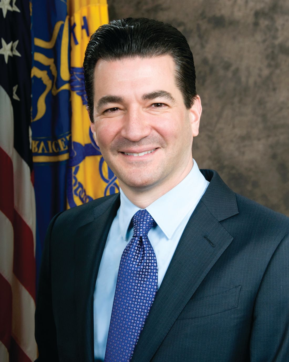
Dr. Gottlieb was deputy commissioner of the FDA from 2005-2007, and he has worked as an advisor and analyst for GlaxoSmithKline, the American Enterprise Institute, Vertex Pharmaceuticals, and Avilene Health.
Dr. Gottlieb earned his medical degree from the Icahn School of Medicine at Mount Sinai, New York, and completed his residency at Mount Sinai Hospital. He has worked as a hospitalist at New York University’s Tisch Hospital, the Hospital for Joint Diseases, and Stamford (Conn.) Hospital.
Patrick Conway, MD, was listed at number 23 on Modern Healthcare’s 50 Most Influential Physician Executives and Leaders. Formerly the deputy administrator for innovation and quality at the Centers for Medicare & Medicaid Services, Dr. Conway recently became president and chief executive officer of Blue Cross and Blue Shield of North Carolina.

Dr. Conway is known for his ability to develop and promote alternative payment models. He was elected to the National Academy of Medicine’s Institute of Medicine in 2014 and was selected as a Master of Hospital Medicine by the Society of Hospital Medicine.
Lynn Massingale, MD, the cofounder and chairman of TeamHealth, was named one of the 50 Most Influential Physician Executives and Leaders for a third year running, coming in at number 27 on the list. Dr. Massingale, who also recently was named to the Tennessee Healthcare Hall of Fame, founded TeamHealth in 1979 and was its chief executive officer for 30 years before assuming the role of chairman in 2008.
TeamHealth provides outsourced emergency medicine, hospitalist, critical care, anesthesiology, and acute care surgery services, among other specialties, at more than 3,200 facilities and physician groups across the United States.
Veeravat Taecharvongphairoj, MD, a veteran internist and hospitalist at Hemet Valley Medical Center in Hemet, Calif., has been honored by the International Association of Healthcare Professionals in its Leading Physicians of the World publication.
Dr. Taecharvongphairoj completed his residency at the University of Hawaii, Honolulu, before accepting a fellowship in hospital and palliative care at Cedars-Sinai Medical Center, Los Angeles. He is a member of the American Academy of Hospice and Palliative Medicine.
Sean Bain, MD, has been selected to the Glen Falls (N.Y.) Hospital Foundation Board of Trustees for 2018. Dr. Bain works as a hospitalist/internist at Glen Falls Hospital, where he is the president of medical staff. He manages the credentialing, continuing education, and policies and practices for the staff’s providers.
Dr. Bain received his medical degree at Albany (N.Y.) Medical College and served his residency at Wake Forest Baptist Medical Center, Winston-Salem, N.C.
George Harrison, MD, has been tabbed the new chief medical officer at Fairview Park Hospital in Dublin, Ga. Dr. Harrison will be charged with managing clinical quality and patient safety, staff relations, and clinical integration strategies at the hospital.
Prior to his appointment, Dr. Harrison was the codirector of the hospitalist program at Fairview Park. The Georgia native previously worked in management roles at urgent care centers, family practice centers, and hospitalist programs in North Carolina, South Carolina, and Georgia. He is a member of the American Academy of Family Physicians, the Society of Hospital Medicine, and the American Academy of Physician Leaders.
Dr. Harrison taught high school geometry and chemistry before earning his medical degree at the Morehouse School of Medicine, Atlanta. He did his residency at Duke University Medical Center, Durham, N.C.
BUSINESS MOVES
U.S. Acute Care Solutions (Canton, Ohio), a physician-owned, national provider of emergency medicine and hospitalist services, has extended its relationship with Central Health of Colorado and western Kansas. USACS has acquired the physicians of Front Range Emergency Specialists (Colorado Springs, Colo.), Southwest Emergency Physicians (Durango, Colo.), and Southern Colorado Emergency Specialists (Pueblo, Colo.).
USACS’s acquisition of these three physician groups adds care to more than 175,000 patients each year in central and southwest Colorado. USACS cares for more than 6 million patients per year at more than 200 locations across the United States.
VEP Healthcare (Concord, Calif.), an emergency medicine and hospitalist staffing company, has signed on to manage hospitalist and ED services at City Hospital at White Rock in Dallas. Its goals are to increase patient satisfaction, decrease wait times in seeing providers, raise recommendation rates, and lower malpractice claims.
White Rock is a 218-bed, community hospital providing care to East Texas since 1959.
Modern Healthcare recently announced its list of the 50 Most Influential Physician Executives and Leaders, and hospital medicine was well represented among the honorees. The honored physicians were selected by a panel of experts and peers for their leadership and impact on the profession.
Topping the list was Scott Gottlieb, MD, the commissioner of the Food and Drug Administration. Dr. Gottlieb was confirmed to his position in May 2017 and, in his first year, has focused on price transparency and the approval of generic medications.
Dr. Gottlieb was deputy commissioner of the FDA from 2005-2007, and he has worked as an advisor and analyst for GlaxoSmithKline, the American Enterprise Institute, Vertex Pharmaceuticals, and Avilene Health.
Dr. Gottlieb earned his medical degree from the Icahn School of Medicine at Mount Sinai, New York, and completed his residency at Mount Sinai Hospital. He has worked as a hospitalist at New York University’s Tisch Hospital, the Hospital for Joint Diseases, and Stamford (Conn.) Hospital.
Patrick Conway, MD, was listed at number 23 on Modern Healthcare’s 50 Most Influential Physician Executives and Leaders. Formerly the deputy administrator for innovation and quality at the Centers for Medicare & Medicaid Services, Dr. Conway recently became president and chief executive officer of Blue Cross and Blue Shield of North Carolina.
Dr. Conway is known for his ability to develop and promote alternative payment models. He was elected to the National Academy of Medicine’s Institute of Medicine in 2014 and was selected as a Master of Hospital Medicine by the Society of Hospital Medicine.
Lynn Massingale, MD, the cofounder and chairman of TeamHealth, was named one of the 50 Most Influential Physician Executives and Leaders for a third year running, coming in at number 27 on the list. Dr. Massingale, who also recently was named to the Tennessee Healthcare Hall of Fame, founded TeamHealth in 1979 and was its chief executive officer for 30 years before assuming the role of chairman in 2008.
TeamHealth provides outsourced emergency medicine, hospitalist, critical care, anesthesiology, and acute care surgery services, among other specialties, at more than 3,200 facilities and physician groups across the United States.
Veeravat Taecharvongphairoj, MD, a veteran internist and hospitalist at Hemet Valley Medical Center in Hemet, Calif., has been honored by the International Association of Healthcare Professionals in its Leading Physicians of the World publication.
Dr. Taecharvongphairoj completed his residency at the University of Hawaii, Honolulu, before accepting a fellowship in hospital and palliative care at Cedars-Sinai Medical Center, Los Angeles. He is a member of the American Academy of Hospice and Palliative Medicine.
Sean Bain, MD, has been selected to the Glen Falls (N.Y.) Hospital Foundation Board of Trustees for 2018. Dr. Bain works as a hospitalist/internist at Glen Falls Hospital, where he is the president of medical staff. He manages the credentialing, continuing education, and policies and practices for the staff’s providers.
Dr. Bain received his medical degree at Albany (N.Y.) Medical College and served his residency at Wake Forest Baptist Medical Center, Winston-Salem, N.C.
George Harrison, MD, has been tabbed the new chief medical officer at Fairview Park Hospital in Dublin, Ga. Dr. Harrison will be charged with managing clinical quality and patient safety, staff relations, and clinical integration strategies at the hospital.
Prior to his appointment, Dr. Harrison was the codirector of the hospitalist program at Fairview Park. The Georgia native previously worked in management roles at urgent care centers, family practice centers, and hospitalist programs in North Carolina, South Carolina, and Georgia. He is a member of the American Academy of Family Physicians, the Society of Hospital Medicine, and the American Academy of Physician Leaders.
Dr. Harrison taught high school geometry and chemistry before earning his medical degree at the Morehouse School of Medicine, Atlanta. He did his residency at Duke University Medical Center, Durham, N.C.
BUSINESS MOVES
U.S. Acute Care Solutions (Canton, Ohio), a physician-owned, national provider of emergency medicine and hospitalist services, has extended its relationship with Central Health of Colorado and western Kansas. USACS has acquired the physicians of Front Range Emergency Specialists (Colorado Springs, Colo.), Southwest Emergency Physicians (Durango, Colo.), and Southern Colorado Emergency Specialists (Pueblo, Colo.).
USACS’s acquisition of these three physician groups adds care to more than 175,000 patients each year in central and southwest Colorado. USACS cares for more than 6 million patients per year at more than 200 locations across the United States.
VEP Healthcare (Concord, Calif.), an emergency medicine and hospitalist staffing company, has signed on to manage hospitalist and ED services at City Hospital at White Rock in Dallas. Its goals are to increase patient satisfaction, decrease wait times in seeing providers, raise recommendation rates, and lower malpractice claims.
White Rock is a 218-bed, community hospital providing care to East Texas since 1959.
Half as many people are trying heroin, but marijuana use grows
Some good news from the front lines of the heroin crisis: Half as many people tried heroin for the first time in 2017 as in 2016. That’s according to data released Sept. 14 from the government’s annual National Survey on Drug Use and Health.

“This is what we were hoping for,” said Elinore McCance-Katz, MD, who directs the Substance Abuse and Mental Health Services Administration. “It tells us that we are getting the word out to the American people of the risks of heroin,” especially when the drug is tainted with additional powerful opioids, fentanyl or carfentanil.
The survey found that marijuana use, however, increased in 2017, especially among pregnant women and young adults. Dr. McCance-Katz said the increase was likely linked to the growing number of states that have legalized marijuana and the misperception that marijuana is harmless.
Dr. McCance-Katz attributed the drop in new heroin users to increased government funding for prevention and public messaging on the local, state and federal levels.
David Kan, MD, president of the California Society of Addiction Medicine, was surprised by the heroin finding. “This report seems to run counter to the common wisdom that everyone is migrating from prescription medications to heroin,” he said. Still, the number of drug overdose deaths continued to climb to a staggering 72,000 in 2017, with the sharpest increase among people who used fentanyl or other synthetic opioids. “All it takes is one exposure to fentanyl to die,” Dr. Kan said.
The survey also found a small increase in the number of people with substance use disorders who receive specialty treatment, particularly heroin and opioid users. Nonetheless, 92% of people with substance use disorders do not receive it.
“It’s unacceptable,” said Greg Williams, executive vice president of Facing Addiction, a nonprofit group that advocates for people struggling with substance use disorders. “We’ve had a 90% treatment gap in America for the two decades we’ve been tracking it, and we have not been able to close it.” Despite all the news coverage of the drug crisis, he said, “the response has been woefully inadequate.”
As for marijuana, it appears that public health messaging has not been as effective as marketing efforts by the burgeoning cannabis industry. “When you have an industry that does nothing but blanket our society with messages about the medicinal value of marijuana, people get the idea this is a safe substance to use. And that’s not true,” said Dr. McCance-Katz.
Cannabis does appear to have medical benefits – in June, for example, the FDA approved the first cannabinoid-derived medication for the treatment of epilepsy. But Dr. McCance-Katz said there is already ample evidence that the drug can pose serious health risks, particularly for teenagers, young adults and pregnant women.
The survey found that during 2015-2017, the percentage of pregnant women who reported marijuana use more than doubled, to 7.1%. Often, they use it to combat nausea and pain, believing it is safer than the drugs prescribed by their doctors and approved of by the Food and Drug Administration. Mounting evidence, however, suggests that marijuana can cause preterm birth and long-term neurologic problems in the babies of mothers who use it during pregnancy.
“I’m going to talk about it every chance I get,” said Dr. McCance-Katz. “Americans have the right to know that marijuana has risks.”
Kaiser Health News is a nonprofit national health policy news service. It is an editorially independent program of the Henry J. Kaiser Family Foundation that is not affiliated with Kaiser Permanente.
Some good news from the front lines of the heroin crisis: Half as many people tried heroin for the first time in 2017 as in 2016. That’s according to data released Sept. 14 from the government’s annual National Survey on Drug Use and Health.
“This is what we were hoping for,” said Elinore McCance-Katz, MD, who directs the Substance Abuse and Mental Health Services Administration. “It tells us that we are getting the word out to the American people of the risks of heroin,” especially when the drug is tainted with additional powerful opioids, fentanyl or carfentanil.
The survey found that marijuana use, however, increased in 2017, especially among pregnant women and young adults. Dr. McCance-Katz said the increase was likely linked to the growing number of states that have legalized marijuana and the misperception that marijuana is harmless.
Dr. McCance-Katz attributed the drop in new heroin users to increased government funding for prevention and public messaging on the local, state and federal levels.
David Kan, MD, president of the California Society of Addiction Medicine, was surprised by the heroin finding. “This report seems to run counter to the common wisdom that everyone is migrating from prescription medications to heroin,” he said. Still, the number of drug overdose deaths continued to climb to a staggering 72,000 in 2017, with the sharpest increase among people who used fentanyl or other synthetic opioids. “All it takes is one exposure to fentanyl to die,” Dr. Kan said.
The survey also found a small increase in the number of people with substance use disorders who receive specialty treatment, particularly heroin and opioid users. Nonetheless, 92% of people with substance use disorders do not receive it.
“It’s unacceptable,” said Greg Williams, executive vice president of Facing Addiction, a nonprofit group that advocates for people struggling with substance use disorders. “We’ve had a 90% treatment gap in America for the two decades we’ve been tracking it, and we have not been able to close it.” Despite all the news coverage of the drug crisis, he said, “the response has been woefully inadequate.”
As for marijuana, it appears that public health messaging has not been as effective as marketing efforts by the burgeoning cannabis industry. “When you have an industry that does nothing but blanket our society with messages about the medicinal value of marijuana, people get the idea this is a safe substance to use. And that’s not true,” said Dr. McCance-Katz.
Cannabis does appear to have medical benefits – in June, for example, the FDA approved the first cannabinoid-derived medication for the treatment of epilepsy. But Dr. McCance-Katz said there is already ample evidence that the drug can pose serious health risks, particularly for teenagers, young adults and pregnant women.
The survey found that during 2015-2017, the percentage of pregnant women who reported marijuana use more than doubled, to 7.1%. Often, they use it to combat nausea and pain, believing it is safer than the drugs prescribed by their doctors and approved of by the Food and Drug Administration. Mounting evidence, however, suggests that marijuana can cause preterm birth and long-term neurologic problems in the babies of mothers who use it during pregnancy.
“I’m going to talk about it every chance I get,” said Dr. McCance-Katz. “Americans have the right to know that marijuana has risks.”
Kaiser Health News is a nonprofit national health policy news service. It is an editorially independent program of the Henry J. Kaiser Family Foundation that is not affiliated with Kaiser Permanente.
Inpatient vs. outpatient addiction treatment: Which is best?
In the course of my general psychiatry practice, there are times when I am unable to manage a patient’s substance abuse issues, and I have referred patients to a higher level of care – often to an intensive outpatient program (IOP) that meets for 3 hours a day, or to an inpatient rehabilitation, usually for 28 days. I’m not always sure who can be managed in which setting, and I usually honor the patient’s wishes. If the patient is motivated, has a support system in place, and is concerned that his job will be in jeopardy if he takes time off work, then I refer to Kolmac Outpatient Recovery Centers, a local outpatient treatment center that gives patients the option of attending in the mornings or evenings and allows most people to continue working. If I think I may have only a single shot at getting a patient engaged in care, and the patient is willing to go to an inpatient setting, I refer to a residential treatment facility. It has occurred to me that this is not a very scientific way of making a treatment decision.

George Kolodner, MD, is the chief clinical officer of Kolmac. He has been a member of the American Society of Addiction Medicine’s (ASAM) treatment criteria committee. When I spoke with Dr. Kolodner, he noted: “Discussions between third-party payers and treatment programs about what is the appropriate level of care for a particular individual have been adversarial. ASAM has spent many years developing the ASAM Criteria, a document that attempts to mediate these disagreements by developing objective criteria for where people ought to be treated. Because it is so comprehensive and the variables are so many, it can be difficult to use. A computerized version, called ‘Continuum,’ has been developed to make the criteria more user-friendly.”
“My 45-year experience,” Dr. Kolodner continued, “is that detoxification and rehabilitation can usually be done successfully on an outpatient basis if an appropriate facility is available and the patient has both a supportive living environment and can get to the treatment. Hospitalization and residential rehabilitation is an essential level of care when those conditions do not exist or when outpatient treatment proves to be insufficient.”
One problem with comparing the success of IOPs to inpatient programs is that these settings differ widely in which services they offer to patients.

“There’s no standardization,” Dr. Kolodner said. “The services may be watered down, they may not have a medical staff or a psychiatrist, and people get sucked into inappropriate treatments. When it comes to both IOPs and inpatient facilities, there is no uniformity, and right now it’s caveat emptor.”
Marc Fishman, MD, is medical director of Maryland Treatment Centers/Mountain Manor Treatment Center, a coeditor of the ASAM Criteria, and, with Dr. Kolodner, a member of ASAM’s treatment criteria committee. He, too, talked about the absence of standardization across treatment settings.
“Bed-based and non–bed-based care exist in many flavors and subflavors. You have to remember,” Dr. Fishman said, “this is a marathon, not a sprint, and one of the most important goals of bed-based care is that it serves as a stepping stone for outpatient treatment.”
Dr. Fishman talked about a list of criteria he uses to decide whether someone can be treated as an outpatient. “First, someone has to be able to access outpatient treatment; it may not be available. Can they get back and forth? How chaotic are their lives? Is there support at home, or is it a toxic environment in which others are using? Are they likely to keep using and drop out? What is the patient’s level of motivation? If a person is very ambivalent, you may need a high-intensity motivational milieu. Are their psychiatric symptoms severe enough to require 24-hour monitoring and supervision? Most detoxification we can do on an outpatient basis, but some complex multisubstance withdrawal may need more monitoring.
“Also, we have an increasing armamentarium of medications to promote abstinence, and sometimes it makes sense to start them in higher-level treatment settings; for example long-acting injectable naltrexone (Vivitrol) needs a 10-day postdetox opioid-free washout before it can be started.”

Dr. Fishman was careful to note that imminent danger is usually not a reason to use an inpatient rehab setting. “When you’re talking about safety issues, then people usually need a hospital. Most rehabs are not equipped to deal with dangerous patients.”
In choosing from the different treatment options, the first question should be to ask which forms of treatment are available with high-quality care. Can the patient access an outpatient center, will he be able to get to treatment, and will he be able to remain sober between visits? Will he be offered a full range of treatment options in that setting? Can substance withdrawal be managed safely? If the patient fails at outpatient care, will he be willing to consider inpatient treatment as a next step? What is the risk associated with relapse in a setting that allows for access to substances between sessions? Is the patient someone who is at high risk for a fatal overdose, or at high risk for endangering others, for example, someone who has been revived from overdoses or has driven while inebriated? Would this patient benefit from more intensive psychotherapeutic care? And the question that always haunts me: If there is a bad outcome, will I regret that I did not recommend more?”
Often, I’m left with the idea that it would be nice if we were all given crystal balls at the end of training. In hindsight, if a patient does well, the treatment that was offered was enough, and perhaps even too much in terms of cost. If a patient does not do well, we may be left to ask if he would have been better off if we had recommended a higher level of care, assuming that care could be financed and accessed, and that the patient complied with the treatment recommendations.
Both experts agree that treatment is often effective, and the news here is good. But treatment only works if a patient actually follows through on it, so the best treatment is often the one the patient is willing to accept.
Dr. Miller is the coauthor of “Committed: The Battle Over Involuntary Psychiatric Care” (Baltimore: Johns Hopkins University Press, 2016).
In the course of my general psychiatry practice, there are times when I am unable to manage a patient’s substance abuse issues, and I have referred patients to a higher level of care – often to an intensive outpatient program (IOP) that meets for 3 hours a day, or to an inpatient rehabilitation, usually for 28 days. I’m not always sure who can be managed in which setting, and I usually honor the patient’s wishes. If the patient is motivated, has a support system in place, and is concerned that his job will be in jeopardy if he takes time off work, then I refer to Kolmac Outpatient Recovery Centers, a local outpatient treatment center that gives patients the option of attending in the mornings or evenings and allows most people to continue working. If I think I may have only a single shot at getting a patient engaged in care, and the patient is willing to go to an inpatient setting, I refer to a residential treatment facility. It has occurred to me that this is not a very scientific way of making a treatment decision.
George Kolodner, MD, is the chief clinical officer of Kolmac. He has been a member of the American Society of Addiction Medicine’s (ASAM) treatment criteria committee. When I spoke with Dr. Kolodner, he noted: “Discussions between third-party payers and treatment programs about what is the appropriate level of care for a particular individual have been adversarial. ASAM has spent many years developing the ASAM Criteria, a document that attempts to mediate these disagreements by developing objective criteria for where people ought to be treated. Because it is so comprehensive and the variables are so many, it can be difficult to use. A computerized version, called ‘Continuum,’ has been developed to make the criteria more user-friendly.”
“My 45-year experience,” Dr. Kolodner continued, “is that detoxification and rehabilitation can usually be done successfully on an outpatient basis if an appropriate facility is available and the patient has both a supportive living environment and can get to the treatment. Hospitalization and residential rehabilitation is an essential level of care when those conditions do not exist or when outpatient treatment proves to be insufficient.”
One problem with comparing the success of IOPs to inpatient programs is that these settings differ widely in which services they offer to patients.
“There’s no standardization,” Dr. Kolodner said. “The services may be watered down, they may not have a medical staff or a psychiatrist, and people get sucked into inappropriate treatments. When it comes to both IOPs and inpatient facilities, there is no uniformity, and right now it’s caveat emptor.”
Marc Fishman, MD, is medical director of Maryland Treatment Centers/Mountain Manor Treatment Center, a coeditor of the ASAM Criteria, and, with Dr. Kolodner, a member of ASAM’s treatment criteria committee. He, too, talked about the absence of standardization across treatment settings.
“Bed-based and non–bed-based care exist in many flavors and subflavors. You have to remember,” Dr. Fishman said, “this is a marathon, not a sprint, and one of the most important goals of bed-based care is that it serves as a stepping stone for outpatient treatment.”
Dr. Fishman talked about a list of criteria he uses to decide whether someone can be treated as an outpatient. “First, someone has to be able to access outpatient treatment; it may not be available. Can they get back and forth? How chaotic are their lives? Is there support at home, or is it a toxic environment in which others are using? Are they likely to keep using and drop out? What is the patient’s level of motivation? If a person is very ambivalent, you may need a high-intensity motivational milieu. Are their psychiatric symptoms severe enough to require 24-hour monitoring and supervision? Most detoxification we can do on an outpatient basis, but some complex multisubstance withdrawal may need more monitoring.
“Also, we have an increasing armamentarium of medications to promote abstinence, and sometimes it makes sense to start them in higher-level treatment settings; for example long-acting injectable naltrexone (Vivitrol) needs a 10-day postdetox opioid-free washout before it can be started.”
Dr. Fishman was careful to note that imminent danger is usually not a reason to use an inpatient rehab setting. “When you’re talking about safety issues, then people usually need a hospital. Most rehabs are not equipped to deal with dangerous patients.”
In choosing from the different treatment options, the first question should be to ask which forms of treatment are available with high-quality care. Can the patient access an outpatient center, will he be able to get to treatment, and will he be able to remain sober between visits? Will he be offered a full range of treatment options in that setting? Can substance withdrawal be managed safely? If the patient fails at outpatient care, will he be willing to consider inpatient treatment as a next step? What is the risk associated with relapse in a setting that allows for access to substances between sessions? Is the patient someone who is at high risk for a fatal overdose, or at high risk for endangering others, for example, someone who has been revived from overdoses or has driven while inebriated? Would this patient benefit from more intensive psychotherapeutic care? And the question that always haunts me: If there is a bad outcome, will I regret that I did not recommend more?”
Often, I’m left with the idea that it would be nice if we were all given crystal balls at the end of training. In hindsight, if a patient does well, the treatment that was offered was enough, and perhaps even too much in terms of cost. If a patient does not do well, we may be left to ask if he would have been better off if we had recommended a higher level of care, assuming that care could be financed and accessed, and that the patient complied with the treatment recommendations.
Both experts agree that treatment is often effective, and the news here is good. But treatment only works if a patient actually follows through on it, so the best treatment is often the one the patient is willing to accept.
Dr. Miller is the coauthor of “Committed: The Battle Over Involuntary Psychiatric Care” (Baltimore: Johns Hopkins University Press, 2016).
FDA approves device for coronary artery perforations
according to an announcement from the agency.

These small tears in the arterial wall are rare but life-threatening complications of certain procedures, such as percutaneous coronary interventions. The device uses a balloon catheter like that used with PCI and, when successful, it can spare patients more invasive procedures, such as open heart surgery.
The approval is based on survey data from 80 patients treated with this device. In 76 of the patients (95%), the device was successfully delivered to the site of the acute coronary arterial perforation, and it successfully healed the tear in 73 patients (91%). Two patients died during the procedure, and five whose perforations were sealed successfully died in the hospital after the procedure, as did one whose perforation was not sealed.
The agency noted that this is the first device approved for the indication in 17 years. More information can be found in the full FDA announcement.
according to an announcement from the agency.
These small tears in the arterial wall are rare but life-threatening complications of certain procedures, such as percutaneous coronary interventions. The device uses a balloon catheter like that used with PCI and, when successful, it can spare patients more invasive procedures, such as open heart surgery.
The approval is based on survey data from 80 patients treated with this device. In 76 of the patients (95%), the device was successfully delivered to the site of the acute coronary arterial perforation, and it successfully healed the tear in 73 patients (91%). Two patients died during the procedure, and five whose perforations were sealed successfully died in the hospital after the procedure, as did one whose perforation was not sealed.
The agency noted that this is the first device approved for the indication in 17 years. More information can be found in the full FDA announcement.
Children with BCP-ALL show inflammatory marker differences at birth
Patients who develop B-cell precursor acute lymphoblastic leukemia (BCP-ALL) in childhood may have dysregulated immune function at birth, according to a study published in Cancer Research.
Investigators evaluated neonatal concentrations of inflammatory markers and found significant differences between children who were later diagnosed with BCP-ALL and leukemia-free control subjects.
“Our findings suggest that children who develop ALL are immunologically disparate already at birth,” said study author Signe Holst Søegaard, PhD, of Statens Serum Institut in Copenhagen. “This may link to other observations suggesting that children who develop ALL respond differently to infections in early childhood, potentially promoting subsequent genetic events required for transformation to ALL, or speculations that they are unable to eliminate preleukemic cells.”
She noted that the study could not determine if the associations shown are causal or consequential so further studies will be needed both to confirm the findings and identify the underlying mechanisms.
For this study, Dr. Søegaard and her colleagues measured concentrations of 10 inflammatory markers on neonatal dried blood spots from 178 patients with BCP-ALL and 178 matched controls. The patients were diagnosed with BCP-ALL at ages 1-9 years.
Compared with controls, children who later developed BCP-ALL had significantly different neonatal concentrations of eight inflammatory markers.
Concentrations of interleukin (IL)–8, soluble receptor sIL-6R alpha, transforming growth factor (TGF)–beta 1, monocyte chemotactic protein (MCP)–1, and C-reactive protein (CRP) were significantly lower among the BCP-ALL patients.
On the other hand, concentrations of IL-6, IL-17, and IL-18 were significantly higher among BCP-ALL patients than controls.
The investigators noted that IL-10 concentrations were too low for accurate measurement in all patients and controls. Additionally, a “large proportion” of patients and controls had IL-6 and IL-17 concentrations that were below the limit of detection.
“We also demonstrated that several previously shown ALL risk factors – namely, birth order, gestational age, and sex – were associated with the neonatal concentrations of inflammatory markers,” Dr. Søegaard said. “These findings raise the interesting possibility that the effects of some known ALL risk factors partly act through prenatal programming of immune function.”
The investigators found that increasing birth order was associated with significantly higher IL-18 and lower CRP concentrations.
Increasing gestational age was associated with significantly lower sIL-6R alpha and TGF-beta 1 concentrations and higher CRP concentrations. And boys had significantly lower sIL-6R alpha and IL-8 concentrations and higher CRP concentrations than girls.
However, none of the following factors were significantly associated with concentrations of inflammatory biomarkers: maternal age at delivery, maternal hospital contact attributable to infection during pregnancy, maternal prescription for antimicrobials during pregnancy, birth weight, and mode of delivery.
“Our findings underline the role the child’s baseline immune characteristics may play in the development of ALL,” Dr. Søegaard said. “However, we cannot yet use our research results to predict who will develop childhood ALL.”
The study was sponsored by the Dagmar Marshall Foundation, the A.P. Møller Foundation, the Danish Childhood Cancer Foundation, the Arvid Nilsson Foundation, and the Danish Cancer Research Foundation. The investigators reported having no conflicts of interest.
SOURCE: Søegaard SH et al. Cancer Res. 2018;78(18);5458-63.
FROM CANCER RESEARCH
Key clinical point:
Major finding: Neonatal concentrations of some inflammatory markers were significantly different between BCP-ALL patients and controls.
Study details: Ten markers were measured in 178 patients with BCP-ALL and 178 matched controls.
Disclosures: The study was sponsored by the Dagmar Marshall Foundation, the A.P. Møller Foundation, the Danish Childhood Cancer Foundation, the Arvid Nilsson Foundation, and the Danish Cancer Research Foundation. The investigators reported having no conflicts of interest.
Source: Søegaard SH et al. Cancer Res. 2018;78(18);5458-63.
Azithromycin for COPD exacerbations may reduce treatment failure
PARIS – In patients with a chronic obstructive pulmonary disease (provided improvement in a variety of outcomes at 90 days, including risk of death, according to a placebo-controlled trial presented as a late-breaker at the annual congress of the European Respiratory Society.

In patients with COPD, “azithromycin initiated in the acute setting and continued for 3 months appears to be safe and potentially effective,” reported Wim Janssens, MD, PhD, division of respiratory medicine, University Hospital, Leuven, Belgium.
The phrase “potentially effective” was used because the primary endpoint, which was time to treatment failure, fell just short of statistical significance (P = .053), but the rate of treatment failures, which was a coprimary endpoint (P = .04), and all of the secondary endpoints, including mortality at 90 days (P = .027), need for treatment intensification (P = .02) and need for an intensive care unit (ICU) admission (P = .003), were significantly lower in the group receiving azithromycin rather than placebo.
In a previous trial, chronic azithromycin therapy on top of usual care in patients frequently hospitalized for COPD was associated with a reduction in the risk of exacerbations and an improvement in quality of life (N Engl J Med. 2011;365:689-98). However, Dr. Janssens explained that this strategy is not commonly used because it was associated with a variety of adverse events, not least of which was QTc prolongation.
The study at the meeting, called the BACE trial, was designed to test whether azithromycin could be employed in a more targeted approach to control exacerbations. In the study, 301 COPD patients hospitalized with an acute exacerbation were randomized within 48 hours of admission to azithromycin or placebo. For the first 3 days, azithromycin was administered in a 500-mg dose. Thereafter, the dose was 250 mg every second day. Treatment was stopped at 90 days.
The primary outcome was time to treatment failure, a novel composite endpoint of any of three events: the need for treatment intensification, the need for step-up hospital care (either ICU admission or hospital readmission), or death by any cause. The two treatment arms were also compared for safety, including QTc prolongation.
The treatment failure rates were 49% in the azithromycin arm and 60% in the placebo arm, producing a hazard ratio (HR) of 0.73. Although this outcome fell short of significance, Dr. Janssens suggested that benefits over the 90 days of treatment are supported by the secondary outcomes. However, he also cautioned that most relative advantages for azithromycin over placebo were found to dissipate over time.
“The maximum separation between the azithromycin and placebo arms [for the primary outcome] occurred at 120 days or 30 days after the medication was stopped,” Dr. Janssens reported. After this point, the two arms converged and eventually overlapped.
However, the acute benefits appeared to be substantial. For example, average hospital stay over the 90-day treatment period was reduced from 40 to 10 days (P = .0061), and the ICU days fell from 11 days to 3 days in the azithromycin relative to the placebo group. According to Dr. Janssens, the difference in hospital stay carries “important health economic potential that deserves further attention.”
Of the three QTc events that occurred during the course of the study, one was observed in the placebo group. There was no significant difference in this or other adverse events, according to Dr. Janssens.
It is notable that the design for the BACE trial called for 500 patients. When enrollment was slow, the design was changed on the basis of power calculations indicating that 300 patients would be sufficient to demonstrate a difference. It is unclear whether a larger study would have permitted the difference in the primary endpoint to advance from a trend.
Dr. Janssens reports no conflicts of interest relevant to this study.
PARIS – In patients with a chronic obstructive pulmonary disease (provided improvement in a variety of outcomes at 90 days, including risk of death, according to a placebo-controlled trial presented as a late-breaker at the annual congress of the European Respiratory Society.
In patients with COPD, “azithromycin initiated in the acute setting and continued for 3 months appears to be safe and potentially effective,” reported Wim Janssens, MD, PhD, division of respiratory medicine, University Hospital, Leuven, Belgium.
The phrase “potentially effective” was used because the primary endpoint, which was time to treatment failure, fell just short of statistical significance (P = .053), but the rate of treatment failures, which was a coprimary endpoint (P = .04), and all of the secondary endpoints, including mortality at 90 days (P = .027), need for treatment intensification (P = .02) and need for an intensive care unit (ICU) admission (P = .003), were significantly lower in the group receiving azithromycin rather than placebo.
In a previous trial, chronic azithromycin therapy on top of usual care in patients frequently hospitalized for COPD was associated with a reduction in the risk of exacerbations and an improvement in quality of life (N Engl J Med. 2011;365:689-98). However, Dr. Janssens explained that this strategy is not commonly used because it was associated with a variety of adverse events, not least of which was QTc prolongation.
The study at the meeting, called the BACE trial, was designed to test whether azithromycin could be employed in a more targeted approach to control exacerbations. In the study, 301 COPD patients hospitalized with an acute exacerbation were randomized within 48 hours of admission to azithromycin or placebo. For the first 3 days, azithromycin was administered in a 500-mg dose. Thereafter, the dose was 250 mg every second day. Treatment was stopped at 90 days.
The primary outcome was time to treatment failure, a novel composite endpoint of any of three events: the need for treatment intensification, the need for step-up hospital care (either ICU admission or hospital readmission), or death by any cause. The two treatment arms were also compared for safety, including QTc prolongation.
The treatment failure rates were 49% in the azithromycin arm and 60% in the placebo arm, producing a hazard ratio (HR) of 0.73. Although this outcome fell short of significance, Dr. Janssens suggested that benefits over the 90 days of treatment are supported by the secondary outcomes. However, he also cautioned that most relative advantages for azithromycin over placebo were found to dissipate over time.
“The maximum separation between the azithromycin and placebo arms [for the primary outcome] occurred at 120 days or 30 days after the medication was stopped,” Dr. Janssens reported. After this point, the two arms converged and eventually overlapped.
However, the acute benefits appeared to be substantial. For example, average hospital stay over the 90-day treatment period was reduced from 40 to 10 days (P = .0061), and the ICU days fell from 11 days to 3 days in the azithromycin relative to the placebo group. According to Dr. Janssens, the difference in hospital stay carries “important health economic potential that deserves further attention.”
Of the three QTc events that occurred during the course of the study, one was observed in the placebo group. There was no significant difference in this or other adverse events, according to Dr. Janssens.
It is notable that the design for the BACE trial called for 500 patients. When enrollment was slow, the design was changed on the basis of power calculations indicating that 300 patients would be sufficient to demonstrate a difference. It is unclear whether a larger study would have permitted the difference in the primary endpoint to advance from a trend.
Dr. Janssens reports no conflicts of interest relevant to this study.
REPORTING FROM THE ERS CONGRESS 2018
Key clinical point: Initiating azithromycin in patients with COPD exacerbation at the time of hospitalization improves short-term outcomes.
Major finding: Relative to placebo, azithromycin provided a borderline reduction in treatment failure (P = .053) while reducing mortality (P = .027).
Study details: Multicenter, placebo-controlled trial.
Disclosures: Dr. Janssens reports no conflicts of interest relevant to this study.
Ob.Gyn. News welcomes Dr. Badell to the Board

Dr. Badell is an assistant professor of gynecology and obstetrics in the division of maternal-fetal medicine at Emory University in Atlanta. She also is director of the Perinatal Center at Emory University Hospital Midtown.
Dr. Badell has been a primary author or coauthor of 35 articles published and accepted in refereed medical publications on topics including Zika infection during pregnancy, HIV infection during pregnancy, use of complementary and alternative medication in obstetrics and gynecology, botulism during pregnancy, maternal and fetal risk associated with assisted reproductive technology, and perinatal outcomes among women with congenital heart disease. She also has written several book chapters, and she currently is involved in research involving pediatric and maternal HIV/AIDS, as well as chronic hypertension during pregnancy.
Dr. Badell serves on a number of committees at Emory for the department of obstetrics and gynecology, including the clinical competency committee and program evaluation committee, as well as similar committees for the maternal-fetal medicine fellowship program. She also is a member of the Emory University Hospital Midtown’s quality enhancement committee.
Dr. Badell graduated with distinction from the University of Rochester (N.Y.), did a clinical research fellowship in obstetrics and gynecology at Baylor College of Medicine in Houston, and a fellowship in maternal-fetal medicine at Emory University. Dr. Badell has won too many honors and awards to mention, but a few of them are membership in Phi Beta Kappa; an outstanding clinician award in maternal-fetal medicine; an excellence in teaching, maternal-fetal medicine award; and a faculty recognition “Hidden Gem” award.