User login
When is it appropriate to remove ovaries in hysterectomy?
LAS VEGAS – The removal of both ovaries during hysterectomy – bilateral salpingo-oophorectomy (BSO) – has declined sharply in popularity as physicians have become more aware of its risks.
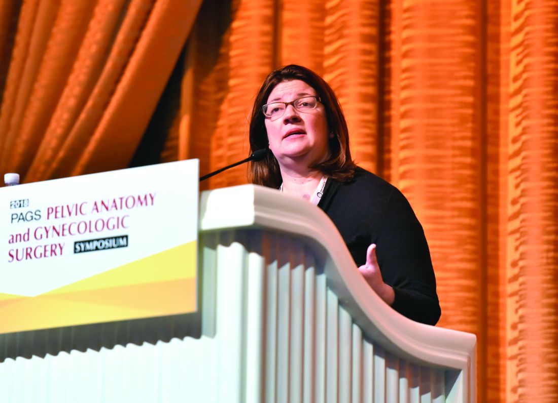
Still, “we’re still seeing a relatively high rate of inappropriate BSO,” Amanda Nickles Fader, MD, said, despite “the many benefits of ovarian conservation. Strong consideration should be made for maintaining normal ovaries in premenopausal women who are not at higher genetic risk of ovarian cancer.”
Dr. Nickles Fader, director of the Kelly gynecologic oncology service and the director of the center for rare gynecologic cancers at Johns Hopkins Hospital, Baltimore, who spoke at the Pelvic Anatomy and Gynecologic Surgery Symposium, urged gynecologists to understand the data about ovarian conservation in hysterectomy and carefully counsel patients.
“We can counsel patients with 100% certainty that BSO absolutely reduces ovarian and fallopian tube cancer rates. That’s a given,” she said. “Women get very excited about that, but you’ve got to be careful to counsel them about the flip side: The overall benefit may not be there when you consider the other morbidity and mortality that may occur because of this removal.”
As she noted, multiple retrospective, prospective, and observational studies have linked ovary removal to a variety of heightened risks, especially on the cardiac front. She highlighted a 2009 study of nearly 30,000 nurses who’d undergone hysterectomy for benign disease, about which the authors wrote that, “compared with ovarian conservation, bilateral oophorectomy at the time of hysterectomy for benign disease is associated with a decreased risk of breast and ovarian cancer but an increased risk of all-cause mortality, fatal and nonfatal coronary heart disease, and lung cancer.” No age group gained a survival benefit from oophorectomy (Obstet Gynecol. 2009 May;113[5]:1027-37 ).
Meanwhile, over the past decade, the “pendulum has swung” toward ovary conservation, at least in premenopausal women, Dr. Nickles Fader said at the meeting jointly provided by Global Academy for Medical Education and the University of Cincinnati. Global Academy and this news organization are owned by the same company.
A 2016 analysis of health statistics in five U.S. Eastern and Midwestern states found that, rates of hospital-based, hysterectomy-alone procedures grew by 15% from 2005 to 2013, while rates of oophorectomy alone and hysterectomy/oophorectomy combination procedures declined by 12% and 29%, respectively.
Still, Dr. Nickles Fader said, as many as 60% of hysterectomies are still performed in conjunction with oophorectomy.
Ovary removal, of course, can be appropriate when patients are at risk of ovarian cancer. Hereditary ovarian cancer accounts for up to 25% of epithelial ovarian cancer, she said, and research suggests that risk-reducing surgery is an effective preventative approach when high-penetrance genes are present. However, the value of the surgery is less clear in regard to moderate-penetrance genes.
Dr. Nickles Fader pointed to guidelines from the National Comprehensive Cancer Network that specify genes and syndromes that should trigger risk-reducing salpingo-oophorectomy, hysterectomy, or hysterectomy and risk-reducing salpingo-oophorectomy after childbirth.
Researchers are exploring salpingectomy – fallopian tube removal – as a possible replacement for oophorectomy. Dr. Nickles Fader highlighted a small pilot study published in 2018 that reported “BRCA mutation carriers who underwent bilateral salpingectomy had no intraoperative complications, were satisfied with their procedure choice, and had decreased cancer worry and anxiety after the procedure.”
Moving forward, she said, research will provide more insight into preventative options such as removing fallopian tubes alone instead of ovaries. “We’re starting to learn, and will probably know in the next 10-15 years, whether oophorectomy is necessary for all high-risk and moderate-risk women or if we can get away with removing their tubes and giving them the maximal health benefits of ovarian conservation.”
Dr. Nickles Fader reported consulting for Ethicon Endosurgery.
LAS VEGAS – The removal of both ovaries during hysterectomy – bilateral salpingo-oophorectomy (BSO) – has declined sharply in popularity as physicians have become more aware of its risks.
Still, “we’re still seeing a relatively high rate of inappropriate BSO,” Amanda Nickles Fader, MD, said, despite “the many benefits of ovarian conservation. Strong consideration should be made for maintaining normal ovaries in premenopausal women who are not at higher genetic risk of ovarian cancer.”
Dr. Nickles Fader, director of the Kelly gynecologic oncology service and the director of the center for rare gynecologic cancers at Johns Hopkins Hospital, Baltimore, who spoke at the Pelvic Anatomy and Gynecologic Surgery Symposium, urged gynecologists to understand the data about ovarian conservation in hysterectomy and carefully counsel patients.
“We can counsel patients with 100% certainty that BSO absolutely reduces ovarian and fallopian tube cancer rates. That’s a given,” she said. “Women get very excited about that, but you’ve got to be careful to counsel them about the flip side: The overall benefit may not be there when you consider the other morbidity and mortality that may occur because of this removal.”
As she noted, multiple retrospective, prospective, and observational studies have linked ovary removal to a variety of heightened risks, especially on the cardiac front. She highlighted a 2009 study of nearly 30,000 nurses who’d undergone hysterectomy for benign disease, about which the authors wrote that, “compared with ovarian conservation, bilateral oophorectomy at the time of hysterectomy for benign disease is associated with a decreased risk of breast and ovarian cancer but an increased risk of all-cause mortality, fatal and nonfatal coronary heart disease, and lung cancer.” No age group gained a survival benefit from oophorectomy (Obstet Gynecol. 2009 May;113[5]:1027-37 ).
Meanwhile, over the past decade, the “pendulum has swung” toward ovary conservation, at least in premenopausal women, Dr. Nickles Fader said at the meeting jointly provided by Global Academy for Medical Education and the University of Cincinnati. Global Academy and this news organization are owned by the same company.
A 2016 analysis of health statistics in five U.S. Eastern and Midwestern states found that, rates of hospital-based, hysterectomy-alone procedures grew by 15% from 2005 to 2013, while rates of oophorectomy alone and hysterectomy/oophorectomy combination procedures declined by 12% and 29%, respectively.
Still, Dr. Nickles Fader said, as many as 60% of hysterectomies are still performed in conjunction with oophorectomy.
Ovary removal, of course, can be appropriate when patients are at risk of ovarian cancer. Hereditary ovarian cancer accounts for up to 25% of epithelial ovarian cancer, she said, and research suggests that risk-reducing surgery is an effective preventative approach when high-penetrance genes are present. However, the value of the surgery is less clear in regard to moderate-penetrance genes.
Dr. Nickles Fader pointed to guidelines from the National Comprehensive Cancer Network that specify genes and syndromes that should trigger risk-reducing salpingo-oophorectomy, hysterectomy, or hysterectomy and risk-reducing salpingo-oophorectomy after childbirth.
Researchers are exploring salpingectomy – fallopian tube removal – as a possible replacement for oophorectomy. Dr. Nickles Fader highlighted a small pilot study published in 2018 that reported “BRCA mutation carriers who underwent bilateral salpingectomy had no intraoperative complications, were satisfied with their procedure choice, and had decreased cancer worry and anxiety after the procedure.”
Moving forward, she said, research will provide more insight into preventative options such as removing fallopian tubes alone instead of ovaries. “We’re starting to learn, and will probably know in the next 10-15 years, whether oophorectomy is necessary for all high-risk and moderate-risk women or if we can get away with removing their tubes and giving them the maximal health benefits of ovarian conservation.”
Dr. Nickles Fader reported consulting for Ethicon Endosurgery.
EXPERT ANALYSIS FROM PAGS
Composite screening measures advocated for asymptomatic PAH
In patients at risk for pulmonary arterial hypertension (PAH) due to a connective tissue disease, composite novel screening methods are improving early detection when employed in the context of traditional tools, such as transthoracic echocardiography (TTE), according to a systematic review of studies published over the last 5 years.
The review was conducted to prepare for a guideline update, according to the authors of this recently published summary in Seminars in Arthritis and Rheumatism.
In a literature review for 2012-2015, the authors evaluated whether new tools or strategies have improved PAH screening in patients with connective tissue disease since the last review was undertaken (Semin Arthritis Rheum 2014;43:536-41).
The latest review found that although TTE and pulmonary function tests (PFT) remain a mainstay of screening, there is growing evidence that composite measures, such as the DETECT and ASIG algorithms, add sensitivity and specificity, compared with guidelines that rely on TTE and PFT alone.
After a literature search, the systematic review included 16 cohort studies and 6 case-control studies. Most of these evaluated PAH screening strategies for patients with systemic sclerosis specifically despite the potential for other connective tissue disease etiologies to lead to PAH.
“We need more longitudinal observational studies to develop and validate screening algorithms for non–systemic sclerosis connective tissue diseases,” stated the authors, led by senior investigator Dinesh Khanna, MD, medical director of ambulatory and chronic disease in the University of Michigan’s Office of Research, Ann Arbor.
Relative to screening primarily based on TTE and PFT as advocated in 2009 joint guidelines from the European Society of Cardiology and the European Respiratory Society (ESC/ERS), the preponderance of data supported the addition of DIRECT and ASIG algorithms to improve the sensitivity and specificity of traditional screening and diagnostic tools, according to the data reviewed.
Several of the studies evaluating DETECT and ASIG compared their sensitivities and specificities to the screening strategy recommended in the 2009 ESC/ERS guidelines because the 2015 ESC/ERS guidelines were not yet available at the time these studies were taking place.
In fact, the advantage of these algorithms has been acknowledged in a set of subsequent ESC/ERS guidelines issued in 2015. These specifically recommend DETECT in selected populations, such as adults with more than a 3-year history of systemic sclerosis and less than 60% diffusing capacity of the lung for carbon monoxide.
Based on the systematic review, the authors did not conclude that there is a single set of optimal tests for PAH screening whether in patients with systemic sclerosis or another connective tissue disease, but the authors did conclude that there has been progress in strategies for early PAH detection.
They also speculated that more progress might be coming. They noted that several newer tests, such as stress TTE to assess cardiopulmonary function during exercise, might further improve PAH detection at early stages.
According to the authors, this work is important, because better screening that results in earlier PAH detection means earlier treatment, which, in turn, “may improve survival.”
SOURCE: Young A et al. Semin Arthritis Rheum. 2018; Oct 14. doi: 10.1016/j.semarthrit.2018.10.010.
FROM SEMINARS IN ARTHRITIS AND RHEUMATISM
Key clinical point: Strategies for early detection of pulmonary arterial hypertension (PAH) are improving with new tools.
Major finding: Screening algorithms are improving sensitivity and specificity for PAH in patients with connective tissue diseases.
Study details: Systematic literature review.
Disclosures: Dr. Khanna has financial relationships with many companies that produce drugs for PAH but no conflicts relative to this study.
Source: Young A et al. Semin Arthritis Rheum. 2018 Oct 14. doi. 10.1016/j.semarthrit.2018.10.010.
AMAROS: Radiation has edge for axillary treatment
SAN ANTONIO – For treatment of the axilla in women with early-stage breast cancer having a positive sentinel node, the risk-benefit calculus tilts toward radiation therapy over axillary lymph node dissection, finds an update of the phase 3, noninferiority, randomized AMAROS trial.

The trial’s previously reported 5-year results showed noninferiority of axillary radiation relative to axillary lymph node dissection (ALND) with respect to axillary recurrences, as well as less lymphedema (Lancet Oncol. 2014 Nov;15(12):1303-10). But the trial was criticized as being underpowered and having insufficient follow-up, according to principal investigator Emiel J. T. Rutgers, MD, PhD, of the Netherlands Cancer Institute in Amsterdam.
Now, at a median follow-up of 10 years, findings were basically the same, with few additional axillary recurrences having occurred in either group, he reported in a session and press conference at the San Antonio Breast Cancer Symposium. There was a nonsignificant difference in the very-low 10-year cumulative incidences of axillary recurrence, and no significant difference in other efficacy outcomes. Meanwhile, an update of the rate of lymphedema at 5 years continued to show that this treatment complication was about half as common with radiation.
The radiation therapy group did have a higher risk of second primaries, with an absolute difference of about 4%, mainly driven by more contralateral breast cancers. But it was unclear whether this difference was related to the radiation, according to Dr. Rutgers.
“Both axillary clearance and radiotherapy provide excellent, comparable locoregional control in patients who have a positive sentinel node in the axilla,” he summarized. “After 10 years [of follow-up], there is significantly less lymphedema after radiation therapy at 5 years, and therefore this can be considered a standard procedure.”
Putting data into practice
SABCS codirector and press conference moderator Virginia Kaklamani, MD, leader of the breast cancer program at University of Texas, San Antonio, wondered how Dr. Rutger’s institution has incorporated findings of the AMAROS trial and findings of the previously reported ACOSOG Z11 trial (JAMA. 2011;305:569-75).
They apply both, on a case-by-case basis, he replied. “If it’s limited node involvement in early breast cancer – and of course that’s subjective, we have some cutoffs for that – we do nothing if the sentinel node is positive. If it’s a larger tumor, high grade, lymphovascular invasion, and there are two positive sentinel nodes, then we irradiate the axilla according to the AMAROS trial. This is a discussion within our tumor board, of course, with the multidisciplinary team and together with the patient.”
Over the past 20 years, the percentage of women at his institute undergoing axillary clearance has fallen sharply, from about 75% to merely 3%, as a result of introduction of and growing evidence on sentinel node biopsy, advances in radiation therapy, and increased use of neoadjuvant chemotherapy, according to Dr. Rutgers. “At our institute, an axillary clearance in breast cancer is a rare operation for our residents. They have to go to the melanoma doctors to learn axillary clearance.” At the same time, the occurrence of lymph node metastases has remained unchanged at about 1% annually.
“This is really the beauty of this sort of deescalation in therapy, where you’re improving the morbidity of our patients – our patients have a better quality of life because they don’t have as much lymphedema – without compromising their outcomes. The chance of them having a local recurrence is very low,” Dr. Kaklamani commented. “So taking 20 and 30 and sometimes 40 lymph nodes out, like we used to do, isn’t needed anymore.”
Still, selecting the right patient for radiation is important, she cautioned, as the findings do not apply to those with a bulky lymph node, for example. “But the majority of patients we see with breast cancer have these small metastases to their axillas, if they do [have any], and in those cases, doing radiation, instead of doing more surgery or not doing anything, is very appropriate.”
Changing the standard in the United States has historically been slow. “It is the longer follow-ups from Z11, from AMAROS that are helping our surgeons cut back on the amount of surgery that they are doing,” Dr. Kaklamani maintained. “I have been surprised because we have had these trials out for 10 years, and we still are doing more axillary node dissections than we should be doing.”
At the global level, trends in use of ALND by country and region have varied depending on national practice patterns and acceptance of the trial data, according to Dr. Rutgers. “Taking a toy away from a surgeon is a difficult thing to do.”
Study details
The AMAROS trial was conducted by the European Organization for Research and Treatment of Cancer Breast Cancer Group and Radiation Oncology Group, in collaboration with the Dutch Breast Cancer Research Group and the ALMANAC Trialists’ Group.
The 1,425 patients randomized had breast cancer that was clinically node negative by either palpation or ultrasound (cT1-2,N0) and were scheduled for breast-conserving surgery or mastectomy.
The updated results showed that the 10-year cumulative incidence of axillary recurrence was 1.82% with axillary radiation and 0.93% with ALND, a nonsignificant difference (hazard ratio, 1.71; P = .365), Dr. Rutgers reported. The women also had statistically indistinguishable rates of disease-free survival (HR, 1.19; P = .105), as well as distant metastasis–free survival and overall survival.
The 10-year cumulative incidence of second primaries was higher with radiation than with ALND: 12.09% versus 8.33% (HR, 1.45; P = .035). “This is to some extent due to contralateral breast cancers,” Dr. Rutgers commented. “We cannot exclude an effect of the radiotherapy to the axilla, but we have to realize that 85% of these patients received radiotherapy anyway because of breast conservation. So for us, it is difficult to see whether the addition of the axillary radiation field would lead to more second primaries.”
The updated 5-year rate of lymphedema (data for this outcome were not collected at 10 years) showed persistence of a large difference in the occurrence of lymphedema as defined by clinical observation and/or treatment: 29.4% with ALND and 14.6% with radiation (P less than .0001).
Dr. Rutgers reported that he had no relevant conflicts of interest. The study was supported by the EORTC Charitable Trust.
SOURCE: Rutgers EJT et al. SABCS 2018, Abstract GS4-01.
SAN ANTONIO – For treatment of the axilla in women with early-stage breast cancer having a positive sentinel node, the risk-benefit calculus tilts toward radiation therapy over axillary lymph node dissection, finds an update of the phase 3, noninferiority, randomized AMAROS trial.
The trial’s previously reported 5-year results showed noninferiority of axillary radiation relative to axillary lymph node dissection (ALND) with respect to axillary recurrences, as well as less lymphedema (Lancet Oncol. 2014 Nov;15(12):1303-10). But the trial was criticized as being underpowered and having insufficient follow-up, according to principal investigator Emiel J. T. Rutgers, MD, PhD, of the Netherlands Cancer Institute in Amsterdam.
Now, at a median follow-up of 10 years, findings were basically the same, with few additional axillary recurrences having occurred in either group, he reported in a session and press conference at the San Antonio Breast Cancer Symposium. There was a nonsignificant difference in the very-low 10-year cumulative incidences of axillary recurrence, and no significant difference in other efficacy outcomes. Meanwhile, an update of the rate of lymphedema at 5 years continued to show that this treatment complication was about half as common with radiation.
The radiation therapy group did have a higher risk of second primaries, with an absolute difference of about 4%, mainly driven by more contralateral breast cancers. But it was unclear whether this difference was related to the radiation, according to Dr. Rutgers.
“Both axillary clearance and radiotherapy provide excellent, comparable locoregional control in patients who have a positive sentinel node in the axilla,” he summarized. “After 10 years [of follow-up], there is significantly less lymphedema after radiation therapy at 5 years, and therefore this can be considered a standard procedure.”
Putting data into practice
SABCS codirector and press conference moderator Virginia Kaklamani, MD, leader of the breast cancer program at University of Texas, San Antonio, wondered how Dr. Rutger’s institution has incorporated findings of the AMAROS trial and findings of the previously reported ACOSOG Z11 trial (JAMA. 2011;305:569-75).
They apply both, on a case-by-case basis, he replied. “If it’s limited node involvement in early breast cancer – and of course that’s subjective, we have some cutoffs for that – we do nothing if the sentinel node is positive. If it’s a larger tumor, high grade, lymphovascular invasion, and there are two positive sentinel nodes, then we irradiate the axilla according to the AMAROS trial. This is a discussion within our tumor board, of course, with the multidisciplinary team and together with the patient.”
Over the past 20 years, the percentage of women at his institute undergoing axillary clearance has fallen sharply, from about 75% to merely 3%, as a result of introduction of and growing evidence on sentinel node biopsy, advances in radiation therapy, and increased use of neoadjuvant chemotherapy, according to Dr. Rutgers. “At our institute, an axillary clearance in breast cancer is a rare operation for our residents. They have to go to the melanoma doctors to learn axillary clearance.” At the same time, the occurrence of lymph node metastases has remained unchanged at about 1% annually.
“This is really the beauty of this sort of deescalation in therapy, where you’re improving the morbidity of our patients – our patients have a better quality of life because they don’t have as much lymphedema – without compromising their outcomes. The chance of them having a local recurrence is very low,” Dr. Kaklamani commented. “So taking 20 and 30 and sometimes 40 lymph nodes out, like we used to do, isn’t needed anymore.”
Still, selecting the right patient for radiation is important, she cautioned, as the findings do not apply to those with a bulky lymph node, for example. “But the majority of patients we see with breast cancer have these small metastases to their axillas, if they do [have any], and in those cases, doing radiation, instead of doing more surgery or not doing anything, is very appropriate.”
Changing the standard in the United States has historically been slow. “It is the longer follow-ups from Z11, from AMAROS that are helping our surgeons cut back on the amount of surgery that they are doing,” Dr. Kaklamani maintained. “I have been surprised because we have had these trials out for 10 years, and we still are doing more axillary node dissections than we should be doing.”
At the global level, trends in use of ALND by country and region have varied depending on national practice patterns and acceptance of the trial data, according to Dr. Rutgers. “Taking a toy away from a surgeon is a difficult thing to do.”
Study details
The AMAROS trial was conducted by the European Organization for Research and Treatment of Cancer Breast Cancer Group and Radiation Oncology Group, in collaboration with the Dutch Breast Cancer Research Group and the ALMANAC Trialists’ Group.
The 1,425 patients randomized had breast cancer that was clinically node negative by either palpation or ultrasound (cT1-2,N0) and were scheduled for breast-conserving surgery or mastectomy.
The updated results showed that the 10-year cumulative incidence of axillary recurrence was 1.82% with axillary radiation and 0.93% with ALND, a nonsignificant difference (hazard ratio, 1.71; P = .365), Dr. Rutgers reported. The women also had statistically indistinguishable rates of disease-free survival (HR, 1.19; P = .105), as well as distant metastasis–free survival and overall survival.
The 10-year cumulative incidence of second primaries was higher with radiation than with ALND: 12.09% versus 8.33% (HR, 1.45; P = .035). “This is to some extent due to contralateral breast cancers,” Dr. Rutgers commented. “We cannot exclude an effect of the radiotherapy to the axilla, but we have to realize that 85% of these patients received radiotherapy anyway because of breast conservation. So for us, it is difficult to see whether the addition of the axillary radiation field would lead to more second primaries.”
The updated 5-year rate of lymphedema (data for this outcome were not collected at 10 years) showed persistence of a large difference in the occurrence of lymphedema as defined by clinical observation and/or treatment: 29.4% with ALND and 14.6% with radiation (P less than .0001).
Dr. Rutgers reported that he had no relevant conflicts of interest. The study was supported by the EORTC Charitable Trust.
SOURCE: Rutgers EJT et al. SABCS 2018, Abstract GS4-01.
REPORTING FROM SABCS 2018
Key clinical point: In patients with a positive sentinel node, axillary radiation has similar efficacy to axillary lymph node dissection and less morbidity.
Major finding: Compared with axillary lymph node dissection, axillary radiation therapy had a similar 10-year cumulative incidence of axillary recurrence (1.82% vs. 0.93%; P = .365) and half the 5-year rate of lymphedema (14.6% vs. 29.4%; P less than .0001).
Study details: A phase 3, noninferiority, randomized, controlled trial among 1,425 women with early-stage breast cancer and a positive sentinel node.
Disclosures: Dr. Rutgers reported that he had no relevant conflicts of interest. The study was supported by the European Organization for Research and Treatment of Cancer Charitable Trust.
Source: Rutgers EJT et al. SABCS 2018, Abstract GS4-01.
Despite risks, exercise is important for patients with sickle cell
SAN DIEGO – Don’t let all your patients with sickle cell anemia (SCA) and sickle cell trait (SCT) off the hook when it comes to exercise. That was the advice from Robert I. Liem, MD, a pediatric hematologist-oncologist who studies fitness.

While some patients may face risk, these conditions should pose less of a barrier to moderate- and even high-intensity exercise, Dr. Liem, of Ann & Robert H. Lurie Children’s Hospital of Chicago and Northwestern University, Chicago, said at the annual meeting of the American Society of Hematology.
“Instead of just focusing on potential harms, we should consider a paradigm shift, especially in sickle cell anemia,” he said. “There, we can start to consider the benefits of exercise, which may include some disease-modifying effects.”
There have been no formal studies into whether high-intensity exercise poses harm in patients with SCA, Dr. Liem noted. Why? There are several reasons, he said, including concern that exercise could increase sickling of red blood cells and assumptions about “sedentary behavior” in SCA.
However, there are indications that factors other than SCA may be reducing fitness in this population, a fact that could potentially be reversed by exercise.
Dr. Liem led a study that found “children and young adults with SCA have reduced exercise capacity attributable to factors independent of anemia.” The study showed that peak VO2 was 30% lower in children and young adults with SCA, compared with controls (Physiol Rep. 2015. doi: 10.14814/phy2.12338).
There are many possible explanations for this, he said, including patient-related factors such as pain during exercise and poor access to fitness resources.
Another study found low fitness in 83% of adults with SCA and identified chronic anemia as the most important factor (Am J Hematol. 2014 Aug;89[8]:819-24).
In patients with sickle cell trait, which Dr. Liem said affects an estimated 6%-9% of African-Americans, early reports of sudden death appeared in the 1960s and 1970s, and recent studies have provided more insight into the risk.
In 2012, a study tracked NCAA student athletes and found that Division I football players with SCT faced a 37-fold higher risk of exertion-related death than did athletes without SCT. Five players with SCT, all black and all Division I football players, had died over a 5-year period (Br J Sports Med 2012 Apr;46[5]:325-30).
A 2016 study, meanwhile, tracked more than 47,000 black soldiers in the U.S. Army and found those with SCT didn’t face a higher risk of death although they did have a “significantly higher risk” of exertional rhabdomyolysis (N Engl J Med. 2016 Aug 4; 375[5]:435-42).
While the cause of exercise-related harm in SCT isn’t fully understood, Dr. Liem said, it’s possible that extreme states such as severe dehydration, acidosis, and hypoxemia may trigger sickling. A combination of SCT, extreme exercise, heat, and genetic predisposition could produce harm by creating a “perfect storm,” he added.
Should patients with SCA or SCT exercise? Yes, Dr. Liem said, pointing to the importance of fitness in the general population.
Exercising to volitional exhaustion appears to be safe in children and adults with SCA, he said, and lack of exercise could lead to a variety of negative effects on growth in children and on quality of life.
There are “limited but promising data” linking exercise to benefits in SCA and SCT populations, he said.
Moving forward, Dr. Liem noted that guidelines from the NCAA and National Athletic Trainers’ Association offer insight into exercise best practices in SCT. They’re designed to promote acclimation during training, access to fluids, and prompt recognition of symptoms of heat-related illness and a condition known as “exercise collapse associated with sickle trait.”
However, there are no guidelines for exercise in SCA and it’s not helpful to let patients set limits on themselves based on the symptoms they experience, he said.
Dr. Liem reported having no relevant financial disclosures.
SAN DIEGO – Don’t let all your patients with sickle cell anemia (SCA) and sickle cell trait (SCT) off the hook when it comes to exercise. That was the advice from Robert I. Liem, MD, a pediatric hematologist-oncologist who studies fitness.
While some patients may face risk, these conditions should pose less of a barrier to moderate- and even high-intensity exercise, Dr. Liem, of Ann & Robert H. Lurie Children’s Hospital of Chicago and Northwestern University, Chicago, said at the annual meeting of the American Society of Hematology.
“Instead of just focusing on potential harms, we should consider a paradigm shift, especially in sickle cell anemia,” he said. “There, we can start to consider the benefits of exercise, which may include some disease-modifying effects.”
There have been no formal studies into whether high-intensity exercise poses harm in patients with SCA, Dr. Liem noted. Why? There are several reasons, he said, including concern that exercise could increase sickling of red blood cells and assumptions about “sedentary behavior” in SCA.
However, there are indications that factors other than SCA may be reducing fitness in this population, a fact that could potentially be reversed by exercise.
Dr. Liem led a study that found “children and young adults with SCA have reduced exercise capacity attributable to factors independent of anemia.” The study showed that peak VO2 was 30% lower in children and young adults with SCA, compared with controls (Physiol Rep. 2015. doi: 10.14814/phy2.12338).
There are many possible explanations for this, he said, including patient-related factors such as pain during exercise and poor access to fitness resources.
Another study found low fitness in 83% of adults with SCA and identified chronic anemia as the most important factor (Am J Hematol. 2014 Aug;89[8]:819-24).
In patients with sickle cell trait, which Dr. Liem said affects an estimated 6%-9% of African-Americans, early reports of sudden death appeared in the 1960s and 1970s, and recent studies have provided more insight into the risk.
In 2012, a study tracked NCAA student athletes and found that Division I football players with SCT faced a 37-fold higher risk of exertion-related death than did athletes without SCT. Five players with SCT, all black and all Division I football players, had died over a 5-year period (Br J Sports Med 2012 Apr;46[5]:325-30).
A 2016 study, meanwhile, tracked more than 47,000 black soldiers in the U.S. Army and found those with SCT didn’t face a higher risk of death although they did have a “significantly higher risk” of exertional rhabdomyolysis (N Engl J Med. 2016 Aug 4; 375[5]:435-42).
While the cause of exercise-related harm in SCT isn’t fully understood, Dr. Liem said, it’s possible that extreme states such as severe dehydration, acidosis, and hypoxemia may trigger sickling. A combination of SCT, extreme exercise, heat, and genetic predisposition could produce harm by creating a “perfect storm,” he added.
Should patients with SCA or SCT exercise? Yes, Dr. Liem said, pointing to the importance of fitness in the general population.
Exercising to volitional exhaustion appears to be safe in children and adults with SCA, he said, and lack of exercise could lead to a variety of negative effects on growth in children and on quality of life.
There are “limited but promising data” linking exercise to benefits in SCA and SCT populations, he said.
Moving forward, Dr. Liem noted that guidelines from the NCAA and National Athletic Trainers’ Association offer insight into exercise best practices in SCT. They’re designed to promote acclimation during training, access to fluids, and prompt recognition of symptoms of heat-related illness and a condition known as “exercise collapse associated with sickle trait.”
However, there are no guidelines for exercise in SCA and it’s not helpful to let patients set limits on themselves based on the symptoms they experience, he said.
Dr. Liem reported having no relevant financial disclosures.
EXPERT ANALYSIS FROM ASH 2018
Developing essential skills at all career stages
SHM Leadership Academy continues to grow
This fall I attended the 2018 Society of Hospital Medicine Leadership Academy, held in Vancouver. Once again, this conference sold out weeks ahead of time, and 300 hospitalists took time out of their busy schedules for learning and fun. There have been about 18 Leadership Academies over the years, with approximately 3,000 total participants, but this one may have been the best to date.
Why was it so good? Here are my top four reasons that Leadership Academy 2018 was the best ever:
Setting: Vancouver is just beautiful. My family has a strong maritime background, and I am a water person with saltwater in my veins. My inner sailor was overjoyed with the hotel’s views of False Creek and Vancouver Harbor, and I loved the mix of yachts and working boats. I even saw a seaplane! The hotel was a great match for the 300 hospitalists who traveled to the JW Marriott for 4 days of learning and relaxing. It was the perfect blend, whether for work or play; the hotel and city did not disappoint.

Networking: What’s more fun than getting to know 300 like-minded, leadership-oriented hospitalists for a few days? I am always energized by seeing old friends and making new ones. I really enjoy hearing about the professional adventures hospitalists at all career points are going through. Plus, I get really good advice on my own career! I also appreciate that a number of hospital medicine leaders (and even giants) come to SHM’s Leadership Academy. Over half of the SHM Board of Directors were there, as were a number of current and previous SHM presidents (Mark Williams, Jeff Wiese, Burke Kealey, Bob Harrington, Nasim Afsar, Rusty Holman, Ron Greeno, Chris Frost, and John Nelson), as well as Larry Wellikson, the CEO who has led our society through its many successes. All of these hospitalist leaders are there, having fun and networking, alongside everyone else.
Faculty: The faculty for all four courses (yes, Leadership Academy junkies, we’ve added a fourth course!) are absolutely phenomenal. I think the faculty are just the right blend of expert hospitalists (Jeff Glasheen, Rusty Holman, Jeff Wiese, Mark Williams, John Nelson) and national experts outside of hospital medicine. For example, Lenny Marcus of Harvard T.H. Chan School of Public Health, Boston, brings his experience coaching the Department of Defense, the White House, the Department of Homeland Security, and many others to the Influential Management and Mastering Teamwork courses. Lenny’s experience working with national leaders through disasters like the Boston Marathon bombing, Hurricane Katrina, and the Ebola outbreak make for more than riveting stories; there are real, tangible lessons for hospitalist leaders trying to improve clinical care. Nancy Spector is a pediatrician, nationally recognized for her work in mentoring, and is the executive director of Drexel University’s Executive Leadership in Academic Medicine. We have been fortunate to have her join the Academies, and Nancy successfully led the first group of hospitalists through the launch of SHM’s fourth leadership course, which I will describe in more detail below.
High energy & continued growth: There continues to be an enormous amount of energy around the Leadership Academy. The Vancouver courses sold out months ahead of the actual meeting! Hospitalists across the country continue to take on leadership roles and have told us that they value the skills they have learned from the courses.
Hospitalist leaders want more
In addition to the current 4-day courses (Strategic Essentials, Influential Management, and Mastering Teamwork), hospitalists are looking for a course that continues skill building once they return home.
That’s why SHM has developed a fourth Leadership Academy course. This course, called the Capstone Course, was launched in Vancouver and consists of 2 days of on-site skill development and team building (during the first 2 days of the traditional Leadership Academy) and 6 months of a longitudinal learning collaborative. The six-month learning collaborative component consists of a learning “pod” of five or six fellow hospitalists and monthly virtual meetings around crucial leadership topics. They are facilitated by an experienced Leadership Academy facilitator.
Dr. Spector is the lead faculty; her expertise made the Capstone launch a huge success. She will work with SHM and the Capstone participants throughout the entire 6 months to ensure the Capstone course is as high-quality as the previous three Academy courses.
If you haven’t been, I invite you to attend our next Leadership Academy. Over the years, despite being course director, I have learned many take-home skills from colleagues and leaders in the field that I use often. Just to name a few:
- Flexing my communications style: Tim Keogh’s lecture opened my eyes to the fact that not everyone is a data-driven introvert. I now know that some people need a social warm up, while others just want the facts, and that there are “huggers and shakers.” (In summary, it’s fine to shake hands with a hugger, but be wary of hugging a shaker.)
- I send birthday emails after I heard Jeff Wiese’s talk.
- Lenny Marcus taught me to be aware when I am “in the basement” emotionally. I now know to wait to send emails or confront others until I can get out of the basement.
And that’s just scratching the surface!
In closing, the Vancouver Leadership Academy was fantastic. Good friends, great professional development, a setting that was amazing, and an Academy that remains relevant and dynamic to our specialty. I can’t wait to see how the 2019 Leadership Academy shapes up for its debut in Nashville. My inner sailor may have to give way to my inner musician! I hope to see you and 300 of my closest friends there.
Learn more about SHM’s Leadership Academy at shmleadershipacademy.org.
Dr. Howell is a professor of medicine at Johns Hopkins University, Baltimore, and chief of the division of hospital medicine at Johns Hopkins Bayview Medical Center. He is also chief operating officer at the Society of Hospital Medicine and course director of the SHM Leadership Academy.
SHM Leadership Academy continues to grow
SHM Leadership Academy continues to grow
This fall I attended the 2018 Society of Hospital Medicine Leadership Academy, held in Vancouver. Once again, this conference sold out weeks ahead of time, and 300 hospitalists took time out of their busy schedules for learning and fun. There have been about 18 Leadership Academies over the years, with approximately 3,000 total participants, but this one may have been the best to date.
Why was it so good? Here are my top four reasons that Leadership Academy 2018 was the best ever:
Setting: Vancouver is just beautiful. My family has a strong maritime background, and I am a water person with saltwater in my veins. My inner sailor was overjoyed with the hotel’s views of False Creek and Vancouver Harbor, and I loved the mix of yachts and working boats. I even saw a seaplane! The hotel was a great match for the 300 hospitalists who traveled to the JW Marriott for 4 days of learning and relaxing. It was the perfect blend, whether for work or play; the hotel and city did not disappoint.
Networking: What’s more fun than getting to know 300 like-minded, leadership-oriented hospitalists for a few days? I am always energized by seeing old friends and making new ones. I really enjoy hearing about the professional adventures hospitalists at all career points are going through. Plus, I get really good advice on my own career! I also appreciate that a number of hospital medicine leaders (and even giants) come to SHM’s Leadership Academy. Over half of the SHM Board of Directors were there, as were a number of current and previous SHM presidents (Mark Williams, Jeff Wiese, Burke Kealey, Bob Harrington, Nasim Afsar, Rusty Holman, Ron Greeno, Chris Frost, and John Nelson), as well as Larry Wellikson, the CEO who has led our society through its many successes. All of these hospitalist leaders are there, having fun and networking, alongside everyone else.
Faculty: The faculty for all four courses (yes, Leadership Academy junkies, we’ve added a fourth course!) are absolutely phenomenal. I think the faculty are just the right blend of expert hospitalists (Jeff Glasheen, Rusty Holman, Jeff Wiese, Mark Williams, John Nelson) and national experts outside of hospital medicine. For example, Lenny Marcus of Harvard T.H. Chan School of Public Health, Boston, brings his experience coaching the Department of Defense, the White House, the Department of Homeland Security, and many others to the Influential Management and Mastering Teamwork courses. Lenny’s experience working with national leaders through disasters like the Boston Marathon bombing, Hurricane Katrina, and the Ebola outbreak make for more than riveting stories; there are real, tangible lessons for hospitalist leaders trying to improve clinical care. Nancy Spector is a pediatrician, nationally recognized for her work in mentoring, and is the executive director of Drexel University’s Executive Leadership in Academic Medicine. We have been fortunate to have her join the Academies, and Nancy successfully led the first group of hospitalists through the launch of SHM’s fourth leadership course, which I will describe in more detail below.
High energy & continued growth: There continues to be an enormous amount of energy around the Leadership Academy. The Vancouver courses sold out months ahead of the actual meeting! Hospitalists across the country continue to take on leadership roles and have told us that they value the skills they have learned from the courses.
Hospitalist leaders want more
In addition to the current 4-day courses (Strategic Essentials, Influential Management, and Mastering Teamwork), hospitalists are looking for a course that continues skill building once they return home.
That’s why SHM has developed a fourth Leadership Academy course. This course, called the Capstone Course, was launched in Vancouver and consists of 2 days of on-site skill development and team building (during the first 2 days of the traditional Leadership Academy) and 6 months of a longitudinal learning collaborative. The six-month learning collaborative component consists of a learning “pod” of five or six fellow hospitalists and monthly virtual meetings around crucial leadership topics. They are facilitated by an experienced Leadership Academy facilitator.
Dr. Spector is the lead faculty; her expertise made the Capstone launch a huge success. She will work with SHM and the Capstone participants throughout the entire 6 months to ensure the Capstone course is as high-quality as the previous three Academy courses.
If you haven’t been, I invite you to attend our next Leadership Academy. Over the years, despite being course director, I have learned many take-home skills from colleagues and leaders in the field that I use often. Just to name a few:
- Flexing my communications style: Tim Keogh’s lecture opened my eyes to the fact that not everyone is a data-driven introvert. I now know that some people need a social warm up, while others just want the facts, and that there are “huggers and shakers.” (In summary, it’s fine to shake hands with a hugger, but be wary of hugging a shaker.)
- I send birthday emails after I heard Jeff Wiese’s talk.
- Lenny Marcus taught me to be aware when I am “in the basement” emotionally. I now know to wait to send emails or confront others until I can get out of the basement.
And that’s just scratching the surface!
In closing, the Vancouver Leadership Academy was fantastic. Good friends, great professional development, a setting that was amazing, and an Academy that remains relevant and dynamic to our specialty. I can’t wait to see how the 2019 Leadership Academy shapes up for its debut in Nashville. My inner sailor may have to give way to my inner musician! I hope to see you and 300 of my closest friends there.
Learn more about SHM’s Leadership Academy at shmleadershipacademy.org.
Dr. Howell is a professor of medicine at Johns Hopkins University, Baltimore, and chief of the division of hospital medicine at Johns Hopkins Bayview Medical Center. He is also chief operating officer at the Society of Hospital Medicine and course director of the SHM Leadership Academy.
Parental leave for residents pales in comparison to that of faculty physicians
Leave policies for residents who become new parents are uneven, oft-ignored by training boards, and provide less time off than similar policies for faculty physicians. Those were the findings of a pair of research letters published in JAMA.
Kirti Magudia, MD, of the department of radiology at Brigham and Women’s Hospital in Boston and her colleagues reviewed childbearing and family leave policies for 15 graduate medical education (GME)–sponsoring institutions, all of which were affiliated with the top 12 U.S. medical schools. Though all 12 schools provided paid childbearing or family leave for faculty physicians, only 8 of the 15 did so for residents (JAMA. 2018 Dec 11;320[22)]:2372-4).
In programs that did provide leave, the average of 6.6 weeks of paid total maternity leave for residents was less than the 8.6 weeks faculty receive. Both are considerably less than proscribed by the Family and Medical Leave Act, which requires large employers to provide 12 weeks of unpaid leave, but only after 12 months of employment.
The research focused on only institutional policies for paid leave; unpaid leave and state policies may extend the average, and departments may offer leave that goes beyond specific policies, Dr. Magudia and her colleagues noted.
Changes in the residency population make now the right time for establishing consistent family leave policies, Dr. Magudia said in an interview. “We have people starting training later; we have more female trainees. And with the Match system, you’re not in control of exactly where you’re going. You may not have a support system where you end up, and a lot of the top training institutions are in high cost-of-living areas. All of those things together can make trainees especially vulnerable, and because trainees are temporary employees, changing policies to benefit them is very challenging.
“Wellness is a huge issue in medicine, and at large in society,” she said. “Making sure people have adequate parental leave goes a long way toward reducing stress levels and helping them cope with normal life transitions. We want to take steps that promote success among a diverse community of physicians; we want to retain as many people in the field as possible, and we want them to feel supported.”
Beyond asking all GME-sponsoring institutions to adopt parental leave policies, Dr. Magudia believes trainees must be better informed. “It should be clear to training program applicants what the policies are at those institutions,” she said. “That information is extremely difficult to obtain, as we’ve discovered. You can imagine that, if you are the applicant, it can be difficult to ask about those policies during the interview process because it may affect how things turn out.”
“If we can see changes like these made in the near future,” she added, “we will be in a good place.”
In the second study, Briony K. Varda, MD, of the department of urology at Boston Children’s Hospital, and her colleagues also noted the complications of balancing parental leave with training requirements from specialty boards. They compared leave policies among American Board of Medical Specialty member organizations and found that less than half specifically mentioned parental leave for resident physicians (JAMA. 2018 Dec 11;320[22]:2374-7).
Dr. Varda and her colleagues reviewed the websites of 24 ABMS boards to determine their leave policies; 22 had policies but only 11 cited parental leave as an option for residents. Twenty boards have time-based training requirements and allow for a median of 6 weeks leave for any reason; none of the boards had a specific policy for parental leave. In addition, only eight boards had “explicit and clear clarifying language” that would allow program directors to seek exemptions for their residents.
Though limitations like not detecting all available policies – and a subjective evaluation of the policies that were reviewed – could have impacted their study, the coauthors reiterated that the median of 6 weeks leave is less than the average leave for faculty physicians. They also emphasized the detriments associated with inadequate parental leave, including delayed childbearing, use of assisted reproduction technology, and difficulty breastfeeding.
Dr. Varda underlined the issues that arise for program directors, who “must weigh potentially conflicting factors such as adhering to board and institutional policies, maintaining adequate clinical service coverage, considering precedent within the program, and ensuring that resident physicians are well trained.” To balance the needs of all involved “novel approaches such as use of competency-based rather than time-based training milestones” to determine certification eligibility and, in return, lessen the stresses for new-parent residents, she noted.
The researchers disclosed no relevant conflicts of interest.
SOURCE: Magudia K et al. JAMA. 2018 Dec 11;320[22)]:2372-4; Varda B et al. JAMA. 2018 Dec 11;320[22]:2374-7.
Recent data by Magudia et al. have highlighted the fact that family leave policies supporting parents during medical training are widely inconsistent and in many instances do not exist. Where trainee policies do exist, benefits are routinely less robust than those of permanent faculty who receive on average 30% more paid leave time. Stratifying physician wellness needs by training status seems to be a misplaced approach.
It is not only the medical field which sees inconsistencies in the way family leave is allocated for different types of jobs. Millions of Americans receive no time off after birth or adoption, at a time when corporate America offers elite benefits for child care. In medicine, however, there is an expectation that paid family leave should be the norm, perhaps because of our mission to improve the quality of health care.
Of course, there are valid distinctions between faculty and trainees: faculty are more permanent, are more professionally differentiated and accomplished than trainees, have greater responsibilities, and are recruited for their expertise. Arguably, faculty deserve better compensation than trainees.
But the importance of parental leave transcends the routine benefits arguments. There is something more universal about how we value parenting. Parental leave policies benefit the health of parent and child, increase career satisfaction, and improve retention. The process of birth or adoption, ensuing fatigue, family bonding needs, and life-restructuring will challenge all parents regardless of career status.
Awareness of the inadequacies of parental leave policies is the first step in remedying the disparities in support for our trainees. Establishing an equal and adequate family leave policy for physicians at any stage is consistent with the goal of success and well-being for us all.
Laurel Fisher, MD, AGAF, professor of clinical internal medicine, division of gastroenterology, University of Pennsylvania, Philadelphia. She reports no conflicts of interest.
Leave policies for residents who become new parents are uneven, oft-ignored by training boards, and provide less time off than similar policies for faculty physicians. Those were the findings of a pair of research letters published in JAMA.
Kirti Magudia, MD, of the department of radiology at Brigham and Women’s Hospital in Boston and her colleagues reviewed childbearing and family leave policies for 15 graduate medical education (GME)–sponsoring institutions, all of which were affiliated with the top 12 U.S. medical schools. Though all 12 schools provided paid childbearing or family leave for faculty physicians, only 8 of the 15 did so for residents (JAMA. 2018 Dec 11;320[22)]:2372-4).
In programs that did provide leave, the average of 6.6 weeks of paid total maternity leave for residents was less than the 8.6 weeks faculty receive. Both are considerably less than proscribed by the Family and Medical Leave Act, which requires large employers to provide 12 weeks of unpaid leave, but only after 12 months of employment.
The research focused on only institutional policies for paid leave; unpaid leave and state policies may extend the average, and departments may offer leave that goes beyond specific policies, Dr. Magudia and her colleagues noted.
Changes in the residency population make now the right time for establishing consistent family leave policies, Dr. Magudia said in an interview. “We have people starting training later; we have more female trainees. And with the Match system, you’re not in control of exactly where you’re going. You may not have a support system where you end up, and a lot of the top training institutions are in high cost-of-living areas. All of those things together can make trainees especially vulnerable, and because trainees are temporary employees, changing policies to benefit them is very challenging.
“Wellness is a huge issue in medicine, and at large in society,” she said. “Making sure people have adequate parental leave goes a long way toward reducing stress levels and helping them cope with normal life transitions. We want to take steps that promote success among a diverse community of physicians; we want to retain as many people in the field as possible, and we want them to feel supported.”
Beyond asking all GME-sponsoring institutions to adopt parental leave policies, Dr. Magudia believes trainees must be better informed. “It should be clear to training program applicants what the policies are at those institutions,” she said. “That information is extremely difficult to obtain, as we’ve discovered. You can imagine that, if you are the applicant, it can be difficult to ask about those policies during the interview process because it may affect how things turn out.”
“If we can see changes like these made in the near future,” she added, “we will be in a good place.”
In the second study, Briony K. Varda, MD, of the department of urology at Boston Children’s Hospital, and her colleagues also noted the complications of balancing parental leave with training requirements from specialty boards. They compared leave policies among American Board of Medical Specialty member organizations and found that less than half specifically mentioned parental leave for resident physicians (JAMA. 2018 Dec 11;320[22]:2374-7).
Dr. Varda and her colleagues reviewed the websites of 24 ABMS boards to determine their leave policies; 22 had policies but only 11 cited parental leave as an option for residents. Twenty boards have time-based training requirements and allow for a median of 6 weeks leave for any reason; none of the boards had a specific policy for parental leave. In addition, only eight boards had “explicit and clear clarifying language” that would allow program directors to seek exemptions for their residents.
Though limitations like not detecting all available policies – and a subjective evaluation of the policies that were reviewed – could have impacted their study, the coauthors reiterated that the median of 6 weeks leave is less than the average leave for faculty physicians. They also emphasized the detriments associated with inadequate parental leave, including delayed childbearing, use of assisted reproduction technology, and difficulty breastfeeding.
Dr. Varda underlined the issues that arise for program directors, who “must weigh potentially conflicting factors such as adhering to board and institutional policies, maintaining adequate clinical service coverage, considering precedent within the program, and ensuring that resident physicians are well trained.” To balance the needs of all involved “novel approaches such as use of competency-based rather than time-based training milestones” to determine certification eligibility and, in return, lessen the stresses for new-parent residents, she noted.
The researchers disclosed no relevant conflicts of interest.
SOURCE: Magudia K et al. JAMA. 2018 Dec 11;320[22)]:2372-4; Varda B et al. JAMA. 2018 Dec 11;320[22]:2374-7.
FROM JAMA
Women in forensic psychiatry making progress but still have ways to go
AUSTIN, TEX. – Women are making progress in equal representation and leadership within the field of forensic psychiatry, but gender parity remains elusive, according to a presentation at the annual meeting of the American Academy of Psychiatry and the Law.
In the presentation, Kelly L. Coffman, MD, MPH, assistant professor of psychiatry and associate program director of the forensic psychiatry fellowship at Emory University, Atlanta, and Helen M. Farrell, MD, a lecturer at Harvard Medical School, Boston, discussed gender bias in the field and in medicine at large.
After reviewing a handful of Supreme Court cases since the 1970s establishing women’s rights to equal opportunities and harassment-free workplaces, Dr. Farrell noted a recent commentary in the New England Journal of Medicine illustrating the challenges women still face. In that commentary, Reshma Jagsi, MD, a professor of radiation oncology at the University of Michigan, Ann Arbor, and director of the Center for Bioethics and Social Sciences in Medicine, shared her own #MeToo experience within the context of such harassment narratives throughout academic medicine (N Engl J Med. 2018;378:209-11).
Harassment found in medicine
Dr. Jagsi had published a study in 2016 on workplace sexual harassment in medicine that surveyed 1,066 recipients of career development grants from the National Institutes of Health (JAMA. 2016;315:2120-1). The average age of respondents was 43 years, and 46% of respondents were women.
While only 22% of men reported perceiving gender bias in their careers, 70% of women reported such bias. Similarly, 66% of women said they had experienced gender bias in their careers, compared with 10% of men (P less than .001). Women also were substantially more likely to have experienced sexual harassment (30% vs. 4%).
Most women (92%) reporting those experiences described sexist remarks or behavior, 41% experienced unwanted sexual advances, and 9% experienced coercive advances.
“Although a lower proportion reported these experiences than in a 1995 sample, the difference appears large given that the women began their careers after the proportion of female medical students exceeded 40%,” Dr. Jagsi and her colleagues reported in the study.
The effects of those experiences were not minor: Among women who reported harassment, 59% said their confidence as professionals took a hit, and 47% said those experiences hurt their career advancement.
Women still underrepresented
One factor in those high rates might be the extent of existing power differentials: Women remain underrepresented in medical leadership despite accounting for more than half of all enrollees in U.S. medical schools in 2017, according to the Association of American Medical Colleges (AAMC). Female enrollment in medical schools hovered around 40% in the mid-90s, yet in 2015, the AAMC reported that women held 37% of all U.S. medical school faculty positions and 20% of full professorships.
“There’s also a large discrepancy in terms of specialties,” said Dr. Farrell, also a staff psychiatrist at Beth Israel Deaconess Medical Center, Boston. “Women tend to go into fields like pediatrics and ob.gyn. at much higher rates than men, and there are very low rates of women going into surgical fields like neurosurgery and orthopedics.”

Dr. Farrell then zeroed in on the field of forensic psychiatry in particular and various ways to consider how gender bias might manifest: opportunities for exposure to forensics in residency; fellowship applications vs. acceptances; experience in court; publications; invitations to present; large- and small-scale organizational leadership representation; and job placement across the settings of clinics, academics, prisons/institutions; and experience in private practice. Then there’s the perception of female forensic psychiatrists in court.
“There’s a really big question about the difference between how men and women are perceived by attorneys who hire us as experts, and by judges and juries in terms of our credibility when we’re testifying,” Dr. Farrell said.
Picking up where Dr. Farrell left off, Dr. Coffman noted that women always have faced an uphill battle – particularly within forensic psychiatry.
“Forensic psychiatry really is the global intersection of medicine, the criminal justice system, and the law – and traditionally, all three of these fields have really been male dominated,” Dr. Coffman said. “We were often told that women should not go into careers like medicine and the law, because if they work too hard, they might ruin their reproductive potential. They were also thought of as being weak and unable to handle seeing blood.”
Fortunately, however, she added: “That’s very different from how we think about things today.” For example,
Gender perceptions matter
Those numbers show progress, though “we’re a little bit behind the trends,” Dr. Coffman said. One reason for this probably is rooted in implicit biases that shape a person’s thinking, without a conscious realization of the sexist ideas about gender roles that have been internalized.

She presented two descriptions of an individual to make her point: one an accomplished scientist, tax attorney, and major political figure, and one a loving parent with a reputation for “always being well-coiffed and tastefully dressed.” Both depictions describe Margaret Thatcher, the first woman to become prime minister of the United Kingdom.
“There’s that real disconnect between the woman and the role, and that that’s where the prejudice lives,” Dr. Coffman said. “The greater the mismatch you see, the greater potential for prejudice.”
Research backs up those assertions. Dr. Coffman shared findings from a study that compared how male and female doctors introduced one another as speakers (J Womens Health [Larchmt]. 2017 May;26[5]:413-9).
Across 321 forms of address, women were more likely to use professional titles when “introducing any speaker during the first form of address, compared with male introducers (96.2% vs. 65.6%).” When the researchers drilled deeper, they found that women introduced others using their professional titles 97.8% of the time (45/46), while men used professional titles to introduce 72.4% of the time (110/152). A disparity was found in mixed-gender introductions: Women used professional titles when introducing men 95% of the time (57/60), but men did the same with women 49.2% of the time (31/63).
Research on perceptions of women as expert witnesses in court is more complicated. In one experiment, for example, mock jurors read a written summary of a civil case where the expert’s opinion was written by either a male or female automobile engineer. The jurors reached the same verdict just as often with female as male engineers – but awarded higher damages when the engineer was a woman.
But that was a written experiment. In similar research where mock jurors viewed video summaries involving cross-examination of a forensic mental health expert, men were found to be more “likable, believable, trustworthy, confident, and credible” than were women.
This and other research underscore a common dilemma for women, Dr. Coffman said: balancing the expectation of being warm and the need to appear competent – both of which can help and harm the way in which women are perceived. But the reality of perception sometimes can surprise.
She pointed to the double-edged sword of eye contact as an example: “If you don’t make eye contact, then you’re perceived as being weak. But if you make too much eye contact, then you’re perceived as being aggressive. So you really can’t win, right?”
Yet the women remained cautiously optimistic, especially noting the large proportion of men attending the session itself.
“It really takes everybody working together to keep the conversation going, finding out where women want to flourish and bloom, and having people to champion us and support that,” Dr. Farrell said.
Dr. Coffman and Dr. Farrell had no relevant conflicts of interest. They will be presenting an expanded version of the presentation at the International Academy of Law and Mental Health meeting in July 2019 in Rome.
AUSTIN, TEX. – Women are making progress in equal representation and leadership within the field of forensic psychiatry, but gender parity remains elusive, according to a presentation at the annual meeting of the American Academy of Psychiatry and the Law.
In the presentation, Kelly L. Coffman, MD, MPH, assistant professor of psychiatry and associate program director of the forensic psychiatry fellowship at Emory University, Atlanta, and Helen M. Farrell, MD, a lecturer at Harvard Medical School, Boston, discussed gender bias in the field and in medicine at large.
After reviewing a handful of Supreme Court cases since the 1970s establishing women’s rights to equal opportunities and harassment-free workplaces, Dr. Farrell noted a recent commentary in the New England Journal of Medicine illustrating the challenges women still face. In that commentary, Reshma Jagsi, MD, a professor of radiation oncology at the University of Michigan, Ann Arbor, and director of the Center for Bioethics and Social Sciences in Medicine, shared her own #MeToo experience within the context of such harassment narratives throughout academic medicine (N Engl J Med. 2018;378:209-11).
Harassment found in medicine
Dr. Jagsi had published a study in 2016 on workplace sexual harassment in medicine that surveyed 1,066 recipients of career development grants from the National Institutes of Health (JAMA. 2016;315:2120-1). The average age of respondents was 43 years, and 46% of respondents were women.
While only 22% of men reported perceiving gender bias in their careers, 70% of women reported such bias. Similarly, 66% of women said they had experienced gender bias in their careers, compared with 10% of men (P less than .001). Women also were substantially more likely to have experienced sexual harassment (30% vs. 4%).
Most women (92%) reporting those experiences described sexist remarks or behavior, 41% experienced unwanted sexual advances, and 9% experienced coercive advances.
“Although a lower proportion reported these experiences than in a 1995 sample, the difference appears large given that the women began their careers after the proportion of female medical students exceeded 40%,” Dr. Jagsi and her colleagues reported in the study.
The effects of those experiences were not minor: Among women who reported harassment, 59% said their confidence as professionals took a hit, and 47% said those experiences hurt their career advancement.
Women still underrepresented
One factor in those high rates might be the extent of existing power differentials: Women remain underrepresented in medical leadership despite accounting for more than half of all enrollees in U.S. medical schools in 2017, according to the Association of American Medical Colleges (AAMC). Female enrollment in medical schools hovered around 40% in the mid-90s, yet in 2015, the AAMC reported that women held 37% of all U.S. medical school faculty positions and 20% of full professorships.
“There’s also a large discrepancy in terms of specialties,” said Dr. Farrell, also a staff psychiatrist at Beth Israel Deaconess Medical Center, Boston. “Women tend to go into fields like pediatrics and ob.gyn. at much higher rates than men, and there are very low rates of women going into surgical fields like neurosurgery and orthopedics.”
Dr. Farrell then zeroed in on the field of forensic psychiatry in particular and various ways to consider how gender bias might manifest: opportunities for exposure to forensics in residency; fellowship applications vs. acceptances; experience in court; publications; invitations to present; large- and small-scale organizational leadership representation; and job placement across the settings of clinics, academics, prisons/institutions; and experience in private practice. Then there’s the perception of female forensic psychiatrists in court.
“There’s a really big question about the difference between how men and women are perceived by attorneys who hire us as experts, and by judges and juries in terms of our credibility when we’re testifying,” Dr. Farrell said.
Picking up where Dr. Farrell left off, Dr. Coffman noted that women always have faced an uphill battle – particularly within forensic psychiatry.
“Forensic psychiatry really is the global intersection of medicine, the criminal justice system, and the law – and traditionally, all three of these fields have really been male dominated,” Dr. Coffman said. “We were often told that women should not go into careers like medicine and the law, because if they work too hard, they might ruin their reproductive potential. They were also thought of as being weak and unable to handle seeing blood.”
Fortunately, however, she added: “That’s very different from how we think about things today.” For example,
Gender perceptions matter
Those numbers show progress, though “we’re a little bit behind the trends,” Dr. Coffman said. One reason for this probably is rooted in implicit biases that shape a person’s thinking, without a conscious realization of the sexist ideas about gender roles that have been internalized.
She presented two descriptions of an individual to make her point: one an accomplished scientist, tax attorney, and major political figure, and one a loving parent with a reputation for “always being well-coiffed and tastefully dressed.” Both depictions describe Margaret Thatcher, the first woman to become prime minister of the United Kingdom.
“There’s that real disconnect between the woman and the role, and that that’s where the prejudice lives,” Dr. Coffman said. “The greater the mismatch you see, the greater potential for prejudice.”
Research backs up those assertions. Dr. Coffman shared findings from a study that compared how male and female doctors introduced one another as speakers (J Womens Health [Larchmt]. 2017 May;26[5]:413-9).
Across 321 forms of address, women were more likely to use professional titles when “introducing any speaker during the first form of address, compared with male introducers (96.2% vs. 65.6%).” When the researchers drilled deeper, they found that women introduced others using their professional titles 97.8% of the time (45/46), while men used professional titles to introduce 72.4% of the time (110/152). A disparity was found in mixed-gender introductions: Women used professional titles when introducing men 95% of the time (57/60), but men did the same with women 49.2% of the time (31/63).
Research on perceptions of women as expert witnesses in court is more complicated. In one experiment, for example, mock jurors read a written summary of a civil case where the expert’s opinion was written by either a male or female automobile engineer. The jurors reached the same verdict just as often with female as male engineers – but awarded higher damages when the engineer was a woman.
But that was a written experiment. In similar research where mock jurors viewed video summaries involving cross-examination of a forensic mental health expert, men were found to be more “likable, believable, trustworthy, confident, and credible” than were women.
This and other research underscore a common dilemma for women, Dr. Coffman said: balancing the expectation of being warm and the need to appear competent – both of which can help and harm the way in which women are perceived. But the reality of perception sometimes can surprise.
She pointed to the double-edged sword of eye contact as an example: “If you don’t make eye contact, then you’re perceived as being weak. But if you make too much eye contact, then you’re perceived as being aggressive. So you really can’t win, right?”
Yet the women remained cautiously optimistic, especially noting the large proportion of men attending the session itself.
“It really takes everybody working together to keep the conversation going, finding out where women want to flourish and bloom, and having people to champion us and support that,” Dr. Farrell said.
Dr. Coffman and Dr. Farrell had no relevant conflicts of interest. They will be presenting an expanded version of the presentation at the International Academy of Law and Mental Health meeting in July 2019 in Rome.
REPORTING FROM THE AAPL ANNUAL MEETING
Social media can help doctors stay up to date
SAN DIEGO –
“People want to see that you’re a person and not strictly a physician,” said Dr. Yates, a pediatric hematologist at Texas Children’s Hospital in Houston, who has had an active Twitter presence for the last few years.
Dr. Yates – whose Twitter handle is @sicklecelldoc – said she dipped a toe in the social media waters because she wanted to bring accurate medical information to patients in the arena where they are seeking information.
“I want families to understand their condition as well as they can on whatever level they can, and so I just found this to be another way to do that ... outside of my clinic,” she said during an interview at the annual meeting of the American Society of Hematology.
But beyond correcting misinformation and serving as an advocate for patients, Dr. Yates said she gets professional benefits from being on Twitter. For instance, she uses the platform to find relevant articles as soon as they publish, without wading through all the journals.
“It’s allowed me to kind of streamline what I read,” she said.
Dr. Yates said Twitter is her social media platform of choice because it provides a simple, succinct way to communicate and provide links to more in-depth resources.
While social media can be fun and rewarding for physicians, Dr. Yates said think before you post. Ask yourself, “would you tell your chairperson this?”
Dr. Yates reported having no relevant financial disclosures.
REPORTING FROM ASH 2018

Dr. Ingrid A. Mayer on neoadjuvant endocrine therapy: The times are changing
SAN ANTONIO – Phase 2 neoadjuvant endocrine therapy breast cancer trials can and should serve as a testing platform to inform the design of phase 3 trials with the ability to change practice, according to Ingrid A. Mayer, MD.
Correlating outcomes such as the Preoperative Endocrine Prognostic Index and Ki67 blood levels from phase 2 trials to larger, similarly designed trials could reduce costs, time, and the need for large numbers of patients, thereby promoting more efficient development of effective breast cancer therapies, she said during a plenary lecture at the San Antonio Breast Cancer Symposium.
In this video interview, Dr. Mayer, director of breast medical oncology at Vanderbilt University, Nashville, Tenn., explained that while neoadjuvant endocrine therapy is suitable for down-staging tumors in women with stage I-III breast cancer, it is not commonly used, as those patients are also good surgery candidates.
However, neoadjuvant endocrine therapy can be “an incredible platform” to test novel combinations of endocrine therapy agents with or without targeted treatments, validate new biomarkers, discover mechanisms of resistance, and predict the best combinations for moving forward in phase 3 trials, she said, adding that “this is something that all academic institutions potentially should be doing.”
Although the direct clinical benefit for patients who participate in such trials is not tremendous, participation has the potential to ultimately improve outcomes for participants and for “their friends and families and daughters,” and to help propel the science forward, Dr. Meyer noted.
“I would argue that we really should be moving forward to designing trials in the neoadjuvant endocrine setting more and more, and really utilizing this platform for that end,” she said.
Dr. Mayer reported having no relevant disclosures.
REPORTING FROM SABCS 2018
Lipoprotein ratio linked to poor survival in metastatic RCC
The ratio of apolipoproteins B and A1 (apo B/A1) is an independent prognostic factor in patients with metastatic renal cell carcinoma, according to authors of a recent retrospective study.
The apo B/A1 ratio, evaluated prior to cytoreductive nephrectomy, was significantly linked with poor progression-free survival and overall survival, according to Fan Zhang, MD, of the Chinese PLA General Hospital in Beijing and coauthors.
Those findings suggest that patients with metastatic RCC should receive “consistent follow-up” that includes evaluation of that ratio, Dr. Zhang and colleagues said.
“As a novel prognostic factor, the preoperative apo B/A1 ratio can be utilized as a supplement to improve the current prognostic evaluation and treatment decision for patients with metastatic RCC,” they wrote in Urologic Oncology.
Apo B and A1, which are predominant components of low-density lipoprotein (LDL) and high-density lipoprotein (HDL), respectively, have “extensive connections” with cardiovascular disease, diabetes, and Alzheimer disease, the authors said.
The apo B/A1 ratio is a known risk index for cardiovascular disease, and in recent studies, it appeared to have some value in prognosis and prediction of gastric and colorectal cancer, among other neoplasms, they added.
In their retrospective study, Dr. Zhang and colleagues analyzed data on 287 metastatic RCC patients who underwent cytoreductive nephrectomy at the Chinese PLA General Hospital. The median age of the patients was 56 years, and the median apo B/A1 ratio was 0.859.
Significantly poorer progression-free survival was seen in the group of patients with a preoperative apo B/A1 ratio over the cutoff of 0.977 (P less than .0001) compared with those under the cutoff, the investigators reported. Likewise, overall survival was poorer for patients with an apo B/A1 ratio over a cutoff of 0.847 (P = .0005).
The apo B/A1 ratio was higher in patients with Fuhrman grade 3-4 versus grade 1-2 patients (P = .010), though the investigators said no significant differences in the ratio were seen in patients stratified by age, body mass index, fatty liver, number of metastases, among other subgroup analyses.
Multivariate analysis revealed that the two independent prognostic factors for progression-free survival were preoperative apo B/A1 ratio and Fuhrman grade, with hazard ratios of 3.131 and 1.906, respectively (P less than .001 for both), while independent prognostic factors for overall survival also included the apo B/A1 ratio (hazard ratio, 2.173; P less than .001) and Fuhrman grade, along with tumor necrosis and receiving targeted therapy.
This retrospective study was limited by relatively small samples from a single center, and the prognostic value of the apo B/A1 ratio needs to be verified in other studies, investigators said.
“Peripheral blood biomarkers only provide a supplement to the traditional prognostic factors in the prediction of the prognosis for patients with metastatic RCC, and are still unable to replace it,” Dr. Zhang and associates said.
The Beijing Natural Science Foundation supported the study. Dr. Zhang and coauthors had no disclosed conflicts of interest related to the work.
SOURCE: Zhang F et al. Urol Oncol. 2018 Nov 30. doi: 10.1016/j.urolonc.2018.11.010.
FROM UROLOGIC ONCOLOGY
Key clinical point: The ratio of apolipoproteins B and A1 (apo B/A1) was an independent prognostic factor in patients with metastatic renal cell carcinoma (RCC).
Major finding: The preoperative apo B/A1 ratio was independently prognostic for progression-free survival and overall survival, with hazard ratios of 3.131 and 2.173, respectively (P less than .001 for both).
Study details: A retrospective, single-center study including 287 patients with metastatic RCC who underwent cytoreductive nephrectomy.
Disclosures: Beijing Natural Science Foundation supported the study. Dr. Zhang and coauthors had no disclosed conflicts of interest.
Source: Zhang F et al. Urol Oncol. 2018 Nov 30. doi: 10.1016/j.urolonc.2018.11.010.