User login
High-efficacy DMTs may reduce depressive symptoms in MS
SEATTLE – , according to an interim analysis described at the annual meeting of the Consortium of Multiple Sclerosis Centers.
Depression, a common psychiatric comorbidity in MS, is associated with excess morbidity and mortality in that population. Treatment with disease-modifying therapy (DMT) could influence the cause and symptoms of depression.
Youkyung S. Roh, a medical student at Johns Hopkins University in Baltimore, and colleagues hypothesized that lower-efficacy DMTs may increase the risk of depression, whereas higher-efficacy DMTs may have antidepressive properties, possibly because of their anti-inflammatory effects.
To test this hypothesis, Ms. Roh and her colleagues examined data from MS Partners Advancing Technology and Health Solutions (MS PATHS), an ongoing, 10-site, longitudinal study. They compared the rates of change in depressive symptom severity, as measured by the Quality of Life in Neurological Disorders (Neuro-QoL) depression subscale, between patients initiating a lower-efficacy DMT and those initiating a higher-efficacy DMT. Eligible participants had relapsing-remitting MS, were new initiators of DMTs, and completed baseline and follow-up Neuro-QoL depression scales. The researchers defined lower-efficacy DMTs as interferons, glatiramer acetate, fingolimod, and dimethyl fumarate. Higher-efficacy DMTs were defined as natalizumab, rituximab, ocrelizumab, and alemtuzumab.
The investigators used multivariable-adjusted, mixed-effects regression models to compare the rates of change in Neuro-QoL depression scores for patients with MS who were initiating higher-efficacy DMTs versus those initiating lower-efficacy DMTs. Secondary analyses excluded interferons from the lower-efficacy DMT category.
In addition, the investigators compared the rates of depression between therapies within each class of DMT efficacy. Other analyses were stratified by sex, race, disability status, and antidepressant use.
The investigators’ interim analyses included 1,501 participants who initiated a new therapy and who had baseline and follow-up Neuro-QoL depression scores. The mean age of the study population was 45.1 years, and 76% of participants were women. Average follow-up duration was just under 1 year.
In all, 922 participants initiated lower-efficacy DMTs, and 579 participants initiated higher-efficacy DMTs. In preliminary analyses, initiation of higher-efficacy DMT was associated with a multivariable-adjusted 0.58-points/year reduction in Neuro-QoL depression scores, compared with initiation of lower-efficacy DMT. The result was statistically significant.
In addition, results were consistent in analyses that excluded interferons from the category of lower-efficacy DMTs. In those analyses, the reduction in depression scores was 0.63 points/year, which was also statistically significant.
Analyses incorporating additional follow-up data from this cohort are ongoing, said the authors.
The study was not supported by outside funding. Ellen M. Mowry, MD, one of the investigators, reported having performed contracted research for Biogen, Genzyme, and Sun Pharma. Ms Youkyung and another two investigators reported no disclosures or conflicts of interest.
REPORTING FROM CMSC 2019
Imaging of Neuroinflammation in Migraine with Aura
Migraine with aura is associated with neuroimmune activation/neuroinflammation, a new study found. Thirteen migraineurs with aura and 16 healthy controls received integrated PET/MRI brain scans with [11C]PBR28, a radioligand that binds to the 18 kDa translocator protein, a marker of glial activation. Standardized uptake value ratio (SUVR) was compared between groups. Researchers found:
- Compared to healthy controls, migraineurs demonstrated SUVR elevations in nociceptive processing areas, as well as in areas previously shown to be involved in cortical spreading depression.
- SUVR levels in frontoinsular cortex, primary/secondary somatosensory cortices, and basal ganglia were correlated with frequency of migraine attacks.
Albrecht DS, et al. Imaging of neuroinflammation in migraine with aura: A [11C]PBR28 PET/MRI study. [Published online ahead of print March 27, 2019]. Neurology. doi:10.1212/WNL.0000000000007371.
Patients with MS who consider using marijuana are more likely to engage in risky behaviors
SEATTLE – based on survey results presented at the annual meeting of the Consortium of Multiple Sclerosis Centers (CMSC).
“Discussing marijuana use with patients should include discussion of better health behaviors overall,” said Stacey S. Cofield, PhD, associate professor of biostatistics at the University of Alabama at Birmingham.
In August 2014, Dr. Cofield and colleagues invited 12,260 active participants in the North American Research Committee on Multiple Sclerosis (NARCOMS) registry to complete an online, anonymous questionnaire about current behaviors and attitudes regarding marijuana use and legality. Marijuana use was defined as smoking or ingesting marijuana, as well as using controlled substances derived from marijuana or synthetic marijuana. The questionnaire also asked participants about other potentially risky health behaviors, including alcohol consumption, smoking, and seat belt use.
About 78% of the 5,481 survey respondents were women. The median age at response time was 57 years, and median age at diagnosis was 37 years. In addition, 26.5% reported having active relapsing disease, 42.9% reported stable relapsing disease (that is, no relapse in at least 2 years), 20.9% reported progressive disease that formerly had been relapsing, and 9.8% reported progressive disease without relapses.
Most respondents (91.5%) thought that marijuana should be legal, 58.1% thought that it should require a prescription, and 52.9% considered using it for MS. Although 25.4% of respondents have used marijuana for their MS, 20.0% had discussed it with their doctor, and 16.1% were currently using some form of marijuana.
Nearly half of respondents, 48%, reported never using tobacco, 39.4% were former smokers, and 12.5% were current smokers. About 25% reported never consuming alcohol, with 32% consuming it monthly or less, 19% consuming it two to four times per month, and 24% consuming alcohol at least weekly. Approximately 93% of participants reported always wearing a seat belt, 4.7% nearly always used it, and more than 1% used it sometimes, seldom, or never, respectively.
When Dr. Cofield and colleagues adjusted the data for age and gender, they found that current marijuana users and those who said they had considered marijuana use were significantly more likely to be current tobacco users and to consume alcohol; they were nominally less likely to wear seat belts.
NARCOMS is funded in part by the CMSC and the Foundation of the CMSC. The present study had no funding support. Dr. Cofield reported receiving a consulting fee from the U.S. Department of Defense.
REPORTING FROM CMSC 2019
Effects of Galcanezumab After Treatment Cessation in Migraine
During posttreament periods, galcanezumab treatment effects in patients with migraine were reduced, but did not return to baseline, a new study found. Adults with episodic migraine were enrolled into EVOLVE-1 and EVOLVE-, which randomized 858 and 915 patients, respectively, to galcanezumab 120 mg, galcanezumab 240 mg, or placebo, administered subcutaneously once monthly for 6 months. After treatment completion or discontinuation, patients entered a 4-month posttreatment period. Researchers found:
- There were 740 patients (EVOLVE-1) and 830 (EVOLVE-2) patients entered into the posttreatment periods.
- About 95% (EVOLVE-1) and 96% (EVOLVE-2) of patients completed their respective trial.
- In both trials, change from pre-randomization baseline in monthly migraine headache days decreased over the posttreatment period.
- During posttreatment periods, 1.6% (EVOLVE-1) and 2.3% (EVOLVE-2) of patients initiated migraine preventive treatments.
- There were no unexpected adverse events after galcanezumab cessation.
Stauffer VL, et al. Effect of galcanezumab following treatment cessation in patients with migraine: Results from 2 randomized phase 3 trials. [Published online ahead of print April 3, 2019]. Headache. doi: 10.1111/head.13508.
Hospitalists can help alleviate rising drug costs
Four key actions providers can take
Because of the increasing costs of prescription drugs and medical therapies, many patients are unable to afford the treatment they need that could improve their health or even save their lives. In the United States, drug manufacturers can set their own prices – a policy that has resulted in overall medicine costs being far higher than in other places around the globe. Increasingly, insurers are passing the costs along to patients through higher deductibles, and pharmaceutical companies are making record profits.

Something needs to change in order to achieve the right balance between maintaining pharmaceutical innovation and ensuring patients have proper access to treatments they need. Waiting for legislation, regulation, or the courts is not an effective short-term solution. Instead, hospitalists can take immediate actions to help by alleviating the costs for as many patients possible.
Historical context
Many might be wondering how prescription costs became so imbalanced in the first place. Here are a few important factors that played a role in the dramatic price increase of pharmaceuticals:
Entrance of generic drugs: Around 2012 the entrance of generic drugs caused major unexpected competition in the medical industry. During this time, many insurers were promoting the generic drugs and not allowing brand names to be covered when a generic substitute was available.
“Orphan drugs” and manufacturer pricing: In 2014, 33 new brand-name drugs were launched in the United States, and only 8 had a direct price competitor at the time they were introduced. In addition, manufacturers were free to set their prices. Over the past decade, introductory prices for brand name drugs have reached unprecedented levels. Furthermore, manufacturers use the patent protections to increase their prices every year, even when no significant improvements have been made to the drug.
Expiring patents: According to research, there are 182 drugs that no longer have patent protection or any associated generics available. This creates opportunities for manufacturers to maintain patent-era pricing or even engage in price gouging.
Lack of robust competition: Several high-priced blockbuster drugs hit the market to treat serious diseases, most of which do not have generic brand substitutes, which leaves only one option for patients – and it’s usually not affordable. According to research, more than 500 drugs have only one marketed generic. In addition, manufacturer mergers and acquisitions have occurred, which has led to a more concentrated and less competitive market for pricing.
Stricter Food and Drug Administration policies: American consumers have access to the safest and most advanced pharmaceutical system in the world, which requires several trials and testing before the drug can be approved and brought to the market. Despite the benefits of these strict procedures, the downside means higher costs for the brand and manufacturer that they will want to recoup through the price of the drug on the market.
Number of new drugs allowed to enter the market: New drugs that enter the market in the United States do so more quickly than in most other countries. Research shows the U.S. pharmaceutical market contributes to 45% of the global pharmaceutical market. The $76 billion in research and development that pharmaceutical companies claim overlooks the ways that U.S. employers and taxpayers pay for at least 44% through tax subsidies and credits. What makes it worse is that research shows most corporate research and development is directed at minimally innovative new drugs, using the system to secure patents and charge monopoly prices.
Compared with other high-income countries, the United States spends the most per capita on prescription drugs. While insured U.S. patients often pay little or nothing for generic prescriptions, they can be billed tens of thousands of dollars for certain high-priced medicines. The United States has the highest rate of insured patients skipping or not filling prescriptions because of cost. For example, the price of EpiPens, a drug delivery system that is crucial for persons experiencing life-threatening allergic reactions, has increased more than $500 in just 9 years.
How to alleviate rising drug costs
The good news is that hospitalists can do something about the high costs of pharmaceuticals.

Understand and offer alternative ways for drug intake: Many patients admitted to a hospital with severe infections are initially started with intravenous medications. Although conversion from intravenous to oral therapy is inappropriate for a patient who is critically ill or has an inability to absorb oral medications, every hospital will have a certain number of patients who are eligible for a switch from intravenous to oral therapy.
The World Health Organization (WHO) reports that the irrational use of medicines is a major problem worldwide, including antibiotics. Switching from IV to oral enables one to select a cheaper or older antibiotic that is as effective as the IV antibiotic. However, this requires breaking the belief that many physicians still have that IV medications’ bioavailability is stronger and creates less susceptibility to the illness reoccurring in the patient. For many medications, essentially the same amount of drug is found in the blood when given intravenously or orally. In addition, research has shown several benefits beyond cost reduction for oral over IV, such as earlier discharge and reduced risk of infections.
Limit unnecessary antibiotic prescriptions and consider antibiotics stewardship programs: The Center for Disease Control reports that one in three (47 million) antibiotic prescriptions are unnecessary. Most of these unnecessary antibiotics are prescribed for respiratory conditions caused by viruses including common colds, viral sore throats, bronchitis, and sinus and ear infections that do not respond to antibiotics. Although the White House released The National Action Plan for Combating Antibiotic-Resistant Bacteria (CARB) in 2015, which set a goal of reducing inappropriate outpatient antibiotic use by at least half by 2020, hospitalists can still do more by being extremely cautious with prescribing drugs to patients. Use appropriate consultants whenever necessary to suggest the right drug. For example, consider an infectious disease specialist to suggest the appropriate type and length of time for an antibiotic. In addition, hospital-based programs dedicated to improving antibiotic use, known as antibiotic stewardship programs (ASPs), have been shown to optimize the treatment of infections and reduce adverse events associated with antibiotic use.
Review labs and vitals carefully and encourage a higher level of patient care beyond the digital tools available: Studies have shown an oversight in an exam (a “miss”) can result in real consequences, including death. Our $3.4 trillion health care system is responsible for more than a quarter of a million deaths per year because of medical error. Much of that is a result of poorly coordinated care, poor communication, patients falling through the cracks, or knowledge not being transferred. “True clinical judgment is more than addressing the avalanche of blood work, imaging, and lab tests; it is about using human skills to understand where the patient is in the trajectory of a life and the disease, what the nature of the patient’s family and social circumstances is, and how much they want done,” wrote Dr. Abraham Verghese in the New York Times in 2018 (“How Tech Can Turn Doctors into Clerical Workers”). This also means understanding whether the patient is on any other type of medication and, as a result, having knowledge of possible consequences for drug interactions. Always look for safe medications or discontinue the use of any unnecessary drugs the patient is currently taking.
Allow pharmacies to automatically substitute less expensive equivalent drugs: When prescribing pharmaceuticals for patients, determine if there are any substitutes that can help alleviate costs while delivering equivalent care to the patient. This requires excellent ongoing communication with pharmacists and understanding the substitutes available, as well as any side effects or consequences.
Hospitalists can make a difference
There are many variables that play a role in rising pharmaceutical costs in the United States. One of the most significant is that there are no strategies in place to control pricing of drugs and the profits made by the pharmaceutical companies.
Although finding new drugs that can cure major life-threatening diseases or illnesses is important, so is ensuring that more patients have access to such drugs at a reasonable cost. While there are several ways that the government can and should help with enabling and supporting this balance, it most likely requires such large changes that it will take a long time. As a result, it is important for hospitalists to find effective short-term solutions that can be implemented right away to alleviate the rising costs of pharmaceuticals and provide proper patient care regardless of their economic status – all of which requires better research, analysis, and comparison before prescribing treatment to patients.
Dr. Kasarla is a hospitalist with APOGEE Physicians at Wise Surgical at Parkway in Fort Worth, Tex. He did his internal medicine residency at Mercy Hospital & Medical Center, Chicago. Contact him at [email protected]. Dr. Devireddy is a hospitalist based at Sri Ramachandra Medical Centre, Porur, Tamilnadu, India. Contact her at [email protected].
FURTHER READING
Olson and Sheiner (2017). “The Hutchins Center Explains: Prescription drug spending” Brooking.edu
Lo, Chris (2018). “Cost control: drug pricing policies around the world,” Pharmaceutical-Technology.com
Center for Disease Control and Prevention. (2016). 1 in 3 antibiotic prescriptions unnecessary. Retrieved Jan 31, 2019, from https://www.cdc.gov/media/releases/2016/p0503-unnecessary-prescriptions.html
Verghese, Abraham (2018). “How Tech Can Turn Doctors Into Clerical Workers” NYTimes.Com
Waxman, Corr, Martin et al (2017). “Getting to the Root of High Prescription Drug Prices” Commonwealthfund.org
American Council on Science and Health. (2018). Government Is The Big Reason EpiPen And Other Generics Are So Expensive. Retrieved Jan 31, 2019, from https://www.acsh.org/news/2018/06/23/government-big-reason-epipen-and-other-generics-are-so-expensive-13114
Statista. (2018). U.S. Pharmaceutical Industry – Statistics & Facts. Retrieved Jan 31, 2019, from https://www.statista.com/topics/1719/pharmaceutical-industry/
Four key actions providers can take
Four key actions providers can take
Because of the increasing costs of prescription drugs and medical therapies, many patients are unable to afford the treatment they need that could improve their health or even save their lives. In the United States, drug manufacturers can set their own prices – a policy that has resulted in overall medicine costs being far higher than in other places around the globe. Increasingly, insurers are passing the costs along to patients through higher deductibles, and pharmaceutical companies are making record profits.
Something needs to change in order to achieve the right balance between maintaining pharmaceutical innovation and ensuring patients have proper access to treatments they need. Waiting for legislation, regulation, or the courts is not an effective short-term solution. Instead, hospitalists can take immediate actions to help by alleviating the costs for as many patients possible.
Historical context
Many might be wondering how prescription costs became so imbalanced in the first place. Here are a few important factors that played a role in the dramatic price increase of pharmaceuticals:
Entrance of generic drugs: Around 2012 the entrance of generic drugs caused major unexpected competition in the medical industry. During this time, many insurers were promoting the generic drugs and not allowing brand names to be covered when a generic substitute was available.
“Orphan drugs” and manufacturer pricing: In 2014, 33 new brand-name drugs were launched in the United States, and only 8 had a direct price competitor at the time they were introduced. In addition, manufacturers were free to set their prices. Over the past decade, introductory prices for brand name drugs have reached unprecedented levels. Furthermore, manufacturers use the patent protections to increase their prices every year, even when no significant improvements have been made to the drug.
Expiring patents: According to research, there are 182 drugs that no longer have patent protection or any associated generics available. This creates opportunities for manufacturers to maintain patent-era pricing or even engage in price gouging.
Lack of robust competition: Several high-priced blockbuster drugs hit the market to treat serious diseases, most of which do not have generic brand substitutes, which leaves only one option for patients – and it’s usually not affordable. According to research, more than 500 drugs have only one marketed generic. In addition, manufacturer mergers and acquisitions have occurred, which has led to a more concentrated and less competitive market for pricing.
Stricter Food and Drug Administration policies: American consumers have access to the safest and most advanced pharmaceutical system in the world, which requires several trials and testing before the drug can be approved and brought to the market. Despite the benefits of these strict procedures, the downside means higher costs for the brand and manufacturer that they will want to recoup through the price of the drug on the market.
Number of new drugs allowed to enter the market: New drugs that enter the market in the United States do so more quickly than in most other countries. Research shows the U.S. pharmaceutical market contributes to 45% of the global pharmaceutical market. The $76 billion in research and development that pharmaceutical companies claim overlooks the ways that U.S. employers and taxpayers pay for at least 44% through tax subsidies and credits. What makes it worse is that research shows most corporate research and development is directed at minimally innovative new drugs, using the system to secure patents and charge monopoly prices.
Compared with other high-income countries, the United States spends the most per capita on prescription drugs. While insured U.S. patients often pay little or nothing for generic prescriptions, they can be billed tens of thousands of dollars for certain high-priced medicines. The United States has the highest rate of insured patients skipping or not filling prescriptions because of cost. For example, the price of EpiPens, a drug delivery system that is crucial for persons experiencing life-threatening allergic reactions, has increased more than $500 in just 9 years.
How to alleviate rising drug costs
The good news is that hospitalists can do something about the high costs of pharmaceuticals.
Understand and offer alternative ways for drug intake: Many patients admitted to a hospital with severe infections are initially started with intravenous medications. Although conversion from intravenous to oral therapy is inappropriate for a patient who is critically ill or has an inability to absorb oral medications, every hospital will have a certain number of patients who are eligible for a switch from intravenous to oral therapy.
The World Health Organization (WHO) reports that the irrational use of medicines is a major problem worldwide, including antibiotics. Switching from IV to oral enables one to select a cheaper or older antibiotic that is as effective as the IV antibiotic. However, this requires breaking the belief that many physicians still have that IV medications’ bioavailability is stronger and creates less susceptibility to the illness reoccurring in the patient. For many medications, essentially the same amount of drug is found in the blood when given intravenously or orally. In addition, research has shown several benefits beyond cost reduction for oral over IV, such as earlier discharge and reduced risk of infections.
Limit unnecessary antibiotic prescriptions and consider antibiotics stewardship programs: The Center for Disease Control reports that one in three (47 million) antibiotic prescriptions are unnecessary. Most of these unnecessary antibiotics are prescribed for respiratory conditions caused by viruses including common colds, viral sore throats, bronchitis, and sinus and ear infections that do not respond to antibiotics. Although the White House released The National Action Plan for Combating Antibiotic-Resistant Bacteria (CARB) in 2015, which set a goal of reducing inappropriate outpatient antibiotic use by at least half by 2020, hospitalists can still do more by being extremely cautious with prescribing drugs to patients. Use appropriate consultants whenever necessary to suggest the right drug. For example, consider an infectious disease specialist to suggest the appropriate type and length of time for an antibiotic. In addition, hospital-based programs dedicated to improving antibiotic use, known as antibiotic stewardship programs (ASPs), have been shown to optimize the treatment of infections and reduce adverse events associated with antibiotic use.
Review labs and vitals carefully and encourage a higher level of patient care beyond the digital tools available: Studies have shown an oversight in an exam (a “miss”) can result in real consequences, including death. Our $3.4 trillion health care system is responsible for more than a quarter of a million deaths per year because of medical error. Much of that is a result of poorly coordinated care, poor communication, patients falling through the cracks, or knowledge not being transferred. “True clinical judgment is more than addressing the avalanche of blood work, imaging, and lab tests; it is about using human skills to understand where the patient is in the trajectory of a life and the disease, what the nature of the patient’s family and social circumstances is, and how much they want done,” wrote Dr. Abraham Verghese in the New York Times in 2018 (“How Tech Can Turn Doctors into Clerical Workers”). This also means understanding whether the patient is on any other type of medication and, as a result, having knowledge of possible consequences for drug interactions. Always look for safe medications or discontinue the use of any unnecessary drugs the patient is currently taking.
Allow pharmacies to automatically substitute less expensive equivalent drugs: When prescribing pharmaceuticals for patients, determine if there are any substitutes that can help alleviate costs while delivering equivalent care to the patient. This requires excellent ongoing communication with pharmacists and understanding the substitutes available, as well as any side effects or consequences.
Hospitalists can make a difference
There are many variables that play a role in rising pharmaceutical costs in the United States. One of the most significant is that there are no strategies in place to control pricing of drugs and the profits made by the pharmaceutical companies.
Although finding new drugs that can cure major life-threatening diseases or illnesses is important, so is ensuring that more patients have access to such drugs at a reasonable cost. While there are several ways that the government can and should help with enabling and supporting this balance, it most likely requires such large changes that it will take a long time. As a result, it is important for hospitalists to find effective short-term solutions that can be implemented right away to alleviate the rising costs of pharmaceuticals and provide proper patient care regardless of their economic status – all of which requires better research, analysis, and comparison before prescribing treatment to patients.
Dr. Kasarla is a hospitalist with APOGEE Physicians at Wise Surgical at Parkway in Fort Worth, Tex. He did his internal medicine residency at Mercy Hospital & Medical Center, Chicago. Contact him at [email protected]. Dr. Devireddy is a hospitalist based at Sri Ramachandra Medical Centre, Porur, Tamilnadu, India. Contact her at [email protected].
FURTHER READING
Olson and Sheiner (2017). “The Hutchins Center Explains: Prescription drug spending” Brooking.edu
Lo, Chris (2018). “Cost control: drug pricing policies around the world,” Pharmaceutical-Technology.com
Center for Disease Control and Prevention. (2016). 1 in 3 antibiotic prescriptions unnecessary. Retrieved Jan 31, 2019, from https://www.cdc.gov/media/releases/2016/p0503-unnecessary-prescriptions.html
Verghese, Abraham (2018). “How Tech Can Turn Doctors Into Clerical Workers” NYTimes.Com
Waxman, Corr, Martin et al (2017). “Getting to the Root of High Prescription Drug Prices” Commonwealthfund.org
American Council on Science and Health. (2018). Government Is The Big Reason EpiPen And Other Generics Are So Expensive. Retrieved Jan 31, 2019, from https://www.acsh.org/news/2018/06/23/government-big-reason-epipen-and-other-generics-are-so-expensive-13114
Statista. (2018). U.S. Pharmaceutical Industry – Statistics & Facts. Retrieved Jan 31, 2019, from https://www.statista.com/topics/1719/pharmaceutical-industry/
Health State Utilities and Migraine Severity
Health state utilities associated with migraine treatment estimated from a general population sample may be used to represent route of administration and adverse events (AEs) in cost-utility models, a new study found. In time trade-off interviews, patients with migraine and general population participants in the United Kingdom valued health state vignettes based on literature, medication labels, and clinician interviews. All respondents valued migraine health states varying in route of administration. Researchers found:
- A total of 400 participants completed interviews (200 general population, 200 migraine patients).
- In the general population sample, mean utilities of health states without aura were 0.79 with daily oral medication, 0.78 with 1 injection per month, and 0.72 with between 31 and 39 injections once every 3 months.
- The greatest disutilities were for AEs associated with oral medications.
Matza LS, et al. Health state utilities associated with attributes of migraine preventive treatments based on patient and general population preferences. [Published online ahead of print March 28, 2019]. Qual Life Res. doi: 10.1007/s11136-019-02163-3.
Stock inhalers at school effectively meet students’ rescue medication needs
DALLAS – Allowing public and private schools to store multiuse stock albuterol inhalers for students with asthma is a legally and medically feasible way to provide students with rescue medication without their need to leave school, according to a recent study.

“Stakeholder coalitions can facilitate the large-scale adoption of stock inhaler programs in schools,” concluded Ashley A. Lowe, MSPH, a senior research specialist and PhD candidate at the University of Arizona, Tucson, and colleagues in a poster at the American Thoracic Society’s international conference.“These programs improve access to rescue medication while returning students back to their classroom.”
The Arizona legislature passed H.B. 2208, “Stock Inhalers for Schools” in March 2017 to allow schools to store and administer albuterol sulfate while indemnifying trained staff against liability when they allowed students to use the inhaler in good faith. A stock inhaler can used by different students because of its disposable valved-holding chambers.
“Such laws allow schools to overcome the legal obstacles that make it difficult for them to ensure such medication is readily available to all children experiencing respiratory distress,” the authors wrote. They assessed the use and outcomes of schools’ storage of stock inhalers during the 2017-2018 school year in Pima County, Arizona.
Of the 213 public, 90 charter, and 61 private/parochial schools in Pima County, 246 (67%) total schools participated, including nearly all of the public schools (93%), nearly half the private/parochial schools (49%), and 17% of the charter schools. A total of 134,251 students had access to a stock inhaler at school.
Each participating school received a kit containing a 60-dose albuterol sulfate inhaler, 10 valved-holding chambers, a signed standing medical order, a standardized emergency protocol for albuterol use, access to an online training curriculum and template resources, along with technical support.
Each time a school used the stock inhaler, they documented whether an asthma diagnosis was known or not, total puffs administered and where the student went next – returned to class, sent home with caregiver, 911 call without transport, or 911 call with EMS transport.
Based on data analyzed from 240 schools, the stock inhalers were used 1,032 times at 152 schools during the study period, predominantly at public schools (97%) and by students with a known asthma diagnosis (82%). In 12.2% of cases, the student did not have a known asthma diagnosis, and 5.8% of the time, asthma diagnosis status was unknown. The students received a mean 2.7 puffs at each use.
Ethnicity and race data of those students who used the inhalers was not complete. Most of the students for whom ethnicity data were available (n = 343) and who used the inhaler were Hispanic/Latino (69.8%) independent of race. Based only on the 437 students for whom data on race were available, students using the inhaler included 41% white, 11.7% black, 3.1% Native American/Alaskan Native, 1% Asian and 0.6% Native Hawaiian/Pacific Islander.
Among the 915 uses of the inhaler for which subsequent student location was available, the majority of students (84%) returned to their classroom after using the inhaler. Only five were transported to a medical facility via EMS following a 911 call, and 911 was called for one student who did not receive EMS transport.
According to the Allergy & Asthma Network, the following states have school stock albuterol laws: Arizona, Colorado, Georgia, Illinois, Missouri, New Hampshire, New Mexico, Oklahoma, Ohio, Texas, Utah, and West Virginia.*
The research was funded by Banner–University Medical Center Tucson, Thayer Medical Corporation, and the Asthma & Airway Disease Research Center. The authors had no disclosures.
SOURCE: Lowe AA et al. ATS 2019, Abstract A4070.
* This article was updated on July 15, 2019.
DALLAS – Allowing public and private schools to store multiuse stock albuterol inhalers for students with asthma is a legally and medically feasible way to provide students with rescue medication without their need to leave school, according to a recent study.
“Stakeholder coalitions can facilitate the large-scale adoption of stock inhaler programs in schools,” concluded Ashley A. Lowe, MSPH, a senior research specialist and PhD candidate at the University of Arizona, Tucson, and colleagues in a poster at the American Thoracic Society’s international conference.“These programs improve access to rescue medication while returning students back to their classroom.”
The Arizona legislature passed H.B. 2208, “Stock Inhalers for Schools” in March 2017 to allow schools to store and administer albuterol sulfate while indemnifying trained staff against liability when they allowed students to use the inhaler in good faith. A stock inhaler can used by different students because of its disposable valved-holding chambers.
“Such laws allow schools to overcome the legal obstacles that make it difficult for them to ensure such medication is readily available to all children experiencing respiratory distress,” the authors wrote. They assessed the use and outcomes of schools’ storage of stock inhalers during the 2017-2018 school year in Pima County, Arizona.
Of the 213 public, 90 charter, and 61 private/parochial schools in Pima County, 246 (67%) total schools participated, including nearly all of the public schools (93%), nearly half the private/parochial schools (49%), and 17% of the charter schools. A total of 134,251 students had access to a stock inhaler at school.
Each participating school received a kit containing a 60-dose albuterol sulfate inhaler, 10 valved-holding chambers, a signed standing medical order, a standardized emergency protocol for albuterol use, access to an online training curriculum and template resources, along with technical support.
Each time a school used the stock inhaler, they documented whether an asthma diagnosis was known or not, total puffs administered and where the student went next – returned to class, sent home with caregiver, 911 call without transport, or 911 call with EMS transport.
Based on data analyzed from 240 schools, the stock inhalers were used 1,032 times at 152 schools during the study period, predominantly at public schools (97%) and by students with a known asthma diagnosis (82%). In 12.2% of cases, the student did not have a known asthma diagnosis, and 5.8% of the time, asthma diagnosis status was unknown. The students received a mean 2.7 puffs at each use.
Ethnicity and race data of those students who used the inhalers was not complete. Most of the students for whom ethnicity data were available (n = 343) and who used the inhaler were Hispanic/Latino (69.8%) independent of race. Based only on the 437 students for whom data on race were available, students using the inhaler included 41% white, 11.7% black, 3.1% Native American/Alaskan Native, 1% Asian and 0.6% Native Hawaiian/Pacific Islander.
Among the 915 uses of the inhaler for which subsequent student location was available, the majority of students (84%) returned to their classroom after using the inhaler. Only five were transported to a medical facility via EMS following a 911 call, and 911 was called for one student who did not receive EMS transport.
According to the Allergy & Asthma Network, the following states have school stock albuterol laws: Arizona, Colorado, Georgia, Illinois, Missouri, New Hampshire, New Mexico, Oklahoma, Ohio, Texas, Utah, and West Virginia.*
The research was funded by Banner–University Medical Center Tucson, Thayer Medical Corporation, and the Asthma & Airway Disease Research Center. The authors had no disclosures.
SOURCE: Lowe AA et al. ATS 2019, Abstract A4070.
* This article was updated on July 15, 2019.
REPORTING FROM ATS 2019
Mental illness in MS: ‘Follow the why’
SEATTLE – , a neuropsychiatrist cautioned colleagues who treat MS.
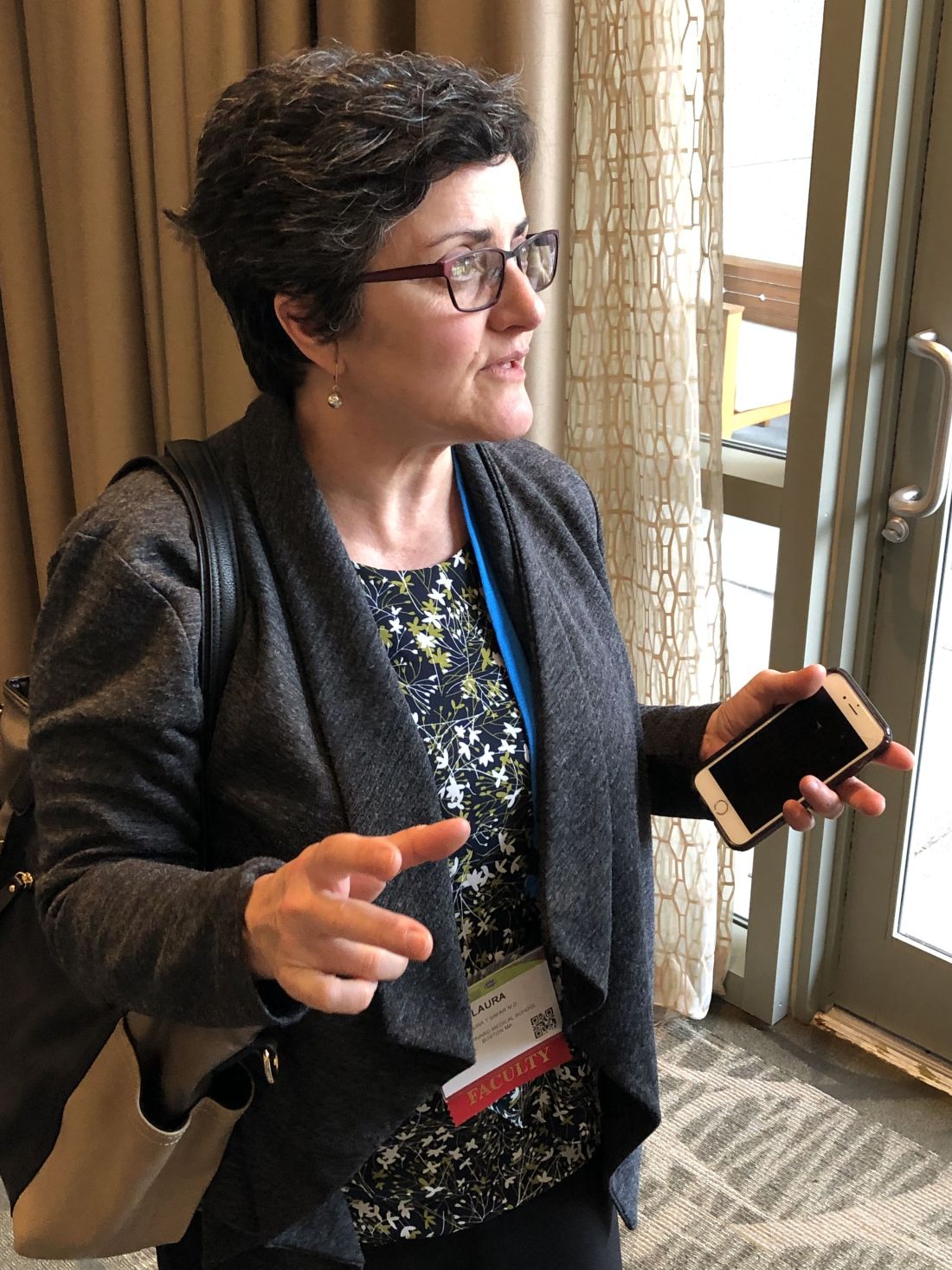
For example, depression may strike a patient as a primary condition, just as it could in anyone. But it may also be a manifestation of MS itself, or a side effect of an MS medication, or spurred by the fatigue and pain caused by MS, said Laura T. Safar, MD, a psychiatrist affiliated with Brigham and Women's Hospital, Boston*. As a result, popular psychiatric treatments such as SSRIs might not necessarily be the best approach, said Dr. Safar, who spoke in an interview and during a presentation at the annual meeting of the Consortium of Multiple Sclerosis Centers.
“You need to follow the why,” she said in the interview, adding that it is crucial to view neurologic and mental health as one and the same in MS. “More integration,” she said, “continues to be the way to go.”
Here are some pearls and tips from Dr. Safar’s presentation on treating psychiatric conditions in patients with MS:
Mental illness incidence
Depression is estimated to affect 25%-45% of people with MS over their lifetimes, while bipolar disorder is thought to affect 6% of patients and a quarter are estimated to have anxiety.
Researchers also believe as many as 10% of patients are affected by pathological laughing and crying during their lives.
Psychiatric side effects
Interferon drugs are notoriously linked to depression and psychosis. Glatiramer acetate (Copaxone) and natalizumab (Tysabri) are also thought to cause psychiatric side effects in some cases – anxiety and depression, respectively. But drug-modifying therapies can also provide relief on the psychiatric front, Dr. Safar said.
Meanwhile, dozens of other drugs used to treat aspects of MS such as spasticity, pain, and fatigue have possible psychiatric side effects.
Alternatives to SSRIs
SSRIs are often a first option in psychiatric patients, but those with MS may need another option because so many – an estimated 80% – also have fatigue, Dr. Safar said.
Alternatives for patients with MS include serotonin and norepinephrine reuptake inhibitors (SNRIs), which may have an advantage over SSRIs, she said. Specifically, SNRIs and bupropion (Wellbutrin) may be better for patients with fatigue and cognitive problems, she said, while vortioxetine (Trintellix) may benefit cognition.
Treating anxiety
There are no data regarding the best drug treatment for anxiety in patients with MS, she said, and SSRIs are typically the starting point. Consider SNRIs and duloxetine, respectively, when patients also have significant fatigue and cognitive symptoms. Use benzodiazepines only in occasional cases (such as anxiety regarding an MRI) and severe cases, she said.
MS-specific side effects
Beware of MS-specific side effects, Dr. Safar said. Some common psychiatric drugs, especially citalopram (Celexa) and escitalopram (Lexapro), may increase the QTc interval and shouldn’t be used in combination with the MS drug fingolimod (Gilenya).
And, she said, bupropion is “a very helpful agent” but poses a rare risk of seizures. Dr. Safar said she has seen this side effect a couple times over 10 years, but both were in patients with “other factors involved.” Still, “it’s something to keep in mind.”
Also understand that serotonergic agents can worsen restless legs syndrome, which is more common in patients with MS. Dr. Safar advises monitoring for the condition.
Pathological laughing, crying
Episodes of so-called pathological laughing, crying, or both tend to be brief, frequent, and intense. They may be sparked by nothing at all, and more often feature crying.
Certain SSRIs have proved helpful for the condition in MS, Dr. Safar said. Research also supports a combination of dextromethorphan (cough suppressant) and quinidine (a drug used to treat arrhythmias and malaria). The combination is sold together as Nuedexta.
Other agents such as venlafaxine (Effexor) and duloxetine (Cymbalta) have very limited data and shouldn’t be first-line treatment, she said.
Dr. Safar reports no relevant disclosures.
Correction, 5/31/19: An earlier version of this article misstated Dr. Safar's hospital affiliation.
SEATTLE – , a neuropsychiatrist cautioned colleagues who treat MS.
For example, depression may strike a patient as a primary condition, just as it could in anyone. But it may also be a manifestation of MS itself, or a side effect of an MS medication, or spurred by the fatigue and pain caused by MS, said Laura T. Safar, MD, a psychiatrist affiliated with Brigham and Women's Hospital, Boston*. As a result, popular psychiatric treatments such as SSRIs might not necessarily be the best approach, said Dr. Safar, who spoke in an interview and during a presentation at the annual meeting of the Consortium of Multiple Sclerosis Centers.
“You need to follow the why,” she said in the interview, adding that it is crucial to view neurologic and mental health as one and the same in MS. “More integration,” she said, “continues to be the way to go.”
Here are some pearls and tips from Dr. Safar’s presentation on treating psychiatric conditions in patients with MS:
Mental illness incidence
Depression is estimated to affect 25%-45% of people with MS over their lifetimes, while bipolar disorder is thought to affect 6% of patients and a quarter are estimated to have anxiety.
Researchers also believe as many as 10% of patients are affected by pathological laughing and crying during their lives.
Psychiatric side effects
Interferon drugs are notoriously linked to depression and psychosis. Glatiramer acetate (Copaxone) and natalizumab (Tysabri) are also thought to cause psychiatric side effects in some cases – anxiety and depression, respectively. But drug-modifying therapies can also provide relief on the psychiatric front, Dr. Safar said.
Meanwhile, dozens of other drugs used to treat aspects of MS such as spasticity, pain, and fatigue have possible psychiatric side effects.
Alternatives to SSRIs
SSRIs are often a first option in psychiatric patients, but those with MS may need another option because so many – an estimated 80% – also have fatigue, Dr. Safar said.
Alternatives for patients with MS include serotonin and norepinephrine reuptake inhibitors (SNRIs), which may have an advantage over SSRIs, she said. Specifically, SNRIs and bupropion (Wellbutrin) may be better for patients with fatigue and cognitive problems, she said, while vortioxetine (Trintellix) may benefit cognition.
Treating anxiety
There are no data regarding the best drug treatment for anxiety in patients with MS, she said, and SSRIs are typically the starting point. Consider SNRIs and duloxetine, respectively, when patients also have significant fatigue and cognitive symptoms. Use benzodiazepines only in occasional cases (such as anxiety regarding an MRI) and severe cases, she said.
MS-specific side effects
Beware of MS-specific side effects, Dr. Safar said. Some common psychiatric drugs, especially citalopram (Celexa) and escitalopram (Lexapro), may increase the QTc interval and shouldn’t be used in combination with the MS drug fingolimod (Gilenya).
And, she said, bupropion is “a very helpful agent” but poses a rare risk of seizures. Dr. Safar said she has seen this side effect a couple times over 10 years, but both were in patients with “other factors involved.” Still, “it’s something to keep in mind.”
Also understand that serotonergic agents can worsen restless legs syndrome, which is more common in patients with MS. Dr. Safar advises monitoring for the condition.
Pathological laughing, crying
Episodes of so-called pathological laughing, crying, or both tend to be brief, frequent, and intense. They may be sparked by nothing at all, and more often feature crying.
Certain SSRIs have proved helpful for the condition in MS, Dr. Safar said. Research also supports a combination of dextromethorphan (cough suppressant) and quinidine (a drug used to treat arrhythmias and malaria). The combination is sold together as Nuedexta.
Other agents such as venlafaxine (Effexor) and duloxetine (Cymbalta) have very limited data and shouldn’t be first-line treatment, she said.
Dr. Safar reports no relevant disclosures.
Correction, 5/31/19: An earlier version of this article misstated Dr. Safar's hospital affiliation.
EXPERT ANALYSIS FROM CMSC 2019
Low baseline heart rate may not increase cardiac risk when starting fingolimod
SEATTLE – , according to data presented at the annual meeting of the Consortium of Multiple Sclerosis Centers. In addition, the data “provide further evidence that first-dose cardiac events with fingolimod are rare,” regardless of whether the first dose is given in a clinic or a patient’s home, the study researchers said.
Transient heart rate decreases are an anticipated effect of starting fingolimod, and the U.S. prescribing information for the drug requires first-dose observation of heart rate and blood pressure for at least 6 hours. Heart rate and blood pressure may be monitored in a clinic or at home via the Gilenya@Home program.
To examine whether low baseline heart rate is associated with the likelihood of certain cardiac events during the first-dose observation period, John Osborne, MD, of State of the Heart Cardiology in Grapevine, Tex., and colleagues analyzed retrospective, first-dose observation data from Gilenya@Home between October 2014 and July 2017 and from Gilenya Assessment Network clinics between July 2010 and December 2016.
The investigators sought to determine whether baseline heart rate predicts the risk of documented bradycardia, new-onset second-degree atrioventricular block, or ED transfer for additional monitoring. In addition, they examined whether patients with heart rates above a certain threshold may be at risk of first-dose cardiac events.
Dr. Osborne and colleagues reviewed data from 5,572 in-home and 15,025 in-clinic first-dose observation procedures. They classified patients as having marked bradycardia (under 50 beats per minute), mild bradycardia (50-59 bpm), or a normal heart rate (at least 60 bpm) at baseline. During the 20,001 procedures with available data, 182 cardiac events occurred, including 28 instances of documented bradycardia, 13 instances of second-degree atrioventricular block, and 141 instances of ED transfer for extended monitoring; 40 events occurred during at-home monitoring, and 142 events occurred in clinic.
About 87.0% of the cardiac events occurred in patients with a normal baseline heart rate, 11.5% occurred in patients with mild bradycardia, and 1.1% occurred in patients with marked bradycardia. The two cardiac events in patients with marked bradycardia at baseline were ED transfers of patients whose first-dose observations occurred in clinics. “The threshold heart rate above which patients did not experience a cardiac event was 80 bpm, well within the normal range of 60-100 bpm,” the authors said.
“These data suggest that patients with a low baseline heart rate may be at no more risk of cardiac events than patients with a heart rate in the normal range, nor is there a baseline heart rate threshold below which a patient is at greater risk of cardiac events,” Dr. Osborne and colleagues concluded.
Dr. Osborne reporting receiving a consulting fee from Novartis, which markets Gilenya (fingolimod), and his coauthors are employees of Novartis.
REPORTING FROM CMSC 2019
Music shows promise for inpatient agitation
SAN FRANCISCO – In a proof-of-concept study, music provided an alternative to oral psychotropic medication in calming agitated patients at an inpatient psychiatric facility. Music has been studied as a treatment for agitation in dementia patients but not so much in psychiatric patients, according to Trevor Scudamore, MD, who is a resident fellow at State University of New York, Syracuse.
“The other thing is that music has been more looked at in group therapy settings than as adjunct therapy, or as an option as an as-needed medication for agitation,” Dr. Scudamore said in an interview. He presented the study at a poster session at the annual meeting of the American Psychiatric Association.
When agitation arose, the program allowed patients to choose between an oral medication or music, which entailed a 30-minute session listening to a preset playlist using a wireless headphone. Playlist options included a variety of musical genres, and participants could sit in one place or roam around while listening.
Traditionally, agitated patients had the choice of an oral medication. If the patient refused and then escalated, they had to accept an intravenous medication. “Now there’s a choice between an oral medication and music, almost like a third layer in defusing agitation in the patient. If they refuse music, then they could go for oral medication, and then [IV medication]. They’re given a little bit more options. Maybe they don’t feel so confined, which is an interesting way of helping possibly defuse anxiety from a situation. That needs to be explored further,” Dr. Scudamore said.
The study had a two-phase, cross-sectional design. The first 3 months, the study included 71 patients, who were used to establish a baseline of agitation and psychotropic medication use. They introduced the music intervention during the second 3-month period, with 101 participants. After they listened to music, the patients completed a self-report form using the Likert scale, and nursing staff observed the patients status during the initial anxiety/agitation, while they listened to music, and 15 minutes after the listening session.
That need for commitment from nurses presented a challenge to implementation. They had to hand out headphones, keep track of them, and make sure they got the headphones back. “It is a lot more work than just giving 50 mg of hydroxyzine,” said coauthor Christopher Botash, MD, who also is a resident at the university.
Study participants had a range of psychiatric ills, including substance abuse (61%), depression (51%), psychosis (28%), trauma (26%), personality disorder (20%), and anxiety (17%).
As part of a preliminary report, the researchers presented data from surveys completed by nine nurses and 31 patients. The two most commonly prescribed antipsychotics saw a decrease in administrations, from 3.37 to 2.93/month for haloperidol, and from 3.83 to 2.73 administrations/month for olanzapine.
A total of 56% of the nursing staff stated that the music therapy program helped calm down the patients. Nurses who disagreed cited the tendency for patients to intrude at the nursing station asking for the music, though this improved as patients learned the routine. Ninety-six percent of the patients reported satisfaction with the experience.
It was challenging for the researchers to implement the study, since offering music was a break in the routine. “The staff really does rely a lot on the meds, so oftentimes we would have to say, ‘Hey, have you offered the music yet?’ It’s a bit of a culture change,” Dr. Botash said.
The study, which Dr. Scudamore and Dr. Botash coauthored with Nekpen S. Ekure, MD, did not receive external funding. Dr. Scudamore and Dr. Botash had no relevant financial disclosures.
REPORTING FROM APA 2019
