User login
FDA approves mepolizumab for severe eosinophilic asthma in younger kids
according to a release from GlaxoSmithKline, which developed the drug. This is the first targeted biologic approved for this condition in this age group.

The approval is supported by both an open-label study in children aged 6-11 years and evidence from other trials conducted in adults and adolescents. The 52-week, long-term study in these younger patients investigated pharmacokinetics, pharmacodynamics, and safety, the last of which was shown to be similar to that seen in older patients.
Hypersensitivity reactions, such as anaphylaxis, rash, and bronchospasm, have been associated with mepolizumab. It should not be used to treat acute bronchospasm or status asthmaticus, nor should systemic or inhaled corticosteroids be stopped abruptly after initiating mepolizumab treatment. Common adverse events include headache, injection-site reactions, back pain, and fatigue. Injection site reactions (such as pain, erythema, and itching) occurred in 8% of mepolizumab patients treated with 100 mg of the drug versus 3% of placebo patients.
The monoclonal antibody targeting interleukin-5 was first approved for severe eosinophilic asthma in 2015 for ages 12 years and older and in ages 6 years and older in the European Union in August 2018. It inhibits IL-5 from binding to eosinophils, which reduces the presence of eosinophils in blood without completely eliminating them.
according to a release from GlaxoSmithKline, which developed the drug. This is the first targeted biologic approved for this condition in this age group.
The approval is supported by both an open-label study in children aged 6-11 years and evidence from other trials conducted in adults and adolescents. The 52-week, long-term study in these younger patients investigated pharmacokinetics, pharmacodynamics, and safety, the last of which was shown to be similar to that seen in older patients.
Hypersensitivity reactions, such as anaphylaxis, rash, and bronchospasm, have been associated with mepolizumab. It should not be used to treat acute bronchospasm or status asthmaticus, nor should systemic or inhaled corticosteroids be stopped abruptly after initiating mepolizumab treatment. Common adverse events include headache, injection-site reactions, back pain, and fatigue. Injection site reactions (such as pain, erythema, and itching) occurred in 8% of mepolizumab patients treated with 100 mg of the drug versus 3% of placebo patients.
The monoclonal antibody targeting interleukin-5 was first approved for severe eosinophilic asthma in 2015 for ages 12 years and older and in ages 6 years and older in the European Union in August 2018. It inhibits IL-5 from binding to eosinophils, which reduces the presence of eosinophils in blood without completely eliminating them.
FDA approves tenapanor for IBS with constipation
The approval is based on a pair of phase 3, randomized, double-blind, placebo-controlled trials in patients meeting the Rome III criteria for IBS-C. Both trials had identical 12-week treatment phases, while trial 1 had a 14-week continuation phase, and trial 2 included a 4-week withdrawal period. The primary endpoint was proportion of responders in the 12-week treatment period; this was defined as a 30% reduction in abdominal pain score and an increase of at least one complete spontaneous bowel movement on average weekly for at least 6 of the first 12 treatment weeks, compared with placebo. Both trials met this endpoint, with trial 1 showing a 37% response rate with treatment versus 24% with placebo, and trial 2 showing rates of 27% and 19%, respectively. Improvements were seen as early as week 1 and were maintained through the end of treatment.
The most common treatment-related adverse event was diarrhea, with severe diarrhea reported in 2.5% of treated patients versus 0.2% of placebo patients. Discontinuation rates were low. Tenapanor is contraindicated in IBS-C patients younger than 6 years because of concerns about dehydration, and use should be avoided in patients aged 6-12 years. Safety and efficacy has not been established in patients younger than 18 years. Tenapanor is also contraindicated in patients with known or suspected mechanical gastrointestinal obstruction.
The full prescribing information can be found on the FDA website.
Introducing SHM’s president-elect
Hoping to expand membership beyond the traditional ‘core’
It is with great pleasure that I enter my president-elect year for the Society of Hospital Medicine! I am hopeful that this year will allow me time to get to know the organization even better than I already do, and truly understand the needs of our members so I can focus on meeting and exceeding your expectations!

I have been a hospitalist now for 17 years and have practiced in both academic tertiary care and community hospital settings. As a chief quality officer, I also work with improving quality and safety in all health care settings, including ambulatory, nursing homes, home health, and surgical centers. As such, I hope I can bring a broad lens of the medical industry to this position, improving the lives and careers of hospitalists and the patients and families they serve.
As we all know, the demands placed on hospitalists are greater than ever. With shortening length of stay, rising acuity and complexity, increasing administrative burdens, and high emphasis on care transitions, our skills (and our patience) need to rise to these increasing demands. As a member-based society, SHM (and the board of directors) seeks to ensure we are helping hospitalists be the very best they can be, regardless of hospitalist type or practice setting.
The good news is that we are still in high demand. Within the medical industry, there has been an explosive growth in the need for hospitalists, as we now occupy almost every hospital setting in the United States. But as a current commodity, it is imperative that we continue to prove the value we are adding to our patients and their families, the systems in which we work, and the industry as a whole. That is where our board and SHM come into play – to provide the resources you need to improve health care.
These resources come in the form of education and training (live or on demand); leadership and professional development; practice management assistance; advocacy work; mentored quality improvement; networking and project work (through special interest groups, local chapter meetings, and committee work); stimulation of research, new knowledge, and innovation; and promotion of evidence-based practice through our educational resources, publications, and other communications. The purpose of our existence is to provide you what you need to improve your work lives and your patients’ health.
SHM has always fostered a “big-tent” philosophy, so we will continue to explore ways to expand membership beyond “the core” of internal medicine, family medicine, and pediatrics, and reach a better understanding of what our constituents need and how we can add value to their work lives and careers. In addition to expanding membership within our borders, other expansions already include working with international chapters and members, with an “all teach, all learn” attitude to better understand mutually beneficial partnerships with international members. Through all these expansions, we will come closer to truly realizing our mission at SHM, which is to “promote exceptional care for hospitalized patients.”
My humble hope, as it is with any of my leadership positions, is to leave SHM better than I found it. As such, please contact me at any time if you have ideas or suggestions on how we can better help you be successful in improving the care for your patients, your systems, and health care as a whole. I look forward to serving you in this incredible journey and mission.
Dr. Scheurer is chief quality officer and professor of medicine at the Medical University of South Carolina, Charleston. She is the medical editor of the Hospitalist, and president-elect of SHM.
Hoping to expand membership beyond the traditional ‘core’
Hoping to expand membership beyond the traditional ‘core’
It is with great pleasure that I enter my president-elect year for the Society of Hospital Medicine! I am hopeful that this year will allow me time to get to know the organization even better than I already do, and truly understand the needs of our members so I can focus on meeting and exceeding your expectations!
I have been a hospitalist now for 17 years and have practiced in both academic tertiary care and community hospital settings. As a chief quality officer, I also work with improving quality and safety in all health care settings, including ambulatory, nursing homes, home health, and surgical centers. As such, I hope I can bring a broad lens of the medical industry to this position, improving the lives and careers of hospitalists and the patients and families they serve.
As we all know, the demands placed on hospitalists are greater than ever. With shortening length of stay, rising acuity and complexity, increasing administrative burdens, and high emphasis on care transitions, our skills (and our patience) need to rise to these increasing demands. As a member-based society, SHM (and the board of directors) seeks to ensure we are helping hospitalists be the very best they can be, regardless of hospitalist type or practice setting.
The good news is that we are still in high demand. Within the medical industry, there has been an explosive growth in the need for hospitalists, as we now occupy almost every hospital setting in the United States. But as a current commodity, it is imperative that we continue to prove the value we are adding to our patients and their families, the systems in which we work, and the industry as a whole. That is where our board and SHM come into play – to provide the resources you need to improve health care.
These resources come in the form of education and training (live or on demand); leadership and professional development; practice management assistance; advocacy work; mentored quality improvement; networking and project work (through special interest groups, local chapter meetings, and committee work); stimulation of research, new knowledge, and innovation; and promotion of evidence-based practice through our educational resources, publications, and other communications. The purpose of our existence is to provide you what you need to improve your work lives and your patients’ health.
SHM has always fostered a “big-tent” philosophy, so we will continue to explore ways to expand membership beyond “the core” of internal medicine, family medicine, and pediatrics, and reach a better understanding of what our constituents need and how we can add value to their work lives and careers. In addition to expanding membership within our borders, other expansions already include working with international chapters and members, with an “all teach, all learn” attitude to better understand mutually beneficial partnerships with international members. Through all these expansions, we will come closer to truly realizing our mission at SHM, which is to “promote exceptional care for hospitalized patients.”
My humble hope, as it is with any of my leadership positions, is to leave SHM better than I found it. As such, please contact me at any time if you have ideas or suggestions on how we can better help you be successful in improving the care for your patients, your systems, and health care as a whole. I look forward to serving you in this incredible journey and mission.
Dr. Scheurer is chief quality officer and professor of medicine at the Medical University of South Carolina, Charleston. She is the medical editor of the Hospitalist, and president-elect of SHM.
NDMA found in samples of ranitidine, FDA says
According to the Food and Drug Administration,

“NDMA is a known environmental contaminant and found in water and foods, including meats, dairy products, and vegetables,” Janet Woodcock, MD, director of the FDA’s Center for Drug Evaluation and Research, said in a prepared statement issued on Sept. 13, 2019. “The FDA has been investigating NDMA and other nitrosamine impurities in blood pressure and heart failure medicines called Angiotensin II Receptor Blockers (ARBs) since last year. In the case of ARBs, the FDA has recommended numerous recalls as it discovered unacceptable levels of nitrosamines.”
Dr. Woodcock said that the agency is working with industry partners to determine whether the low levels of NDMA in ranitidine pose a risk to patients, and it plans to post that information when it becomes available. For now, “patients should be able to trust that their medicines are as safe as they can be and that the benefits of taking them outweigh any risk to their health,” she said. “Although NDMA may cause harm in large amounts, the levels the FDA is finding in ranitidine from preliminary tests barely exceed amounts you might expect to find in common foods.”
Dr. Woodcock emphasized that the FDA is not suggesting that individuals stop taking ranitidine at this time. “However, patients taking prescription ranitidine who wish to discontinue use should talk to their health care professional about other treatment options,” she said. “People taking OTC ranitidine could consider using other OTC medicines approved for their condition. There are multiple drugs on the market that are approved for the same or similar uses as ranitidine.”

She advised consumers and health care professionals to report any adverse reactions with ranitidine to the FDA’s MedWatch program to help the agency better understand the problem.
According to the Food and Drug Administration,
“NDMA is a known environmental contaminant and found in water and foods, including meats, dairy products, and vegetables,” Janet Woodcock, MD, director of the FDA’s Center for Drug Evaluation and Research, said in a prepared statement issued on Sept. 13, 2019. “The FDA has been investigating NDMA and other nitrosamine impurities in blood pressure and heart failure medicines called Angiotensin II Receptor Blockers (ARBs) since last year. In the case of ARBs, the FDA has recommended numerous recalls as it discovered unacceptable levels of nitrosamines.”
Dr. Woodcock said that the agency is working with industry partners to determine whether the low levels of NDMA in ranitidine pose a risk to patients, and it plans to post that information when it becomes available. For now, “patients should be able to trust that their medicines are as safe as they can be and that the benefits of taking them outweigh any risk to their health,” she said. “Although NDMA may cause harm in large amounts, the levels the FDA is finding in ranitidine from preliminary tests barely exceed amounts you might expect to find in common foods.”
Dr. Woodcock emphasized that the FDA is not suggesting that individuals stop taking ranitidine at this time. “However, patients taking prescription ranitidine who wish to discontinue use should talk to their health care professional about other treatment options,” she said. “People taking OTC ranitidine could consider using other OTC medicines approved for their condition. There are multiple drugs on the market that are approved for the same or similar uses as ranitidine.”
She advised consumers and health care professionals to report any adverse reactions with ranitidine to the FDA’s MedWatch program to help the agency better understand the problem.
IMpower131: Improvement in OS for stage IV NSCLC+high-PD-L1 expression
BARCELONA – Final overall survival (OS) did not differ significantly among patients with stage IV squamous non–small cell lung carcinoma who were treated with either first-line atezolizumab + chemotherapy or chemotherapy alone in the phase 3 IMpower131 trial.
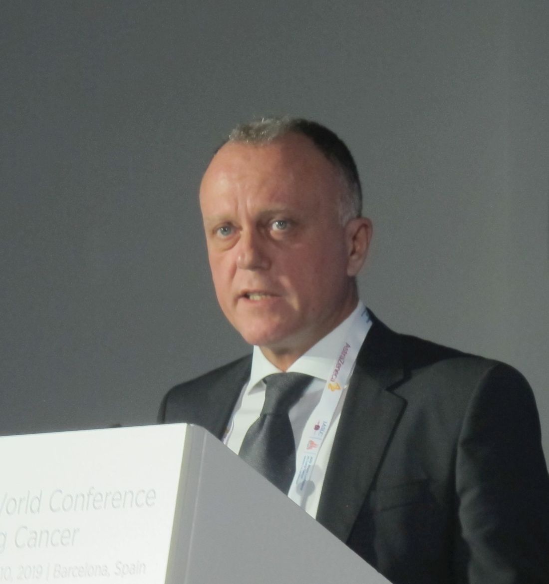
Median OS in the intent-to-treat population was 14.2 months vs. 13.5 months with vs. without the programmed death-ligand 1 (PD-L1) inhibitor atezolizumab, respectively (hazard ratio, 0.88), Federico Cappuzzo, MD, director of medical oncology at Azienda Unità Sanitaria Locale della Romagna-Ravenna, Italy, reported at the World Conference on Lung Cancer.
However, patients with high PD-L1 expression (14% and 13% of patients in the groups, respectively), experienced a clinically meaningful improvement in OS with atezolizumab + chemotherapy vs. chemotherapy alone (median of 23.4 vs. 10.2 months; HR, 0.48), Dr. Cappuzzo said at the conference, which was sponsored by the International Association for the Study of Lung Cancer.
“This means we had a reduction in the risk of death that was more than 50%,” he said, adding that no new or unexpected safety signals were reported.
IMpower131 randomized 1,021 patients with a median age of 65 years 1:1:1 to receive either atezolizumab (1,200 mg) + carboplatin (area under the curve 6) + paclitaxel (200 mg/m2) every 3 weeks, or atezolizumab + carboplatin + nab-paclitaxel (100 mg/m2 every week), or carboplatin + nab-paclitaxel for four or six cycles. Patients in the first two arms (A and B) received atezolizumab maintenance therapy until loss of clinical benefit or progressive disease occurred, and those in latter arm (C) received best supportive care after completing the treatment cycles.
The current analysis compared only the OS outcomes for arms B and C. Results of the primary analysis of investigator-assessed progression-free survival (PFS) – a coprimary endpoint of the trial – were reported in 2018 at the annual meeting of the American Society of Clinical Oncology and showed a statistically significant improvement in median PFS in arm B vs. arm C (6.3 vs. 5.6 months; HR, 0.715). The PFS benefit was seen in all PD-L1-positive subgroups.
“This was a very incredible trial, because it was conducted in a group of patients in which we need additional therapies – patients with squamous histology,” Dr. Cappuzzo said during a press briefing at the conference. “IMpower131 is certainly a positive study; we had PFS, as an independent coprimary endpoint, improve significantly, with a meaningful survival difference in the group of patients with strongly positive PD-L1 tumors.
“So these data clearly suggest that [patients with high PD-L1 expression), specifically, may benefit from the combination of chemotherapy and atezolizumab.”
He further noted in a press statement that “the findings provide additional evidence of the efficacy of immunotherapy in patients with lung cancer, and highlight the relevance of biomarkers for patient selection.”
Dr. Cappuzzo reported having no disclosures.
SOURCE: Cappuzzo F et al. WCLC 2019, Abstract OS14.02 .
BARCELONA – Final overall survival (OS) did not differ significantly among patients with stage IV squamous non–small cell lung carcinoma who were treated with either first-line atezolizumab + chemotherapy or chemotherapy alone in the phase 3 IMpower131 trial.
Median OS in the intent-to-treat population was 14.2 months vs. 13.5 months with vs. without the programmed death-ligand 1 (PD-L1) inhibitor atezolizumab, respectively (hazard ratio, 0.88), Federico Cappuzzo, MD, director of medical oncology at Azienda Unità Sanitaria Locale della Romagna-Ravenna, Italy, reported at the World Conference on Lung Cancer.
However, patients with high PD-L1 expression (14% and 13% of patients in the groups, respectively), experienced a clinically meaningful improvement in OS with atezolizumab + chemotherapy vs. chemotherapy alone (median of 23.4 vs. 10.2 months; HR, 0.48), Dr. Cappuzzo said at the conference, which was sponsored by the International Association for the Study of Lung Cancer.
“This means we had a reduction in the risk of death that was more than 50%,” he said, adding that no new or unexpected safety signals were reported.
IMpower131 randomized 1,021 patients with a median age of 65 years 1:1:1 to receive either atezolizumab (1,200 mg) + carboplatin (area under the curve 6) + paclitaxel (200 mg/m2) every 3 weeks, or atezolizumab + carboplatin + nab-paclitaxel (100 mg/m2 every week), or carboplatin + nab-paclitaxel for four or six cycles. Patients in the first two arms (A and B) received atezolizumab maintenance therapy until loss of clinical benefit or progressive disease occurred, and those in latter arm (C) received best supportive care after completing the treatment cycles.
The current analysis compared only the OS outcomes for arms B and C. Results of the primary analysis of investigator-assessed progression-free survival (PFS) – a coprimary endpoint of the trial – were reported in 2018 at the annual meeting of the American Society of Clinical Oncology and showed a statistically significant improvement in median PFS in arm B vs. arm C (6.3 vs. 5.6 months; HR, 0.715). The PFS benefit was seen in all PD-L1-positive subgroups.
“This was a very incredible trial, because it was conducted in a group of patients in which we need additional therapies – patients with squamous histology,” Dr. Cappuzzo said during a press briefing at the conference. “IMpower131 is certainly a positive study; we had PFS, as an independent coprimary endpoint, improve significantly, with a meaningful survival difference in the group of patients with strongly positive PD-L1 tumors.
“So these data clearly suggest that [patients with high PD-L1 expression), specifically, may benefit from the combination of chemotherapy and atezolizumab.”
He further noted in a press statement that “the findings provide additional evidence of the efficacy of immunotherapy in patients with lung cancer, and highlight the relevance of biomarkers for patient selection.”
Dr. Cappuzzo reported having no disclosures.
SOURCE: Cappuzzo F et al. WCLC 2019, Abstract OS14.02 .
REPORTING FROM WCLC 2019
Encourage participation in team sports
Participation in sports, competitive team sports in particular, is very good for the physical well-being and emotional development of children and adolescents. Specifically, there is growing evidence that sports promote healthy development socially and emotionally, protecting against drug use, poor body image, and against psychiatric illness in youth.

Sustaining academic productivity and team sports is demanding. By the middle of autumn, the amount of homework can begin to wear on teenagers, and the burden of getting them to practices and games can wear on parents. It can be very tempting for youth and their parents to drop team sports in high school, and turn their time and effort more completely to the serious work of school. But advocating for your patients and their parents to protect the time for team sports participation will pay dividends in the health and well-being of your patients and may even support rather than detract from academic performance.
The benefits of regular exercise for physical health are well established. Most teenagers do not get the recommended 60 minutes daily of moderate to vigorous physical activity. Participating in a team sport enforces this level of activity, in ways that parents typically don’t have to enforce. This level of physical activity typically promotes healthy eating and a healthy weight. Daily exercise promotes adequate, restful sleep, one of the most critical (and usually compromised) components of adolescent health. These exercise habits are easier to maintain into adulthood – when they protect against cardiovascular and inflammatory diseases – if they have been established early.

Beyond physical health, participation in team sports has been shown to promote good mental health and protect against psychiatric illnesses. They generally are less likely to use drugs and more likely to have a healthy body image than are their nonathlete peers. It is worth noting that the mental health benefits of team sports are even more robust than the benefits of solitary exercise in teenagers,1 possibly because of the social connections to peers and adults that grow out of them.
In the Monitoring the Future surveys (biannual national surveys of high school student health and behaviors funded by the National Institutes of Health) from 2010 to 2015, teenagers who participated in team sports were more likely to describe higher self-esteem and lower levels of loneliness. It is important to note that it has been difficult to establish the causal direction of the association between team sports and mental health in youth. We need more prospective randomized controlled trials to assert that the benefit is not simply an artifact of healthier youth choosing to participate in sports, but actually an active consequence of that choice. For now, though, we can say with confidence that physical activity promotes good mental health in youth and may protect against mental illness.

While student athletes benefit from the opportunity to develop deep social connections – ones forged in the intense setting of competition, collaboration, and sustained teamwork – they also benefit from strong mentorship connections with adults, including coaches, trainers, and even the parents of teammates who participate in all of the efforts that go into team sports in youth. While it might seem that all of the mental and physical benefits must be offset by lower academic performance, it turns out that is not the case. It is well established that regular exercise promotes healthy cognitive function, including processing speed, working memory, and even creativity. According to data from the Monitoring the Future survey, adolescents who participated in team sports were more likely to have As and to plan on attending a 4-year college than were their nonathlete peers.
Beyond the physiologic and social benefits of exercise, team sports provide adolescents with a powerful opportunity to get comfortable with failure. Even the best athletes cannot win all the time, and sports are unique in building failure into the work. Practice is almost entirely about failure, gradually getting better at something that is difficult. While everyone aims to win, they also prepare to struggle and lose. Athletes must learn how to persevere through a match that they are losing, and then pick themselves up and prepare again for the next match. When young people get comfortable with facing and managing challenges, managing setbacks and failure, they are ready to face the larger challenges, setbacks, and failures of adult life.
Team sports enable young people to learn what they are actually capable of managing – they build resilience. This promotion of resilience is illustrated in recent research that demonstrated that team sports may be especially protective for young people who have experienced trauma (adverse childhood experiences, or “ACEs”). Researchers at the University of California, Los Angeles, followed teenagers with and without high ACE scores into their mid 20s. They found that those with high ACE scores who participated in team sports as adolescents were 24% less likely to have depression and 30% less likely to have anxiety diagnoses as adults, compared with their peers who did not participate in team sports.2
Of course, the details matter in team sports. If your patients are participating and they or their parents are worried about spending so much time on something other than homework, talk to them about all of these exceptional benefits of team sports. But the culture of the team matters also. Some teams may be focused on winning at all costs, or have a practice culture that is humiliating or bullying. Some teams may have a culture of partying after games, with binge drinking and drug use. Ask your patients about whether they feel they are respected members of the team, and if effort and sportsmanship are valued as well as performance. Do they trust their coaches? Do they believe their coaches know and care about them? If your patients are not participating in a team sport, encourage them to find one (or more) that engage their interests. The benefits of track and field, crew, and tennis can be just as robust as the benefits of football or soccer. Speak with your patients and their parents about the payoff for their physical, mental, and developmental health the time and effort they are putting into a team sport can provide.
Dr. Swick is physician in chief at Ohana, Center for Child and Adolescent Behavioral Health, Community Hospital of the Monterey (Calif.) Peninsula. Dr. Jellinek is professor emeritus of psychiatry and pediatrics, Harvard Medical School, Boston. Email them at [email protected].
References
1. Int J Nutr Phys Act. 2013 Aug 15. doi: 10.1186/1479-5868-10-98.
2. JAMA Pediatr. 2019 Jul 1;173(7):681-8.
Participation in sports, competitive team sports in particular, is very good for the physical well-being and emotional development of children and adolescents. Specifically, there is growing evidence that sports promote healthy development socially and emotionally, protecting against drug use, poor body image, and against psychiatric illness in youth.
Sustaining academic productivity and team sports is demanding. By the middle of autumn, the amount of homework can begin to wear on teenagers, and the burden of getting them to practices and games can wear on parents. It can be very tempting for youth and their parents to drop team sports in high school, and turn their time and effort more completely to the serious work of school. But advocating for your patients and their parents to protect the time for team sports participation will pay dividends in the health and well-being of your patients and may even support rather than detract from academic performance.
The benefits of regular exercise for physical health are well established. Most teenagers do not get the recommended 60 minutes daily of moderate to vigorous physical activity. Participating in a team sport enforces this level of activity, in ways that parents typically don’t have to enforce. This level of physical activity typically promotes healthy eating and a healthy weight. Daily exercise promotes adequate, restful sleep, one of the most critical (and usually compromised) components of adolescent health. These exercise habits are easier to maintain into adulthood – when they protect against cardiovascular and inflammatory diseases – if they have been established early.
Beyond physical health, participation in team sports has been shown to promote good mental health and protect against psychiatric illnesses. They generally are less likely to use drugs and more likely to have a healthy body image than are their nonathlete peers. It is worth noting that the mental health benefits of team sports are even more robust than the benefits of solitary exercise in teenagers,1 possibly because of the social connections to peers and adults that grow out of them.
In the Monitoring the Future surveys (biannual national surveys of high school student health and behaviors funded by the National Institutes of Health) from 2010 to 2015, teenagers who participated in team sports were more likely to describe higher self-esteem and lower levels of loneliness. It is important to note that it has been difficult to establish the causal direction of the association between team sports and mental health in youth. We need more prospective randomized controlled trials to assert that the benefit is not simply an artifact of healthier youth choosing to participate in sports, but actually an active consequence of that choice. For now, though, we can say with confidence that physical activity promotes good mental health in youth and may protect against mental illness.
While student athletes benefit from the opportunity to develop deep social connections – ones forged in the intense setting of competition, collaboration, and sustained teamwork – they also benefit from strong mentorship connections with adults, including coaches, trainers, and even the parents of teammates who participate in all of the efforts that go into team sports in youth. While it might seem that all of the mental and physical benefits must be offset by lower academic performance, it turns out that is not the case. It is well established that regular exercise promotes healthy cognitive function, including processing speed, working memory, and even creativity. According to data from the Monitoring the Future survey, adolescents who participated in team sports were more likely to have As and to plan on attending a 4-year college than were their nonathlete peers.
Beyond the physiologic and social benefits of exercise, team sports provide adolescents with a powerful opportunity to get comfortable with failure. Even the best athletes cannot win all the time, and sports are unique in building failure into the work. Practice is almost entirely about failure, gradually getting better at something that is difficult. While everyone aims to win, they also prepare to struggle and lose. Athletes must learn how to persevere through a match that they are losing, and then pick themselves up and prepare again for the next match. When young people get comfortable with facing and managing challenges, managing setbacks and failure, they are ready to face the larger challenges, setbacks, and failures of adult life.
Team sports enable young people to learn what they are actually capable of managing – they build resilience. This promotion of resilience is illustrated in recent research that demonstrated that team sports may be especially protective for young people who have experienced trauma (adverse childhood experiences, or “ACEs”). Researchers at the University of California, Los Angeles, followed teenagers with and without high ACE scores into their mid 20s. They found that those with high ACE scores who participated in team sports as adolescents were 24% less likely to have depression and 30% less likely to have anxiety diagnoses as adults, compared with their peers who did not participate in team sports.2
Of course, the details matter in team sports. If your patients are participating and they or their parents are worried about spending so much time on something other than homework, talk to them about all of these exceptional benefits of team sports. But the culture of the team matters also. Some teams may be focused on winning at all costs, or have a practice culture that is humiliating or bullying. Some teams may have a culture of partying after games, with binge drinking and drug use. Ask your patients about whether they feel they are respected members of the team, and if effort and sportsmanship are valued as well as performance. Do they trust their coaches? Do they believe their coaches know and care about them? If your patients are not participating in a team sport, encourage them to find one (or more) that engage their interests. The benefits of track and field, crew, and tennis can be just as robust as the benefits of football or soccer. Speak with your patients and their parents about the payoff for their physical, mental, and developmental health the time and effort they are putting into a team sport can provide.
Dr. Swick is physician in chief at Ohana, Center for Child and Adolescent Behavioral Health, Community Hospital of the Monterey (Calif.) Peninsula. Dr. Jellinek is professor emeritus of psychiatry and pediatrics, Harvard Medical School, Boston. Email them at [email protected].
References
1. Int J Nutr Phys Act. 2013 Aug 15. doi: 10.1186/1479-5868-10-98.
2. JAMA Pediatr. 2019 Jul 1;173(7):681-8.
PBRM1 mutations not quite up to snuff as ccRCC biomarkers
Loss-of-function mutations in the PBRM1 gene are more common among patients with metastatic clear cell renal cell carcinoma (ccRCC) who have clinical responses to immune checkpoint inhibitors (ICIs), but the mutations fall a little short of serving as clinical biomarkers, investigators say.
Among patients with metastatic ccRCC enrolled in a randomized, phase 3 clinical trial comparing nivolumab (Opdivo) with everolimus (Afinitor) in patients who had previously received antiangiogenic therapy, patients with responses to nivolumab were more than twice as likely as nonresponders to have a truncating loss-of-function mutation in PBRM1, reported Toni K. Choueiri, MD, and colleagues from the Dana-Farber Cancer Institute in Boston.
Patients with PBRM1 mutations were also more likely to have a clinical benefit (complete/partial response, or stable disease with tumor shrinkage and progression-free survival [PFS] of at least 6 months) as well as better PFS and overall survival than patients without mutations.
“The association of PBRM1 truncating mutations with response to anti–[programmed death-1] therapy was confirmed in an independent ccRCC cohort. However, key limitations restrict use of PBRM1 mutations as a clinical biomarker,” they wrote in a research letter to JAMA Oncology.
Those limitations include an only modest effect of mutations on response and survival, a lack of evidence for a PBRM1 mutation effect in the first-line setting, and a possible association between mutations and benefit from prior antiangiogenic therapies, they acknowledged.
Dr. Choueiri was a coauthor of a previous study of genomic correlates of response to ICIs in ccRCC, which found that PBRM1 loss may influence response to checkpoint inhibitors by altering global tumor-cell expression.
In the current study, he and his colleagues looked at archival tumor tissue from an independent cohort of 382 patients who were part of a larger phase 3 trial. Of this group, 189 were treated with nivolumab, and 193 were treated with everolimus. PBRM1 mutations were identified in 55 of the nivolumab-treated patients (29%) and in 45 of the everolimus-treated patients (23%).
When they looked at clinical responses, they found that 15 of 38 patients with response to nivolumab (39%) had truncating PBRM1 mutations, compared with 16 of 74 nonresponding patients (22%), which translated into an odds ratio for response of 2.34 (P = .04).
Similarly, PBRM1 mutations among nivolumab-treated patients were significantly associated with clinical benefit (OR, 2.14; P = .0497), PFS (hazard ratio for progression, 0.67; P = .03), and overall survival (HR, 0.65; P = .03).
In contrast, there were no significant associations in everolimus-treated patients between PBRM1 mutations and either response, PFS, or overall survival.
“The concomitant presence of other cellular or molecular features may further influence the findings described herein. Nonetheless, this validated association between PBRM1 alterations and ICI response in a large randomized study represents a further step toward the development of genomic predictors for immunotherapies in advanced RCC,” the investigators concluded.
The study was supported by Department of Defense Congressionally Directed Medical Research Programs and Bristol-Myers Squibb. Dr. Choueiri disclosed personal fees from Bristol-Myers Squibb and fees and grants from other companies. One coauthor is a Bristol-Myers Squibb employee and shareholder.
SOURCE: Choueiri TK et al. JAMA Oncol. 2019 Sep 5. doi: 10.1001/jamaoncol.2019.3158.
FROM JAMA ONCOLOGY
Morinda citrifolia (Noni) tree: Many names, even more applications
, which has been in use on the islands for two millennia.1-4 The plant, found abundantly in Southeast Asia, Australia, the Pacific Basin, and the Caribbean, is called Great Morinda or cheese fruit in Australia, Nono in Tahiti, Indian Mulberry in India, and Ba ji tian in China.4-6 It is also deployed for a wide range of health purposes in Brazil.7

Noni has been credited with conferring various salutary benefits against arthritis, diabetes, fever, gingivitis, headaches, infections, inflammation, respiratory illnesses, and tuberculosis.3,8 In alternative medicine, the fruit juice, which has been found to be safe, is used for multiple indications, with a slew of studies presenting evidence for anti-inflammatory, antioxidant, and apoptosis-inducing benefits against cancer.5,6 All parts of M. citrifolia – leaves, fruits, roots, bark, flowers, and seeds – have been used in traditional medical practices.8 This column will focus on recent research into the broad array of biologic activities attributed to the plant and possible dermatologic uses.
Diverse biologic properties
In 2007, Nayak et al. showed that the juice of M. citrifolia fruit significantly lowered sugar levels in diabetic rats and facilitated their wound healing.1
Three years later, Thani et al. determined that the leaves of M. citrifolia exert antiproliferative and antioxidative activities, with chemopreventive benefits seen against epidermoid and cervical cancers.9
In 2011, Serafini et al. confirmed the antibacterial, anti-inflammatory, antioxidant, and antinociceptive qualities of the aqueous extract from M. citrifolia leaves, with the extract shown to significantly lower leukocyte migration in doses of 200 and 400 mg/kg. Mild antibacterial properties were seen as was an antinociceptive effect at the higher dose in the acetic-acid-induced writhing test.3
A comprehensive literature review in 2017 by Torres et al. identified a varied and extensive list of biological activities of M. citrifolia, including immunostimulatory, antitumor, antidiabetic, antiobesity, antibacterial and antiseptic, antifungal, antiviral, anti-inflammatory, antinociceptive and analgesic, antioxidant, neuroprotective, wound healing, antiallergic, photoprotective, and antiwrinkle among several others. Despite its use in disease prevention and treatment around the world, the researchers call for more in vitro and in vivo models in addition to clinical trials to further examine the health benefits of Noni.7
Early in 2019, De La Cruz-Sánchez et al. determined that the methanolic extract of M. citrifolia displayed marked activity against methicillin-resistant Staphylococcus aureus (MRSA), thus supporting its continuing applications in traditional medical practice.2
Photoprotection and antiaging potential
Based on their prior work demonstrating that M. citrifolia fruit upregulates the production of type I collagen and glycosaminoglycans in primary cultures of normal human fibroblasts, Kim et al. isolated anthraquinone from the fruit and showed that it dose-dependently decreased the expression of collagenase matrix metalloproteinase-1 in human dermal fibroblasts. The investigators also found that an anthraquinone-containing nano-emulsion raised type I procollagen in nude mouse skin. They concluded, in this 2005 study, that Noni extract warrants consideration as an antiwrinkle agent given its proclivity to induce the production of collagen.10
In 2009, West et al. assessed a carbomer gel base containing the ethanol extract and juice pressed from Noni leaves for possible allergenic activity in a repeat-insult patch test in 49 volunteers. They also used a UVB-induced erythema model in 25 subjects to test the topical photoprotective potential of the ethanol extract and leaf juice. The investigators reported no allergic potential evinced by the patch tests, and in a histamine H-1 receptor antagonism assay, the leaves hindered receptor binding by 57%, suggesting anti-inflammatory activity. In the UVB test, the dose necessary to engender erythema was nearly 3.5 times higher than in untreated skin. The team concluded that M. citrifolia leaves are safe for topical application and show promise in lessening UVB-induced skin damage.11
A 2014 study on mice by Serafini et al. showed that the dorsal skin of mice treated for 7 days with topical M. citrifolia was protected from damage by exposure to UVA-UVB radiation as measured by skin thickness, transepidermal water loss, erythema, and histological changes.12
Conclusion
Morinda citrifolia has been used in traditional medicine for at least 2,000 years. Its reported list of uses covers an impressive gamut of indications.

Modern medicine is beginning to catch up with new research conducted on this copious and beloved plant. That said, much more data, particularly from human clinical trials, are necessary to elucidate the most appropriate dermatologic roles for M. citrifolia. I just started growing a Noni tree in my yard because some patients have reported using it on their skin. I will report back and let you know how it goes. It is flowering now!
Dr. Baumann is a private practice dermatologist, researcher, author, and entrepreneur who practices in Miami. She founded the Cosmetic Dermatology Center at the University of Miami in 1997. Dr. Baumann wrote two textbooks: “Cosmetic Dermatology: Principles and Practice” (New York: McGraw-Hill, 2002), and “Cosmeceuticals and Cosmetic Ingredients” (New York: McGraw-Hill, 2014), and a New York Times Best Sellers book for consumers, “The Skin Type Solution” (New York: Bantam Dell, 2006). Dr. Baumann has received funding for advisory boards and/or clinical research trials from Allergan, Evolus, Galderma, and Revance. She is the founder and CEO of Skin Type Solutions Franchise Systems LLC. Write to her at [email protected].
References
1. Nayak BS et al. J Wound Care. 2007 Feb;16(2):83-6.
2. De La Cruz-Sánchez NG et al. Microb Pathog. 2019 Mar;128:347-53.
3. Serafini MR et al. J Med Food. 2011 Oct;14(10):1159-66.
4. Wang MY, Su C. Ann N Y Acad Sci. 2001 Dec;952:161-8.
5. Gupta RK, Patel AK. Asian Pac J Cancer Prev. 2013;14(8):4495-9.
6. Brown AC. Phytother Res. 2012 Oct;26(10):1427-40.
7. Torres MAO et al. Phytother Res. 2017 Jul;31(7):971-9.
8. Potterat O, Hamburger M. Planta Med. 2007 Mar;73(3):191-9.
9. Thani W et al. Southeast Asian J Trop Med Public Health. 2010 Mar;41(2):482-9.
10. Kim SW et al. J Med Food. 2005 Winter;8(4):552-5.
11. West BJ et al. J Nat Med. 2009 Jul;63(3):351-4.
12. Serafini MR et al. Biomed Res Int. 2014;2014:587819. doi: 10.1155/2014/587819.
, which has been in use on the islands for two millennia.1-4 The plant, found abundantly in Southeast Asia, Australia, the Pacific Basin, and the Caribbean, is called Great Morinda or cheese fruit in Australia, Nono in Tahiti, Indian Mulberry in India, and Ba ji tian in China.4-6 It is also deployed for a wide range of health purposes in Brazil.7
Noni has been credited with conferring various salutary benefits against arthritis, diabetes, fever, gingivitis, headaches, infections, inflammation, respiratory illnesses, and tuberculosis.3,8 In alternative medicine, the fruit juice, which has been found to be safe, is used for multiple indications, with a slew of studies presenting evidence for anti-inflammatory, antioxidant, and apoptosis-inducing benefits against cancer.5,6 All parts of M. citrifolia – leaves, fruits, roots, bark, flowers, and seeds – have been used in traditional medical practices.8 This column will focus on recent research into the broad array of biologic activities attributed to the plant and possible dermatologic uses.
Diverse biologic properties
In 2007, Nayak et al. showed that the juice of M. citrifolia fruit significantly lowered sugar levels in diabetic rats and facilitated their wound healing.1
Three years later, Thani et al. determined that the leaves of M. citrifolia exert antiproliferative and antioxidative activities, with chemopreventive benefits seen against epidermoid and cervical cancers.9
In 2011, Serafini et al. confirmed the antibacterial, anti-inflammatory, antioxidant, and antinociceptive qualities of the aqueous extract from M. citrifolia leaves, with the extract shown to significantly lower leukocyte migration in doses of 200 and 400 mg/kg. Mild antibacterial properties were seen as was an antinociceptive effect at the higher dose in the acetic-acid-induced writhing test.3
A comprehensive literature review in 2017 by Torres et al. identified a varied and extensive list of biological activities of M. citrifolia, including immunostimulatory, antitumor, antidiabetic, antiobesity, antibacterial and antiseptic, antifungal, antiviral, anti-inflammatory, antinociceptive and analgesic, antioxidant, neuroprotective, wound healing, antiallergic, photoprotective, and antiwrinkle among several others. Despite its use in disease prevention and treatment around the world, the researchers call for more in vitro and in vivo models in addition to clinical trials to further examine the health benefits of Noni.7
Early in 2019, De La Cruz-Sánchez et al. determined that the methanolic extract of M. citrifolia displayed marked activity against methicillin-resistant Staphylococcus aureus (MRSA), thus supporting its continuing applications in traditional medical practice.2
Photoprotection and antiaging potential
Based on their prior work demonstrating that M. citrifolia fruit upregulates the production of type I collagen and glycosaminoglycans in primary cultures of normal human fibroblasts, Kim et al. isolated anthraquinone from the fruit and showed that it dose-dependently decreased the expression of collagenase matrix metalloproteinase-1 in human dermal fibroblasts. The investigators also found that an anthraquinone-containing nano-emulsion raised type I procollagen in nude mouse skin. They concluded, in this 2005 study, that Noni extract warrants consideration as an antiwrinkle agent given its proclivity to induce the production of collagen.10
In 2009, West et al. assessed a carbomer gel base containing the ethanol extract and juice pressed from Noni leaves for possible allergenic activity in a repeat-insult patch test in 49 volunteers. They also used a UVB-induced erythema model in 25 subjects to test the topical photoprotective potential of the ethanol extract and leaf juice. The investigators reported no allergic potential evinced by the patch tests, and in a histamine H-1 receptor antagonism assay, the leaves hindered receptor binding by 57%, suggesting anti-inflammatory activity. In the UVB test, the dose necessary to engender erythema was nearly 3.5 times higher than in untreated skin. The team concluded that M. citrifolia leaves are safe for topical application and show promise in lessening UVB-induced skin damage.11
A 2014 study on mice by Serafini et al. showed that the dorsal skin of mice treated for 7 days with topical M. citrifolia was protected from damage by exposure to UVA-UVB radiation as measured by skin thickness, transepidermal water loss, erythema, and histological changes.12
Conclusion
Morinda citrifolia has been used in traditional medicine for at least 2,000 years. Its reported list of uses covers an impressive gamut of indications.
Modern medicine is beginning to catch up with new research conducted on this copious and beloved plant. That said, much more data, particularly from human clinical trials, are necessary to elucidate the most appropriate dermatologic roles for M. citrifolia. I just started growing a Noni tree in my yard because some patients have reported using it on their skin. I will report back and let you know how it goes. It is flowering now!
Dr. Baumann is a private practice dermatologist, researcher, author, and entrepreneur who practices in Miami. She founded the Cosmetic Dermatology Center at the University of Miami in 1997. Dr. Baumann wrote two textbooks: “Cosmetic Dermatology: Principles and Practice” (New York: McGraw-Hill, 2002), and “Cosmeceuticals and Cosmetic Ingredients” (New York: McGraw-Hill, 2014), and a New York Times Best Sellers book for consumers, “The Skin Type Solution” (New York: Bantam Dell, 2006). Dr. Baumann has received funding for advisory boards and/or clinical research trials from Allergan, Evolus, Galderma, and Revance. She is the founder and CEO of Skin Type Solutions Franchise Systems LLC. Write to her at [email protected].
References
1. Nayak BS et al. J Wound Care. 2007 Feb;16(2):83-6.
2. De La Cruz-Sánchez NG et al. Microb Pathog. 2019 Mar;128:347-53.
3. Serafini MR et al. J Med Food. 2011 Oct;14(10):1159-66.
4. Wang MY, Su C. Ann N Y Acad Sci. 2001 Dec;952:161-8.
5. Gupta RK, Patel AK. Asian Pac J Cancer Prev. 2013;14(8):4495-9.
6. Brown AC. Phytother Res. 2012 Oct;26(10):1427-40.
7. Torres MAO et al. Phytother Res. 2017 Jul;31(7):971-9.
8. Potterat O, Hamburger M. Planta Med. 2007 Mar;73(3):191-9.
9. Thani W et al. Southeast Asian J Trop Med Public Health. 2010 Mar;41(2):482-9.
10. Kim SW et al. J Med Food. 2005 Winter;8(4):552-5.
11. West BJ et al. J Nat Med. 2009 Jul;63(3):351-4.
12. Serafini MR et al. Biomed Res Int. 2014;2014:587819. doi: 10.1155/2014/587819.

Inflammatory markers may start in later stages of bipolar disorder
Interleukin-6, IL-1 receptor antagonist (IL-1RA), and tumor necrosis factor–alpha (TNF-alpha) activity was associated with inflammation and neurodegeneration in patients with chronic bipolar disorder, according to Sercan Karabulut, MD, of Kepez State Hospital in Antalya, Turkey, and associates.
In a study published in the Turkish Journal of Psychiatry, the investigators collected enzyme-linked immunosorbent assays from 30 patients with early-stage bipolar disorder, 77 with chronic disease, and 30 healthy controls. Early-stage disease patients were significantly younger than chronic patients (25.3 years vs. 37.8 years). reported Dr. Karabulut and associates.
Patients with chronic bipolar disorder had significantly increased levels of all measured markers, compared with those with early-stage disease and the healthy controls. IL-6 and IL-1RA levels correlated with neuron-specific enolase and S100B, biomarkers that are associated with glial alterations and neuronal damage. TNF-alpha correlated with scores on the Clinical Global Impressions Scale and other measures.
“The present findings support in part the hypothesis that inflammation starts at later stages of [bipolar disorder], presumably as an associated effect of gliosis and neuronal loss, which appear to be particularly associated with IL-1RA and IL-6 activity,” the investigators wrote. “TNF-alpha ... might be a useful prognostic marker in patients with [bipolar disorder].”
No conflicts of interest were reported.
SOURCE: Karabulut S et al. Turk Psikiyatri Derg. 2019 Winter;30(2):75-81.
FROM THE TURKISH JOURNAL OF PSYCHIATRY
Average daily step count reflects disability in MS
STOCKHOLM – according to research presented at the annual congress of the European Committee for Treatment and Research in MS. Remote gait monitoring using a popular fitness tracker may offer a surrogate measure of MS disability in clinical trials, the researchers reported.
Many outcome measures in MS are evaluated in controlled contexts and do not indicate how patients are functioning outside of the clinical setting, said Valerie J. Block, PhD, from the Weill Institute for Neurosciences at the University of California, San Francisco. Patient-reported outcome measures are subject to recall bias and uneven perception of deficits. Remote ambulatory monitoring, on the other hand, could be a more objective measure that provides continuous information in the real-world setting, said Dr. Block. She and her colleagues have proposed remote ambulatory activity monitoring as an outcome measure for clinical trials.
The investigators chose this measure as an exploratory endpoint for SPI2, a phase 3 trial investigating the efficacy and safety of MD1003 (high-dose pharmaceutical-grade biotin) in patients with inactive primary progressive MS and secondary progressive MS. “To our knowledge, this is the first major clinical trial in progressive MS to include continuous remote step count monitoring as an exploratory endpoint,” said Dr. Block.
In the SPI2 study, patients received either MD1003 (300 mg/day) or placebo. To examine the relationship between ambulatory monitoring and clinical disability and MRI measures, the researchers remotely monitored participants’ ambulatory activity for 27 months using a fitness tracker. The investigators used the average daily step count from the first 30 days as the baseline activity measure. At first, they set a low daily step-count goal to minimize the influence of motivation on ambulatory activity. Participants later were taught how to change the goal independently.
Dr. Block and colleagues created LASSO subset selection regression models to correlate average daily step count with sex, age, disease duration, age at onset, disease course, and various MRI models (such as upper cervical cord area, gray matter volume, normalized brain volume, thalamic volume, and T1 and T2 lesion volumes). They performed least squares regression models on the subset selection results. Finally, the researchers calculated Spearman correlations between average daily step count and clinical disability, as measured by Expanded Disability Status Scale (EDSS) and timed 25-foot walk, and the Physical and Mental Health Composite measures of the MS Quality of Life scale (MSQoL-29).
As of April 23, 2019, the researchers had enrolled 492 patients (262 women) with full data at 90 centers (40 in the United States, 39 in Europe, 8 in Canada, and 3 in Australia). In all, 311 patients (63%) had secondary progressive MS, and 181 had primary progressive MS. Participants had moderate disability; the median EDSS score was 6.0. Median disease duration was 10.6 years. The mean daily step count during the first month was 3,699.
Greater step count was correlated with lower EDSS score, faster completion of the timed 25-foot walk, better Physical Health Composite score, better Symbol Digit Modalities Test score, and better Mental Health Composite score. Furthermore, greater mean daily step count also correlated with greater upper cervical cord area, greater normalized brain volume, greater gray matter volume, and lower T1 lesion volume. The correlations between step count and thalamic volume and T2 lesion volume were not significant. “These data support the study of steps as an exploratory outcome measure in clinical trials for progressive MS,” said Dr. Block.
Dr. Block received reimbursement for travel expenses related to this study from MedDay Pharmaceuticals. Coinvestigators received research support and compensation from companies such as Abbvie, Alexion, Biogen, Genentech, MedDay Pharmaceuticals, Novartis, and Sanofi Genzyme. One investigator is an employee of MedDay Pharmaceuticals.
SOURCE: Block V et al. ECTRIMS 2019, Abstract 217.
REPORTING FROM ECTRIMS 2019