User login
Health Policy Q&A

Pediatric Vaccines & Infectious Diseases: September 2019
Click here to read the supplement.

The 2019 Pediatric Vaccines & Infectious Diseases supplement features a selection of articles published in Pediatric News , with commentary by Kristina A. Bryant, MD.
Dr. Bryant is a pediatrician specializing in infectious diseases at the University of Louisville (Ky.) and Norton Children’s Hospital, also in Louisville. She reported serving as a clinical investigator on trials of pneumococcal conjugate vaccines and the MenB vaccine funded by Pfizer.
Highlights include:
- Combo vaccines for infants provides lasting hepatitis B immunity for teens
- Most doctors don’t discuss MenB vaccines
- Combo respiratory test misses pertussis. Use PCR
Click here to read the supplement.
Click here to read the supplement.
The 2019 Pediatric Vaccines & Infectious Diseases supplement features a selection of articles published in Pediatric News , with commentary by Kristina A. Bryant, MD.
Dr. Bryant is a pediatrician specializing in infectious diseases at the University of Louisville (Ky.) and Norton Children’s Hospital, also in Louisville. She reported serving as a clinical investigator on trials of pneumococcal conjugate vaccines and the MenB vaccine funded by Pfizer.
Highlights include:
- Combo vaccines for infants provides lasting hepatitis B immunity for teens
- Most doctors don’t discuss MenB vaccines
- Combo respiratory test misses pertussis. Use PCR
Click here to read the supplement.
Dapagliflozin given Fast Track status for HF therapy

The decision is based on results from two phase 3 trials – DAPA-HF and DELIVER – that assessed dapagliflozin in patients with HFrEF and HFpEF, respectively.
Dapagliflozin, an oral, once-daily sodium-glucose transporter 2 inhibitor, was first approved as monotherapy and as part of combination therapy for the improvement of glycemic control in adults with type 2 diabetes. It was also granted Fast Track designation in August 2019 as a therapy for chronic renal disease, both to slow progression of renal failure and to prevent cardiovascular and renal death.
“Heart failure affects approximately 64 million people worldwide, and about half will die within 5 years of diagnosis,” Mene Pangalos, executive vice president of biopharmaceuticals research and development, said in the AstraZeneca press release. “This Fast Track designation for Farxiga brings us closer to fulfilling our ambition to help prevent, treat and cure heart failure, and we look forward to working with the FDA to explore Farxiga as a potential new treatment option for heart failure patients.”
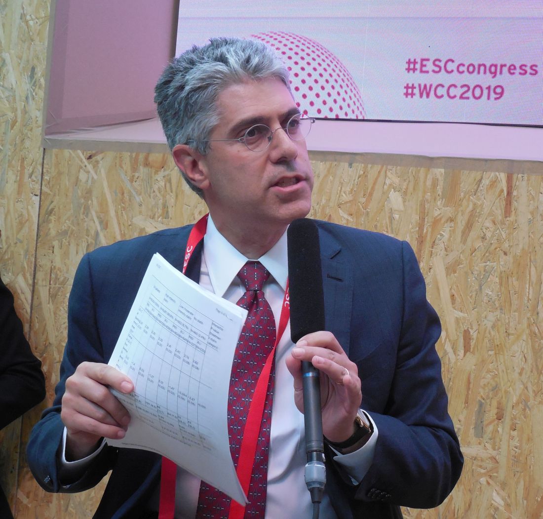
European cholesterol guidelines push LDL targets below 55 mg/dL
PARIS – The 2019 dyslipidemia management guidelines from the European Society of Cardiology set an LDL cholesterol target for very-high-risk people of less than 55 mg/dL (as well as at least a 50% cut from baseline), a class I recommendation. This marks the first time a cardiology society has either recommended a target goal for this measure below 70 mg/dL or endorsed treating patients to still-lower cholesterol once their level was already under 70 mg/dL.*
The guidelines went further by suggesting consideration of an even lower treatment target for LDL-cholesterol in very-high-risk, secondary prevention patients who have already had at least two atherosclerotic cardiovascular disease events during the past 2 years, a setting that could justify an LDL-cholesterol goal of less than 40 mg/dL (along with a cut from baseline of at least 50%), a class IIb recommendation that denotes a “may be considered,” endorsement.
“In all the trials, lower was better. There was no lower level of LDL cholesterol that’s been studied that was not better” for patient outcomes, Colin Baigent, BMBCH, said while presenting the new guideline at the annual congress of the European Society of Cardiology (ESC). “It’s very clear” that the full treatment benefit from lowering LDL-cholesterol extends to getting very-high risk patients below these levels, said Dr. Baigent, professor of cardiology at Oxford (England) University and one of three chairs of the ESC’s dyslipidemia guideline-writing panel.
While this change was seen as a notably aggressive goal and too fixed on a specific number by at least one author of the 2018 American Heart Association/American College of Cardiology cholesterol management guideline (J Am Coll Cardiol. 2019 Jun;73[24]:e285-e350), it was embraced by another U.S. expert not involved in writing the most recent U.S. recommendations.

“A goal for LDL-cholesterol of less than 55 mg/dL is reasonable; it’s well documented” by trial evidence “and I support it,” said Robert H. Eckel, MD, an endocrinologist and professor of medicine at the University of Colorado in Aurora. Dr. Eckel added that he “also supports” an LDL-cholesterol of less than 40 mg/dL in very-high-risk patients with a history of multiple events or with multiple residual risk factors, and he said he has applied this lower LDL-cholesterol goal in his practice for selected patients. But Dr. Eckel acknowledged in an interview that the evidence for it was less clear-cut than was the evidence behind a goal of less than 55 mg/dL. He also supported the concept of including a treatment goal in U.S. lipid recommendations, which in recent versions has been missing. “I fall back on a cholesterol goal for practical purposes” of making the success of cholesterol-lowering treatment easier to track.
The new ESC goal was characterized as “arbitrary” by Neil J. Stone, MD, vice-chair of the panel that wrote the 2018 AHA/ACC guideline, which relied on treating secondary-prevention patients at high risk to an LDL-cholesterol at least 50% less than before treatment, and recommended continued intensification for patients whose LDL-cholesterol level remained at or above 70 mg/dL.

“If the patient is at 58 mg/dL I’m not sure anyone can tell me what the difference is,” compared with reaching less than 55 mg/dL, Dr. Stone said in an interview. “I worry about focusing on a number and not on the concept that people at the very highest risk deserve the most intensive treatment; the Europeans agree, but they have a different way of looking at it. Despite this difference in approach, the new ESC guidelines and the 2018 U.S. guideline “are more similar than different,” stressed Dr. Stone, professor of medicine and preventive medicine at Northwestern University, Chicago.
However, other experts see an important difference in the risk faced by patients who reach the ESC’s recommended treatment goals and those who fall just short.
“It’s hard to lower an LDL-cholesterol that is already relatively low. People who are close to their cholesterol target need the most intensified treatment” to reach their goal, said Rory Collins, F.Med.Sci., professor of epidemiology at Oxford University. He was not on the ESC guidelines panel.

“It’s a mind shift that clinicians need to be most aggressive in treating patients with the highest risk” even when their LDL-cholesterol is low but not yet at the target level, Dr. Collins said during a discussion session at the congress.
The new ESC guidelines is about “both getting the LDL-cholesterol down to a certain level and also about achieving a big [at least 50%] change” from baseline. “I think the ESC guidelines make that crystal clear,” said Marc S. Sabatine, MD, professor of medicine at Harvard Medical School, Boston, and the sole American to participate in the ESC guidelines-writing panel.
The ESC also broke new ground by advocating an aggressive path toward achieving these LDL-cholesterol goals by elevating the newest and most potent class of approved LDL-cholesterol-lowering drugs, the PCSK9 (proprotein convertase subtilisin/kexin type 9) inhibitors, to a top-tier, class I recommendation (“is recommended”) for secondary prevention in very-high-risk patients not reaching their goal LDL-cholesterol level on a maximally tolerated statin plus ezetimibe. This recommendation to unequivocally add a PCSK9 inhibitor for this patient population contrasts with the 2018 AHA/ACC guideline that deemed adding a PCSK9 inhibitor a IIa recommendation (“is reasonable”).
A similar uptick in treatment aggressiveness appeared in the ESC’s recommendations for managing very-high-risk patients in a primary prevention setting, including those without familial hypercholesterolemia. For these people, the ESC panel, which worked in concert with the European Atherosclerosis Society, pegged adding a PCSK9 inhibitor as a IIb (“may be considered”) recommendation when these very-high-risk people fail to reach their LDL-cholesterol target on a maximally tolerated statin and ezetimibe. Once again, this opening to use a PCSK9 inhibitor contrasted with the 2018 U.S. guideline, which never mentioned an option of adding a PCSK9 inhibitor for primary prevention except when someone also has familial hypercholesterolemia and starts treatment with an LDL level of at least 190 mg/dL (a IIb recommendation). The new European guidelines proposed using a PCSK9 inhibitor as a second-line option to consider when needed for people whose very high risk derives primarily from older age and other factors such as smoking or hypertension that give them at least a 10% 10-year risk for cardiovascular death as estimated with the European-oriented SCORE risk calculator tables.
Updated SCORE risk designations appear in the new ESC dyslipidemia guidelines, and they show, for example, that in lower-risk European countries (mostly Western European nations) virtually all men who are at least 70 years old would fall into the very-high-risk category that makes them potential candidates for treatment with a PCSK9 inhibitor regardless of any other risk they may or may not have. In higher-risk (mostly Eastern European) countries this designation kicks in for most men once they reach the age of 65.
Several Congress attendees who came to a discussion session on the guidelines voiced concerns that the new revision will lead to substantially increased use of the these drugs and hence will significantly boost medical costs, because these drugs today are priced at about $6,000 annually to treat one patient. In response, members of the guideline-writing panel defended their decision as unavoidable given what’s been reported on the clinical impact of PCSK9 inhibitors when lowering LDL cholesterol and cutting atherosclerotic cardiovascular disease events.
“I commend the [ESC] guideline for focusing on the science and on what is best for patients. The U.S. guidelines conflated the science and the cost, and the recommendations got watered down by cost considerations,” said Dr. Sabatine, who has led several studies of PCSK9 inhibitors.
Dr. Baigent added that the panel “deliberated long and hard on cost, but we felt that we had to focus on the evidence. The cost will shift” in the future, he predicted.
Other U.S. physicians highlighted the need to take drug cost into account when writing public health policy documents such as lipid-management guidelines and questioned whether this more liberal use of PCSK9 inhibitors was justified.
“I think that in the absence of familial hypercholesterolemia you need to waffle around the edges to justify a PCSK9 inhibitor,” said Dr. Eckel. “The cost of PCSK9 inhibitors has come down, but at $6,000 per year you can’t ignore their cost.”
“In the U.S. we need to be mindful of the cost of treatment,” said Dr. Stone. “The ESC guidelines are probably more aggressive” than the 2018 U.S. guideline. “They use PCSK9 inhibitors perhaps more than we do; we [in the United States] prefer generic ezetimibe. A lot has to do with the definitions of risk. The European guidelines have a lot of risk definitions that differ” from the U.S. guideline, he said.
Members of the ESC guidelines panel acknowledged that the SCORE risk-assessment charts could overestimate risk in older people who need primary prevention treatment, as well as underestimate the risk in younger adults.
This inherent age bias in the SCORE risk tables make it “extremely important to contextualize” a person’s risk “by considering other risk factors,” advised Brian A. Ference, MD, an interventional cardiologist and professor at Cambridge (England) University who was a member of the ESC guidelines writing group.
The new ESC guidelines say that risk categorization “must be interpreted in light of the clinician’s knowledge and experience, and of the patient’s pretest likelihood” of cardiovascular disease.”
Dr. Baigent has received research funding from Boehringer Ingelheim, Novartis, and Pfizer. Dr. Eckel has been an expert witness on behalf of Sanofi/Regeneron. Dr. Sabatine and Dr. Ference have received honoraria and research funding from several companies including those that market lipid-lowering drugs. Dr. Stone and Dr. Collins had no disclosures.
*Correction, 9/20/19: A previous version of this article incorrectly stated that the ESC guidelines were the first by a medical society to recommend the lower cholesterol goals. The American Association of Clinical Endocrinologists included targets below 55 mg/dL in their 2017 dyslipidemia management guidelines.
SOURCE: Mach F et al. Eur Heart J. 2019 Aug 31. doi: 10.1093/eurheartj/ehz455.
The new ESC dyslipidemia guidelines recently presented at the society’s annual congress are a welcome addition to the lipid disorder treatment guidelines available to clinicians. These guidelines follow the groundbreaking recommendation in 2017 by AACE in their updated guidelines that introduced an LDL goal of <55 mg/dL in “extreme risk” patients. The ESC guidelines now also recommend an LDL goal of <55 mg/dL in “very-high-risk” patients but go further by also requiring a 50% reduction in LDL. Furthermore, they have established an LDL goal of <40 mg/dL in patients who experienced a second vascular event in the past 2 years while on maximally tolerated statin dose.
The ESC very-high-risk category shares many features with AACE’s extreme-risk category but is broader in that it includes patients without a clinical event who display unequivocal evidence of arteriosclerotic cardiovascular disease (ASCVD) on imaging and patients with severe chronic kidney disease (GFR <30 mL/min ) without known ASCVD. There are substantial differences between the ESC and AHA-ACC 2018 guidelines in the very-high-risk category. The AHA very-high-risk is directed toward secondary prevention only and requires two major ASCVD events or one major and at least two high-risk conditions. Moreover, elements of both major ASCVD events and high-risk conditions as well as the very-high-risk eligibility requirements could mean that some patients, who would clearly be classified by both ESC and AACE as candidates for an LDL goal of <55, may not qualify for threshold consideration for maximal LDL lowering below 70 mg/dL including the use of PCSK9 inhibitors. Relative to this point, the AHA-ACC guidelines do not classify past CABG or PCI as a major ASCVD event, nor is a TIA considered a major event or a high-risk condition.
For LDL, “lower is better” is supported by years of statin clinical trial evidence, along with the robust findings in the 2010 Cholesterol Trialists Collaboration. The goal of <55 mg/dL is supported by the IMPROVE-IT, FOURIER, and ODYSSEY trials. The ESC guidelines appropriately take this body of evidence and applies it to an aggressive treatment platform that, like AACE, sets clinically useful LDL goals for clinicians and patients. It takes early, aggressive LDL-lowering treatment to stay ahead of atherosclerotic plaque development in patients who are at very high or extreme risk. Following AACE’s lead, the ESC guidelines are the newest tool available to clinicians addressing this issue with the promise of further decreasing CVD events and extending lives.
Dr. Jellinger is a member of the editorial advisory board for Clinical Endocrinology News. He is professor of clinical medicine on the voluntary faculty at the University of Miami Miller School of Medicine and a practicing endocrinologist at The Center for Diabetes & Endocrine Care in Hollywood, Fla. He is past president of the American Association of Clinical Endocrinologists and the American College of Endocrinology and was chair of the writing committee for the 2017 AACE-ACE lipid guidelines.
PARIS – The 2019 dyslipidemia management guidelines from the European Society of Cardiology set an LDL cholesterol target for very-high-risk people of less than 55 mg/dL (as well as at least a 50% cut from baseline), a class I recommendation. This marks the first time a cardiology society has either recommended a target goal for this measure below 70 mg/dL or endorsed treating patients to still-lower cholesterol once their level was already under 70 mg/dL.*
The guidelines went further by suggesting consideration of an even lower treatment target for LDL-cholesterol in very-high-risk, secondary prevention patients who have already had at least two atherosclerotic cardiovascular disease events during the past 2 years, a setting that could justify an LDL-cholesterol goal of less than 40 mg/dL (along with a cut from baseline of at least 50%), a class IIb recommendation that denotes a “may be considered,” endorsement.
“In all the trials, lower was better. There was no lower level of LDL cholesterol that’s been studied that was not better” for patient outcomes, Colin Baigent, BMBCH, said while presenting the new guideline at the annual congress of the European Society of Cardiology (ESC). “It’s very clear” that the full treatment benefit from lowering LDL-cholesterol extends to getting very-high risk patients below these levels, said Dr. Baigent, professor of cardiology at Oxford (England) University and one of three chairs of the ESC’s dyslipidemia guideline-writing panel.
While this change was seen as a notably aggressive goal and too fixed on a specific number by at least one author of the 2018 American Heart Association/American College of Cardiology cholesterol management guideline (J Am Coll Cardiol. 2019 Jun;73[24]:e285-e350), it was embraced by another U.S. expert not involved in writing the most recent U.S. recommendations.
“A goal for LDL-cholesterol of less than 55 mg/dL is reasonable; it’s well documented” by trial evidence “and I support it,” said Robert H. Eckel, MD, an endocrinologist and professor of medicine at the University of Colorado in Aurora. Dr. Eckel added that he “also supports” an LDL-cholesterol of less than 40 mg/dL in very-high-risk patients with a history of multiple events or with multiple residual risk factors, and he said he has applied this lower LDL-cholesterol goal in his practice for selected patients. But Dr. Eckel acknowledged in an interview that the evidence for it was less clear-cut than was the evidence behind a goal of less than 55 mg/dL. He also supported the concept of including a treatment goal in U.S. lipid recommendations, which in recent versions has been missing. “I fall back on a cholesterol goal for practical purposes” of making the success of cholesterol-lowering treatment easier to track.
The new ESC goal was characterized as “arbitrary” by Neil J. Stone, MD, vice-chair of the panel that wrote the 2018 AHA/ACC guideline, which relied on treating secondary-prevention patients at high risk to an LDL-cholesterol at least 50% less than before treatment, and recommended continued intensification for patients whose LDL-cholesterol level remained at or above 70 mg/dL.
“If the patient is at 58 mg/dL I’m not sure anyone can tell me what the difference is,” compared with reaching less than 55 mg/dL, Dr. Stone said in an interview. “I worry about focusing on a number and not on the concept that people at the very highest risk deserve the most intensive treatment; the Europeans agree, but they have a different way of looking at it. Despite this difference in approach, the new ESC guidelines and the 2018 U.S. guideline “are more similar than different,” stressed Dr. Stone, professor of medicine and preventive medicine at Northwestern University, Chicago.
However, other experts see an important difference in the risk faced by patients who reach the ESC’s recommended treatment goals and those who fall just short.
“It’s hard to lower an LDL-cholesterol that is already relatively low. People who are close to their cholesterol target need the most intensified treatment” to reach their goal, said Rory Collins, F.Med.Sci., professor of epidemiology at Oxford University. He was not on the ESC guidelines panel.
“It’s a mind shift that clinicians need to be most aggressive in treating patients with the highest risk” even when their LDL-cholesterol is low but not yet at the target level, Dr. Collins said during a discussion session at the congress.
The new ESC guidelines is about “both getting the LDL-cholesterol down to a certain level and also about achieving a big [at least 50%] change” from baseline. “I think the ESC guidelines make that crystal clear,” said Marc S. Sabatine, MD, professor of medicine at Harvard Medical School, Boston, and the sole American to participate in the ESC guidelines-writing panel.
The ESC also broke new ground by advocating an aggressive path toward achieving these LDL-cholesterol goals by elevating the newest and most potent class of approved LDL-cholesterol-lowering drugs, the PCSK9 (proprotein convertase subtilisin/kexin type 9) inhibitors, to a top-tier, class I recommendation (“is recommended”) for secondary prevention in very-high-risk patients not reaching their goal LDL-cholesterol level on a maximally tolerated statin plus ezetimibe. This recommendation to unequivocally add a PCSK9 inhibitor for this patient population contrasts with the 2018 AHA/ACC guideline that deemed adding a PCSK9 inhibitor a IIa recommendation (“is reasonable”).
A similar uptick in treatment aggressiveness appeared in the ESC’s recommendations for managing very-high-risk patients in a primary prevention setting, including those without familial hypercholesterolemia. For these people, the ESC panel, which worked in concert with the European Atherosclerosis Society, pegged adding a PCSK9 inhibitor as a IIb (“may be considered”) recommendation when these very-high-risk people fail to reach their LDL-cholesterol target on a maximally tolerated statin and ezetimibe. Once again, this opening to use a PCSK9 inhibitor contrasted with the 2018 U.S. guideline, which never mentioned an option of adding a PCSK9 inhibitor for primary prevention except when someone also has familial hypercholesterolemia and starts treatment with an LDL level of at least 190 mg/dL (a IIb recommendation). The new European guidelines proposed using a PCSK9 inhibitor as a second-line option to consider when needed for people whose very high risk derives primarily from older age and other factors such as smoking or hypertension that give them at least a 10% 10-year risk for cardiovascular death as estimated with the European-oriented SCORE risk calculator tables.
Updated SCORE risk designations appear in the new ESC dyslipidemia guidelines, and they show, for example, that in lower-risk European countries (mostly Western European nations) virtually all men who are at least 70 years old would fall into the very-high-risk category that makes them potential candidates for treatment with a PCSK9 inhibitor regardless of any other risk they may or may not have. In higher-risk (mostly Eastern European) countries this designation kicks in for most men once they reach the age of 65.
Several Congress attendees who came to a discussion session on the guidelines voiced concerns that the new revision will lead to substantially increased use of the these drugs and hence will significantly boost medical costs, because these drugs today are priced at about $6,000 annually to treat one patient. In response, members of the guideline-writing panel defended their decision as unavoidable given what’s been reported on the clinical impact of PCSK9 inhibitors when lowering LDL cholesterol and cutting atherosclerotic cardiovascular disease events.
“I commend the [ESC] guideline for focusing on the science and on what is best for patients. The U.S. guidelines conflated the science and the cost, and the recommendations got watered down by cost considerations,” said Dr. Sabatine, who has led several studies of PCSK9 inhibitors.
Dr. Baigent added that the panel “deliberated long and hard on cost, but we felt that we had to focus on the evidence. The cost will shift” in the future, he predicted.
Other U.S. physicians highlighted the need to take drug cost into account when writing public health policy documents such as lipid-management guidelines and questioned whether this more liberal use of PCSK9 inhibitors was justified.
“I think that in the absence of familial hypercholesterolemia you need to waffle around the edges to justify a PCSK9 inhibitor,” said Dr. Eckel. “The cost of PCSK9 inhibitors has come down, but at $6,000 per year you can’t ignore their cost.”
“In the U.S. we need to be mindful of the cost of treatment,” said Dr. Stone. “The ESC guidelines are probably more aggressive” than the 2018 U.S. guideline. “They use PCSK9 inhibitors perhaps more than we do; we [in the United States] prefer generic ezetimibe. A lot has to do with the definitions of risk. The European guidelines have a lot of risk definitions that differ” from the U.S. guideline, he said.
Members of the ESC guidelines panel acknowledged that the SCORE risk-assessment charts could overestimate risk in older people who need primary prevention treatment, as well as underestimate the risk in younger adults.
This inherent age bias in the SCORE risk tables make it “extremely important to contextualize” a person’s risk “by considering other risk factors,” advised Brian A. Ference, MD, an interventional cardiologist and professor at Cambridge (England) University who was a member of the ESC guidelines writing group.
The new ESC guidelines say that risk categorization “must be interpreted in light of the clinician’s knowledge and experience, and of the patient’s pretest likelihood” of cardiovascular disease.”
Dr. Baigent has received research funding from Boehringer Ingelheim, Novartis, and Pfizer. Dr. Eckel has been an expert witness on behalf of Sanofi/Regeneron. Dr. Sabatine and Dr. Ference have received honoraria and research funding from several companies including those that market lipid-lowering drugs. Dr. Stone and Dr. Collins had no disclosures.
*Correction, 9/20/19: A previous version of this article incorrectly stated that the ESC guidelines were the first by a medical society to recommend the lower cholesterol goals. The American Association of Clinical Endocrinologists included targets below 55 mg/dL in their 2017 dyslipidemia management guidelines.
SOURCE: Mach F et al. Eur Heart J. 2019 Aug 31. doi: 10.1093/eurheartj/ehz455.
REPORTING FROM THE ESC CONGRESS 2019
Managing alcohol withdrawal in the hospitalized patient
Symptom-triggered therapy has multiple benefits
Case
A 57-year-old man with a history of alcohol abuse (no history of seizures) presents to the ED “feeling awful.” He claims his last drink was 1 day prior. Initial vital signs are: T = 99.1°F, HR 102 bpm, BP 162/85 mm Hg, respirations 18/minute, and 99% oxygen saturation. He is tremulous, diaphoretic, and has an unsteady gait. What is the best way to manage his symptoms while hospitalized?
Brief overview of the issue
With over 15 million people with alcohol use disorder (AUD) in the United States alone, alcohol dependence and misuse remain significant issues among hospitalized patients.1 It is estimated that over 20% of admitted patients meet DSM-5 criteria for AUD and that over 2 million will withdraw each year.2,3 Acute withdrawal includes a spectrum of symptoms ranging from mild anxiety and diaphoresis to hallucinations, seizures, and delirium tremens. Onset of these symptoms ranges from 24 hours up to 5 days.

Severe alcohol withdrawal syndrome (SAWS) attributable to abrupt discontinuation of alcohol leads to increased morbidity and mortality; therefore, early detection and prevention in the acute care setting is critical. Several factors can help predict who may withdraw, and once detected, pharmacological treatment is necessary.4 Thorough evaluation and treatment can help reduce mortality from the most severe forms of alcohol withdrawal including delirium tremens, which has up to 40% mortality if left untreated.5
Overview of the data
How do we use benzodiazepines to treat alcohol withdrawal?
Benzodiazepines are the mainstay of alcohol withdrawal treatment. Benzodiazepines work by stimulating the gamma-aminobutyric acid (GABA) receptor resulting in a reduction of neuronal activity. This leads to a sedative effect and thus slows the progression of withdrawal symptoms.

Long-acting benzodiazepines, such as chlordiazepoxide and diazepam, are the preferred choices for most patients. Their active metabolites have a rapid onset of action and their long half-lives allow for a lower incidence of breakthrough symptoms and rebound phenomena such as seizures.6 Benzodiazepines with shorter half-lives, such as lorazepam and oxazepam, are preferred in patients with liver dysfunction and those prone to respiratory depression.
Intravenous administration has a rapid onset of action and is the standard administration route of choice in patients with acute severe withdrawal, delirium tremens, and seizure activity. In patients with mild withdrawal symptoms or those in the outpatient setting, oral administration is generally effective.6

The Clinical Institute Withdrawal Assessment (CIWA) is one commonly used titration model that requires calculation of a symptom-based withdrawal score. Data have consistently demonstrated that a symptom-triggered method results in administration of less total benzodiazepines over a significantly shorter duration, thereby reducing cost and duration of treatment and minimizing side effects. This regimen may also reduce the risk of undermedicating or overmedicating a patient since the dosing is based upon an individual’s symptoms.7,8
The efficacy of symptom-triggered regimens however, depends on the reliability and accuracy of the patient assessment. A fixed-interval benzodiazepine-dosing approach where benzodiazepines are administered regardless of symptoms is useful when frequent monitoring and reassessment are not feasible or are unreliable.
What about phenobarbital?
Phenobarbital has similar pharmacokinetics to the benzodiazepines frequently used for alcohol withdrawal, including simultaneous effects on gamma-aminobutyric acid (GABA) and N-methyl-D-aspartate (NMDA) receptors, and has been proposed as a treatment option for delirium tremens.

In 2019, as reported in the American Journal of Emergency Medicine, Nelson et al. found that incorporating phenobarbital into a benzodiazepine-based protocol or as sole agent led to similar rates of ICU admission, length of stay, and need for mechanical ventilation in patients treated for alcohol withdrawal in the emergency department.9 The authors concluded that “phenobarbital (was) a safe and effective treatment alternative for alcohol withdrawal.” The systematic review by Hammond et al. in 2017 found that phenobarbital, either as monotherapy or in conjunction with benzodiazepines, could have comparable or superior results in comparison to other treatments, including benzodiazepines monotherapy.10 Further studies are needed to determine dosing and the most effective way to incorporate the use of phenobarbital in treatment of Alcohol Withdrawal Syndrome (AWS).
Should gabapentin or any other medications be added to his treatment regimen?
Chronic alcohol use induces a reduction in GABA activity (the major inhibitory neurotransmitter in the brain) and alcohol cessation results in decreased inhibitory tone. This physiologic imbalance contributes to the syndrome of alcohol withdrawal. As such, gabapentin has emerged as a promising treatment option in AWS and may help reduce the need for benzodiazepines.

Gabapentin has few drug-drug interactions and is safe for use in patients with impaired liver function; however, dosage adjustment is required for renal dysfunction (CrCl less than 60 mL/min). Gabapentin’s neuroprotective effects may also help decrease the neurotoxic effects associated with AWS. Common side effects of gabapentin include dizziness, drowsiness, ataxia, diarrhea, nausea, and vomiting. The potential for misuse has been reported.
In several small studies, gabapentin monotherapy was found to be comparable to benzodiazepines in the treatment of mild to moderate AWS. Gabapentin is efficacious in reducing cravings as well as improving mood, anxiety, and sleep, and showed an advantage over benzodiazepines in preventing relapse with no difference in length of hospital stay.6,11 Given the small sample sizes of these studies and the differing methods, settings, and inclusion/ exclusion criteria used, the generalizability of these findings to patients with significant medical and/or psychiatric comorbidities remains limited. Additional studies are needed to standardize dosing protocols and treatment strategies for both inpatients and outpatients.

Alternative agents such as antipsychotics (e.g., haloperidol), centrally acting alpha-2 agonists (e.g., clonidine), beta-blockers, and an agonist of the GABA-B receptor (e.g., baclofen) may also attenuate the symptoms of withdrawal. Since these all have limited evidence of their efficacy and have potential for harm, such as masking symptoms of progressive withdrawal and lowering seizure threshold, these agents are not routinely recommended for use. Valproic acid/divalproex, levetiracetam, topiramate, and zonisamide have also showed some efficacy in reducing symptoms of alcohol withdrawal in limited studies. The data on prevention of withdrawal seizures or delirium tremens when used as monotherapy is less robust.12
A daily multivitamin and folate are ordered. What about thiamine? Does the route matter?
Alarmingly, 80% of people who chronically abuse alcohol are thiamine deficient.13 This deficiency is attributable to several factors including inadequate oral intake, malabsorption, and decreased cellular utilization. Thiamine is a crucial factor in multiple enzymatic and metabolic pathways. Its deficiency can lead to free radical production, neurotoxicity, impaired glucose metabolism, and ultimately, cell death.14 A clinical concern stemming from thiamine deficiency is the development of Wernicke’s encephalopathy (WE), which is potentially reversible with prompt recognition and treatment, in comparison to its irreversible amnestic sequela, Korsakoff’s syndrome.

Wernicke’s encephalopathy had been defined as a triad of ataxia, ophthalmoplegia, and global confusion. However, Harper et al. discovered that only 16% of patients presented with the classic triad and 19% had none of these signs.15 Diagnosis is clinical since thiamine serology results do not accurately represent brain storage.
Currently, there are no consistent guidelines regarding repletion of thiamine administration in the treatment or prevention of WE attributable to alcohol overuse. Thiamine has a safe toxicity profile as excess thiamine is excreted in the urine. Outside of rare reports of anaphylactoid reactions involving large parenteral doses, there is no concern for overtreatment. As Wernicke-Korsakoff syndrome is associated with significant morbidity and mortality, high doses such as 200 to 500 mg are recommended to ensure blood-brain barrier passage. The intravenous route is optimal over oral administration to bypass concerns of gastrointestinal malabsorption. Thiamine 100 mg by mouth daily for ongoing supplementation can be considered for patients who are at risk for WE. It is also important to recognize that magnesium and thiamine are intertwined in several key enzymatic pathways. To optimize the responsiveness of thiamine repletion, magnesium levels should be tested and repleted if low.
Application of the data to our patient
Nurses are able to frequently monitor the patient so he is started on symptom-triggered treatment with chlordiazepoxide using the CIWA protocol. This strategy will help limit the amount of benzodiazepines he receives and shorten his treatment duration. Given the ataxia, the patient is also started on high-dose IV thiamine three times a day to treat possible Wernicke’s encephalopathy. Gabapentin is added to his regimen to help manage his moderate alcohol withdrawal syndrome.
Bottom line
Long-acting benzodiazepines using symptom-triggered administration when feasible are the mainstay of treating alcohol withdrawal. Other medications such as gabapentin, carbamazepine, and phenobarbital can be considered as adjunctive agents. Given the high rate of thiamine deficiency and the low risk of overreplacement, intravenous thiamine can be considered for inpatients with AWS.
Dr. Agrawal, Dr. Chernyavsky, Dr. Dharapak, Dr. Grabscheid, Dr. Merrill, Dr. Pillay, and Dr. Rizk are hospitalists at Mount Sinai Beth Israel in New York.
References
1. CDC - Fact Sheets: “Alcohol Use And Health – Alcohol.” Centers for Disease Control and Prevention, Centers for Disease Control and Prevention, 3 Jan. 2018.
2. Rawlani V et al. Treatment of the hospitalized alcohol-dependent patient with alcohol withdrawal syndrome. Internet J Intern Med. 2008;8(1).
3. Grant BF et al. Epidemiology of DSM-5 alcohol use disorder: Results from the National Epidemiologic Survey on Alcohol and Related Conditions III. JAMA Psychiatry. 2015;72:757.
4. Wood E et al. Will this hospitalized patient develop severe alcohol withdrawal syndrome?: The Rational Clinical Examination Systematic Review. JAMA. 2018;320:825.
5. Sarkar S et al. Risk factors for the development of delirium in alcohol dependence syndrome: Clinical and neurobiological implications. Indian J Psychiatry. 2017 Jul-Sep;59(3):300-5.
6. Sachdeva A et al. Alcohol withdrawal syndrome: Benzodiazepines and beyond. J Clinical Diagn Res. 2015 Sep 9(9).
7. Sullivan JT et al. Benzodiazepine requirements during alcohol withdrawal syndrome: Clinical implications of using a standardized withdrawal scale. J Clin Psychopharmacol. 1991;11:291-5.
8. Saitz R et al. Individualized treatment for alcohol withdrawal. A randomized double-blind controlled trial. JAMA. 1994;272(7):519.
9. Nelson AC et al. Benzodiazepines vs. barbiturates for alcohol withdrawal: Analysis of 3 different treatment protocols. Am J Emerg Med. 2019 Jan 3.
10. Hammond DA et al. Patient outcomes associated with phenobarbital use with or without benzodiazepines for alcohol withdrawal syndrome: A systematic review. Hosp Pharm. 2017 Oct;52(9):607-16.
11. Mo Y et al. Current practice patterns in the management of alcohol withdrawal syndrome. P T. 2018 Mar;43(3):158-62.
12. Leung JG et al. The role of gabapentin in the management of alcohol withdrawal and dependence. Ann Pharmacother. 2015 Aug;49(8):897-906.
13. Martin P et al. The role of thiamine deficiency in alcoholic brain disease. Alcohol Res Health. 2003:27(2):134-42.
14. Flannery A et al. Unpeeling the evidence for the banana bag: Evidence-based recommendations for the management of alcohol-associated vitamin and electrolyte deficiencies in the ICU. Crit Care Med. 2016 Aug:44(8):1545-52.
15. Harper CG et al. Clinical signs in Wernicke Korsakoff complex: A retrospective analysis of 131 cases diagnosed at autopsy. J Neurol Neurosurg Psychiatry. 1986;49(4):341-5.
Key points
- Alcohol use disorder and alcohol withdrawal are significant problems in hospitalized patients; early detection and treatment are crucial in preventing high morbidity and mortality.
- Long acting benzodiazepines with active metabolites such as chlordiazepoxide and diazepam are the preferred treatment for alcohol withdrawal except for patients with advanced liver disease or those prone to respiratory depression.
- Symptom-triggered therapy decreases the amount of medication, shortens treatment duration, and decreases inpatient length of stay, compared with fixed schedule dosing.
- Gabapentin may be effective in the treatment of mild to moderate AWS but cannot yet be routinely recommended as monotherapy in severe withdrawal, in patients with seizure history, or in patients who are at high risk for progression to delirium tremens.
- Thiamine deficiency is common in chronic alcohol use disorders; thiamine repletion should be considered for patients at risk or when Wernicke’s encephalopathy and Korsakoff’s syndrome are suspected.
Additional reading
1. Perry EC. Inpatient management of acute alcohol withdrawal syndrome. CNS Drugs. 2014;28(5):401-10.
2. Mayo-Smith MF. Pharmacological management of alcohol withdrawal: A meta-analysis and evidence-based practice guideline. JAMA. 1997;278(2):144-51.
3. Michael F. Mayo-Smith, MD, MPH et al. for the Working Group on the Management of Alcohol Withdrawal Delirium, Practice Guidelines Committee, American Society of Addiction Medicine. Management of alcohol withdrawal delirium: An evidence-based practice guideline. Arch Intern Med. 2004;164(13):1405-12.
Quiz
A 51-year-old female with a history of hypertension and continuous alcohol abuse presents to the hospital with fever and cough. She is found to have community-acquired pneumonia and is admitted for treatment. How else would you manage this patient?
A. Start scheduled benzodiazepines and oral thiamine.
B. Start CIWA protocol using a long-acting benzodiazepine and oral thiamine.
C. Start scheduled benzodiazepines and IV thiamine.
D. Start CIWA protocol using a long-acting benzodiazepine and consider IV or oral thiamine.
Answer: D. Symptom-triggered benzodiazepine therapy is favored as is consideration for thiamine repletion in the treatment of AWS.
Symptom-triggered therapy has multiple benefits
Symptom-triggered therapy has multiple benefits
Case
A 57-year-old man with a history of alcohol abuse (no history of seizures) presents to the ED “feeling awful.” He claims his last drink was 1 day prior. Initial vital signs are: T = 99.1°F, HR 102 bpm, BP 162/85 mm Hg, respirations 18/minute, and 99% oxygen saturation. He is tremulous, diaphoretic, and has an unsteady gait. What is the best way to manage his symptoms while hospitalized?
Brief overview of the issue
With over 15 million people with alcohol use disorder (AUD) in the United States alone, alcohol dependence and misuse remain significant issues among hospitalized patients.1 It is estimated that over 20% of admitted patients meet DSM-5 criteria for AUD and that over 2 million will withdraw each year.2,3 Acute withdrawal includes a spectrum of symptoms ranging from mild anxiety and diaphoresis to hallucinations, seizures, and delirium tremens. Onset of these symptoms ranges from 24 hours up to 5 days.
Severe alcohol withdrawal syndrome (SAWS) attributable to abrupt discontinuation of alcohol leads to increased morbidity and mortality; therefore, early detection and prevention in the acute care setting is critical. Several factors can help predict who may withdraw, and once detected, pharmacological treatment is necessary.4 Thorough evaluation and treatment can help reduce mortality from the most severe forms of alcohol withdrawal including delirium tremens, which has up to 40% mortality if left untreated.5
Overview of the data
How do we use benzodiazepines to treat alcohol withdrawal?
Benzodiazepines are the mainstay of alcohol withdrawal treatment. Benzodiazepines work by stimulating the gamma-aminobutyric acid (GABA) receptor resulting in a reduction of neuronal activity. This leads to a sedative effect and thus slows the progression of withdrawal symptoms.
Long-acting benzodiazepines, such as chlordiazepoxide and diazepam, are the preferred choices for most patients. Their active metabolites have a rapid onset of action and their long half-lives allow for a lower incidence of breakthrough symptoms and rebound phenomena such as seizures.6 Benzodiazepines with shorter half-lives, such as lorazepam and oxazepam, are preferred in patients with liver dysfunction and those prone to respiratory depression.
Intravenous administration has a rapid onset of action and is the standard administration route of choice in patients with acute severe withdrawal, delirium tremens, and seizure activity. In patients with mild withdrawal symptoms or those in the outpatient setting, oral administration is generally effective.6
The Clinical Institute Withdrawal Assessment (CIWA) is one commonly used titration model that requires calculation of a symptom-based withdrawal score. Data have consistently demonstrated that a symptom-triggered method results in administration of less total benzodiazepines over a significantly shorter duration, thereby reducing cost and duration of treatment and minimizing side effects. This regimen may also reduce the risk of undermedicating or overmedicating a patient since the dosing is based upon an individual’s symptoms.7,8
The efficacy of symptom-triggered regimens however, depends on the reliability and accuracy of the patient assessment. A fixed-interval benzodiazepine-dosing approach where benzodiazepines are administered regardless of symptoms is useful when frequent monitoring and reassessment are not feasible or are unreliable.
What about phenobarbital?
Phenobarbital has similar pharmacokinetics to the benzodiazepines frequently used for alcohol withdrawal, including simultaneous effects on gamma-aminobutyric acid (GABA) and N-methyl-D-aspartate (NMDA) receptors, and has been proposed as a treatment option for delirium tremens.
In 2019, as reported in the American Journal of Emergency Medicine, Nelson et al. found that incorporating phenobarbital into a benzodiazepine-based protocol or as sole agent led to similar rates of ICU admission, length of stay, and need for mechanical ventilation in patients treated for alcohol withdrawal in the emergency department.9 The authors concluded that “phenobarbital (was) a safe and effective treatment alternative for alcohol withdrawal.” The systematic review by Hammond et al. in 2017 found that phenobarbital, either as monotherapy or in conjunction with benzodiazepines, could have comparable or superior results in comparison to other treatments, including benzodiazepines monotherapy.10 Further studies are needed to determine dosing and the most effective way to incorporate the use of phenobarbital in treatment of Alcohol Withdrawal Syndrome (AWS).
Should gabapentin or any other medications be added to his treatment regimen?
Chronic alcohol use induces a reduction in GABA activity (the major inhibitory neurotransmitter in the brain) and alcohol cessation results in decreased inhibitory tone. This physiologic imbalance contributes to the syndrome of alcohol withdrawal. As such, gabapentin has emerged as a promising treatment option in AWS and may help reduce the need for benzodiazepines.
Gabapentin has few drug-drug interactions and is safe for use in patients with impaired liver function; however, dosage adjustment is required for renal dysfunction (CrCl less than 60 mL/min). Gabapentin’s neuroprotective effects may also help decrease the neurotoxic effects associated with AWS. Common side effects of gabapentin include dizziness, drowsiness, ataxia, diarrhea, nausea, and vomiting. The potential for misuse has been reported.
In several small studies, gabapentin monotherapy was found to be comparable to benzodiazepines in the treatment of mild to moderate AWS. Gabapentin is efficacious in reducing cravings as well as improving mood, anxiety, and sleep, and showed an advantage over benzodiazepines in preventing relapse with no difference in length of hospital stay.6,11 Given the small sample sizes of these studies and the differing methods, settings, and inclusion/ exclusion criteria used, the generalizability of these findings to patients with significant medical and/or psychiatric comorbidities remains limited. Additional studies are needed to standardize dosing protocols and treatment strategies for both inpatients and outpatients.
Alternative agents such as antipsychotics (e.g., haloperidol), centrally acting alpha-2 agonists (e.g., clonidine), beta-blockers, and an agonist of the GABA-B receptor (e.g., baclofen) may also attenuate the symptoms of withdrawal. Since these all have limited evidence of their efficacy and have potential for harm, such as masking symptoms of progressive withdrawal and lowering seizure threshold, these agents are not routinely recommended for use. Valproic acid/divalproex, levetiracetam, topiramate, and zonisamide have also showed some efficacy in reducing symptoms of alcohol withdrawal in limited studies. The data on prevention of withdrawal seizures or delirium tremens when used as monotherapy is less robust.12
A daily multivitamin and folate are ordered. What about thiamine? Does the route matter?
Alarmingly, 80% of people who chronically abuse alcohol are thiamine deficient.13 This deficiency is attributable to several factors including inadequate oral intake, malabsorption, and decreased cellular utilization. Thiamine is a crucial factor in multiple enzymatic and metabolic pathways. Its deficiency can lead to free radical production, neurotoxicity, impaired glucose metabolism, and ultimately, cell death.14 A clinical concern stemming from thiamine deficiency is the development of Wernicke’s encephalopathy (WE), which is potentially reversible with prompt recognition and treatment, in comparison to its irreversible amnestic sequela, Korsakoff’s syndrome.
Wernicke’s encephalopathy had been defined as a triad of ataxia, ophthalmoplegia, and global confusion. However, Harper et al. discovered that only 16% of patients presented with the classic triad and 19% had none of these signs.15 Diagnosis is clinical since thiamine serology results do not accurately represent brain storage.
Currently, there are no consistent guidelines regarding repletion of thiamine administration in the treatment or prevention of WE attributable to alcohol overuse. Thiamine has a safe toxicity profile as excess thiamine is excreted in the urine. Outside of rare reports of anaphylactoid reactions involving large parenteral doses, there is no concern for overtreatment. As Wernicke-Korsakoff syndrome is associated with significant morbidity and mortality, high doses such as 200 to 500 mg are recommended to ensure blood-brain barrier passage. The intravenous route is optimal over oral administration to bypass concerns of gastrointestinal malabsorption. Thiamine 100 mg by mouth daily for ongoing supplementation can be considered for patients who are at risk for WE. It is also important to recognize that magnesium and thiamine are intertwined in several key enzymatic pathways. To optimize the responsiveness of thiamine repletion, magnesium levels should be tested and repleted if low.
Application of the data to our patient
Nurses are able to frequently monitor the patient so he is started on symptom-triggered treatment with chlordiazepoxide using the CIWA protocol. This strategy will help limit the amount of benzodiazepines he receives and shorten his treatment duration. Given the ataxia, the patient is also started on high-dose IV thiamine three times a day to treat possible Wernicke’s encephalopathy. Gabapentin is added to his regimen to help manage his moderate alcohol withdrawal syndrome.
Bottom line
Long-acting benzodiazepines using symptom-triggered administration when feasible are the mainstay of treating alcohol withdrawal. Other medications such as gabapentin, carbamazepine, and phenobarbital can be considered as adjunctive agents. Given the high rate of thiamine deficiency and the low risk of overreplacement, intravenous thiamine can be considered for inpatients with AWS.
Dr. Agrawal, Dr. Chernyavsky, Dr. Dharapak, Dr. Grabscheid, Dr. Merrill, Dr. Pillay, and Dr. Rizk are hospitalists at Mount Sinai Beth Israel in New York.
References
1. CDC - Fact Sheets: “Alcohol Use And Health – Alcohol.” Centers for Disease Control and Prevention, Centers for Disease Control and Prevention, 3 Jan. 2018.
2. Rawlani V et al. Treatment of the hospitalized alcohol-dependent patient with alcohol withdrawal syndrome. Internet J Intern Med. 2008;8(1).
3. Grant BF et al. Epidemiology of DSM-5 alcohol use disorder: Results from the National Epidemiologic Survey on Alcohol and Related Conditions III. JAMA Psychiatry. 2015;72:757.
4. Wood E et al. Will this hospitalized patient develop severe alcohol withdrawal syndrome?: The Rational Clinical Examination Systematic Review. JAMA. 2018;320:825.
5. Sarkar S et al. Risk factors for the development of delirium in alcohol dependence syndrome: Clinical and neurobiological implications. Indian J Psychiatry. 2017 Jul-Sep;59(3):300-5.
6. Sachdeva A et al. Alcohol withdrawal syndrome: Benzodiazepines and beyond. J Clinical Diagn Res. 2015 Sep 9(9).
7. Sullivan JT et al. Benzodiazepine requirements during alcohol withdrawal syndrome: Clinical implications of using a standardized withdrawal scale. J Clin Psychopharmacol. 1991;11:291-5.
8. Saitz R et al. Individualized treatment for alcohol withdrawal. A randomized double-blind controlled trial. JAMA. 1994;272(7):519.
9. Nelson AC et al. Benzodiazepines vs. barbiturates for alcohol withdrawal: Analysis of 3 different treatment protocols. Am J Emerg Med. 2019 Jan 3.
10. Hammond DA et al. Patient outcomes associated with phenobarbital use with or without benzodiazepines for alcohol withdrawal syndrome: A systematic review. Hosp Pharm. 2017 Oct;52(9):607-16.
11. Mo Y et al. Current practice patterns in the management of alcohol withdrawal syndrome. P T. 2018 Mar;43(3):158-62.
12. Leung JG et al. The role of gabapentin in the management of alcohol withdrawal and dependence. Ann Pharmacother. 2015 Aug;49(8):897-906.
13. Martin P et al. The role of thiamine deficiency in alcoholic brain disease. Alcohol Res Health. 2003:27(2):134-42.
14. Flannery A et al. Unpeeling the evidence for the banana bag: Evidence-based recommendations for the management of alcohol-associated vitamin and electrolyte deficiencies in the ICU. Crit Care Med. 2016 Aug:44(8):1545-52.
15. Harper CG et al. Clinical signs in Wernicke Korsakoff complex: A retrospective analysis of 131 cases diagnosed at autopsy. J Neurol Neurosurg Psychiatry. 1986;49(4):341-5.
Key points
- Alcohol use disorder and alcohol withdrawal are significant problems in hospitalized patients; early detection and treatment are crucial in preventing high morbidity and mortality.
- Long acting benzodiazepines with active metabolites such as chlordiazepoxide and diazepam are the preferred treatment for alcohol withdrawal except for patients with advanced liver disease or those prone to respiratory depression.
- Symptom-triggered therapy decreases the amount of medication, shortens treatment duration, and decreases inpatient length of stay, compared with fixed schedule dosing.
- Gabapentin may be effective in the treatment of mild to moderate AWS but cannot yet be routinely recommended as monotherapy in severe withdrawal, in patients with seizure history, or in patients who are at high risk for progression to delirium tremens.
- Thiamine deficiency is common in chronic alcohol use disorders; thiamine repletion should be considered for patients at risk or when Wernicke’s encephalopathy and Korsakoff’s syndrome are suspected.
Additional reading
1. Perry EC. Inpatient management of acute alcohol withdrawal syndrome. CNS Drugs. 2014;28(5):401-10.
2. Mayo-Smith MF. Pharmacological management of alcohol withdrawal: A meta-analysis and evidence-based practice guideline. JAMA. 1997;278(2):144-51.
3. Michael F. Mayo-Smith, MD, MPH et al. for the Working Group on the Management of Alcohol Withdrawal Delirium, Practice Guidelines Committee, American Society of Addiction Medicine. Management of alcohol withdrawal delirium: An evidence-based practice guideline. Arch Intern Med. 2004;164(13):1405-12.
Quiz
A 51-year-old female with a history of hypertension and continuous alcohol abuse presents to the hospital with fever and cough. She is found to have community-acquired pneumonia and is admitted for treatment. How else would you manage this patient?
A. Start scheduled benzodiazepines and oral thiamine.
B. Start CIWA protocol using a long-acting benzodiazepine and oral thiamine.
C. Start scheduled benzodiazepines and IV thiamine.
D. Start CIWA protocol using a long-acting benzodiazepine and consider IV or oral thiamine.
Answer: D. Symptom-triggered benzodiazepine therapy is favored as is consideration for thiamine repletion in the treatment of AWS.
Postdural puncture headache linked to increased risk of subdural hematoma
Postdural puncture headache in women who have undergone neuraxial anesthesia in childbirth may be associated with a small but significant increase in the risk of being diagnosed with intracranial subdural hematoma, research findings suggest.
A cohort study, published online in JAMA Neurology, looked at the incidence of intracranial subdural hematoma within 2 months of delivery in 22,130,815 women, using data from the U.S. Agency for Healthcare Research and Quality’s National Readmission Database.
The overall rate of postdural puncture headaches was 309 per 100,000 deliveries, and the overall incidence of subdural hematoma was 1.5 per 100,000 deliveries. Among the women with postdural puncture headache, however, the unadjusted rate of subdural hematoma was 147 per 100,000. After adjusting for confounding factors, women who experienced postdural puncture headache had a nearly 200-fold higher risk of subdural hematoma (odds ratio, 199; P less than .001), representing an absolute risk increase of 130 per 100,000 deliveries.
“This was a small absolute increase because of the rarity of this outcome in this population,” wrote Dr. Albert R. Moore of the Royal Victoria Hospital at McGill University, Montreal, and coauthors. “However, this is an important and devastating outcome for a common exposure in young and usually healthy mothers.”
The authors noted that these findings confirmed other reports linking postdural puncture headache and intracranial subdural hematoma. The proposed mechanism connecting the two conditions was that decreased intracranial pressure from cerebrospinal fluid leakages leads to “sagging” of the brain and tension on the veins between the dura and arachnoid, which in turn could trigger a rupture and formation of a subdural hematoma.
Other risk factors for subdural hematoma included coagulopathy, arteriovenous malformation, and delayed blood patch. The investigators also found that obesity was associated with a lower risk of headache after postdural puncture, which might be the result of increased intracranial pressure providing resistance to the development of subdural hematoma.
There was a significant interaction between postdural puncture headache, severe preeclampsia, and chronic hypertension. In the absence of postdural puncture headache, severe preeclampsia and chronic hypertension were both independently associated with significant increases in the risk of subdural hematoma, Dr. Moore and associates noted.
In women who experienced postdural puncture headache, only chronic hypertension was significantly associated with subdural hematoma, they said.
The study was limited in being observational and at risk of misclassification. In addition, there was a risk of surveillance bias in that women with postdural puncture headaches might be more likely to receive brain imaging that would pick up minor subdural hematomas, the investigators said.
The study was supported by McGill University Health Center’s department of anesthesia. The authors reported no conflicts of interest.
SOURCE: Moore A et al. JAMA Neurol. 2019 Sep 16. doi: 10.1001/jamaneurol.2019.2995.
FROM JAMA NEUROLOGY
Key clinical point:
Major finding: The subdural hematoma rate increased slightly, but significantly, to 147 per 100,000 deliveries.
Study details: A cohort study in 22,130,815 patients.
Disclosures: The study was supported by McGill University Health Center’s department of anesthesia. The authors reported no conflicts of interest.
Source: Moore A et al. JAMA Neurol. 2019 Sep 16. doi: 10.1001/jamaneurol.2019.2995.
Pulegone levels in e-liquids, smokeless tobacco products exceed FDA limits
A group of mint- and menthol-flavored e-liquids and smokeless tobacco products contained significantly more pulegone – a known carcinogen that causes hepatic carcinomas, pulmonary metaplasia, and other neoplasms – than the Food and Drug Administration considers acceptable, according to new findings.
Pulegone, an oil extract from mint plants such as peppermint, spearmint, and pennyroyal, was banned as a food additive by the agency in 2018, and the tobacco industry has taken steps to minimize pulegone levels in cigarettes because of the toxicity concerns.
Studies from the Centers for Disease Control and Prevention, however, have indicated that mint- and menthol-flavored e-cigarette liquids and smokeless tobacco products marketed in the United States contain substantial amounts of the substance, Sairam V. Jabba, DVM, PhD, and Sven-Eric Jordt, PhD, said in a research letter published in JAMA Internal Medicine.
Dr. Jabba and Dr. Jordt, both with the department of anesthesiology at Duke University, Durham, N.C., calculated the margin of exposure in five e-liquids (V2 Menthol, V2 Peppermint, Premium Menthol, South Beach Smoke Menthol, and South Beach Smoke Peppermint) and one smokeless tobacco product (Skoal Xtra Mint snuff) by dividing the no–observed adverse event level (13.39 mg/kg of bodyweight per day) by the mean human exposure to e-liquids or smokeless tobacco. The FDA considers margin-of-exposure values of 10,000 or less to require mitigation strategies.
The six products included in the analysis had pulegone concentration levels ranging from 25.7 to 119.0 mcg/g (a menthol cigarette has a pulegone concentration of 0.037-0.290 mcg/g). Based on those levels, light daily use (5 mL e-liquid, 10 g smokeless tobacco, half a pack of cigarettes) exposed e-cigarette users to 44-198 times more pulegone, compared with menthol cigarettes, and exposed smokeless tobacco users to 168-1,319 times as much pulegone. The margin of exposure ranged from 1,298 to 6,012, all below 10,000 threshhold the FDA deems acceptable.
For heavy daily use (20 mL e-liquid, 30 g smokeless tobacco, two packs of cigarettes), e-cigarette users were exposed to 282-1,608 times more pulegone, compared with menthol cigarettes; smokeless tobacco users were exposed to 126-990 times more pulegone. The margin of exposure ranged from 325 to 1,503.
The study findings “appear to establish health risks associated with pulegone intake and concerns that the FDA should address before suggesting mint- and menthol-flavored e-cigarettes and smokeless tobacco products as alternatives for people who use combustible tobacco products,” Dr. Jabba and Dr. Jordt concluded.
The study was funded by a grant from the National Institute of Environmental Health Sciences. Dr. Jordt reported receiving grants from the NIEHS and the National Institute on Drug Abuse, personal fees from Hydra Biosciences and Sanofi, and nonfinancial support from GlaxoSmithKline. Dr. Jabba reported no disclosures.
SOURCE: Jabba SV, Jordt S-E. JAMA Intern Med. 2019 Sep 16. doi: 10.1001/jamainternmed.2019.3649.
FROM JAMA INTERNAL MEDICINE
Key clinical point: Mint- and menthol-flavored e-liquids and smokeless tobacco contain levels of pulegone that are much higher than those deemed acceptable by the Food and Drug Administration.
Major finding:
Study details: An assessment of pulegone in five e-liquids and one brand of smokeless tobacco.
Disclosures: The study was funded by a grant from the National Institute of Environmental Health Sciences. Dr. Jordt reported receiving grants from the NIEHS and the National Institute on Drug Abuse, personal fees from Hydra Biosciences and Sanofi, and nonfinancial support from GlaxoSmithKline. Dr. Jabba reported no disclosures.
Source: Jabba SV, Jordt S-E. JAMA Intern Med. 2019 Sep 16. doi: 10.1001/jamainternmed.2019.3649.
Apply for the Travel Advocacy Scholarship by Oct. 31
Oct. 31 is the deadline for the Vascular Surgery Trainee Advocacy Travel Scholarship. The primary purpose of this award is to provide the recipient with an opportunity to participate in Capitol Hill visits and learn more about the SVS’ health policy and advocacy activities. The awardee will receive $1,500 that can be used toward the cost of travel, housing and subsistence during the visits. Reports from past recipients are available on the SVS website here. For questions, email [email protected] or telephone 800-258-7188.
Oct. 31 is the deadline for the Vascular Surgery Trainee Advocacy Travel Scholarship. The primary purpose of this award is to provide the recipient with an opportunity to participate in Capitol Hill visits and learn more about the SVS’ health policy and advocacy activities. The awardee will receive $1,500 that can be used toward the cost of travel, housing and subsistence during the visits. Reports from past recipients are available on the SVS website here. For questions, email [email protected] or telephone 800-258-7188.
Oct. 31 is the deadline for the Vascular Surgery Trainee Advocacy Travel Scholarship. The primary purpose of this award is to provide the recipient with an opportunity to participate in Capitol Hill visits and learn more about the SVS’ health policy and advocacy activities. The awardee will receive $1,500 that can be used toward the cost of travel, housing and subsistence during the visits. Reports from past recipients are available on the SVS website here. For questions, email [email protected] or telephone 800-258-7188.
Ofatumumab has superior efficacy in relapsing-remitting MS, compared with teriflunomide
STOCKHOLM – (MS), according to research presented at the annual congress of the European Committee for Treatment and Research in MS. Ofatumumab also reduces the risk of 3-month and 6-month confirmed disability worsening, compared with teriflunomide. The former therapy has a favorable safety profile, and the investigators did not observe any unexpected safety findings.
Ofatumumab is a fully human anti-CD20 monoclonal antibody that Novartis is developing as a monthly 20-mg subcutaneous infusion. “Targeting CD20-bearing B-cells has been shown to be an effective therapeutic against MS,” said Stephen Hauser, MD, professor of neurology at the University of California, San Francisco. “CD20 targeting is very effective at nearly complete depletion of B-cells in the blood, but [performs] only partial depletion in lymph nodes. This may explain, in part, its safety profile.”
Two concurrent phase 3 trials
Dr. Hauser and colleagues conducted two contemporaneous phase 3 trials, ASCLEPIOS I and ASCLEPIOS II, to compare the efficacy and safety of ofatumumab with those of teriflunomide. The two multicenter trials were double blinded and had a parallel-group design. Eligible patients were between ages 18 and 55 years; had an Expanded Disability Status Scale (EDSS) score of 0 to 5.5; and had had one or more relapse in the previous year, two or more relapses in the previous 2 years, or a positive gadolinium-enhancing MRI scan during the year before randomization. Patients with progressive MS, neuromyelitis optica, or progressive multifocal leukoencephalopathy were excluded.
The investigators randomized patients in equal groups to receive 20-mg subcutaneous injections of ofatumumab every 4 weeks (plus a daily oral placebo) or 14 mg/day of teriflunomide orally (plus placebo subcutaneous injections). Participants randomized to ofatumumab underwent an initial loading regimen of 20-mg subcutaneous doses on days 1, 7, and 14. The trials had flexible durations: The number of events determined the length of the studies, which could last for as long as 30 months. At study completion, participants were enrolled into an ongoing, open-label extension study.
The primary endpoint of both trials was the annualized relapse rate (ARR). Among the secondary endpoints were 3- and 6-month confirmed disability worsening, 6-month confirmed disability improvement, MRI-related outcomes, and serum neurofilament light chain (NfL) levels.
Ofatumumab improved clinical and imaging outcomes
In all, 927 patients were randomized in ASCLEPIOS I (462 to teriflunomide and 465 to ofatumumab), and 955 patients were randomized in ASCLEPIOS II (474 to teriflunomide and 481 to ofatumumab). The study completion rate was approximately 85% in ASCLEPIOS I and approximately 82% in ASCLEPIOS II. The two studies had similar populations, and the two treatment arms in each trial were well balanced. Overall, mean age was approximately 38 years, 68% of patients were female, mean disease duration was approximately 8 years, and mean EDSS score was about 3.
Compared with teriflunomide, ofatumumab reduced the ARR by 50.5% in ASCLEPIOS I and by 58.5% in ASCLEPIOS II. Ofatumumab reduced the risk of 3-month confirmed disability worsening by 34.4%, compared with teriflunomide, and reduced the risk of 6-month confirmed disability worsening by 32.5%, compared with teriflunomide. All of these differences were statistically significant. Ofatumumab tended to increase the likelihood of confirmed disability improvement, compared with teriflunomide, but the result was not statistically significant.
Ofatumumab was superior to teriflunomide on imaging and laboratory measures, as well. Compared with teriflunomide, ofatumumab significantly reduced gadolinium-enhancing T1 lesions by 97.5% in ASCLEPIOS I and by 93.8% in ASCLEPIOS II. In addition, ofatumumab significantly reduced new or enlarging T2 lesions by 82.0% in ASCLEPIOS I and by 84.5% in ASCLEPIOS II. At month 24, ofatumumab reduced serum NfL levels by 23% in ASCLEPIOS I and by 24% in ASCLEPIOS II, compared with teriflunomide.
In both studies, adverse events and serious adverse events were well balanced between treatment groups. The ofatumumab groups had a slight increase in injection-related reactions, compared with the teriflunomide groups. Most systemic injection reactions were mild to moderate.
Novartis Pharma funded the research. Dr. Hauser has received travel reimbursement from F. Hoffmann-La Roche and Novartis for meetings and presentations related to anti-CD20 therapies.
REPORTING FROM ECTRIMS 2019
Hospital-acquired C. diff. tied to four ‘high-risk’ antibiotic classes
The use of four antibiotic classes designated “high risk” was found to be an independent predictor of hospital-acquired Clostridioides difficile (CDI), based upon an analysis of microbiologic and pharmacy data from 171 hospitals in the United States.

The high-risk antibiotic classes were second-, third-, and fourth-generation cephalosporins, fluoroquinolones, carbapenems, and lincosamides, according to a report by Ying P. Tabak, PhD, of Becton Dickinson in Franklin Lakes, N.J., and colleagues published in Infection Control & Hospital Epidemiology.
Of the 171 study sites studied, 66 (39%) were teaching hospitals and 105 (61%) were nonteaching hospitals. The high-risk antibiotics most frequently used were cephalosporins (47.9%), fluoroquinolones (31.6%), carbapenems (13.0%), and lincosamides (7.6%). The sites were distributed across various regions of the United States. The hospital-level antibiotic use was measured as days of therapy (DOT) per 1,000 days present (DP).
The study was not able to determine specific links to individual antibiotic classes but to the use of high-risk antibiotics as a whole, except for cephalosporins, which were significantly correlated with hospital-acquired CDI (r = 0.23; P less than .01).
The overall correlation of high-risk antibiotic use and hospital-acquired CDI was 0.22 (P = .003). Higher correlation was observed in teaching hospitals (r = 0.38; P = .002) versus nonteaching hospitals (r = 0.19; P = .055), according to the researchers. The authors attributed this to the possibility of teaching hospitals dealing with more elderly and sicker patients.
After adjusting for significant confounders, the use of high-risk antibiotics was still independently associated with significant risk for hospital-acquired CDI. “For every 100-day increase of DOT per 1,000 DP in high-risk antibiotic use, there was a 12% increase in [hospital-acquired] CDI (RR, 1.12; 95% [confidence interval], 1.04-1.21; P = .002),” according to the authors. This translated to four additional hospital-acquired CDI cases with every 100 DOT increase per 1,000 DP.
“Using a large and current dataset, we found an independent impact of hospital-level high-risk antibiotic use on [hospital-acquired] CDI even after adjusting for confounding factors such as community CDI pressure, proportion of patients aged 65 years or older, average length of stay, and hospital teaching status,” the researchers concluded.
Funding was provided by Nabriva Therapeutics, an antibiotic development company. Four of the authors are full-time employees of Becton Dickinson, which sells diagnostics for infectious diseases, including CDI, and one author was an employee of Nabriva Therapeutics.
SOURCE: Tabak YP et al. Infect Control Hosp Epidemiol. 2019 Sep 16. doi: 10.1017/ice.2019.236.
The use of four antibiotic classes designated “high risk” was found to be an independent predictor of hospital-acquired Clostridioides difficile (CDI), based upon an analysis of microbiologic and pharmacy data from 171 hospitals in the United States.
The high-risk antibiotic classes were second-, third-, and fourth-generation cephalosporins, fluoroquinolones, carbapenems, and lincosamides, according to a report by Ying P. Tabak, PhD, of Becton Dickinson in Franklin Lakes, N.J., and colleagues published in Infection Control & Hospital Epidemiology.
Of the 171 study sites studied, 66 (39%) were teaching hospitals and 105 (61%) were nonteaching hospitals. The high-risk antibiotics most frequently used were cephalosporins (47.9%), fluoroquinolones (31.6%), carbapenems (13.0%), and lincosamides (7.6%). The sites were distributed across various regions of the United States. The hospital-level antibiotic use was measured as days of therapy (DOT) per 1,000 days present (DP).
The study was not able to determine specific links to individual antibiotic classes but to the use of high-risk antibiotics as a whole, except for cephalosporins, which were significantly correlated with hospital-acquired CDI (r = 0.23; P less than .01).
The overall correlation of high-risk antibiotic use and hospital-acquired CDI was 0.22 (P = .003). Higher correlation was observed in teaching hospitals (r = 0.38; P = .002) versus nonteaching hospitals (r = 0.19; P = .055), according to the researchers. The authors attributed this to the possibility of teaching hospitals dealing with more elderly and sicker patients.
After adjusting for significant confounders, the use of high-risk antibiotics was still independently associated with significant risk for hospital-acquired CDI. “For every 100-day increase of DOT per 1,000 DP in high-risk antibiotic use, there was a 12% increase in [hospital-acquired] CDI (RR, 1.12; 95% [confidence interval], 1.04-1.21; P = .002),” according to the authors. This translated to four additional hospital-acquired CDI cases with every 100 DOT increase per 1,000 DP.
“Using a large and current dataset, we found an independent impact of hospital-level high-risk antibiotic use on [hospital-acquired] CDI even after adjusting for confounding factors such as community CDI pressure, proportion of patients aged 65 years or older, average length of stay, and hospital teaching status,” the researchers concluded.
Funding was provided by Nabriva Therapeutics, an antibiotic development company. Four of the authors are full-time employees of Becton Dickinson, which sells diagnostics for infectious diseases, including CDI, and one author was an employee of Nabriva Therapeutics.
SOURCE: Tabak YP et al. Infect Control Hosp Epidemiol. 2019 Sep 16. doi: 10.1017/ice.2019.236.
FROM INFECTION CONTROL & HOSPITAL EPIDEMIOLOGY
Key clinical point:
Major finding: For every 100-day increase in high-risk antibiotic therapy, there was a 12% increase in hospital-acquired C. difficile.
Study details: Microbiological and pharmacy data from 171 hospitals comparing hospitalwide use of four antibiotics classes on hospital-acquired C. difficile.
Disclosures: Funding was provided Nabriva Therapeutics, an antibiotic development company. Four of the authors are full-time employees of Becton Dickinson, which sells diagnostics for infectious diseases, including C. difficile, and one author was an employee of Nabriva Therapeutics.
Source: Tabak YP et al. Infect Control Hosp Epidemiol. 2019 Sep 16. doi: 10.1017/ice.2019.236.