User login
Don’t Take the Fall With Head Injuries
In the early morning hours of June 10, 2009, a 77-year-old man who had been undergoing chemotherapy for multiple myeloma took sleep medication. He then fell down a flight of stairs in his split-level home.
The patient sustained a laceration to his scalp but returned to bed and waited until later that morning to call his internist for an appointment. Later that day, the physician placed 11 sutures for the scalp laceration and performed a neurologic examination; he did not note any abnormalities. The patient complained of back pain, so the physician ordered a back x-ray, which revealed a TI2 fracture that had occurred from the fall. No further treatment was provided for the scalp injury, except removal of the stitches about a week later.
Six days after the fall and doctor visit, the patient’s condition began to deteriorate rapidly, with noted slurred speech and loss of consciousness. He was transported to an emergency department, where CT revealed a massive subdural hematoma. An immediate craniotomy was performed. However, on June 27, 2009, the patient died as a result of the brain bleed.
His estate filed suit against the physician and his practice, alleging medical malpractice and violations in the standard of care. The estate alleged that the standard of care required the physician to obtain a CT scan and that, had one been performed, it would have revealed a small subdural hematoma in time for it to have been successfully treated (ie, before the massive second related bleed). The estate’s theory of the case did not rest on the presentation of clinical symptoms. A medical expert who testified for the estate stated that the subdural hematoma began at the time of the fall.
The defense denied any violations in the standard of care. The physician contended that the patient had presented with no symptoms other than a head laceration, and there were no criteria for ordering CT. Further, the defense asserted that the patient was symptom free for 6 days post-fall. According to the defense, the patient experienced a sudden arterial bleed that was not caused by the fall and would not have been revealed on CT ordered at the time of initial presentation, because it did not occur until 6 days later.
VERDICT
After a 10-day trial and 25 minutes’ deliberation, the jury returned a defense verdict.
COMMENTARY
The 25-minute deliberation suggests that terms such as “bridging veins” and “shearing injury” were unlikely bandied about in the jury room. The jury was likely dismissive of the plaintiff’s claim owing to his cancer diagnosis, and perhaps rightly so. But if we eliminate the multiple myeloma diagnosis, the jury might have decided differently.
Continue to: The defendant physician...
The defendant physician did a good job of documenting a negative neurologic exam, which helped him convince the jury that the patient did not have any signs or symptoms when first evaluated. But in this patient, was imaging to rule out intracranial bleeding indicated?
As an oversimplification, we tend to think of intracranial hemorrhage in 2 varieties: the insidious and the bold. Subdural hematomas are stealthy, they are sneaky, and they prey on the old. They step out of the shadows to cause symptoms. They are the ninjas of intracranial hemorrhage. Beware.
Epidural hematomas and subarachnoid hemorrhage (SAH) are the opposite. They classically present with a sudden and severe symptom complex: with epidural hematoma, the loss of consciousness, lucid interval, and final loss of consciousness; with SAH, the “worst in your life” thunder-clap headache, which may be heralded by a sentinel headache.1 When manifesting this way, they are brash, direct, and unsubtle to the point of being obnoxious—the Steven Stifler of intracranial bleeding.
This generalization is made to highlight the potentially sneaky nature of subdural hemorrhage. There are circumstances in which the clinical presentation of epidural hematoma and SAH will be more challenging. The question here is whether a negative initial neurologic exam can adequately screen for a potentially stealthy subdural hematoma.
Subdural hemorrhage is caused by rapidly changing velocity that may stretch and tear small bridging veins.2,3 Subdural hematoma is more common in the elderly, those who abuse alcohol, and those with a prior history of head trauma.4 As the brain shrinks with age or atrophy, the subdural space enlarges and traversing veins are stretched to cover a wider distance—rendering them vulnerable to rupture.5 These structures may also weaken as a result of low cerebrospinal fluid (intracranial hypotension); as pressure decreases (eg, from a leak), the brain’s buoyancy is reduced, causing traction on anchoring and supporting structures (eg, bridging veins).5 Injury to bridging veins can even occur as a result of a coup-contrecoup mechanism in the absence of direct physical impact.6,7 Bottom line: the injury itself may be subtle, requiring an index of suspicion to make the diagnosis.
Continue to: The case patient was...
The case patient was elderly. He had a chronic malignancy and sustained a fall down the stairs. He was taking sleeping pills, which may have slowed reflexive protective mechanisms after he started to fall (resulting in greater force imparted to his head). Multiple myeloma can predispose a patient to coagulopathy, and we don’t know in this case if this patient’s multiple myeloma made him more susceptible to bleeding—but it certainly didn’t help.8 The patient’s age, the mechanism of injury, and the history of malignancy made this a setup for hemorrhage.
Interestingly, we are not given details about how the patient looked during his suture removal. We are told the time between the initial fall and deterioration was 6 days. Scalp sutures were removed “about a week later,” which was after the deterioration—so this can’t be correct. Removing scalp sutures after 5 days seems premature, but that is the only possibility if 6 days elapsed between the fall and the deterioration.
In short, these are difficult cases. If intracranial bleeding can be subtle and delayed, how can we be sure a patient is not experiencing a bleed? We can only apply the relevant standard of care using all the clinical information we have. The Canadian CT Head Rule and New Orleans Criteria are clinical tools designed to help providers determine when to image (see Table for details).9

Applying the Canadian CT Head Rule to the facts of this case, we would image the patient because he fell down a “flight” of stairs (which is > 5 stairs) and he is 77 years old (older than 65). The New Orleans Criteria require head CT for minor injury with any positive findings.9 Because the patient is older than 60, he would be scanned according this rule.
In this case, the tools indicate scanning would have been appropriate. The patient’s multiple myeloma might have further impelled a decision to image. However, the jury was persuaded that the defendant’s negative neurologic exam was reasonable under the circumstances. This was likely made possible by the physician’s good recordkeeping and demonstrated genuine concern for the patient’s well-being—as well as a differing viewpoint of the patient’s age and health status.
Continue to: Finally, a word about...
Finally, a word about falls and the elderly: We’ve all heard the 80s advertising catchphrase (which lives on as a present-day meme) “I’ve fallen, and I can’t get up!” The problem is, many don’t. It would be more clinically accurate to say, “I’ve fallen, and I’ll be hospitalized for an extended period of time, then transferred to a skilled nursing facility, but I won’t survive to discharge.” The reality is that falls kill, and the severity is underestimated.10 If it were a “brain-eating amoeba,” the media would be all over it. With falls, not so much. We tend to pay less attention.
Risk factors for a fall include postural hypotension; use of benzodiazepines or other sedative-hypnotic drugs; use of ≥ 4 medications; environmental hazards for tripping; impairment in balance and transfer skills; and gait impairment.11 Home setup also contributes—loose throw rugs, uneven carpet edges, cracked sidewalks, clutter and furniture, cables and wires and cords, oh my.
Do your older patients a favor by reinforcing fall risk. Instruct them to rise slowly from seated or recumbent positions; always consider central nervous system sedation and/or the coordination-hampering properties of medications, particularly in combination. Raise the issue of home safety. A brief 10-second comment from you may plant a seed in a family member’s head to do what you cannot: scan and make safe the patient’s living environment.
1. de Falco FA. Sentinel headache. Neurol Sci. 2004;25(suppl 3):S215-S217.
2. Miller JD, Nader R. Acute subdural hematoma from bridging vein rupture: a potential mechanism for growth. J Neurosurg. 2014;120(6):1378-1384.
3. Victor M, Ropper A. Craniocerebral trauma. In: Victor M, Ropper A, eds. Adams and Victor’s Principles of Neurology. 7th ed. New York, NY: McGraw-Hill; 2001:925.
4. McBridde W. Subdural hematoma in adults: etiology, clinical features, and diagnosis. UpToDate website. www.uptodate.com/contents/subdural-hematoma-in-adults-etiology-clinical-features-and-diagnosis? search=subdural%20hematoma. Published December 10, 2018. Accessed September 23, 2019.
5. US National Library of Medicine. Subdural hematoma. Medline Plus website. https://medlineplus.gov/ency/article/000713.htm. Accessed September 23, 2019.
6. Besenski N. Traumatic injuries: imaging of head injuries. Eur Radiol. 2002;12(6):1237-1252.
7. Mayer S, Rowland L. Head injury. In: Rowland L, ed. Merritt’s Neurology. Philadelphia, PA: Lippincott Williams & Wilkins; 2000:401.
8. Saif MW, Allegra CJ, Greenberg B. Bleeding diathesis in multiple myeloma. J Hematother Stem Cell Res. 2001;10(5):657-660.
9. Stiell IG, Clement CM, Rowe BH, et al. Comparison of the Canadian CT Head Rule and the New Orleans Criteria in patients with minor head injury. JAMA. 2005;294(12):1511-1518.
10. Abdelrahman H, Almadani A, El-Menyar A, et al. Home-related falls: an underestimated mechanism of injury. J Family Community Med. 2018; 25(1):48-51.
11. Fuller GF. Falls in the elderly. Am Fam Physician. 2000;61(7):2159-2168.
In the early morning hours of June 10, 2009, a 77-year-old man who had been undergoing chemotherapy for multiple myeloma took sleep medication. He then fell down a flight of stairs in his split-level home.
The patient sustained a laceration to his scalp but returned to bed and waited until later that morning to call his internist for an appointment. Later that day, the physician placed 11 sutures for the scalp laceration and performed a neurologic examination; he did not note any abnormalities. The patient complained of back pain, so the physician ordered a back x-ray, which revealed a TI2 fracture that had occurred from the fall. No further treatment was provided for the scalp injury, except removal of the stitches about a week later.
Six days after the fall and doctor visit, the patient’s condition began to deteriorate rapidly, with noted slurred speech and loss of consciousness. He was transported to an emergency department, where CT revealed a massive subdural hematoma. An immediate craniotomy was performed. However, on June 27, 2009, the patient died as a result of the brain bleed.
His estate filed suit against the physician and his practice, alleging medical malpractice and violations in the standard of care. The estate alleged that the standard of care required the physician to obtain a CT scan and that, had one been performed, it would have revealed a small subdural hematoma in time for it to have been successfully treated (ie, before the massive second related bleed). The estate’s theory of the case did not rest on the presentation of clinical symptoms. A medical expert who testified for the estate stated that the subdural hematoma began at the time of the fall.
The defense denied any violations in the standard of care. The physician contended that the patient had presented with no symptoms other than a head laceration, and there were no criteria for ordering CT. Further, the defense asserted that the patient was symptom free for 6 days post-fall. According to the defense, the patient experienced a sudden arterial bleed that was not caused by the fall and would not have been revealed on CT ordered at the time of initial presentation, because it did not occur until 6 days later.
VERDICT
After a 10-day trial and 25 minutes’ deliberation, the jury returned a defense verdict.
COMMENTARY
The 25-minute deliberation suggests that terms such as “bridging veins” and “shearing injury” were unlikely bandied about in the jury room. The jury was likely dismissive of the plaintiff’s claim owing to his cancer diagnosis, and perhaps rightly so. But if we eliminate the multiple myeloma diagnosis, the jury might have decided differently.
Continue to: The defendant physician...
The defendant physician did a good job of documenting a negative neurologic exam, which helped him convince the jury that the patient did not have any signs or symptoms when first evaluated. But in this patient, was imaging to rule out intracranial bleeding indicated?
As an oversimplification, we tend to think of intracranial hemorrhage in 2 varieties: the insidious and the bold. Subdural hematomas are stealthy, they are sneaky, and they prey on the old. They step out of the shadows to cause symptoms. They are the ninjas of intracranial hemorrhage. Beware.
Epidural hematomas and subarachnoid hemorrhage (SAH) are the opposite. They classically present with a sudden and severe symptom complex: with epidural hematoma, the loss of consciousness, lucid interval, and final loss of consciousness; with SAH, the “worst in your life” thunder-clap headache, which may be heralded by a sentinel headache.1 When manifesting this way, they are brash, direct, and unsubtle to the point of being obnoxious—the Steven Stifler of intracranial bleeding.
This generalization is made to highlight the potentially sneaky nature of subdural hemorrhage. There are circumstances in which the clinical presentation of epidural hematoma and SAH will be more challenging. The question here is whether a negative initial neurologic exam can adequately screen for a potentially stealthy subdural hematoma.
Subdural hemorrhage is caused by rapidly changing velocity that may stretch and tear small bridging veins.2,3 Subdural hematoma is more common in the elderly, those who abuse alcohol, and those with a prior history of head trauma.4 As the brain shrinks with age or atrophy, the subdural space enlarges and traversing veins are stretched to cover a wider distance—rendering them vulnerable to rupture.5 These structures may also weaken as a result of low cerebrospinal fluid (intracranial hypotension); as pressure decreases (eg, from a leak), the brain’s buoyancy is reduced, causing traction on anchoring and supporting structures (eg, bridging veins).5 Injury to bridging veins can even occur as a result of a coup-contrecoup mechanism in the absence of direct physical impact.6,7 Bottom line: the injury itself may be subtle, requiring an index of suspicion to make the diagnosis.
Continue to: The case patient was...
The case patient was elderly. He had a chronic malignancy and sustained a fall down the stairs. He was taking sleeping pills, which may have slowed reflexive protective mechanisms after he started to fall (resulting in greater force imparted to his head). Multiple myeloma can predispose a patient to coagulopathy, and we don’t know in this case if this patient’s multiple myeloma made him more susceptible to bleeding—but it certainly didn’t help.8 The patient’s age, the mechanism of injury, and the history of malignancy made this a setup for hemorrhage.
Interestingly, we are not given details about how the patient looked during his suture removal. We are told the time between the initial fall and deterioration was 6 days. Scalp sutures were removed “about a week later,” which was after the deterioration—so this can’t be correct. Removing scalp sutures after 5 days seems premature, but that is the only possibility if 6 days elapsed between the fall and the deterioration.
In short, these are difficult cases. If intracranial bleeding can be subtle and delayed, how can we be sure a patient is not experiencing a bleed? We can only apply the relevant standard of care using all the clinical information we have. The Canadian CT Head Rule and New Orleans Criteria are clinical tools designed to help providers determine when to image (see Table for details).9
Applying the Canadian CT Head Rule to the facts of this case, we would image the patient because he fell down a “flight” of stairs (which is > 5 stairs) and he is 77 years old (older than 65). The New Orleans Criteria require head CT for minor injury with any positive findings.9 Because the patient is older than 60, he would be scanned according this rule.
In this case, the tools indicate scanning would have been appropriate. The patient’s multiple myeloma might have further impelled a decision to image. However, the jury was persuaded that the defendant’s negative neurologic exam was reasonable under the circumstances. This was likely made possible by the physician’s good recordkeeping and demonstrated genuine concern for the patient’s well-being—as well as a differing viewpoint of the patient’s age and health status.
Continue to: Finally, a word about...
Finally, a word about falls and the elderly: We’ve all heard the 80s advertising catchphrase (which lives on as a present-day meme) “I’ve fallen, and I can’t get up!” The problem is, many don’t. It would be more clinically accurate to say, “I’ve fallen, and I’ll be hospitalized for an extended period of time, then transferred to a skilled nursing facility, but I won’t survive to discharge.” The reality is that falls kill, and the severity is underestimated.10 If it were a “brain-eating amoeba,” the media would be all over it. With falls, not so much. We tend to pay less attention.
Risk factors for a fall include postural hypotension; use of benzodiazepines or other sedative-hypnotic drugs; use of ≥ 4 medications; environmental hazards for tripping; impairment in balance and transfer skills; and gait impairment.11 Home setup also contributes—loose throw rugs, uneven carpet edges, cracked sidewalks, clutter and furniture, cables and wires and cords, oh my.
Do your older patients a favor by reinforcing fall risk. Instruct them to rise slowly from seated or recumbent positions; always consider central nervous system sedation and/or the coordination-hampering properties of medications, particularly in combination. Raise the issue of home safety. A brief 10-second comment from you may plant a seed in a family member’s head to do what you cannot: scan and make safe the patient’s living environment.
1. de Falco FA. Sentinel headache. Neurol Sci. 2004;25(suppl 3):S215-S217.
2. Miller JD, Nader R. Acute subdural hematoma from bridging vein rupture: a potential mechanism for growth. J Neurosurg. 2014;120(6):1378-1384.
3. Victor M, Ropper A. Craniocerebral trauma. In: Victor M, Ropper A, eds. Adams and Victor’s Principles of Neurology. 7th ed. New York, NY: McGraw-Hill; 2001:925.
4. McBridde W. Subdural hematoma in adults: etiology, clinical features, and diagnosis. UpToDate website. www.uptodate.com/contents/subdural-hematoma-in-adults-etiology-clinical-features-and-diagnosis? search=subdural%20hematoma. Published December 10, 2018. Accessed September 23, 2019.
5. US National Library of Medicine. Subdural hematoma. Medline Plus website. https://medlineplus.gov/ency/article/000713.htm. Accessed September 23, 2019.
6. Besenski N. Traumatic injuries: imaging of head injuries. Eur Radiol. 2002;12(6):1237-1252.
7. Mayer S, Rowland L. Head injury. In: Rowland L, ed. Merritt’s Neurology. Philadelphia, PA: Lippincott Williams & Wilkins; 2000:401.
8. Saif MW, Allegra CJ, Greenberg B. Bleeding diathesis in multiple myeloma. J Hematother Stem Cell Res. 2001;10(5):657-660.
9. Stiell IG, Clement CM, Rowe BH, et al. Comparison of the Canadian CT Head Rule and the New Orleans Criteria in patients with minor head injury. JAMA. 2005;294(12):1511-1518.
10. Abdelrahman H, Almadani A, El-Menyar A, et al. Home-related falls: an underestimated mechanism of injury. J Family Community Med. 2018; 25(1):48-51.
11. Fuller GF. Falls in the elderly. Am Fam Physician. 2000;61(7):2159-2168.

OS benefit with pembrolizumab endures long-term in advanced NSCLC
BARCELONA – First-line pembrolizumab provides a durable long-term overall survival (OS) benefit, compared with that of chemotherapy, in patients with advanced nonsquamous non–small cell lung cancer (NSCLC), according to 3-year data from the phase 3 Keynote-024 trial.

As previously reported, first-line treatment with the programmed death-1 (PD-1) inhibitor significantly improved progression-free survival (PFS) and OS, compared with those of platinum-based chemotherapy, and had fewer adverse events after a median of 11.2 months of follow-up in the open-label trial. In 305 patients with advanced NSCLC, high PD-L1 expression, and an absence of targetable epidermal growth factor or anaplastic lymphoma kinase gene alterations, median PFS was 10.3 months vs. 6.0 months, and estimated OS was 80.2% vs. 72.4% in the groups, respectively (hazard ratios, 0.50 and 0.60, respectively).
At 3 years after treatment initiation, median OS was 26.3 months vs. 14.2 months in patients treated with pembrolizumab or chemotherapy (HR, 0.65), respectively, and the OS rates were 43.7% and 24.9%, Martin Reck, MD, reported at the World Conference on Lung Cancer.
This was despite 98 of 151 patients assigned to chemotherapy crossing over to pembrolizumab, Dr. Reck, head of the department of thoracic oncology and the clinical trial department in the department of thoracic oncology at the Lung Clinic Grosshansdorf (Germany), noted at the conference, which was sponsored by the International Association for the Study of Lung Cancer.
Additionally, despite longer mean treatment duration in the pembrolizumab arm than in the chemotherapy arm (11.1 vs. 4.4 months), grade 3-5 treatment-related adverse events were less frequent with pembrolizumab than with chemotherapy (31.2% vs. 53.3%), he said.
Of 38 patients in the pembrolizumab arm who completed 2 years of therapy, 34 were alive at 3 years, and 31 (81.6%) had an objective response, including 2 who had a complete response. Median duration of response was not reached, and OS was 97.4%.
Grade 3-5 adverse events occurred in 5 (13.2%) of those patients; no fatal treatment-related adverse events occurred.
Of note, 7 of 10 patients who completed 2 years of treatment, but who subsequently progressed, experienced an objective response with a second course of pembrolizumab, and 8 remain alive, he said.
Patients in KEYNOTE-024 were randomized to receive 200 mg of pembrolizumab every 3 weeks for 2 years or investigator’s choice of platinum doublet for 4-6 cycles plus optional maintenance, with stratification by performance status, tumor histology, and region.
The findings confirm the long-term efficacy of pembrolizumab, compared with platinum-based chemotherapy, demonstrate “consistent benefit in overall survival ... despite a crossover of 65%,” and show the first signs of efficacy with reexposure to pembrolizumab at the time of progression.
“This is work in progress; currently we have data from 10 [reexposed] patients, but what we do see is clinical activity even after [reexposure] to pembrolizumab, so we do see a stabilization of response to the disease in 70% of the patients, and 50% are ongoing.”
The findings highlight a new reality: “There are really some patients who have this disease as a chronic disease induced by immunotherapy.” Dr. Reck said.
KEYNOTE-024 was funded by Merck Sharp & Dohme. Dr. Reck reported receiving personal fees/honoraria for consultancy and lectures from Amgen, AstraZeneca, Bristol-Myers Squibb, Boehringer Ingelheim, Celgene, Merck Sharp & Dohme, MSD, Eli Lilly, Pfizer, AbbVie, Roche, and Novartis.
SOURCE: Reck M et al. WCLC 2019, Abstract OA14.01.
BARCELONA – First-line pembrolizumab provides a durable long-term overall survival (OS) benefit, compared with that of chemotherapy, in patients with advanced nonsquamous non–small cell lung cancer (NSCLC), according to 3-year data from the phase 3 Keynote-024 trial.
As previously reported, first-line treatment with the programmed death-1 (PD-1) inhibitor significantly improved progression-free survival (PFS) and OS, compared with those of platinum-based chemotherapy, and had fewer adverse events after a median of 11.2 months of follow-up in the open-label trial. In 305 patients with advanced NSCLC, high PD-L1 expression, and an absence of targetable epidermal growth factor or anaplastic lymphoma kinase gene alterations, median PFS was 10.3 months vs. 6.0 months, and estimated OS was 80.2% vs. 72.4% in the groups, respectively (hazard ratios, 0.50 and 0.60, respectively).
At 3 years after treatment initiation, median OS was 26.3 months vs. 14.2 months in patients treated with pembrolizumab or chemotherapy (HR, 0.65), respectively, and the OS rates were 43.7% and 24.9%, Martin Reck, MD, reported at the World Conference on Lung Cancer.
This was despite 98 of 151 patients assigned to chemotherapy crossing over to pembrolizumab, Dr. Reck, head of the department of thoracic oncology and the clinical trial department in the department of thoracic oncology at the Lung Clinic Grosshansdorf (Germany), noted at the conference, which was sponsored by the International Association for the Study of Lung Cancer.
Additionally, despite longer mean treatment duration in the pembrolizumab arm than in the chemotherapy arm (11.1 vs. 4.4 months), grade 3-5 treatment-related adverse events were less frequent with pembrolizumab than with chemotherapy (31.2% vs. 53.3%), he said.
Of 38 patients in the pembrolizumab arm who completed 2 years of therapy, 34 were alive at 3 years, and 31 (81.6%) had an objective response, including 2 who had a complete response. Median duration of response was not reached, and OS was 97.4%.
Grade 3-5 adverse events occurred in 5 (13.2%) of those patients; no fatal treatment-related adverse events occurred.
Of note, 7 of 10 patients who completed 2 years of treatment, but who subsequently progressed, experienced an objective response with a second course of pembrolizumab, and 8 remain alive, he said.
Patients in KEYNOTE-024 were randomized to receive 200 mg of pembrolizumab every 3 weeks for 2 years or investigator’s choice of platinum doublet for 4-6 cycles plus optional maintenance, with stratification by performance status, tumor histology, and region.
The findings confirm the long-term efficacy of pembrolizumab, compared with platinum-based chemotherapy, demonstrate “consistent benefit in overall survival ... despite a crossover of 65%,” and show the first signs of efficacy with reexposure to pembrolizumab at the time of progression.
“This is work in progress; currently we have data from 10 [reexposed] patients, but what we do see is clinical activity even after [reexposure] to pembrolizumab, so we do see a stabilization of response to the disease in 70% of the patients, and 50% are ongoing.”
The findings highlight a new reality: “There are really some patients who have this disease as a chronic disease induced by immunotherapy.” Dr. Reck said.
KEYNOTE-024 was funded by Merck Sharp & Dohme. Dr. Reck reported receiving personal fees/honoraria for consultancy and lectures from Amgen, AstraZeneca, Bristol-Myers Squibb, Boehringer Ingelheim, Celgene, Merck Sharp & Dohme, MSD, Eli Lilly, Pfizer, AbbVie, Roche, and Novartis.
SOURCE: Reck M et al. WCLC 2019, Abstract OA14.01.
REPORTING FROM WCLC 2019
CBD in beauty products
Cannabidiol (CBD) seems to be everywhere now. Since the Farm Bill of 2018 legalizing the cultivation of hemp was signed into law last December, many CBD-based products have hit the market. The advent of and changed the public conversation about cannabis. That, and with the surge in legal availability, its use is more commonplace now – even in elderly populations and regions of the country where products thought to be associated with the marijuana plant would have once been considered taboo. A recent Gallup poll found that 14% of Americans say they now use CBD. As the benefits of CBD are demonstrated and perceptions change, having background knowledge of the manufacturing and available data on CBD will be helpful when patients ask about these products for skin care, to provide an evidenced-based approach.

CBD is one of over a hundred phytocannabinoids, which are naturally occurring cannabinoids found in the oily resin of the flower or “bud” (and to a lesser extent the leaves) of the cannabis plant. This is opposed to synthetic cannabinoids, as well as endocannabinoids (cannabinoid receptors found in humans and animals). Both CBD and THC (delta9-tetrahydrocannabinol), another phytocannabinoid, can provide anti-inflammatory and pain-control benefits; the main difference is that THC has psychoactive effects and CBD does not.
Cannabis is a genus of flowering plants in the Cannabaceae family, made up of three primary species: Cannabis sativa, Cannabis indica, and Cannabis ruderalis. CBD can be harvested from either Cannabis sativa or Cannabis indica. People often confuse hemp as equal to Cannabis sativa species and marijuana as equal to Cannabis indica, but neither hemp or marijuana are specific strains or species of cannabis plants, they are broad classifications of cannabis that do not indicate a specific strain.

Hemp, a term used to classify varieties of cannabis that contain trace amounts of THC, has generally been used to describe nonintoxicating cannabis harvested for the industrial use of its derived products, such as textiles, paper, food (hemp seeds), building materials, and skin care. While both “hemp” and “marijuana” can produce high amounts of CBD, CBD products sourced from hemp contain 0.3% THC or less (the legal allowance), while CBD products derived from “marijuana” typically contain 5%-35% THC. Since the 2018 Farm Act legalized the production of hemp in all 50 states, but not marijuana, most CBD nationwide is sourced from hemp. CBD from a marijuana source or a product containing both CBD and over 0.3% THC can only be sold in states where marijuana is legal. At this time, 11 states have legalized marijuana.

Marijuana varieties, grown to maximize the amount or quality of THC, are selectively bred in controlled environments designed to optimize the breed’s characteristics and produce female plants that yield budding flowers. In contrast, because of hemp’s diverse uses, it is grown to maximize its size and yield and is typically grown outdoors and does not require the level of control and attention needed to grow marijuana.
While there is some debate about whether CBD derived from hemp or marijuana differs, medical observations to date are that CBD derived from either source has the same mechanism of action; however, whether CBD has more therapeutic benefits in products alone or in combination with THC and other cannabis components remains to be determined. Of note, CBD is also absent in the roots or the seeds of cannabis and hemp. While hemp seeds are a good source of protein and omega-3 fatty acids, companies that claim they derive CBD from hemp stalk, hemp seeds, or hemp seed oil are making false claims because these parts of the plants contain no CBD, no THC, and no known plant cannabinoids.
CBD binds to endocannabinoid receptor CB2, whereas THC binds to both CB1 and CB2. CB1 receptors are primarily found in the central nervous system, affecting neurotransmitters leading to CNS depression, euphoria, psychosis, impaired memory, and increased appetite and have antiemetic effects, whereas CB2 is mostly found in peripheral organs and primarily affects the immune system resulting in decreased pain and anti-inflammatory and antioxidant effects.
The skin has the highest amount and concentration of CB2 receptors in the body. As detailed in Dr. Leslie Baumann’s column “Primer on cannabis for cosmeceuticals” in Dermatology News, June 2019, skin-specific studies indicate that, when applied topically, CBD decreases sebum production and has anti-inflammatory effects. There is also evidence that CBD has antioxidant effects. Therefore, in the correct formulation, CBD may have potential in treating common sometimes debilitating skin conditions such as acne, as well as other inflammatory skin conditions.
For acne, beauty products containing CBD have the potential to help overall complexion and prevent acne scars. Because most degradation of collagen involves inflammation – whether the inflammation is secondary to excessive UV exposure, diet, poor health, or stress – the anti-inflammatory and antioxidant effects also have potential benefit in treating and preventing signs of aging. Of note, the CB2 receptor has also been shown to be upregulated in melanoma and squamous cell carcinoma. In a recent study of keratinocytes irradiated with UVA and UVB light, CBD demonstrated antioxidant activity through nuclear factor erythroid 2–related factor 2 (Nrf2) activation, as well as anti-inflammatory properties as an inhibitor of the nuclear factor NF-kappa-B. Whether topical CBD can effectively prevent or treat cutaneous tumorigenesis is promising, but large scale data are still needed.
So far, the benefits of CBD in beauty products and topical skin formulations for treatment of skin disease are based on preclinical information, and there is a corresponding lack of high-quality randomized, controlled trials that evaluate their effects on skin-specific issues. Now, with the 2018 Farm Act in place, large-scale, randomized, controlled trials with cannabinoids should be able to be performed more easily to demonstrate the dermatologic benefits of this promising compound.
Dr. Wesley and Dr. Talakoub are cocontributors to this column. Dr. Wesley practices dermatology in Beverly Hills, Calif. Dr. Talakoub is in private practice in McLean, Va. This month’s column is by Dr. Wesley. Write to them at [email protected]. They had no relevant disclosures.
Resources
Gallup. “14% of Americans Say They Use CBD Products.” https://news.gallup.com/poll/263147/americans-say-cbd-products.aspx.
Project CBD. “What is CBD?” www.projectcbd.org/cbd-101-what-is-cbd.
Palmieri B et al. Clin Ter. 2019 Mar-Apr;170(2):e93-e99.
Jastrząb A et al. Cells. 2019 Aug 3;8(8).
Cannabidiol (CBD) seems to be everywhere now. Since the Farm Bill of 2018 legalizing the cultivation of hemp was signed into law last December, many CBD-based products have hit the market. The advent of and changed the public conversation about cannabis. That, and with the surge in legal availability, its use is more commonplace now – even in elderly populations and regions of the country where products thought to be associated with the marijuana plant would have once been considered taboo. A recent Gallup poll found that 14% of Americans say they now use CBD. As the benefits of CBD are demonstrated and perceptions change, having background knowledge of the manufacturing and available data on CBD will be helpful when patients ask about these products for skin care, to provide an evidenced-based approach.
CBD is one of over a hundred phytocannabinoids, which are naturally occurring cannabinoids found in the oily resin of the flower or “bud” (and to a lesser extent the leaves) of the cannabis plant. This is opposed to synthetic cannabinoids, as well as endocannabinoids (cannabinoid receptors found in humans and animals). Both CBD and THC (delta9-tetrahydrocannabinol), another phytocannabinoid, can provide anti-inflammatory and pain-control benefits; the main difference is that THC has psychoactive effects and CBD does not.
Cannabis is a genus of flowering plants in the Cannabaceae family, made up of three primary species: Cannabis sativa, Cannabis indica, and Cannabis ruderalis. CBD can be harvested from either Cannabis sativa or Cannabis indica. People often confuse hemp as equal to Cannabis sativa species and marijuana as equal to Cannabis indica, but neither hemp or marijuana are specific strains or species of cannabis plants, they are broad classifications of cannabis that do not indicate a specific strain.
Hemp, a term used to classify varieties of cannabis that contain trace amounts of THC, has generally been used to describe nonintoxicating cannabis harvested for the industrial use of its derived products, such as textiles, paper, food (hemp seeds), building materials, and skin care. While both “hemp” and “marijuana” can produce high amounts of CBD, CBD products sourced from hemp contain 0.3% THC or less (the legal allowance), while CBD products derived from “marijuana” typically contain 5%-35% THC. Since the 2018 Farm Act legalized the production of hemp in all 50 states, but not marijuana, most CBD nationwide is sourced from hemp. CBD from a marijuana source or a product containing both CBD and over 0.3% THC can only be sold in states where marijuana is legal. At this time, 11 states have legalized marijuana.
Marijuana varieties, grown to maximize the amount or quality of THC, are selectively bred in controlled environments designed to optimize the breed’s characteristics and produce female plants that yield budding flowers. In contrast, because of hemp’s diverse uses, it is grown to maximize its size and yield and is typically grown outdoors and does not require the level of control and attention needed to grow marijuana.
While there is some debate about whether CBD derived from hemp or marijuana differs, medical observations to date are that CBD derived from either source has the same mechanism of action; however, whether CBD has more therapeutic benefits in products alone or in combination with THC and other cannabis components remains to be determined. Of note, CBD is also absent in the roots or the seeds of cannabis and hemp. While hemp seeds are a good source of protein and omega-3 fatty acids, companies that claim they derive CBD from hemp stalk, hemp seeds, or hemp seed oil are making false claims because these parts of the plants contain no CBD, no THC, and no known plant cannabinoids.
CBD binds to endocannabinoid receptor CB2, whereas THC binds to both CB1 and CB2. CB1 receptors are primarily found in the central nervous system, affecting neurotransmitters leading to CNS depression, euphoria, psychosis, impaired memory, and increased appetite and have antiemetic effects, whereas CB2 is mostly found in peripheral organs and primarily affects the immune system resulting in decreased pain and anti-inflammatory and antioxidant effects.
The skin has the highest amount and concentration of CB2 receptors in the body. As detailed in Dr. Leslie Baumann’s column “Primer on cannabis for cosmeceuticals” in Dermatology News, June 2019, skin-specific studies indicate that, when applied topically, CBD decreases sebum production and has anti-inflammatory effects. There is also evidence that CBD has antioxidant effects. Therefore, in the correct formulation, CBD may have potential in treating common sometimes debilitating skin conditions such as acne, as well as other inflammatory skin conditions.
For acne, beauty products containing CBD have the potential to help overall complexion and prevent acne scars. Because most degradation of collagen involves inflammation – whether the inflammation is secondary to excessive UV exposure, diet, poor health, or stress – the anti-inflammatory and antioxidant effects also have potential benefit in treating and preventing signs of aging. Of note, the CB2 receptor has also been shown to be upregulated in melanoma and squamous cell carcinoma. In a recent study of keratinocytes irradiated with UVA and UVB light, CBD demonstrated antioxidant activity through nuclear factor erythroid 2–related factor 2 (Nrf2) activation, as well as anti-inflammatory properties as an inhibitor of the nuclear factor NF-kappa-B. Whether topical CBD can effectively prevent or treat cutaneous tumorigenesis is promising, but large scale data are still needed.
So far, the benefits of CBD in beauty products and topical skin formulations for treatment of skin disease are based on preclinical information, and there is a corresponding lack of high-quality randomized, controlled trials that evaluate their effects on skin-specific issues. Now, with the 2018 Farm Act in place, large-scale, randomized, controlled trials with cannabinoids should be able to be performed more easily to demonstrate the dermatologic benefits of this promising compound.
Dr. Wesley and Dr. Talakoub are cocontributors to this column. Dr. Wesley practices dermatology in Beverly Hills, Calif. Dr. Talakoub is in private practice in McLean, Va. This month’s column is by Dr. Wesley. Write to them at [email protected]. They had no relevant disclosures.
Resources
Gallup. “14% of Americans Say They Use CBD Products.” https://news.gallup.com/poll/263147/americans-say-cbd-products.aspx.
Project CBD. “What is CBD?” www.projectcbd.org/cbd-101-what-is-cbd.
Palmieri B et al. Clin Ter. 2019 Mar-Apr;170(2):e93-e99.
Jastrząb A et al. Cells. 2019 Aug 3;8(8).
CONCLUDE data inconclusive on hypoglycemia risk for degludec vs. glargine
BARCELONA – data presented at the annual meeting of the European Association for the Study of Diabetes.

During a 36-week maintenance treatment period, the rate ratio (RR) for the primary endpoint of the study – severe or confirmed symptomatic hypoglycemia, defined as a blood-glucose level of less than 3.1 mmol/L (56 mg/dL) – was 0.88 (95% confidence interval, 0.73-1.06; P = .17) in a comparison the two long-acting insulins.
There were some differences that favored insulin degludec over insulin glargine when other endpoints were considered, but because a statistical testing hierarchy was used, “no confirmatory results can be made from CONCLUDE,” one of the study investigators, Athena Philis-Tsimikas, MD, of Scripps Whittier Diabetes Institute, San Diego, said during a special symposium at the meeting.
“If the primary endpoint was not achieved, the testing hierarchy stopped after the first occurrence of nonsignificance,” she explained. Although that occurred, the other endpoints could still be analyzed as they were all prespecified, she argued. These showed that during the maintenance treatment period, there were lower rates of nocturnal symptomatic hypoglycemic episodes (RR, 0.63; 95% CI, 0.48-0.84; P = .0014) and severe episodes (those requiring third-party assistance; RR, 0.20; 95% CI, 0.07-0.57; P = .0027) with insulin degludec, compared with insulin glargine.
The trial came under fire, however, from Stefano Del Prato, MD, professor of endocrinology in the department of endocrinology metabolism and chief of the section of diabetes at the University of Pisa, Italy, who provided the EASD-invited independent commentary on the study’s findings. “This is the CONCLUDE trial. You’d could expect a CONCLUDE trial to be conclusive in its conclusions!” he said, noting that there were “too many uncertainties” in the trial.
CONCLUDE was a randomized, open-label, head-to-head study of insulin degludec versus insulin glargine in 1,609 adult patients with type 2 diabetes who were inadequately treated with basal insulin with or without oral antidiabetic drugs. The aim of the trial was to evaluate the risk of hypoglycemia associated with the two long-acting insulins.
“Hypoglycemia is a very common event in both type 1 and type 2 diabetes,” Thomas Pieber, MD, Medical University Graz, Austria, reminded the audience. Severe hypoglycemia is associated with insulin use, and data show that patients with type 2 diabetes are at as much risk as are those with type 1 diabetes in the longer term (Diabetologia. 2007;50:1140-7).

The rationale for the CONCLUDE study comes from evidence from other trials, he said. Specifically, there was a pharmacodynamic/pharmacokinetic study comparing insulin degludec U200 with insulin glargine U300 that showed four times lower day-to-day variability in favor of insulin degludec (Diabetes Obes Metab. 2017;19:1032-9). Dr. Pieber noted that there was also a 30% lower potency of insulin glargine U300 versus insulin degludec U200. Those findings, together with findings from the SWITCH 2 (JAMA. 2017;318:45-56) and EDITION II (Diabetes Care. 2014;37:3235-43) trials, led to the hypothesis that there might be lower rates of hypoglycemia with insulin degludec U200 than with insulin glargine U300.
Dr. Pieber noted that one early issue with the trial was how patients’ blood glucose had been initially measured, because the blood glucose monitoring system that patients were first using seemed to display falsely higher values than what was actually expected, potentially putting patients’ safety at risk. This called for a protocol amendment (J Diabetes Sci Technol. 2019;13:498-506) and a new blood glucose monitoring system being used.
A similar proportion of patients in each group discontinued treatment during the study – 12.3% in the insulin degludec arm, and 11.4% in the insulin glargine arm – for reasons including adverse events (2.9% vs. 2.1%, respectively) and lack of efficacy (1.0% vs. 1.6%), he said.
Post hoc analyses hinted at slight benefits of insulin degludec over insulin glargine in terms of end of treatment hemoglobin A1c and the daily observed insulin dose, but less weight gain for insulin glargine.
Commenting further on the study, Dr. Del Prato said there was much debate around the pharmacokinetics and pharmacodynamic differences between insulin degludec and insulin glargine, and that the data should be interpreted “with a lot of caution.”

He said that it was not clear from the findings why there might be a lower risk of hypoglycemia with insulin degludec. He noted that the rate of diurnal hypoglycemia should be considered, and that the statistical interpretation of the data may be “a matter of uncertainty as well.”
Dr. Del Prato agreed that “prevention of hypoglycemia remained a major task in treating type 2 diabetic patients, particularly those on insulin therapy.” He proposed that differentiating between new basal insulin analogs may need “better tools for characterization of PK/PD, rigorous interpretation of the results, careful assessment of the generalizability of results from randomized controlled trials performed in selected study populations, and independent research and careful real-world studies to be performed.”
Furthermore, he said translating the potential benefit of new insulin analogs “cannot just rely on their properties, rather it requires validation over the time of the accuracy of glucose meters, and adequate patient education, and even more, education reinforcement.”
All of the speakers disclosed ties with Novo Nordisk, which funded the study, as well as other pharmaceutical companies.
SOURCES: Philis-Tsimikas A. EASD 2019, Oral Presentation 90; Pieber T. EASD 2019, Oral Presentation S38.1; Philis-Tsimikas A. EASD 2019, Oral Presentation S38.2; Del Prato S. EASD 2019. Oral Presentation S38.3.
BARCELONA – data presented at the annual meeting of the European Association for the Study of Diabetes.
During a 36-week maintenance treatment period, the rate ratio (RR) for the primary endpoint of the study – severe or confirmed symptomatic hypoglycemia, defined as a blood-glucose level of less than 3.1 mmol/L (56 mg/dL) – was 0.88 (95% confidence interval, 0.73-1.06; P = .17) in a comparison the two long-acting insulins.
There were some differences that favored insulin degludec over insulin glargine when other endpoints were considered, but because a statistical testing hierarchy was used, “no confirmatory results can be made from CONCLUDE,” one of the study investigators, Athena Philis-Tsimikas, MD, of Scripps Whittier Diabetes Institute, San Diego, said during a special symposium at the meeting.
“If the primary endpoint was not achieved, the testing hierarchy stopped after the first occurrence of nonsignificance,” she explained. Although that occurred, the other endpoints could still be analyzed as they were all prespecified, she argued. These showed that during the maintenance treatment period, there were lower rates of nocturnal symptomatic hypoglycemic episodes (RR, 0.63; 95% CI, 0.48-0.84; P = .0014) and severe episodes (those requiring third-party assistance; RR, 0.20; 95% CI, 0.07-0.57; P = .0027) with insulin degludec, compared with insulin glargine.
The trial came under fire, however, from Stefano Del Prato, MD, professor of endocrinology in the department of endocrinology metabolism and chief of the section of diabetes at the University of Pisa, Italy, who provided the EASD-invited independent commentary on the study’s findings. “This is the CONCLUDE trial. You’d could expect a CONCLUDE trial to be conclusive in its conclusions!” he said, noting that there were “too many uncertainties” in the trial.
CONCLUDE was a randomized, open-label, head-to-head study of insulin degludec versus insulin glargine in 1,609 adult patients with type 2 diabetes who were inadequately treated with basal insulin with or without oral antidiabetic drugs. The aim of the trial was to evaluate the risk of hypoglycemia associated with the two long-acting insulins.
“Hypoglycemia is a very common event in both type 1 and type 2 diabetes,” Thomas Pieber, MD, Medical University Graz, Austria, reminded the audience. Severe hypoglycemia is associated with insulin use, and data show that patients with type 2 diabetes are at as much risk as are those with type 1 diabetes in the longer term (Diabetologia. 2007;50:1140-7).
The rationale for the CONCLUDE study comes from evidence from other trials, he said. Specifically, there was a pharmacodynamic/pharmacokinetic study comparing insulin degludec U200 with insulin glargine U300 that showed four times lower day-to-day variability in favor of insulin degludec (Diabetes Obes Metab. 2017;19:1032-9). Dr. Pieber noted that there was also a 30% lower potency of insulin glargine U300 versus insulin degludec U200. Those findings, together with findings from the SWITCH 2 (JAMA. 2017;318:45-56) and EDITION II (Diabetes Care. 2014;37:3235-43) trials, led to the hypothesis that there might be lower rates of hypoglycemia with insulin degludec U200 than with insulin glargine U300.
Dr. Pieber noted that one early issue with the trial was how patients’ blood glucose had been initially measured, because the blood glucose monitoring system that patients were first using seemed to display falsely higher values than what was actually expected, potentially putting patients’ safety at risk. This called for a protocol amendment (J Diabetes Sci Technol. 2019;13:498-506) and a new blood glucose monitoring system being used.
A similar proportion of patients in each group discontinued treatment during the study – 12.3% in the insulin degludec arm, and 11.4% in the insulin glargine arm – for reasons including adverse events (2.9% vs. 2.1%, respectively) and lack of efficacy (1.0% vs. 1.6%), he said.
Post hoc analyses hinted at slight benefits of insulin degludec over insulin glargine in terms of end of treatment hemoglobin A1c and the daily observed insulin dose, but less weight gain for insulin glargine.
Commenting further on the study, Dr. Del Prato said there was much debate around the pharmacokinetics and pharmacodynamic differences between insulin degludec and insulin glargine, and that the data should be interpreted “with a lot of caution.”
He said that it was not clear from the findings why there might be a lower risk of hypoglycemia with insulin degludec. He noted that the rate of diurnal hypoglycemia should be considered, and that the statistical interpretation of the data may be “a matter of uncertainty as well.”
Dr. Del Prato agreed that “prevention of hypoglycemia remained a major task in treating type 2 diabetic patients, particularly those on insulin therapy.” He proposed that differentiating between new basal insulin analogs may need “better tools for characterization of PK/PD, rigorous interpretation of the results, careful assessment of the generalizability of results from randomized controlled trials performed in selected study populations, and independent research and careful real-world studies to be performed.”
Furthermore, he said translating the potential benefit of new insulin analogs “cannot just rely on their properties, rather it requires validation over the time of the accuracy of glucose meters, and adequate patient education, and even more, education reinforcement.”
All of the speakers disclosed ties with Novo Nordisk, which funded the study, as well as other pharmaceutical companies.
SOURCES: Philis-Tsimikas A. EASD 2019, Oral Presentation 90; Pieber T. EASD 2019, Oral Presentation S38.1; Philis-Tsimikas A. EASD 2019, Oral Presentation S38.2; Del Prato S. EASD 2019. Oral Presentation S38.3.
REPORTING FROM EASD 2019
Continuation of natalizumab treatment reduces risk of MS relapses during pregnancy
STOCKHOLM – according to research presented at the annual congress of the European Committee for Treatment and Research in Multiple Sclerosis. Continuation of treatment is not associated with major fetal risks.
Pregnancy influences the choice of treatment for patients with MS. Not all therapies are compatible with pregnancy; the newer, more effective treatments in particular may pose risks in this regard. “There is a need to accumulate new data on how to manage our highly active patients with MS – in particular, patients who are treated with natalizumab,” said Doriana Landi, MD, PhD, a research fellow at the University of Rome Tor Vergata.
Data suggest that suspension of natalizumab before pregnancy is associated with significant disease worsening. In 2018, Portaccio et al. found that suspending natalizumab treatment before conception is associated with a higher risk of relapses during pregnancy (Neurology. 2018 Mar 6;90[10]:e832-9). Suspending natalizumab treatment at conception, however, reduced the risk of relapse by a factor of three. Other data have suggested that continuing natalizumab treatment until the 30th week of gestation could protect mothers and guarantee fetal safety.
Comparing two cohorts of women with MS
To investigate this question further, Dr. Landi and colleagues investigated a cohort of 29 women with MS who were receiving natalizumab and became pregnant. They compared this cohort’s outcomes with those of the cohort that Portaccio et al. had studied. The investigators categorized participants in both cohorts into three groups according to the time of their last infusion of natalizumab in relation to their last menstrual period. Group 0 had their last infusion before their last menstrual period, group 1 had their last infusion during the first trimester of pregnancy, and group 2 continued treatment after the first trimester. All women restarted natalizumab post partum and had at least 12 months of follow-up after delivery.
Dr. Landi and colleagues calculated the annualized relapse rate during pregnancy and during 12 months post partum for all groups. They also compared the outcomes of newborns between groups.
Prematurity may have influenced rate of hematologic complications
In all, the researchers analyzed 90 completed pregnancies in 86 women with MS. There were no significant demographic differences between groups. The overall population’s mean age was approximately 31 years, and its median Expanded Disability Status Scale (EDSS) score was about 2. The women gave birth to 94 newborns. Mean gestational age was 38.42 weeks, mean birth weight was 2,878.08 mg, and mean length was 48.23 cm.
Group 0 included 31 patients, group 1 included 30 patients, and group 2 included 28 patients. The median interval between the last dose of natalizumab and last menstrual period was 70 days for group 0, 21 days for group 1, and 197 days for group 2. Group 2 received a median of five natalizumab infusions during pregnancy, with a median interval of 80 days between the last prepartum dose and delivery. The median interval between last prepartum dose and the first postpartum dose was 411 days in group 0, 288 in group 1, and 103 in group 2.
Most pregnancies in group 2 were exposed to natalizumab for at least 28 weeks of gestation. Women in group 2 restarted natalizumab treatment significantly earlier, compared with women in the other groups.
Group 0 had a significantly increased ARR during and after pregnancy, compared with the other groups, and group 2 had the lowest ARR during pregnancy. The mean ARR during pregnancy was 1.06 in group 0, 0.49 in group 1, and 0.09 in group 2. The ARR decreased to 0 in pregnancies exposed to natalizumab for more than 90 days. The mean ARR post partum was 0.39 in group 0, 0.23 in group 1, and 0.10 in group 2. Women in group 0 and group 1 had a higher EDSS during pregnancy than before conception.
Newborns’ mean gestational age, birth weight, and length did not differ significantly between groups. Five newborns in group 2, three of whom were premature, had anemia. This outcome is consistent with previous findings, and prematurity may be a confounding factor, said Dr. Landi. The frequency of birth defects was higher in group 2 than in the other groups, but most of them occurred in one twin, said Dr. Landi. The investigators observed malformations in one group 0 newborn (minor), four group 1 newborns (five minor and one major), and four (two minor and four major) group 2 newborns.
In pregnancy during which natalizumab treatment is prolonged until the third trimester, treatment should be restarted after delivery within 2 weeks of the last infusion to avoid potential doubled rebound, said Dr. Landi. Future studies should examine whether extended dosing is as effective as regular dosing. A larger sample size is needed to estimate risks correctly for newborns in exposed pregnancies and counsel patients appropriately.
Dr. Landi reported receiving travel funding from Biogen, Merck Serono, Sanofi-Genzyme, and Teva; honoraria for speaking from Sanofi-Genzyme and Teva; and consultation fees from Merck Serono and Teva. She is an investigator in clinical trials being conducted for Biogen, Merck Serono, Novartis, Roche, and Teva.
SOURCE: Landi D et al. ECTRIMS 2019, Abstract 338.
REPORTING FROM ECTRIMS 2019
ASCT may cure follicular lymphoma for some rituximab-naive patients
Prompt autologous stem cell transplantation (ASCT) is often curative in rituximab-naive patients with follicular lymphoma who have experienced early failure of first-line therapy and achieved a response to second-line therapy, suggest results from a registry-based study conducted by GELTAMO (the Spanish Lymphoma and Bone Marrow Transplant Group).

“Overall, our results suggest that, whereas some patients might benefit from more aggressive therapies, such as allogenic stem cell transplantations, or novel drugs, such as immunomodulatory agents, monoclonal antibodies, phosphoinositide 3-kinase inhibitors, or even the application of bispecific T-cell engagers and chimeric antigen receptor T cells, there are a considerable number of patients in this high-risk [early therapy failure] subgroup that can be cured with ASCT, even in the absence of rituximab,” Ana Jiménez-Ubieto, MD, PhD, of the Hospital Universitario, 12 de Octubre, Madrid, Spain, and colleagues wrote.
The results are more favorable when ASCT is performed in patients experiencing early therapy failure, with less than 1 year from first relapse after primary treatment to ASCT.
“Early ASCT could be a hopeful option in patients with difficult access to rituximab,” the researchers wrote in Hematology/Oncology and Stem Cell Therapy.
Patients with follicular lymphoma who experience relapse or progression during or soon after first-line therapy have poor overall survival, and there is no standard therapy for this population, according to the researchers. Previous research has shown that ASCT prolongs survival in those who have received rituximab before transplantation, but benefit in the absence of this agent is unknown.
Dr. Jiménez-Ubieto and colleagues conducted a multicenter registry-based retrospective cohort study of 134 patients with nontransformed follicular lymphoma who underwent ASCT during 1989-2007 while in second complete or partial response to rescue chemotherapy and had not received rituximab.
Overall, 65% of the patients had experienced early therapy failure (relapse or progression within 2 years of starting first-line chemotherapy). Within this group, 78% underwent ASCT within 1 year, and 67% underwent ASCT while in second complete response. Median posttransplantation follow-up for the entire study cohort was 13.4 years.
Study results showed that patients who had experienced early therapy failure versus who had not had poorer 5-year progression-free survival (43% vs. 57%; P = .048) but similar 5-year overall survival (69% vs. 77%; P = .4). However, those patients with early therapy failure who underwent ASCT within 1 year had a statistically indistinguishable 5-year progression-free survival relative to counterparts without early therapy failure (48% vs. 66%; P = .44).
Additionally, the 48% progression-free survival seen in this subset was almost identical to the 49% seen in a historical cohort of patients with early therapy failure who similarly underwent ASCT within 1 year of first relapse but received rituximab before transplantation (Hematol Oncol. 2018;36[5]:765-72). This suggests “that the possible synergistic effect of rituximab plus ASCT is not as relevant if ASCT is offered soon in the course of the disease,” the researchers wrote.
Patients who had experienced early therapy failure achieved better overall survival if they underwent ASCT while in second complete response, as opposed to second partial response. Notably, 56% of those who underwent ASCT while in second complete response were alive at 13.7 years of follow-up and remained so long term.
The study was funded by the Foundation Research Institute at the Hospital Universitario 12 de Octubre. The researchers reported having no relevant conflicts of interest.
SOURCE: Jiménez-Ubieto A et al. Hematol Oncol Stem Cell Ther. 2019 Jul 9. doi: 10.1016/j.hemonc.2019.06.001.
Prompt autologous stem cell transplantation (ASCT) is often curative in rituximab-naive patients with follicular lymphoma who have experienced early failure of first-line therapy and achieved a response to second-line therapy, suggest results from a registry-based study conducted by GELTAMO (the Spanish Lymphoma and Bone Marrow Transplant Group).
“Overall, our results suggest that, whereas some patients might benefit from more aggressive therapies, such as allogenic stem cell transplantations, or novel drugs, such as immunomodulatory agents, monoclonal antibodies, phosphoinositide 3-kinase inhibitors, or even the application of bispecific T-cell engagers and chimeric antigen receptor T cells, there are a considerable number of patients in this high-risk [early therapy failure] subgroup that can be cured with ASCT, even in the absence of rituximab,” Ana Jiménez-Ubieto, MD, PhD, of the Hospital Universitario, 12 de Octubre, Madrid, Spain, and colleagues wrote.
The results are more favorable when ASCT is performed in patients experiencing early therapy failure, with less than 1 year from first relapse after primary treatment to ASCT.
“Early ASCT could be a hopeful option in patients with difficult access to rituximab,” the researchers wrote in Hematology/Oncology and Stem Cell Therapy.
Patients with follicular lymphoma who experience relapse or progression during or soon after first-line therapy have poor overall survival, and there is no standard therapy for this population, according to the researchers. Previous research has shown that ASCT prolongs survival in those who have received rituximab before transplantation, but benefit in the absence of this agent is unknown.
Dr. Jiménez-Ubieto and colleagues conducted a multicenter registry-based retrospective cohort study of 134 patients with nontransformed follicular lymphoma who underwent ASCT during 1989-2007 while in second complete or partial response to rescue chemotherapy and had not received rituximab.
Overall, 65% of the patients had experienced early therapy failure (relapse or progression within 2 years of starting first-line chemotherapy). Within this group, 78% underwent ASCT within 1 year, and 67% underwent ASCT while in second complete response. Median posttransplantation follow-up for the entire study cohort was 13.4 years.
Study results showed that patients who had experienced early therapy failure versus who had not had poorer 5-year progression-free survival (43% vs. 57%; P = .048) but similar 5-year overall survival (69% vs. 77%; P = .4). However, those patients with early therapy failure who underwent ASCT within 1 year had a statistically indistinguishable 5-year progression-free survival relative to counterparts without early therapy failure (48% vs. 66%; P = .44).
Additionally, the 48% progression-free survival seen in this subset was almost identical to the 49% seen in a historical cohort of patients with early therapy failure who similarly underwent ASCT within 1 year of first relapse but received rituximab before transplantation (Hematol Oncol. 2018;36[5]:765-72). This suggests “that the possible synergistic effect of rituximab plus ASCT is not as relevant if ASCT is offered soon in the course of the disease,” the researchers wrote.
Patients who had experienced early therapy failure achieved better overall survival if they underwent ASCT while in second complete response, as opposed to second partial response. Notably, 56% of those who underwent ASCT while in second complete response were alive at 13.7 years of follow-up and remained so long term.
The study was funded by the Foundation Research Institute at the Hospital Universitario 12 de Octubre. The researchers reported having no relevant conflicts of interest.
SOURCE: Jiménez-Ubieto A et al. Hematol Oncol Stem Cell Ther. 2019 Jul 9. doi: 10.1016/j.hemonc.2019.06.001.
FROM HEMATOLOGY/ONCOLOGY AND STEM CELL THERAPY
Using ultrasound guidance for adult abdominal paracentesis
Background: Abdominal paracentesis is a commonly performed procedure, and with appropriate training, hospitalists can deliver similar outcomes when compared to interventional radiologists.

Study design: Position statement.
Setting: The Society of Hospital Medicine Point-of-Care Ultrasound (POCUS) Task Force developed these guidelines after reviewing available literature and voted on the appropriateness and consensus of a recommendation.
Synopsis: A total of 794 articles were screened, and 91 articles were included and incorporated into the recommendations. The 12 recommendations fall into three categories (clinical outcomes, technique, and training), and all 12 recommendations achieved consensus as strong recommendations.
To improve clinical outcomes, the authors recommended ultrasound guidance in performing paracentesis to reduce the risk of serious complications, to avoid attempting paracentesis with insufficient fluid, and to improve overall procedure success.
The authors advocated for several technique recommendations, including using the ultrasound to assess volume and location of intraperitoneal fluid, to identify the needle insertion site and confirm in multiple planes, to use color flow Doppler to identify abdominal wall vessels, to mark the insertion site immediately prior to the procedure, and to consider real-time ultrasound guidance.
When health care professionals are learning ultrasound-guided paracentesis, the authors recommended use of dedicated training sessions with simulation if available and that competency should be demonstrated before independently attempting the procedure.
Bottom line: These recommendations from SHM POCUS Task Force provides consensus guidelines on the use of ultrasound guidance when performing or learning abdominal paracentesis.
Citation: Cho J et al. Recommendations on the use of ultrasound guidance for adult abdominal paracentesis: A position statement of the Society of Hospital Medicine. 2019 Jan 2. doi: 10.12788/jhm.3095.
Dr. Schmit is an associate professor of medicine in the division of general and hospital medicine at UT Health San Antonio and a hospitalist at South Texas Veterans Health Care System, also in San Antonio.
Background: Abdominal paracentesis is a commonly performed procedure, and with appropriate training, hospitalists can deliver similar outcomes when compared to interventional radiologists.
Study design: Position statement.
Setting: The Society of Hospital Medicine Point-of-Care Ultrasound (POCUS) Task Force developed these guidelines after reviewing available literature and voted on the appropriateness and consensus of a recommendation.
Synopsis: A total of 794 articles were screened, and 91 articles were included and incorporated into the recommendations. The 12 recommendations fall into three categories (clinical outcomes, technique, and training), and all 12 recommendations achieved consensus as strong recommendations.
To improve clinical outcomes, the authors recommended ultrasound guidance in performing paracentesis to reduce the risk of serious complications, to avoid attempting paracentesis with insufficient fluid, and to improve overall procedure success.
The authors advocated for several technique recommendations, including using the ultrasound to assess volume and location of intraperitoneal fluid, to identify the needle insertion site and confirm in multiple planes, to use color flow Doppler to identify abdominal wall vessels, to mark the insertion site immediately prior to the procedure, and to consider real-time ultrasound guidance.
When health care professionals are learning ultrasound-guided paracentesis, the authors recommended use of dedicated training sessions with simulation if available and that competency should be demonstrated before independently attempting the procedure.
Bottom line: These recommendations from SHM POCUS Task Force provides consensus guidelines on the use of ultrasound guidance when performing or learning abdominal paracentesis.
Citation: Cho J et al. Recommendations on the use of ultrasound guidance for adult abdominal paracentesis: A position statement of the Society of Hospital Medicine. 2019 Jan 2. doi: 10.12788/jhm.3095.
Dr. Schmit is an associate professor of medicine in the division of general and hospital medicine at UT Health San Antonio and a hospitalist at South Texas Veterans Health Care System, also in San Antonio.
Hormone therapy may do more harm than good for men with low PSA before early salvage radiation
For men with prostate cancer who have a low prostate-specific antigen (PSA) level prior to salvage radiation therapy (SRT), adding an antiandrogen may increase the risk of other-cause mortality by twofold or more, according to investigators.

This finding was drawn from a secondary analysis of the NRG Oncology/RTOG 9601 phase 3 trial, a practice-changing study that showed that 2 years of antiandrogen therapy with bicalutamide improved overall survival when added to SRT, compared with that of SRT alone.
Results from the present analysis paint a more complex picture, revealing that patients with low PSA levels who received hormone therapy had a higher rate of other-cause mortality, primarily because of high-grade cardiac and neurologic events, reported lead author Daniel Spratt, MD, of the University of Michigan Rogel Cancer Center, Ann Arbor, who presented findings at the annual meeting of the American Society for Radiation Oncology.
Dr. Spratt described how treatment paradigms have changed since RTOG 9601 began in 1998, which prompted a revisitation of the trial. “Almost half of the men [in the trial] had a persistently elevated PSA after they underwent their initial surgery,” Dr. Spratt said. “This is uncommonly seen today. Additionally, about 60% of patients received what we call late salvage radiation therapy, where PSA was monitored and continued to rise beyond 0.5 [ng/mL]; again, this is not what is recommended to be used today.”
In the present analysis, the investigators stratified patients by PSA level. Of the 760 men involved, 85% had a PSA of 0.2-1.5 ng/mL. Patients were further subgrouped by those with a PSA of 0.2-0.6 ng/mL and those with a very low PSA, of 0.2-0.3 ng/mL. Multiple endpoints were assessed, including overall survival, other-cause mortality, distant metastasis, and rates of grade 3-5 cardiac or neurologic events.
The analysis showed that men with a PSA greater than 1.5 ng/mL had improved survival (hazard ratio, 0.45; 95% confidence interval, 0.25-0.81) when treated with bicalutamide, but those with PSA of 1.5 ng/mL or less did not (HR, 0.87; 95% CI, 0.66-1.16). Looking more closely at patients with a PSA of 1.5 ng/mL or less, those with a PSA of 0.2-0.6 ng/mL had a twofold increased rate of other-cause mortality (subdistribution HR, 1.94; P = .009). The picture became even more concerning for patients with a PSA of 0.2-0.3 ng/mL who were treated with bicalutamide: They had a fourfold increased risk of other cause mortality (sHR, 4.14). Among the cases with elevated other-cause mortality, grade 3-4 cardiac and neurologic events were likely to be blamed.
“What is likely driving this, and of concern, is that for [patients with a] PSA of less than 1.5 [ng/mL] … there was a three- to four-and-a-half-fold increased risk of high grade cardiac events,” Dr. Spratt said.
“The current guidelines recommend that all men be offered hormone therapy when receiving salvage radiation therapy, but our data demonstrate that men with lower PSA’s are probably more harmed than helped by long-term hormone therapy,” Dr. Spratt concluded. “We now have three randomized trials with over 2,400 men in total, [none of which showed that] short- or long-term hormone therapy improves overall survival in men [with a low PSA level who receive] early salvage radiotherapy. Thus, PSA prior to salvage radiation actually is not only prognostic, it predicts who will benefit most from hormone therapy, and guidelines should now be updated to reflect this finding.”

The session moderator, Anthony Zietman, MD, of Massachusetts General Hospital, Boston, helped put the findings in perspective: “This really suggests that we’ve got to hold back a little,” Dr. Zietman said. “There are some people who really benefit [from hormone therapy], some who don’t benefit, and some who just might be harmed, so I think we can be much more thoughtful and cautious in the future. … Thirty thousand men a year are in this situation who could be receiving this treatment. You could fill Fenway Park with that many people. So there are some big downstream implications. From here on out, I’m going to be a lot more cautious with my patients.”
The study was primarily funded by AstraZeneca with additional support from the Korea Institute of Radiological and Medical Sciences. The investigators disclosed relationships with Novartis, Roche, Amgen, and others.
SOURCE: Spratt et al. ASTRO 2019, Abstract LBA1.
For men with prostate cancer who have a low prostate-specific antigen (PSA) level prior to salvage radiation therapy (SRT), adding an antiandrogen may increase the risk of other-cause mortality by twofold or more, according to investigators.
This finding was drawn from a secondary analysis of the NRG Oncology/RTOG 9601 phase 3 trial, a practice-changing study that showed that 2 years of antiandrogen therapy with bicalutamide improved overall survival when added to SRT, compared with that of SRT alone.
Results from the present analysis paint a more complex picture, revealing that patients with low PSA levels who received hormone therapy had a higher rate of other-cause mortality, primarily because of high-grade cardiac and neurologic events, reported lead author Daniel Spratt, MD, of the University of Michigan Rogel Cancer Center, Ann Arbor, who presented findings at the annual meeting of the American Society for Radiation Oncology.
Dr. Spratt described how treatment paradigms have changed since RTOG 9601 began in 1998, which prompted a revisitation of the trial. “Almost half of the men [in the trial] had a persistently elevated PSA after they underwent their initial surgery,” Dr. Spratt said. “This is uncommonly seen today. Additionally, about 60% of patients received what we call late salvage radiation therapy, where PSA was monitored and continued to rise beyond 0.5 [ng/mL]; again, this is not what is recommended to be used today.”
In the present analysis, the investigators stratified patients by PSA level. Of the 760 men involved, 85% had a PSA of 0.2-1.5 ng/mL. Patients were further subgrouped by those with a PSA of 0.2-0.6 ng/mL and those with a very low PSA, of 0.2-0.3 ng/mL. Multiple endpoints were assessed, including overall survival, other-cause mortality, distant metastasis, and rates of grade 3-5 cardiac or neurologic events.
The analysis showed that men with a PSA greater than 1.5 ng/mL had improved survival (hazard ratio, 0.45; 95% confidence interval, 0.25-0.81) when treated with bicalutamide, but those with PSA of 1.5 ng/mL or less did not (HR, 0.87; 95% CI, 0.66-1.16). Looking more closely at patients with a PSA of 1.5 ng/mL or less, those with a PSA of 0.2-0.6 ng/mL had a twofold increased rate of other-cause mortality (subdistribution HR, 1.94; P = .009). The picture became even more concerning for patients with a PSA of 0.2-0.3 ng/mL who were treated with bicalutamide: They had a fourfold increased risk of other cause mortality (sHR, 4.14). Among the cases with elevated other-cause mortality, grade 3-4 cardiac and neurologic events were likely to be blamed.
“What is likely driving this, and of concern, is that for [patients with a] PSA of less than 1.5 [ng/mL] … there was a three- to four-and-a-half-fold increased risk of high grade cardiac events,” Dr. Spratt said.
“The current guidelines recommend that all men be offered hormone therapy when receiving salvage radiation therapy, but our data demonstrate that men with lower PSA’s are probably more harmed than helped by long-term hormone therapy,” Dr. Spratt concluded. “We now have three randomized trials with over 2,400 men in total, [none of which showed that] short- or long-term hormone therapy improves overall survival in men [with a low PSA level who receive] early salvage radiotherapy. Thus, PSA prior to salvage radiation actually is not only prognostic, it predicts who will benefit most from hormone therapy, and guidelines should now be updated to reflect this finding.”
The session moderator, Anthony Zietman, MD, of Massachusetts General Hospital, Boston, helped put the findings in perspective: “This really suggests that we’ve got to hold back a little,” Dr. Zietman said. “There are some people who really benefit [from hormone therapy], some who don’t benefit, and some who just might be harmed, so I think we can be much more thoughtful and cautious in the future. … Thirty thousand men a year are in this situation who could be receiving this treatment. You could fill Fenway Park with that many people. So there are some big downstream implications. From here on out, I’m going to be a lot more cautious with my patients.”
The study was primarily funded by AstraZeneca with additional support from the Korea Institute of Radiological and Medical Sciences. The investigators disclosed relationships with Novartis, Roche, Amgen, and others.
SOURCE: Spratt et al. ASTRO 2019, Abstract LBA1.
FROM ASTRO 2019
Symmetrical Pruriginous Nasal Rash
The Diagnosis: Irritant Contact Dermatitis
A slang term for volatile alkyl nitrites, poppers are inhaled for recreational purposes. They produce rapid-onset euphoria and sexual arousal, as well as relax anal and vaginal sphincters, facilitating sexual intercourse. Alkyl nitrites initially were developed to treat coronary disease and angina but were replaced by more potent drugs.1 Because of their psychoactive effects and smooth muscle relaxation properties, they are widely used by homosexual and bisexual men.1-3 The term poppers was originated by the sound generated when the glass vials are crushed; currently, they also may be found in other formats.1
Nausea, hypotension, and headache are mild common adverse effects of volatile alkyl nitrites1; cardiac arrhythmia, oxidative hemolysis,4 and poppers maculopathy5,6 with permanent eye damage also have been reported.7 On the skin, volatile alkyl nitrites induce irritant contact dermatitis that heals without scarring, characteristically involving the face and upper thoracic region, as they are volatile vapors.2 However, the reaction can occur elsewhere. There have been reports of contact dermatitis on other locations, such as the thigh or the ankle, due to vials broken while stored in pockets or on the cuff of the socks.1 There also is a report of irritant contact dermatitis manifesting as a penile ulcer.3 Albeit rare, allergic contact dermatitis to volatile alkyl nitrites and other nitrites also can occur.8
The abuse of alkyl nitrites may increase the risk for sexually transmitted infections (STIs), as they may decrease safer sexual practices and increase the propensity to engage in risky sexual behavior. It has been suggested to screen for STIs in patients with history of volatile alkyl nitrite use. In the past, volatile alkyl nitrites were believed to be a potential vector of human immunodeficiency virus.9 Other popular drugs used in social context or "club drugs," such as 3,4-methylenedioxymethamphetamine, gamma hydroxybutyrate, methamphetamine, and ketamine, do not produce irritant dermatitis as an adverse cutaneous reaction.10 The differential diagnosis in our patient included herpes simplex virus and contagious impetigo1 as well as bullous lupus erythematosus and periorificial dermatitis; however, the clinical picture, acute onset of the reaction, and the patient's medical history were critical in making the correct diagnosis.
The patient was treated with topical hydrocortisone and fusidic acid cream twice daily for 7 days with complete response. Sexually transmitted infection screening was unremarkable. We suggest performing an STI workup on patients with history of volatile alkyl nitrite use.
- Schauber J, Herzinger T. 'Poppers' dermatitis. Clin Exp Dermatol. 2012;37:587-588.
- Foroozan M, Studer M, Splingard B, et al. Facial dermatitis due to inhalation of poppers [in French]. Ann Dermatol Venereol. 2009;136:298-299.
- Latini A, Lora V, Zaccarelli M, et al. Unusual presentation of poppers dermatitis. JAMA Dermatol. 2017;153:233-234.
- Shortt J, Polizzotto MN, Opat SS, et al. Oxidative haemolysis due to 'poppers'. Br J Haematol. 2008;142:328.
- Davies AJ, Kelly SP, Naylor SG, et al. Adverse ophthalmic reaction in poppers users: case series of 'poppers maculopathy'. Eye (Lond). 2012;26:1479-1486.
- Davies AJ, Kelly SP, Bhatt PR. 'Poppers maculopathy'--an emerging ophthalmic reaction to recreational substance abuse. Eye (Lond). 2012;26:888.
- Vignal-Clermont C, Audo I, Sahel JA, et al. Poppers-associated retinal toxicity. N Engl J Med. 2010;363:1583-1585.
- Bos JD, Jansen FC, Timmer JG. Allergic contact dermatitis to amyl nitrite ('poppers'). Contact Dermatitis. 1985;12:109.
- Stratford M, Wilson PD. Agitation effects on microbial cell-cell interactions. Lett Appl Microbiol. 1990;11:1-6.
- Romanelli F, Smith KM, Thornton AC, et al. Poppers: epidemiology and clinical management of inhaled nitrite abuse. Pharmacotherapy. 2004;24:69-78.
The Diagnosis: Irritant Contact Dermatitis
A slang term for volatile alkyl nitrites, poppers are inhaled for recreational purposes. They produce rapid-onset euphoria and sexual arousal, as well as relax anal and vaginal sphincters, facilitating sexual intercourse. Alkyl nitrites initially were developed to treat coronary disease and angina but were replaced by more potent drugs.1 Because of their psychoactive effects and smooth muscle relaxation properties, they are widely used by homosexual and bisexual men.1-3 The term poppers was originated by the sound generated when the glass vials are crushed; currently, they also may be found in other formats.1
Nausea, hypotension, and headache are mild common adverse effects of volatile alkyl nitrites1; cardiac arrhythmia, oxidative hemolysis,4 and poppers maculopathy5,6 with permanent eye damage also have been reported.7 On the skin, volatile alkyl nitrites induce irritant contact dermatitis that heals without scarring, characteristically involving the face and upper thoracic region, as they are volatile vapors.2 However, the reaction can occur elsewhere. There have been reports of contact dermatitis on other locations, such as the thigh or the ankle, due to vials broken while stored in pockets or on the cuff of the socks.1 There also is a report of irritant contact dermatitis manifesting as a penile ulcer.3 Albeit rare, allergic contact dermatitis to volatile alkyl nitrites and other nitrites also can occur.8
The abuse of alkyl nitrites may increase the risk for sexually transmitted infections (STIs), as they may decrease safer sexual practices and increase the propensity to engage in risky sexual behavior. It has been suggested to screen for STIs in patients with history of volatile alkyl nitrite use. In the past, volatile alkyl nitrites were believed to be a potential vector of human immunodeficiency virus.9 Other popular drugs used in social context or "club drugs," such as 3,4-methylenedioxymethamphetamine, gamma hydroxybutyrate, methamphetamine, and ketamine, do not produce irritant dermatitis as an adverse cutaneous reaction.10 The differential diagnosis in our patient included herpes simplex virus and contagious impetigo1 as well as bullous lupus erythematosus and periorificial dermatitis; however, the clinical picture, acute onset of the reaction, and the patient's medical history were critical in making the correct diagnosis.
The patient was treated with topical hydrocortisone and fusidic acid cream twice daily for 7 days with complete response. Sexually transmitted infection screening was unremarkable. We suggest performing an STI workup on patients with history of volatile alkyl nitrite use.
- Schauber J, Herzinger T. 'Poppers' dermatitis. Clin Exp Dermatol. 2012;37:587-588.
- Foroozan M, Studer M, Splingard B, et al. Facial dermatitis due to inhalation of poppers [in French]. Ann Dermatol Venereol. 2009;136:298-299.
- Latini A, Lora V, Zaccarelli M, et al. Unusual presentation of poppers dermatitis. JAMA Dermatol. 2017;153:233-234.
- Shortt J, Polizzotto MN, Opat SS, et al. Oxidative haemolysis due to 'poppers'. Br J Haematol. 2008;142:328.
- Davies AJ, Kelly SP, Naylor SG, et al. Adverse ophthalmic reaction in poppers users: case series of 'poppers maculopathy'. Eye (Lond). 2012;26:1479-1486.
- Davies AJ, Kelly SP, Bhatt PR. 'Poppers maculopathy'--an emerging ophthalmic reaction to recreational substance abuse. Eye (Lond). 2012;26:888.
- Vignal-Clermont C, Audo I, Sahel JA, et al. Poppers-associated retinal toxicity. N Engl J Med. 2010;363:1583-1585.
- Bos JD, Jansen FC, Timmer JG. Allergic contact dermatitis to amyl nitrite ('poppers'). Contact Dermatitis. 1985;12:109.
- Stratford M, Wilson PD. Agitation effects on microbial cell-cell interactions. Lett Appl Microbiol. 1990;11:1-6.
- Romanelli F, Smith KM, Thornton AC, et al. Poppers: epidemiology and clinical management of inhaled nitrite abuse. Pharmacotherapy. 2004;24:69-78.
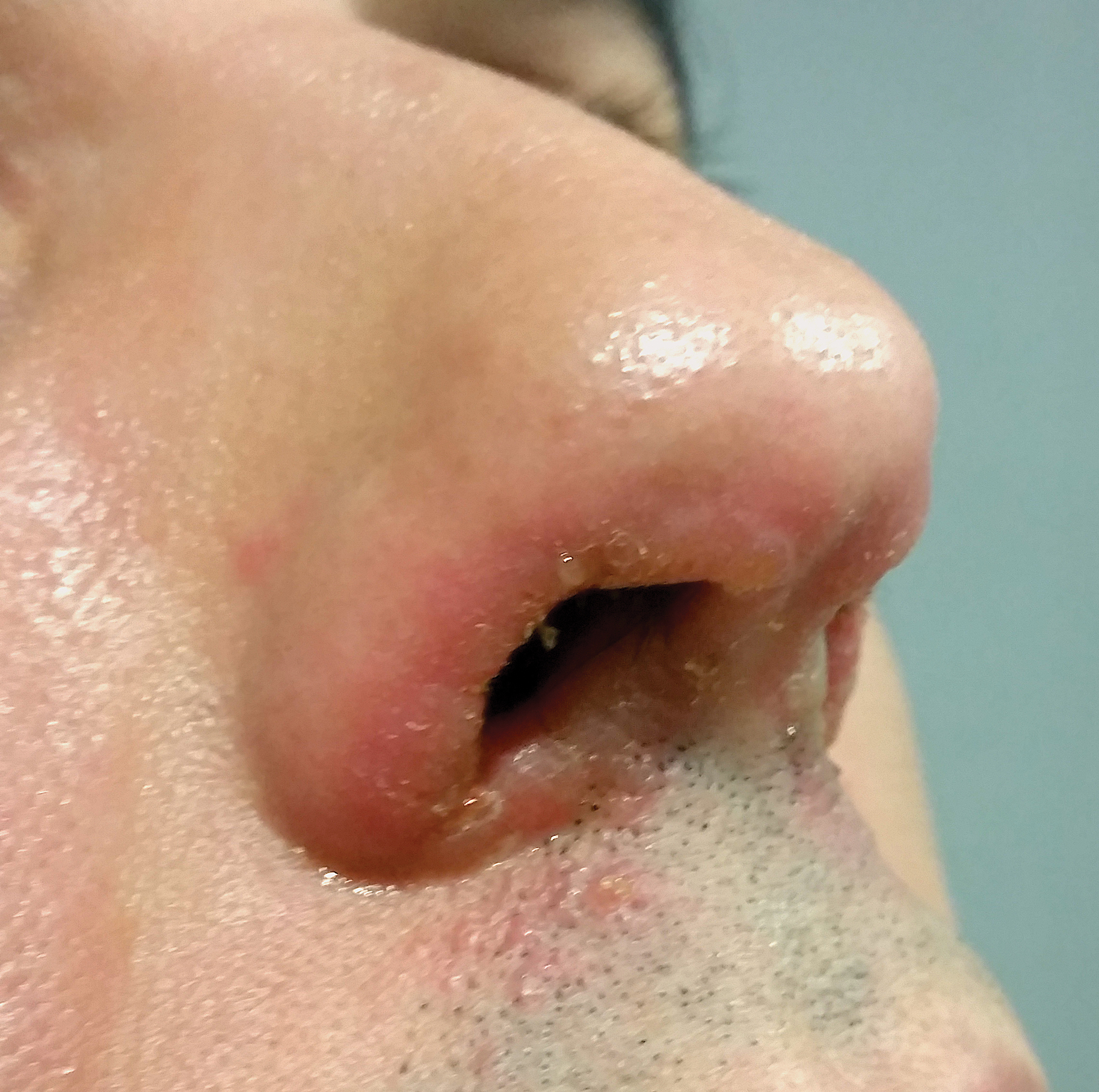
A 44-year-old man was referred to the department of dermatology for a pruriginous nasal rash. Physical examination revealed vesicles with clear content and crusts symmetrically in both nostrils and philtra. The remainder of the examination was otherwise unremarkable. The patient reported inhalation of poppers the prior night during a party. No history of connective tissue diseases was present. The patient was in overall good health with no fever or chills.
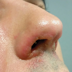
U.S. adults eating fewer carbs, more protein and fat
American diets have improved in the last several decades, with declines in consumption of low-quality carbohydrates and increases in plant protein and healthy fats, based on data from a nationally representative sample of 43,996 adults.
Changes in the economy, food policies, and food processing can affect diet over time, but trends in consumption of macronutrients in the U.S. have not been well studied, wrote Zhilei Shan, MD, of Harvard T.H. Chan School of Public Health, Boston, and colleagues.
In a study published in JAMA, the researchers reviewed data from nine consecutive cycles of the National Health and Nutrition Examination Survey (NHANES) from 1999 to 2016 to determine trends in macronutrient intake. The study population included adults aged 20 years and older who could provide at least 1 valid dietary recall questionnaire. The average age was 47 years, and 52% were women.
Overall, total carbohydrate consumption decreased from 52.5% to 50.5%, the estimated energy from high-quality carbohydrates increased from 7.42% to 8.65%, and the estimated energy from low-quality carbohydrates decreased from 45.1% to 41.8%. Total protein consumption increased from 15.5% to 16.4%, and plant protein consumption increased from 5.38% to 5.76%. Total fat consumption increased from 32.0% to 33.2%, including a 0.36% increase in saturated fatty acids from 11.5% to 11.9% and a 0.65% increase in polyunsaturated fatty acids from 7.58% to 8.23%.
Although the changes in total carbohydrates, total protein, and total fat were significant, 42% of energy intake came from low-quality carbohydrates and 10% came from saturated fat, the researchers noted. Also, the overall diet quality as measured by the Healthy Eating Index 2015 (HEI-2015) improved from 55.7 in 1999 to 57.7 in 2016. The HEI-2015 measures how diet data adheres to recommendations in the 2015-2020 Dietary Guidelines for Americans on a scale of 0-100.
“Improvements in intakes of whole grains and plant protein are encouraging, but how much do popular foods such as pizza, fast food sandwiches and burgers, and foods categorized as ‘snacks’ and ‘desserts’ contribute to the U.S. eating pattern?” wrote Linda Van Horn, PhD, RDN, and Marilyn C. Cornelis, PhD, of Northwestern University, Chicago, in an editorial accompanying the study. They cited a closer look at the NHANES data in the USDA’s “What We Eat in America” report and noted the differences in ethnicity with regard to macronutrient consumption.
“Non-Hispanic whites consume more alcohol, pizza, and fast food sandwiches (24% of total energy), while non-Hispanic blacks consume more salty snacks and sweet desserts (17% of total energy), and Hispanics consume more sugar-sweetened beverages (8% of total energy) and the least alcohol compared with the other racial/ethnic groups,” they said. “Public health efforts to educate, inform, and incent better adherence to the recommended nutrient-dense food groups are clearly needed,” they emphasized.
The study findings were limited by several factors, including the use of self-reports and changes in dietary assessments over time, the researchers noted. However, the results are strengthened by the large sample size and suggest improvements in the overall American diet, but also highlight the need for continuing public education and intervention, they said.
The study was supported by the National Institutes of Health and the Young Scientists Fund of the National Natural Science Foundation of China. Dr. Shan had no financial conflicts to disclose. Dr. Van Horn disclosed being a member of the 2020 US Dietary Guidelines Advisory Committee.
SOURCE: Shan Z et al. JAMA. 2019. doi: 10.1001/jama.2019.13771.
FROM JAMA