User login
Lay press stories about research: Putting them into perspective for patients
I recently had an unusual patient call. A woman I’ve seen for many years for migraines called to tell me her son was being hospitalized for appendicitis. He was scheduled for surgery in the morning.

She called because she’d recently seen a news report about how people without an appendix may have a higher rate of Parkinson’s disease as they age. She was, understandably, concerned about the long-term risks the procedure could pose.
On the surface, as a medical professional, the call sounds frivolous and silly. The risks of untreated acute appendicitis, such as peritonitis and death, are pretty well documented. Surgery offers the best possibility for a cure without recurrence. Compared with the long-term, uncertain, risk of Parkinson’s disease, the benefit-to-risk ratio and options are pretty obvious.
The question of the GI tract’s involvement in neurologic diseases is a legitimate one that needs to be answered. It may provide new insight into their causes and potential treatments. The research she brought up raises some interesting points.
But that doesn’t mean there should be any delay in treating something as easily cured – and potentially serious – as acute appendicitis.
My patient called to ask questions, and I have no issue with that. To someone with no medical training, it’s a legitimate concern. But not everyone will call to ask.

This is a hazard of early stages of medical research making it into the lay press. It may be right, it may be wrong, but it’s too early to tell either way. We have years of training to help us recognize the uncertainties of preliminary data, but the general public doesn’t. Stories like this create interest and raise questions in the medical literature and fear and anxiety in the lay press.
I’m a strong supporter of freedom of the press, and certainly they have every right to air or publish such stories. But they should also be put in perspective at the beginning, not the bottom, and make it clear the findings are far from proven.
Dr. Block has a solo neurology practice in Scottsdale, Ariz.
I recently had an unusual patient call. A woman I’ve seen for many years for migraines called to tell me her son was being hospitalized for appendicitis. He was scheduled for surgery in the morning.
She called because she’d recently seen a news report about how people without an appendix may have a higher rate of Parkinson’s disease as they age. She was, understandably, concerned about the long-term risks the procedure could pose.
On the surface, as a medical professional, the call sounds frivolous and silly. The risks of untreated acute appendicitis, such as peritonitis and death, are pretty well documented. Surgery offers the best possibility for a cure without recurrence. Compared with the long-term, uncertain, risk of Parkinson’s disease, the benefit-to-risk ratio and options are pretty obvious.
The question of the GI tract’s involvement in neurologic diseases is a legitimate one that needs to be answered. It may provide new insight into their causes and potential treatments. The research she brought up raises some interesting points.
But that doesn’t mean there should be any delay in treating something as easily cured – and potentially serious – as acute appendicitis.
My patient called to ask questions, and I have no issue with that. To someone with no medical training, it’s a legitimate concern. But not everyone will call to ask.
This is a hazard of early stages of medical research making it into the lay press. It may be right, it may be wrong, but it’s too early to tell either way. We have years of training to help us recognize the uncertainties of preliminary data, but the general public doesn’t. Stories like this create interest and raise questions in the medical literature and fear and anxiety in the lay press.
I’m a strong supporter of freedom of the press, and certainly they have every right to air or publish such stories. But they should also be put in perspective at the beginning, not the bottom, and make it clear the findings are far from proven.
Dr. Block has a solo neurology practice in Scottsdale, Ariz.
Venous Venous Venous @VEITHsymposium
The Venous Venous Venous @VEITHsymposium program has become a popular staple of the the VEITHsymposium. Now in its 13th year, the Venous Venous Venous program, which will be held on Wednesday, Thursday, and Friday, features a mixture of didactic sessions and workshops to cover the full gamut of venous disorders and their treatments, surgical, endovascular, and medical. It is “considered to be the best and most complete program,” on venous diseases according to conference organizers.
The didactic Program I (Sessions 63-70) on Superficial Venous Disease will be held all day Thursday and will detail the latest developments in venous clinical examinations and imaging, superficial vein strategies and techniques, thermal and non-thermal ablation, and there will be a special session on venous societal issues and governance.
Moderated by Kathleen J. Ozsvath, MD, and Harold J. Welch, MD, this special program will feature discussions on the future of venous reimbursement In a non-fee for service environment, upcoming venous trials, appropriate use criteria to limit inappropriate venous care, and more.
The didactic Program L (Sessions 87-94) on Deep Venous Disease will be all day Friday and the sessions will include discussions on illiocaval stenting, wound care, and surgical and interventional management strategies for thromboembolic events in the venous system. A special medical management session will address topics including statin use, reversal agents for the DOAC, and the potential utility of e-selectin inhibition, and more.
In addition, two special non-CME workshops – “Ask the Experts” sessions – are being offered on Wednesday afternoon this year:
Module 1: Current Superficial Venous Treatment, Wounds, and Edema will focus on endothermal therapy, cyanoacrylate, MOCA, VTE and recanalization, perforators, sclerotherapy, CVI and lymphadema, lipedema, and wound care and compression.
Module 2: Thrombus Management will address thrombolysis and thrombectomy, stents, IVUS, valves, and Nutcracker syndrome.
The workshops will include video case presentations, lectures and demonstrations on vein management by experts, plus hands-on opportunities where participants can rotate through multiple training stations staffed by professionals to assist them.
Caring for the carotids a focus of VEITHsymposium
Developments in carotid artery disease diagnosis and treatment have always been an important component of the VEITHsymposium programs and there will be sessions focusing on this critical area of patient management throughout the entire meeting.
The session will be moderated by Bruce A. Perler, MD, and L. Nelson Hopkins, MD, and include presentations on the value of using biomarkers of brain injury associated with carotid interventions, and how to assess and treat such adverse events as cerebral hyperfusion syndrome and intracranial artery dissection, as well as best medical treatments for carotid patients.
Of particular interest, there will be a debate Tuesday on the need for completion imaging with duplex or angiography with Hans-Henning Eckstein, MD, PhD, and R. Clement Darling III, MD.
Presentations will also address some of the latest treatment techniques for carotid artery disease. For example, on Wednesday morning, Norman H. Kumins, MD, of the Cleveland Medical Center, will present a study on the duration of blood flow reversal during transcarotid artery revascularization (TCAR), an “increasingly popular alternative to carotid endarterctomy and transfemoral artery carotid stenting,” which is designed to provide increased neuroprotection during the placement and angioplasty of the carotid stent. They analyzed the relationship between the length of TCAR flow reversal time (FRT) and major adverse events in 307 patients who underwent TCAR at four high-volume institutions. They separated patients into short (3-7minutes); medium(8-12 minutes); and long group (greater than 12 minutes) FRT. They designated a subset of the long group patients of those with greater than or equal to 20 or more minutes FRT, which they defined as the very long group. The stroke, myocardial infarction, and death rates at 30 days were assessed for all patients and were compared them between groups.
Dr. Kumins will detail how the overall stroke rate was 1.3%, with all strokes considered minor, and all patients showing full recovery. The four strokes occurred in patients with FRT of 6, 7, 11, and 12 minutes, showing no difference in the composite stroke/death or stroke/death/MI rates among the groups, the researchers indicated.
Dr. Kumins will discuss how flow reversal time does not affect stroke rates in patients undergoing TCAR, and suggest that operators should focus on the technical aspects of the procedure during flow reversal rather than being concerned about the amount of FRT.
Assessing and treating lower extremity arterial disease
This year at the VEITHsymposium, lower extremity arterial disease diagnosis and treatment takes pride of place in multiple sessions on each day.
For example, Tuesday will feature a special afternoon program on Hot New Topics In Lower Extremity Occlusive Disease Treatment, and on Wednesday morning, an associate faculty session will be held on Progress In Lower Extremity Occlusive Disease And Its Treatments.
In one particular presentation on Wednesday morning, Arsalan Wafi, MBBS, a clinical researcher at St. George’s Vascular Institute, London, will present a 10-year prospective study demonstrating that the poor mobility, lack of statin use, and socioeconomic deprivation are all associated with worse survival after a major lower limb amputation. Dr. Wafi will discuss how he and his colleagues assessed consecutive 805 major lower limb amputation patients seen in the Roehampton Rehabilitation Center between January 2007 and January 2018, using prospective records, which included demographics, etiologies of limb loss, operative details, medications, and mortality data over a 10-year follow-up period.
A total of 611 (76%) occurred in men, and 194 (24%) in women. Etiologies included diabetes mellitus, peripheral vascular disease, and other causes such as trauma, malignancy, sepsis, and complex regional pain syndrome.
Dr. Wafi will present data showing that living in a deprived area and being further away from the rehabilitation center were both significantly associated with poorer survival. Diabetes mellitus or peripheral vascular disease were associated with significantly shorter survival, compared with other etiologies, and not being on a statin was associated with significantly worse survival among the vascular patients. In addition, poorer overall mobility at discharge from rehabilitation was associated with poorer survival, according to the researchers. However there was no significant difference in survival between below-knee and above-knee amputees, or between unilateral and bilateral amputees.
Thursday will be highlighted by a session on New Devices For Treating Lower Extremity Lesions By Endovascular Or Open Techniques, and Friday will see a session New Developments In The Treatment Of Popliteal Diseases And Aneurysms.
This is only one of many such studies focused on lower extremity arterial disease at this year’s VEITHsymposium.
Vaping-linked lung injury: 2,172 cases, 42 deaths
The Centers for Disease Control and Prevention has from 49 states (all except Alaska), the District of Columbia, and two U.S. territories (Puerto Rico and U.S. Virgin Islands). Forty-two deaths have been confirmed in 24 states and the District of Columbia, the CDC reported.

Laboratory test results of bronchoalveolar lavage fluid samples from 29 patients submitted to CDC from 10 states found vitamin E acetate in all of the samples. This is the first time a chemical of concern has been found in biologic samples from patients with EVALI. These findings provide direct evidence of vitamin E acetate at the primary site of injury within the lungs.
Tetrahydrocannabinol (THC) was identified in 82% of the samples and nicotine was identified in 62% of the samples. Testing continues for other chemicals including plant oils, petroleum distillates like mineral oil, medium-chain triglycerides oil, and terpenes, which are compounds commonly found in or added to THC products. None of these chemicals has been detected in the bronchoalveolar lavage fluid samples tested.
For more information and resources visit For the Public, For Healthcare Providers, and For State and Local Health Departments pages, as well as the CDC’s Publications and Resources page.
The Centers for Disease Control and Prevention has from 49 states (all except Alaska), the District of Columbia, and two U.S. territories (Puerto Rico and U.S. Virgin Islands). Forty-two deaths have been confirmed in 24 states and the District of Columbia, the CDC reported.
Laboratory test results of bronchoalveolar lavage fluid samples from 29 patients submitted to CDC from 10 states found vitamin E acetate in all of the samples. This is the first time a chemical of concern has been found in biologic samples from patients with EVALI. These findings provide direct evidence of vitamin E acetate at the primary site of injury within the lungs.
Tetrahydrocannabinol (THC) was identified in 82% of the samples and nicotine was identified in 62% of the samples. Testing continues for other chemicals including plant oils, petroleum distillates like mineral oil, medium-chain triglycerides oil, and terpenes, which are compounds commonly found in or added to THC products. None of these chemicals has been detected in the bronchoalveolar lavage fluid samples tested.
For more information and resources visit For the Public, For Healthcare Providers, and For State and Local Health Departments pages, as well as the CDC’s Publications and Resources page.
REPORTING FROM CDC
57 Varieties: Sleep-Disordered Breathing Linked to Changes in 1 Important Gene
Blood oxygen levels drop during sleep, but how much and when that happens is mainly hereditary? Now researchers from Brigham and Women’s Hospital and Case Western Reserve University are getting closer to finding the genetic reasons for the fluctuations, which could help people with sleep apnea and other lung illnesses.
In their study, funded by the National Heart, Lung, and Blood Institute (NHLBI), the researchers analyzed whole genome sequence data from the NHLBI’s Trans-Omics for Precision Medicine (TOPMed) program. They also incorporated results for family-based linkage analysis, which maps genes with hereditary traits to their location in the genome.
The researchers identified 57 genetic variations of DLC1, a gene consistently associated with average arterial oxyhemoglobin saturation during sleep. The variants explain almost 1% of the variability in the oxygen levels in European Americans. That is high for complex genetic phenotypes, the researchers say. Of the 57 variants, 51 influence and regulate human lung fibroblast cells.
“This study highlights the advantage of using family data in searching for rare variants,” says James Kiley, PhD, director of the Division of Lung Diseases at NHLBI. “It showed that, when guided by family linkage data, whole genome sequence analysis can identify rare variants that signal disease risks, even with a small sample. In this case, the initial discovery was done with fewer than 500 samples.”
Transfusion-related lung injury is on the rise in elderly patients
SAN ANTONIO – Although there has been a general decline in transfusion-related anaphylaxis and acute infections over time among hospitalized older adults in the United States, incidence rates for both transfusion-related acute lung injury and transfusion-associated circulatory overload have risen over the last decade, according to researchers from the Food and Drug Administration.
Mikhail Menis, PharmD, an epidemiologist at the FDA Center for Biologics Evaluation and Research (CBER) and colleagues queried large Medicare databases to assess trends in transfusion-related adverse events among adults aged 65 years and older.
The investigators saw “substantially higher risk of all outcomes among immunocompromised beneficiaries, which could be related to higher blood use of all blood components, especially platelets, underlying conditions such as malignancies, and treatments such as chemotherapy or radiation, which need further investigation,” Dr. Menis said at the annual meeting of AABB, the group formerly known as the American Association of Blood Banks.
He reported data from a series of studies on four categories of transfusion-related events that may be life-threatening or fatal: transfusion-related anaphylaxis (TRA), transfusion-related acute lung injury (TRALI), transfusion-associated circulatory overload (TACO), and acute infection following transfusion (AIFT).
For each type of event, the researchers looked at overall incidence and the incidence by immune status, calendar year, blood components transfused, number of units transfused, age, sex, and race.
Anaphylaxis (TRA)
TRA may be caused by preformed immunoglobin E (IgE) antibodies to proteins in the plasma in transfused blood products or by preformed IgA antibodies in patients who are likely IgA deficient, Dr. Menis said.
The overall incidence of TRA among 8,833,817 inpatient transfusions stays for elderly beneficiaries from 2012 through 2018 was 7.1 per 100,000 stays. The rate was higher for immunocompromised patients, at 9.6, than it was among nonimmunocompromised patients, at 6.5.
The rates varied by every subgroup measured except immune status. Annual rates showed a downward trend, from 8.7 per 100,000 in 2012, to 5.1 in 2017 and 6.4 in 2018. The decline in occurrence may be caused by a decline in inpatient blood utilization during the study period, particularly among immunocompromised patients.
TRA rates increased with five or more units transfused. The risk was significantly reduced in the oldest group of patients versus the youngest (P less than .001), which supports the immune-based mechanism of action of anaphylaxis, Dr. Menis said.
They also found that TRA rates were substantially higher among patients who had received platelet and/or plasma transfusions, compared with patients who received only red blood cells (RBCs).
Additionally, risk for TRA was significantly higher among men than it was among women (9.3 vs. 5.4) and among white versus nonwhite patients (7.8 vs. 3.8).
The evidence suggested TRA cases are likely to be severe in this population, with inpatient mortality of 7.1%, and hospital stays of 7 days or longer in about 58% of cases, indicating the importance of TRA prevention, Dr. Menis said.
The investigators plan to perform multivariate regression analyses to assess potential risk factors, including underlying comorbidities and health histories for TRA occurrence for both the overall population and by immune status.
Acute lung injury (TRALI)
TRALI is a rare but serious adverse event, a clinical syndrome with onset within 6 hours of transfusion that presents as acute hypoxemia, respiratory distress, and noncardiogenic pulmonary edema.
Among 17,771,193 total inpatient transfusion stays, the overall incidence of TRALI was 33.2 per 100,000. The rate was 55.9 for immunocompromised patients versus 28.4 for nonimmunocompromised patients. The rate ratio was 2.0 (P less than .001).
The difference by immune status may be caused by higher blood utilizations with more units transfused per stay among immunocompromised patients, a higher incidence of prior transfusions among these patients, higher use of irradiated blood components that may lead to accumulation of proinflammatory mediators in blood products during storage, or underlying comorbidities.
The overall rate increased from 14.3 in 2007 to 56.4 in 2018. The rates increased proportionally among both immunocompromised and nonimmunocompromised patients.
As with TRA, the incidence of TRALI was higher in patients with five or more units transfused, while the incidence declined with age, likely caused by declining blood use and age-related changes in neutrophil function, Dr. Menis said.
TRALI rates were slightly higher among men than among women, as well as higher among white patients than among nonwhite patients.
Overall, TRALI rates were higher for patients who received platelets either alone or in combination with RBCs and/or plasma. The highest rates were among patients who received RBCs, plasma and platelets.
Dr. Menis called for studies to determine what effects the processing and storage of blood components may have on TRALI occurrence; he and his colleagues also are planning regression analyses to assess potential risk factors for this complication.
Circulatory overload (TACO)
TACO is one of the leading reported causes of transfusion-related fatalities in the U.S., with onset usually occurring within 6 hours of transfusion, presenting as acute respiratory distress with dyspnea, orthopnea, increased blood pressure, and cardiogenic pulmonary edema.
The overall incidence of TACO among hospitalized patients aged 65 years and older from 2011 through 2018 was 86.3 per 100,000 stays. The incidences were 128.3 in immunocompromised and 76.0 in nonimmunocompromised patients. The rate ratio for TACO in immunocompromised versus nonimmunocompromised patients was 1.70 (P less than .001).
Overall incidence rates of TACO rose from 62 per 100,000 stays in 2011 to 119.8 in 2018. As with other adverse events, incident rates rose with the number of units transfused.
Rates of TACO were significantly higher among women than they were among men (94.6 vs. 75.9 per 100,000; P less than .001), which could be caused by the higher mean age of women and/or a lower tolerance for increased blood volume from transfusion.
The study results also suggested that TACO and TRALI may coexist, based on evidence that 3.5% of all TACO stays also had diagnostic codes for TRALI. The frequency of co-occurrence of these two adverse events also increased over time, which may be caused by improved awareness, Dr. Menis said.
Infections (AIFT)
Acute infections following transfusion can lead to prolonged hospitalizations, sepsis, septic shock, and death. Those most at risk include elderly and immunocompromised patients because of high utilization of blood products, comorbidities, and decreased immune function.
Among 8,833,817 stays, the overall rate per 100,000 stays was 2.1. The rate for immunocompromised patients was 5.4, compared with 1.2 for nonimmunocompromised patients, for a rate ratio of 4.4 (P less than .001).
The incidence rate declined significantly (P = .03) over the study period, with the 3 latest years having the lowest rates.
Rates increased substantially among immunocompromised patients by the number of units transfused, but remained relatively stable among nonimmunocompromised patients.
Infection rates declined with age, from 2.7 per 100,000 stays for patients aged 65-68 years to 1.2 per 100,000 for those aged 85 years and older.
As with other adverse events, AIFT rates were likely related to the blood components transfused, with substantially higher rates for stays during which platelets were transfused either alone or with RBCs, compared with RBCs alone. This could be caused by the room-temperature storage of platelets and higher number of platelets units transfused, compared with RBCs alone, especially among immunocompromised patients.
In all, 51.9% of AIFT cases also had sepsis noted in the medical record, indicating high severity and emphasizing the importance of AIFT prevention, Dr. Menis said.
The studies were funded by the FDA, and Dr. Menis is an FDA employee. He reported having no conflicts of interest.
REPORTING FROM AABB 2019
Nivolumab-ipilimumab combo has ‘robust’ clinical benefit in sorafenib-treated HCC patients
BOSTON – The combination of nivolumab and ipilimumab provided a “robust” clinical benefit and had manageable hepatic adverse events in a phase 2 study of sorafenib-treated patients with advanced hepatocellular carcinoma (HCC), an investigator said at the annual meeting of the American Association for the Study of Liver Diseases.
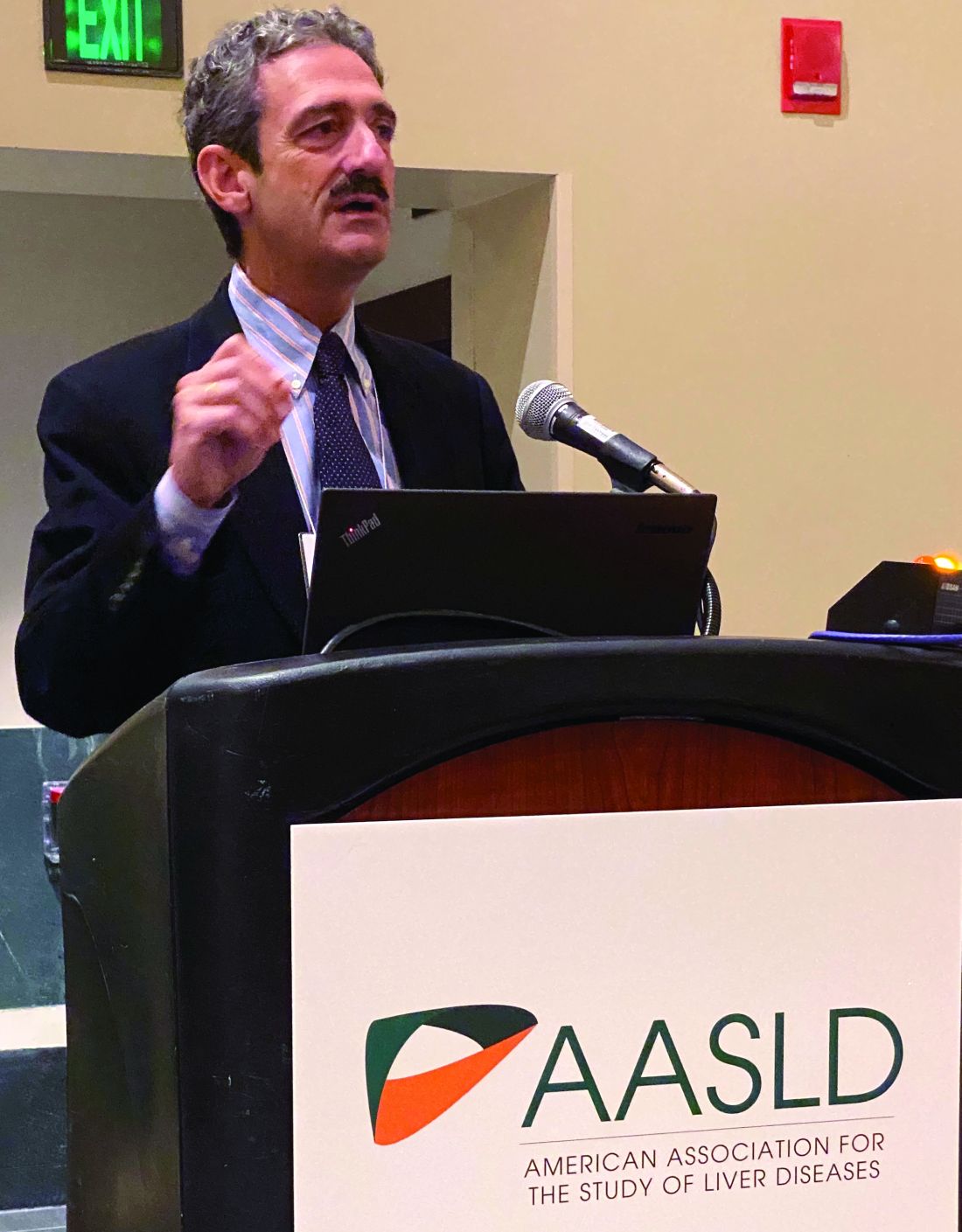
Response rates for the combination treatment exceeded 30% in CheckMate 040, with median overall survival approaching 23 months in one arm of this randomized trial, said Bruno Sangro, MD, PhD, of Clinica Universidad de Navarra, Pamplona, Spain.
The combination had a manageable safety profile with no new safety signals, and most immune-mediated adverse events resolved, including hepatic events, Dr. Sango said in an oral abstract session.
“The favorable benefit/risk profile observed we believe warrants further investigation in patients with HCC,” Dr. Sangro said in his presentation to attendees, adding that a phase 3 study of the combination is already ongoing.
On Nov. 11, 2019, Bristol-Myers Squibb announced its application for nivolumab plus ipilimumab for previously treated advanced HCC had been accepted for priority review by the Food and Drug Administration, which had furthermore granted breakthrough therapy designation for that potential indication.
In September 2017, the FDA approved nivolumab as monotherapy for patients with HCC previously treated with sorafenib. That action was based on results from CheckMate 040 showing on overall response rate of 14% and a median overall survival of 15 months, Dr. Sangro said.
The combination of the programmed death–1 inhibitor nivolumab and the CTLA-4 inhibitor ipilimumab, which has shown durable responses in other tumor types, may promote synergistic immune activity in HCC through their distinct but complementary mechanisms, according to the investigator.
The first report on the combination of nivolumab plus ipilimumab in CheckMate 040, reported earlier this year at the annual meeting of the American Society of Clinical Oncology, indicated that the combination produced responses that were robust and durable.
Dr. Sangro reported data on 148 patients with advanced HCC previously treated with sorafenib randomized to one of three different dosing regimens with nivolumab plus ipilimumab.
Response rates ranged from 31% to 32% in the three arms, while one particular dosing regimen given every 3 weeks for four cycles had a median overall survival of 22.8 months.
“Just to give a perspective, let me remind you that patients receiving placebo post sorafenib in a number of phase 3 trials have very consistently shown median overall survivals of around 8 months,” Dr. Sangro told attendees.
Hepatic treatment-related adverse events of any grade reported within 30 days of the last dose were seen in 39% of patients in that arm, Dr. Sangro said.
Hepatic events thought to be immune mediated were typically managed with a short course of high-dose corticosteroids, according to Dr. Sangro, who said no patients rechallenged with treatment experienced a recurrence of the event.
Most of the hepatic adverse events occurred early, with a median time to onset of 5.6-8.1 weeks, he said, and most resolved at a median of 6.1-7.9 weeks.
In the ongoing phase 3 CheckMate 9DW study, patients with advanced HCC who are naive to systemic therapy are being randomized to the combination of nivolumab plus ipilimumab, or to the investigators’ choice of either sorafenib or lenvatinib, with a primary endpoint of overall survival, Dr. Sangro said.
The study was supported by Bristol-Myers Squibb and ONO Pharmaceutical. Dr. Sangro reported disclosures related to Adaptimmune, AstraZeneca, Bayer, Bristol-Myers Squibb, BTG, Merck, Onxeo, Sirtex Medical, Terumo, H3 Biomedicine, Ipsen, Lilly, Exelixis, Roche, and Ipsen.
SOURCE: Sangro B et al. The Liver Meeting 2019, Abstract 200.
BOSTON – The combination of nivolumab and ipilimumab provided a “robust” clinical benefit and had manageable hepatic adverse events in a phase 2 study of sorafenib-treated patients with advanced hepatocellular carcinoma (HCC), an investigator said at the annual meeting of the American Association for the Study of Liver Diseases.
Response rates for the combination treatment exceeded 30% in CheckMate 040, with median overall survival approaching 23 months in one arm of this randomized trial, said Bruno Sangro, MD, PhD, of Clinica Universidad de Navarra, Pamplona, Spain.
The combination had a manageable safety profile with no new safety signals, and most immune-mediated adverse events resolved, including hepatic events, Dr. Sango said in an oral abstract session.
“The favorable benefit/risk profile observed we believe warrants further investigation in patients with HCC,” Dr. Sangro said in his presentation to attendees, adding that a phase 3 study of the combination is already ongoing.
On Nov. 11, 2019, Bristol-Myers Squibb announced its application for nivolumab plus ipilimumab for previously treated advanced HCC had been accepted for priority review by the Food and Drug Administration, which had furthermore granted breakthrough therapy designation for that potential indication.
In September 2017, the FDA approved nivolumab as monotherapy for patients with HCC previously treated with sorafenib. That action was based on results from CheckMate 040 showing on overall response rate of 14% and a median overall survival of 15 months, Dr. Sangro said.
The combination of the programmed death–1 inhibitor nivolumab and the CTLA-4 inhibitor ipilimumab, which has shown durable responses in other tumor types, may promote synergistic immune activity in HCC through their distinct but complementary mechanisms, according to the investigator.
The first report on the combination of nivolumab plus ipilimumab in CheckMate 040, reported earlier this year at the annual meeting of the American Society of Clinical Oncology, indicated that the combination produced responses that were robust and durable.
Dr. Sangro reported data on 148 patients with advanced HCC previously treated with sorafenib randomized to one of three different dosing regimens with nivolumab plus ipilimumab.
Response rates ranged from 31% to 32% in the three arms, while one particular dosing regimen given every 3 weeks for four cycles had a median overall survival of 22.8 months.
“Just to give a perspective, let me remind you that patients receiving placebo post sorafenib in a number of phase 3 trials have very consistently shown median overall survivals of around 8 months,” Dr. Sangro told attendees.
Hepatic treatment-related adverse events of any grade reported within 30 days of the last dose were seen in 39% of patients in that arm, Dr. Sangro said.
Hepatic events thought to be immune mediated were typically managed with a short course of high-dose corticosteroids, according to Dr. Sangro, who said no patients rechallenged with treatment experienced a recurrence of the event.
Most of the hepatic adverse events occurred early, with a median time to onset of 5.6-8.1 weeks, he said, and most resolved at a median of 6.1-7.9 weeks.
In the ongoing phase 3 CheckMate 9DW study, patients with advanced HCC who are naive to systemic therapy are being randomized to the combination of nivolumab plus ipilimumab, or to the investigators’ choice of either sorafenib or lenvatinib, with a primary endpoint of overall survival, Dr. Sangro said.
The study was supported by Bristol-Myers Squibb and ONO Pharmaceutical. Dr. Sangro reported disclosures related to Adaptimmune, AstraZeneca, Bayer, Bristol-Myers Squibb, BTG, Merck, Onxeo, Sirtex Medical, Terumo, H3 Biomedicine, Ipsen, Lilly, Exelixis, Roche, and Ipsen.
SOURCE: Sangro B et al. The Liver Meeting 2019, Abstract 200.
REPORTING FROM THE LIVER MEETING 2019
Don’t miss neuromuscular complications of cancer immunotherapy
AUSTIN, TEX. – Neuromuscular complications from immunotherapy for cancer are rare, but they occur often enough that it is helpful to know which ones can result from different immunotherapies and how to distinguish them from non–adverse event conditions, according to Christopher Trevino, MD, a neuro-oncologist at Tulane University in New Orleans.
At the annual meeting of the American Association for Neuromuscular and Electrodiagnostic Medicine, Dr. Trevino reviewed immunotherapy types, particularly immune checkpoint inhibitors, and the most common neuromuscular complications – primarily neuropathy, myasthenia gravis (MG), myositis, and encephalitis or meningitis.
“Timing of onset is a critical component to assist in identifying immune checkpoint inhibitor–associated versus non–immune checkpoint inhibitor–associated neuromuscular disease,” Dr. Trevino told attendees. Prompt recognition can be particularly urgent for MG because crisis and death rates are higher when induced by immunotherapy and require quick treatment. “Understanding the mechanisms of action sets a foundation for treatment approach,” he added.
Any part of the nervous system can be affected by immunotherapy toxicity, he said, and syndromes often overlap, with the peripheral nervous system typically more often affected than the central nervous system. Neurologic immune-related adverse events typically occur within four cycles of therapy – about 12 weeks after therapy initiation – but should always involve a work-up to exclude effects from the cancer itself, other neuromuscular diagnoses unrelated to therapy, and other toxicities from chemotherapy.
Recommended first-line treatment is halting immunotherapy with or without corticosteroids, after which most patients improve, often with “rapid, complete resolution of symptoms,” Dr. Trevino said. Restarting immunotherapy treatment is possible in some patients, though.
CAR T-cell and dendritic cell vaccine therapies
Four main types of immunotherapy exist: viral therapy, vaccine therapy, immune checkpoint inhibitors, and adoptive cell transfer, such as chimeric antigen receptor (CAR) T-cell therapy. Dr. Trevino focused on checkpoint inhibitors and adoptive cell transfer.
CAR T-cell therapy is a multistep treatment process that involves first removing blood from the patient to obtain their T cells. These are used to create and grow CAR T cells in the lab so that they can be infused back into the patient. The cells then bind to cancer cells and destroy them. Examples of approved CAR T-cell therapy include Yescarta (axicabtagene ciloleucel) for some types of non-Hodgkin lymphoma and Kymriah (tisagenlecleucel) for acute lymphoblastic leukemia (ALL).
Dendritic cell vaccines are similar to CAR T-cell therapy in that they also use the patient’s own immune cells to create cancer-killing cells that the patient then receives back. The only currently approved dendritic cell vaccine is Provenge (sipuleucel-T) for advanced prostate cancer.
The main toxicity to watch for from CAR T-cell therapy and dendritic cell vaccines is cytokine release syndrome (CRS). It can begin anywhere from 1-14 days after the infusion and involves T-cell expansion in the body that leads to a cytokine storm. Symptoms are wide ranging, including fatigue, fever, loss of appetite, tachycardia, hypotension, pain, rash, diarrhea, headache, confusion, seizures, muscle and joint pain, tachypnea, hypoxia and hallucinations, among others.
Specific central neurotoxicities that can result from CAR T-cell therapy include encephalopathy, cerebral edema, seizures and status epilepticus, cerebral vasospasm, and aphasia.
Immune checkpoint inhibitor toxicities
Immune checkpoint inhibitors are drugs that interrupt a cancer’s ability to hijack the immune system; they block the proteins that hold back T-cells from attacking the cancer, thereby releasing the immune system to go after the malignant cells.
The two most common types of immune checkpoint inhibitors are those targeting the programmed cell death protein 1 (PD-1) and programmed death-ligand 1 (PD-L1) pathways. The three currently approved PD-1 inhibitors are pembrolizumab (Keytruda), nivolumab (Opdivo), and cemiplimab (Libtayo), which can treat nearly a dozen malignancies affecting different organs. Atezolizumab (Tecentriq), avelumab (Bavencio), and durvalumab (Imfinzi) are the three currently approved PD-L1 inhibitors, indicated for urothelial carcinoma and a handful of other cancers, such as small-cell and non–small cell lung cancer and triple negative breast cancer.
The only other type of approved checkpoint inhibitor is ipilimumab (Yervoy), which targets the CTLA-4 protein. A number of other checkpoint inhibitors are in trials, however, such as ones targeting pathways involving OX40, ICOS, TIM3, and LAG-3 (J Hematol Oncol. 2018. doi: 10.1186/s13045-018-0582-8).
Immune-related adverse events are less common with PD-1 or PD-L1 inhibitors – a rate of 5%-10% – compared with adverse events from CTLA-4 inhibitors, which occur in about 15% of patients. Neurologic complications occur even more rarely – about 1%-4% of all immune checkpoint inhibitor therapies – and primarily include MG, Guillain-Barré syndrome (GBS), chronic inflammatory demyelinating polyneuropathy (CIDP), and inflammatory myositis (Muscle Nerve. 2018;58[1]:10-22).
Treatment with multiple checkpoint inhibitors increases the likelihood of severe adverse events, with rates of up to 30%-50% of patients with dual treatment.
Distinguishing features of neuromuscular immunotherapy-related adverse events
MG is the most common neuromuscular immune-related adverse event from immune checkpoint inhibitors and tends to occur 3-12 weeks after beginning treatment, frequently comorbid with inflammatory myopathy or cardiomyopathy, Dr. Trevino said. About two-thirds of cases are de novo, while the remaining one-third involve preexisting MG; no reports of Lambert-Eaton myasthenic syndrome have been linked to checkpoint inhibitors.
Several characteristics distinguish checkpoint inhibitor–associated MG from standard MG. Standard MG can be ocular with or without bulbar or appendicular weakness, whereas immunotherapy-related MG is rarely only ocular (about 18% of cases). Immunotherapy-related MG involves an MG crisis at diagnosis in up to 50% of cases and has high mortality, both of which are rarer with standard MG.
While standard MG can be seronegative or involve AChR, MuSK, or LRP4 antibodies, about two-thirds of immunotherapy-related MG cases are positive for AChR antibodies. LRP4 antibodies are rare with MG from checkpoint inhibitors, and no MuSK antibodies have been reported in these cases. Creatine kinase (CK) or troponin I (TnI) elevation occurs in about 87% of patients with checkpoint inhibitor-induced MG, but standard MG doesn’t typically involve increased CK levels.
Inflammatory myositis (IM), the second most common neuromuscular adverse event from immunotherapy, tends to occur 2-15 weeks after immune checkpoint inhibitor therapy and can involve polymyositis, necrotizing autoimmune myopathy, dermatomyositis, granulomatous myositis, or other nonspecific myositis and myopathies.
Though proximal weakness occurs with IM both associated with immunotherapy and not, ocular symptoms are unique to cases associated with therapy and occur in about half of them. Myalgia, dyspnea, and dysphagia can all occur with checkpoint inhibitor–associated IM but don’t generally occur with standard IM. Immunotherapy-related IM is usually seronegative for myositis antibodies and doesn’t generally cause abnormalities in electromyography, compared with increased exertional activity and early recruitment of myopathic motor units in electromyography with standard IM.
GBS and CIDP are the third most common cause of neuromuscular complications from checkpoint inhibitors. The main distinguishing feature of these conditions from those not related to immunotherapy is that they occur anywhere from 4 to 68 weeks after therapy begins. Presentation is otherwise similar whether related to checkpoint inhibitors or not.
Aside from GBS and CIDP, other neuropathies that can result from immunotherapy complications include acute cranial neuropathies, axonal or demyelinating neuropathies, motor polyradiculopathy, vasculitic neuropathy, and plexopathy.
Neuromuscular complications other than those described above can also occur from checkpoint inhibitor therapy, such as enteric neuropathy, polyradiculitis, and meningo-radiculo-neuritis, but these are much rarer.
Four organizations have developed consensus guidelines for immune checkpoint inhibitor toxicities: the European Society for Medical Oncology (ESMO, 2017), Society for Immunotherapy of Cancer (SITC, 2017), American Society of Clinical Oncology (ASCO, 2018), and National Comprehensive Cancer Network (NCCN, 2019).
Dr Trevino had no disclosures.
EXPERT ANALYSIS FROM AANEM 2019
Gene signature may help guide initial CLL treatment choice
A novel 17-gene expression signature may help guide the choice of initial treatment in patients with IGHV-unmutated chronic lymphocytic leukemia (CLL), according to findings of a retrospective dual cohort study.

“[Fludarabine, cyclophosphamide, and rituximab] was the first regimen to improve progression-free survival and overall survival in patients with chronic lymphocytic leukaemia, and has become a gold-standard chemoimmunotherapy regimen in physically fit patients,” wrote the investigators, who were led by Carmen D. Herling, MD, of the Center for Integrated Oncology, Cologne, Germany; and Kevin R. Coombes, PhD, of Ohio State University, Columbus.
While several studies demonstrate that young, fit patients with mutated IGHV gene and no high-risk cytogenetic abnormalities achieve durable remission with the FCR (fludarabine, cyclophosphamide, and rituximab) regimen, there have been no studies to identify if this is true for patients with unmutated IGHV gene, they reported in the Lancet Oncology.
The investigators performed transcriptional profiling using peripheral blood samples collected from two cohorts of patients with CLL who were treated with frontline FCR.
The discovery and training cohort consisted of 101 patients (65% with IGHV-unmutated disease) treated at the MD Anderson Cancer Center who had a median follow-up of about 12 years. The validation cohort consisted of 109 patients with IGHV-unmutated disease treated on the German CLL8 single-arm trial who had a median follow-up of about 6 years.
A total of 1,136 genes showed a significant univariate association with time to progression. Ultimately, 17 of these genes – most of them involved in purine metabolism and oxidative phosphorylation – were included in the expression signature.
Among patients with IGHV-unmutated CLL, the 17-gene signature discriminated between two groups having differing time to progression after their frontline FCR chemoimmunotherapy: an unfavorable prognosis group and an intermediate prognosis group.
The unfavorable prognosis group had a significantly higher relative risk of progression in both the discovery/training cohort (hazard ratio, 3.83; P less than .0001) and the validation cohort (HR, 1.90; P = .008). In the validation cohort, the median time to progression was 39 months among patients with a signature-defined unfavorable prognosis, compared with 59 months among patients with a signature-defined intermediate prognosis.
“We would recommend testing the value of the 17-gene signature in a prospective study that compares FCR treatment with alternative therapies, such as ibrutinib, as part of a randomised clinical trial,” the investigators wrote.
Dr. Herling reported financial disclosures related to Hoffmann-La Roche, and Dr. Coombes reported grants from the National Institutes of Health. The study was funded by the Chronic Lymphocytic Leukaemia Global Research Foundation and the National Institutes of Health/National Cancer Institute.
SOURCE: Herling CD et al. Lancet Oncol. 2019;20(11):1576-86.
A novel 17-gene expression signature may help guide the choice of initial treatment in patients with IGHV-unmutated chronic lymphocytic leukemia (CLL), according to findings of a retrospective dual cohort study.
“[Fludarabine, cyclophosphamide, and rituximab] was the first regimen to improve progression-free survival and overall survival in patients with chronic lymphocytic leukaemia, and has become a gold-standard chemoimmunotherapy regimen in physically fit patients,” wrote the investigators, who were led by Carmen D. Herling, MD, of the Center for Integrated Oncology, Cologne, Germany; and Kevin R. Coombes, PhD, of Ohio State University, Columbus.
While several studies demonstrate that young, fit patients with mutated IGHV gene and no high-risk cytogenetic abnormalities achieve durable remission with the FCR (fludarabine, cyclophosphamide, and rituximab) regimen, there have been no studies to identify if this is true for patients with unmutated IGHV gene, they reported in the Lancet Oncology.
The investigators performed transcriptional profiling using peripheral blood samples collected from two cohorts of patients with CLL who were treated with frontline FCR.
The discovery and training cohort consisted of 101 patients (65% with IGHV-unmutated disease) treated at the MD Anderson Cancer Center who had a median follow-up of about 12 years. The validation cohort consisted of 109 patients with IGHV-unmutated disease treated on the German CLL8 single-arm trial who had a median follow-up of about 6 years.
A total of 1,136 genes showed a significant univariate association with time to progression. Ultimately, 17 of these genes – most of them involved in purine metabolism and oxidative phosphorylation – were included in the expression signature.
Among patients with IGHV-unmutated CLL, the 17-gene signature discriminated between two groups having differing time to progression after their frontline FCR chemoimmunotherapy: an unfavorable prognosis group and an intermediate prognosis group.
The unfavorable prognosis group had a significantly higher relative risk of progression in both the discovery/training cohort (hazard ratio, 3.83; P less than .0001) and the validation cohort (HR, 1.90; P = .008). In the validation cohort, the median time to progression was 39 months among patients with a signature-defined unfavorable prognosis, compared with 59 months among patients with a signature-defined intermediate prognosis.
“We would recommend testing the value of the 17-gene signature in a prospective study that compares FCR treatment with alternative therapies, such as ibrutinib, as part of a randomised clinical trial,” the investigators wrote.
Dr. Herling reported financial disclosures related to Hoffmann-La Roche, and Dr. Coombes reported grants from the National Institutes of Health. The study was funded by the Chronic Lymphocytic Leukaemia Global Research Foundation and the National Institutes of Health/National Cancer Institute.
SOURCE: Herling CD et al. Lancet Oncol. 2019;20(11):1576-86.
FROM LANCET ONCOLOGY