User login
ESC suspension of Russia, Belarus societies sparks controversy
, provoking a heated discussion on whether medical organizations should become involved in politics.
“In the light of the continued aggression against Ukraine by the leaderships of the Russian Federation and Belarus, the European Society of Cardiology (ESC) has temporarily suspended the memberships of the Russian Society of Cardiology and the Belarussian Society of Cardiologists in the ESC,” the ESC statement reads.
“Individuals based in the Russian Federation or in Belarus are excluded from active participation in any ESC event or activity,” it states.
“The ESC very much regrets the effect this may have on individual Russian and Belarussian cardiologists and scientists, but the message to Russian and Belarussian leadership must be distinct and unequivocal,” it adds.
This action from the ESC has provoked a storm of heated discussions on the issue.
In a Twitter thread on the subject, Italian cardiologist Giuseppe Galati, MD, writes: “An astonishing decision by ESC that’s excluding all the Russian and Belarussian scientists from ESC congresses and activities. Treating doctors and scientists as [if] they are Putin and are responsible for the war.”
Dr. Galati adds: “A strong message that brings us to 70 years ago. ESC is promoting exclusion and not inclusion and diversity.”
Another commentator on the thread says: “It is a very unfortunate decision. Science, medicine should not be involved in politics. We are colleagues gathering together during congresses to exchange information for the sake of our patients. Politics should not overshadow this.”
And another added: “I think most cardiologists from Russia will not be able to participate in the events anyway, since international payments will soon be impossible from Russia. But it is wrong to limit the rights of doctors because of their nationality.”
But others support the ESC’s stance. Polish cardiologist Blazej Michalski, MD, says: “I think it is [a] good decision. Russians if they do not actively support dictatorship of Putin, the silence is also an agreement.” He adds: “I am proud of ESC. They did what they were supposed to do.”
A Twitter poll started by Ali Elzieny, MD, a cardiologist from Boston, titled “Do you agree that ESC suspend membership of Russian Society of Cardiology?” as of March 8 had 1,300 votes, with 61% of respondents disagreeing with the ESC decision and 39% in favor.
Medical societies respond
Several other medical societies have issued communications appearing to disagree with the action by the ESC.
The American College of Cardiology issued a statement saying medicine should be above politics.
“The American College of Cardiology believes that patients come first, and now, more than ever, there is a need to rally around our members across the globe to ensure that they have the support and resources they need to care for their patients,” said ACC President Dipti Itchhaporia, MD.
“Medicine is above politics and ACC will not exclude any of our colleagues who are working toward a shared mission of improving heart health. The College has a long history of working across borders to improve heart health and remains committed to that now and in the future. The ACC continues to express its support and concern for our members in the Ukraine and the patients they are working to treat on the frontlines,” the ACC statement added.
The Tele-Cardiology Working Group of the International Society for Telemedicine & eHealth (ISFTEH) also issued a statement disagreeing with the action from the ESC.
“In light of recent events, the cardiology working group of the ISFTEH will not restrict access to its events to cardiologists with regards to their nationality, religious beliefs or other characteristics that may seem discriminatory. We believe medical information should be widely available for all, especially for those doctors that find themselves in difficulty,” it said.
The European Academy of Neurology (EAN) said: “EAN is looking at ways to give practical support to Ukrainian neurologists and healthcare professionals there. EAN is not considering suspension of any individual member based on country of residence or nationality or any National Society member.”
But one oncology professional group has also cut ties with Russia.
The international cancer specialist network, OncoAlert, issued a statement saying it has severed all cooperation with doctors in Russia as part of the Western sanctions.
“The OncoAlert Network is non-political, but we cannot stand idle and not take a stand against this aggression towards our Ukrainian friends & colleagues,” OncoAlert said, adding that it will be pulling out of all collaborations and congresses in Russia. That statement was also greeted with a barrage of criticism on Twitter, mainly from Russian users.
A version of this article first appeared on Medscape.com.
Intermittent joint aches
Fundamental changes in the initial pharmacologic approach to psoriatic arthritis were made in the 2018 American College Rheumatology/National Psoriasis (ACR/NPF) guidelines for the treatment of psoriatic arthritis. Previously, methotrexate had been widely used as the first-line agent. The 2018 guidelines recommend a tumor necrosis factor (TNF) inhibitor over methotrexate and other oral small molecules (leflunomide, cyclosporine, and apremilast).
Herein is a broad summary of the guidelines:
· Treat with TNF inhibitor over oral small molecule; may consider oral small molecule with mild psoriatic arthritis and psoriasis, patient preference, and/or contraindication to TNF inhibitor
· Treat with TNF inhibitor over interleukin (IL)-17 inhibitor; may consider IL-17 inhibitor with severe psoriasis or contraindication to TNF inhibitor
· Treat with TNF inhibitor over interleukin (IL)-12/23 inhibitor; may consider IL-12/23 inhibitor with severe psoriasis or contraindication to TNF inhibitor
· Treat with oral small molecule over IL-17 inhibitor; may consider IL-17 inhibitor with severe psoriasis and/or psoriatic arthritis
· Treat with oral small molecule over IL-12/23 inhibitor; may consider IL-12/23 inhibitor with severe psoriasis or psoriatic arthritis, or concomitant inflammatory bowel disease
· Treat with methotrexate over nonsteroidal anti-inflammatory drugs; may consider nonsteroidals for mild psoriatic arthritis and psoriasis
· Treat with IL-17 inhibitor over IL-12/23 inhibitor; may consider IL-12/23 inhibitor in a patient with concomitant inflammatory bowel disease
Note that these recommendations are based on conditional evidence (ie, low to very low quality). In fact, in the entire guideline document, only 6% of the recommendations are strong, whereas 96% are conditional. This emphasizes the importance of evaluating each patient individually and engaging in a discussion to choose optimal therapy.
Another set of guidelines, from the Group for Research and Assessment of Psoriasis and Psoriatic Arthritis (GRAPPA), was last updated in 2015. Since then, many of the agents above have been introduced. Updated GRAPPA guidelines are expected to be released later this year.
Herbert S. Diamond, MD, Professor of Medicine (retired), Temple University School of Medicine, University of Pittsburgh; Chairman, Department of Medicine Emeritus, Western Pennsylvania Hospital, Pittsburgh, PA.
Herbert S. Diamond, MD, has disclosed no relevant financial relationships.
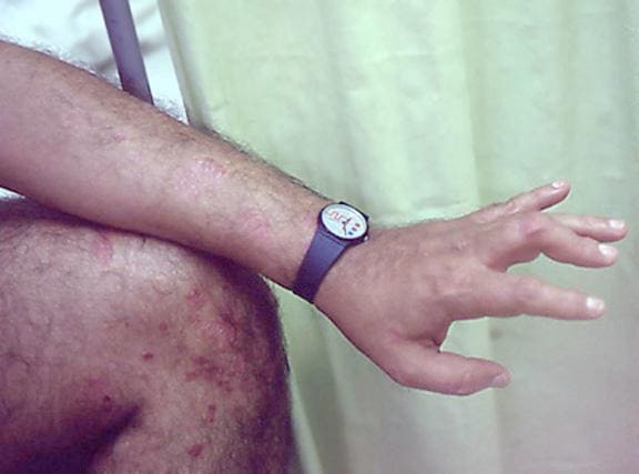
A 56-year-old man presents because of intermittent joint aches and difficulty picking up his grandchild and cleaning his home. He has a 6-year history of scalp psoriasis that he has controlled with a salicylic acid shampoo. On physical examination, he has tenderness over both elbows and in his metacarpophalangeal and proximal interphalangeal (PIP) joints on both hands. Swollen joints are noted in the proximal and distal joints of the right hand. His fingernails show uniform pitting.
Neurologic examination shows no sensory deficits or hyperesthesia. Motor examination is unremarkable, and chest and abdominal findings are unremarkable. Blood pressure is 138/90 mm Hg. Radiographic imaging shows asymmetric erosive changes with very small areas of bony proliferation in the PIP joints.There is asymmetric narrowing of the joint space in the interphalangeal joints. Laboratory findings reveal an erythrocyte sedimentation rate of 35 mm/h, negative rheumatoid factor, negative antinuclear antibody, and C-reactive protein of 7 mg/dL.
These findings are consistent with a diagnosis of psoriatic arthritis. This patient has severe psoriatic arthritis based on radiographic evidence of erosive disease.

Home blood pressure testing better than at clinics: Study
Everyone’s been there. Which one’s right?
The answer: Perhaps neither. Individual measures of blood pressure are not as accurate as taking multiple readings over a day and averaging them.
Blood pressure varies throughout the day – by about 30 points for systolic pressure, or the pressure when the heart beats – and one or two measurements in a doctor’s office may not accurately reflect the average figure, said Beverly B. Green, MD, a senior investigator at Kaiser Permanente Washington Health Research Institute in Seattle.
Average blood pressure reading is the only measurement on which a doctor can accurately diagnose and treat high blood pressure, she said. A new study by Dr. Green and other researchers at Kaiser Permanente showed that giving patients the chance to monitor their blood pressure at home could help get more reliable measurements.
Nearly one in four adults in the United States with high blood pressure are unaware they have the condition and are not getting treatment to control it. Without treatment, the condition can cause heart attacks, strokes, kidney damage, and other potentially life-threatening health problems.
Current guidelines for diagnosing high blood pressure recommend that patients whose pressure is high in the clinic get tested again to confirm the results. While the guidelines recommend home monitoring before diagnosing high blood pressure, research shows that doctors continue to measure blood pressure in their clinics for the second reading.
In their study, Dr. Green and colleagues found that home readings were more accurate than measurements taken in clinics or at pharmacy kiosks.
“Home blood pressure monitoring was a better option, because it was more accurate” than clinic blood pressure readings, Green said. A companion study found that patients preferred taking their blood pressure at home.
For their study, Dr. Green’s group used Kaiser’s electronic health record system to identify people at high risk for high blood pressure based on a recent clinic visit. They then randomly assigned the participants to get their follow-up blood pressure readings in the clinic, at home, or at kiosks in clinics or pharmacies.
Each participant also received a 24-hour ambulatory blood pressure monitor (ABPM). These devices, which people must wear continuously for 24 hours, have cuffs that inflate every 20-0 minutes during the day and every 30-60 minutes at night. Although ABPMs are the preferred test for accurately diagnosing high blood pressure, they aren’t available for widespread use.
The Kaiser researchers found that people’s systolic BP readings at clinics were generally lower than their ABPM measurements, leading to undiagnosed high BP in more than 50% of cases. Kiosk readings were much higher than the ABPM measurements and tended to overdiagnose high BP.
The value of home monitoring
Branden Villavaso, a 48-year-old attorney in New Orleans who was diagnosed with high BP at age 32, attributes his condition to genetics. He says an at-home monitor plus the occasional use of an ABPM finally provided his doctor with an accurate assessment of his condition.
Thanks to this aggressive approach, over the past 3 years, Mr. Villavaso’s diastolic reading has dropped from a previous range of between 90 and 100 to a healthier but not quite ideal value of about 80. Meanwhile, his systolic pressure has dropped to about 120, well below the goal of 130.
Mr. Villavaso said his doctor has relied on the averages of the BP readings to tailor his medication, and he also credited his wife, Chloe, a clinical nurse specialist, for monitoring his progress.
While previous studies have found similar benefits for measuring BP at home, Dr. Green said the latest study may offer the most powerful evidence to date because of the large number of people who took part, the involvement of primary care clinics, and the use of real-world health care professionals to take measurements instead of people who usually do health research. She said this study is the first to compare kiosk and ABPM results.
“The study indicates that assisting patients with getting access to valid blood pressure readings so they can measure their blood pressure at home will give a better picture of the true burden of [high BP],” said Keith C. Ferdinand, MD, a cardiologist at Tulane University, New Orleans.
He recommended patients select a home monitoring device from www.validatebp.org, a noncommercial website that lists home BP systems that have proven to be accurate.
“We know that [high blood pressure] is the most common and powerful cause of heart disease and death,” Dr. Ferdinand said. “Patients are pleased to participate in shared decision-making and actively assist in the control of a potentially deadly disease.”
A version of this article first appeared on WebMD.com.
FROM THE JOURNAL OF GENERAL INTERNAL MEDICINE
Discoid lupus
THE COMPARISON
A Multicolored (pink, brown, and white) indurated plaques in a butterfly distribution on the face of a 30-year-old woman with a darker skin tone.
B Pink, elevated, indurated plaques with hypopigmentation in a butterfly distribution on the face of a 19-year-old woman with a lighter skin tone.
Cutaneous lupus erythematosus may occur with or without systemic lupus erythematosus. Discoid lupus erythematosus (DLE), a form of chronic cutaneous lupus, is most commonly found on the scalp, face, and ears.1

Epidemiology
DLE is most common in adult women (age range, 20–40 years).2 It occurs more frequently in women of African descent.3,4
Key clinical features in people with darker skin tones
Clinical features of DLE lesions include erythema, induration, follicular plugging, dyspigmentation, and scarring alopecia.1 In patients of African descent, lesions may be annular and hypopigmented to depigmented centrally with a border of hyperpigmentation. Active lesions may be painful and/or pruritic.2
DLE lesions occur in photodistributed areas, although not exclusively. Photoprotective clothing and sunscreen are an important part of the treatment plan.1 Although sunscreen is recommended for patients with DLE, those with darker skin tones may find some sunscreens cosmetically unappealing due to a mismatch with their normal skin color.5 Tinted sunscreens may be beneficial additions.
Worth noting
Approximately 5% to 25% of patients with cutaneous lupus go on to develop systemic lupus erythematosus.6
Health disparity highlight
Discoid lesions may cause cutaneous scars that are quite disfiguring and may negatively impact quality of life. Some patients may have a few scattered lesions, whereas others have extensive disease covering most of the scalp. DLE lesions of the scalp have classic clinical features including hair loss, erythema, hypopigmentation, and hyperpigmentation. The clinician’s comfort with performing a scalp examination with cultural humility is an important acquired skill and is especially important when the examination is performed on patients with more tightly coiled hair.7 For example, physicians may adopt the “compliment, discuss, and suggest” method when counseling patients.8
1. Bolognia JL, Jorizzo JJ, Schaffer JV, et al. Dermatology. 3rd ed. Elsevier; 2012.
2. Otberg N, Wu W-Y, McElwee KJ, et al. Diagnosis and management of primary cicatricial alopecia: part I. Skinmed. 2008;7:19-26. doi:10.1111/j.1540-9740.2007.07163.x
3. Callen JP. Chronic cutaneous lupus erythematosus. clinical, laboratory, therapeutic, and prognostic examination of 62 patients. Arch Dermatol. 1982;118:412-416. doi:10.1001/archderm.118.6.412
4. McCarty DJ, Manzi S, Medsger TA Jr, et al. Incidence of systemic lupus erythematosus. race and gender differences. Arthritis Rheum. 1995;38:1260-1270. doi:10.1002/art.1780380914
5. Morquette AJ, Waples ER, Heath CR. The importance of cosmetically elegant sunscreen in skin of color populations. J Cosmet Dermatol. In press.
6. Zhou W, Wu H, Zhao M, et al. New insights into the progression from cutaneous lupus to systemic lupus erythematosus. Expert Rev Clin Immunol. 2020;16:829-837. doi:10.1080/17446 66X.2020.1805316
7. Grayson C, Heath C. An approach to examining tightly coiled hair among patients with hair loss in race-discordant patient-physician interactions. JAMA Dermatol. 2021;157:505-506. doi:10.1001/jamadermatol.2021.0338
8. Grayson C, Heath CR. Counseling about traction alopecia: a “compliment, discuss, and suggest” method. Cutis. 2021;108:20-22.
THE COMPARISON
A Multicolored (pink, brown, and white) indurated plaques in a butterfly distribution on the face of a 30-year-old woman with a darker skin tone.
B Pink, elevated, indurated plaques with hypopigmentation in a butterfly distribution on the face of a 19-year-old woman with a lighter skin tone.
Cutaneous lupus erythematosus may occur with or without systemic lupus erythematosus. Discoid lupus erythematosus (DLE), a form of chronic cutaneous lupus, is most commonly found on the scalp, face, and ears.1
Epidemiology
DLE is most common in adult women (age range, 20–40 years).2 It occurs more frequently in women of African descent.3,4
Key clinical features in people with darker skin tones
Clinical features of DLE lesions include erythema, induration, follicular plugging, dyspigmentation, and scarring alopecia.1 In patients of African descent, lesions may be annular and hypopigmented to depigmented centrally with a border of hyperpigmentation. Active lesions may be painful and/or pruritic.2
DLE lesions occur in photodistributed areas, although not exclusively. Photoprotective clothing and sunscreen are an important part of the treatment plan.1 Although sunscreen is recommended for patients with DLE, those with darker skin tones may find some sunscreens cosmetically unappealing due to a mismatch with their normal skin color.5 Tinted sunscreens may be beneficial additions.
Worth noting
Approximately 5% to 25% of patients with cutaneous lupus go on to develop systemic lupus erythematosus.6
Health disparity highlight
Discoid lesions may cause cutaneous scars that are quite disfiguring and may negatively impact quality of life. Some patients may have a few scattered lesions, whereas others have extensive disease covering most of the scalp. DLE lesions of the scalp have classic clinical features including hair loss, erythema, hypopigmentation, and hyperpigmentation. The clinician’s comfort with performing a scalp examination with cultural humility is an important acquired skill and is especially important when the examination is performed on patients with more tightly coiled hair.7 For example, physicians may adopt the “compliment, discuss, and suggest” method when counseling patients.8
1. Bolognia JL, Jorizzo JJ, Schaffer JV, et al. Dermatology. 3rd ed. Elsevier; 2012.
2. Otberg N, Wu W-Y, McElwee KJ, et al. Diagnosis and management of primary cicatricial alopecia: part I. Skinmed. 2008;7:19-26. doi:10.1111/j.1540-9740.2007.07163.x
3. Callen JP. Chronic cutaneous lupus erythematosus. clinical, laboratory, therapeutic, and prognostic examination of 62 patients. Arch Dermatol. 1982;118:412-416. doi:10.1001/archderm.118.6.412
4. McCarty DJ, Manzi S, Medsger TA Jr, et al. Incidence of systemic lupus erythematosus. race and gender differences. Arthritis Rheum. 1995;38:1260-1270. doi:10.1002/art.1780380914
5. Morquette AJ, Waples ER, Heath CR. The importance of cosmetically elegant sunscreen in skin of color populations. J Cosmet Dermatol. In press.
6. Zhou W, Wu H, Zhao M, et al. New insights into the progression from cutaneous lupus to systemic lupus erythematosus. Expert Rev Clin Immunol. 2020;16:829-837. doi:10.1080/17446 66X.2020.1805316
7. Grayson C, Heath C. An approach to examining tightly coiled hair among patients with hair loss in race-discordant patient-physician interactions. JAMA Dermatol. 2021;157:505-506. doi:10.1001/jamadermatol.2021.0338
8. Grayson C, Heath CR. Counseling about traction alopecia: a “compliment, discuss, and suggest” method. Cutis. 2021;108:20-22.
 indurated plaques in a butterfly distribution on the face of a 30-year-old woman with a darker skin tone")
Mutation drives persistent Pseudomonas in COPD
Pseudomonas aeruginosa persisted in the airways of patients with chronic obstructive pulmonary disease (COPD), based on data from 23 patients over a 1-year period.
P. aeruginosa is cultured in as many as 20% of bacterial exacerbations and has been linked to increased morbidity and mortality in patients with COPD, wrote Josefin Eklöf, MD, of the University of Copenhagen and colleagues. However, its patterns and characteristics have not been well studied, and researchers proposed that P. aerunginosa persists in COPD patients in part because of genetic adaptations in the genes related to antibiotic resistance.
In a study published in Clinical Microbiology and Infection, the researchers identified 23 consecutive patients enrolled in an ongoing randomized clinical trial at four sites in Denmark between Jan. 2018 and Jan. 2020. Participants were randomized 1:1 to targeted antipseudomonal antibiotic treatment for 14 days (between visit day 1 and visit day 14) or no antipseudomonal treatment. Sputum samples were collected at baseline on day 1 and on days 14, 30, 60, 90, and 365.
The researchers sequenced isolates from 23 adult patients over 365 days of follow-up. The recurrence of P. aeruginosa occurred in 19 patients (83%) during this period. Ultimately, a total of 153 isolates were analyzed. The researchers found that each patient carried their own unique lineage, with the except of one patient in whom two distinct lineages were identified.
“Independent mutation of the same gene across multiple lineages may be the result of positive selection of adaptive mutations,” Dr. Eklöf and colleagues wrote. They found 38 genes for P. aeruginosa that were mutated in at least two lineages, which suggested adaptive mutations. Some of the more frequently mutated genes were those important to antibiotic resistance and chronic infections, the researchers said. Specifically, mutations occurred in 40 of 140 pathoadaptive genes, compared with 265 of 5,572 other genes (P < .001). In addition, the 24 total lineages carried 4-6 antibiotic resistance genes, and no evidence suggested that lineages acquired or lost these genes during carriage.
Overall, the results indicate that the recurrence of P. aeruginosa was caused by persistence of the same clonal lineage in each patient. “This pattern of persistence was associated with genetic adaptation related to phenotypes considered important for P. aeruginosa infections,” the researchers said.
The study findings were limited by the relatively small number of samples and isolates per sample, the follow-up of only 1 year, and the inability to account for mutations in the early stage because few patients were naive to P. aeruginosa at the start of the study, the researchers noted. However, the results were strengthened by the relatively large and well-defined study population and high rate of sampling compliance, they said.
Overall, “the findings warrant research to improve therapy, including trial data on possible clinical benefits of attempting antibiotic eradication of P. aeruginosa in this vulnerable group of patients,” they concluded.
The study was supported by the Independent Research Fund Denmark and the Research committee at Copenhagen University Hospital-Herlev and Gentofte Hospital. The researchers had no financial conflicts to disclose.
FROM CLINICAL MICROBIOLOGY AND INFECTION
First ‘before-and-after’ COVID-19 brain imaging study shows structural changes
, a new imaging study shows.
In the first study to use magnetic resonance brain imaging, before and after COVID-19, investigators found “greater reduction in grey matter thickness and tissue-contrast in the orbitofrontal cortex and parahippocampal gyrus, greater changes in markers of tissue damage in regions functionally connected to the primary olfactory cortex and greater reduction in global brain size.” However, the researchers urge caution when interpreting the findings.
Gwenaëlle Douaud, PhD, Wellcome Center for Integrative Neuroimaging, Nuffield Department of Clinical Neurosciences, University of Oxford, England, and colleagues describe these brain changes as “modest.”
“Whether these abnormal changes are the hallmark of the spread of the pathogenic effects in the brain, or of the virus itself, and whether these may prefigure a future vulnerability of the limbic system in particular, including memory, for these participants, remains to be investigated,” the researchers wrote.
The findings were published online March 7 in the journal Nature.
Gray matter loss
The investigators analyzed data from the UK Biobank, a large-scale biomedical database with genetic and health information for about 500,000 individuals living in the UK. They identified 785 adults aged 51-81 years who had undergone two brain MRIs about 3 years apart. Of these, 401 tested positive for SARS-CoV-2 before the second scan.
Participants also completed cognitive tests at the time of both scans.
Biobank centers use identical MRI scans and scanning methods, including six types of MRI scans, to image distinct regions of the brain and brain function. Results showed that although some loss of gray matter over time is normal, individuals who were infected with SARS-CoV-2 showed a 0.2% to 2% brain tissue loss in the parahippocampal gyrus, the orbitofrontal cortex, and the insula – all of which are largely involved in the sense of smell.
Participants who had contracted COVID-19 also showed a greater reduction in overall brain volume and a decrease in cognitive function.
Most of those with COVID-19 had only mild or moderate symptoms. However, the findings held even after the researchers excluded patients who had been hospitalized.
More research needed
“These findings might help explain why some people experience brain symptoms long after the acute infection,” Max Taquet, PhD, National Institute for Health Research Oxford Health BRC senior research fellow, University of Oxford, said in a press release.
Dr. Taquet, who was not a part of the study, noted the causes of these brain changes remain to be determined. Questions remain as to “whether they can be prevented or even reverted, as well as whether similar changes are observed in hospitalized patients,” children, younger adults, and minority groups.
“It is possible that these brain changes are not caused by COVID-19 but represent the natural progression of a disease that itself increased the risk of COVID-19,” Dr. Taquet said.
Other experts expressed concern over the findings and emphasized the need for more research.
“I am very concerned by the alarming use of language in the report with terms such as ‘neurodegenerative,’ “ Alan Carson, MD, professor of neuropsychiatry at the Center for Clinical Brain Sciences at the University of Edinburgh, Scotland, said in a press release. “The size and magnitude of brain changes found is very modest and such changes can be caused by a simple change in mental experience,” Dr. Carson said.
“What this study almost certainly shows is the impact, in terms of neural changes, of being disconnected from one’s sense of smell,” he added.
The study was funded by the Wellcome Trust Collaborative. Full financial conflict information for the study authors is included in the original article. Dr. Taquet has collaborated previously with some of the investigators.
A version of this article first appeared on Medscape.com.
From Nature
Do Not Expect a Patient With MS to Have Just MS

By Ruth Ann Marrie, MD, PhD, FRCPC, FCAHS
Waugh Family Chair in Multiple Sclerosis, Professor of Medicine & Community Health Sciences, Max Rady College of Medicine, Rady Faculty of Health Sciences, University of Manitoba and Director, Multiple Sclerosis Clinic, Winnipeg, Manitoba, Canada.
The diseases and disorders known to coexist with multiple sclerosis (MS), overall, are not passive bystanders. While they have not been proven to cause MS – or vice versa – some of these comorbidities advance MS disease at a quicker pace; some may lead to an earlier death; and others could be, and should be, considered relevant harbingers of a diagnosis to come.
These comorbidities are not isolated to 1 organ system, but rather have been found in the endocrine, cardiovascular, respiratory, central nervous, and immune systems. The more comorbidities someone has, the higher the frequency of relapses in those with relapsing MS, the most common type of MS.1
Temporally speaking, the comorbidities can precede MS diagnosis or develop after diagnosis; they tend to increase in number with age and over time. As for their connection to MS, the very common denominator among many of these comorbidities is their inflammatory characteristic.
There are compelling reasons for specialists – endocrinologists, cardiologists, pulmonologists –and generalists, like primary care physicians, to appreciate the complexities of this disease, both in its prodromal state and beyond.
The literature shows how difficult diagnosis can be. A 2016 study of 4 MS centers found that 110 patients, 33% of the population, had been misdiagnosed for 10 years; their migraines had been misdiagnosed as MS.2 Then again, migraine and MS frequently overlap; a 2012 study reported that 43% of patients with MS also have migraine.3 Considering that females present with relapsing-remitting MS more often than males and deal more with migraines, this observation should not be a big surprise.
Patients come with histories including medical, familial, and lifestyle histories. Exploring that history informs illness; how clinicians incorporate that history is important to disease management and patient outcomes.
What follows is an overview of comorbidities and MS.
MS and the immune system
MS, for which there is no known cure, permanently disables the body and mind by progressively damaging the myelin sheath that protects axons. It is usually diagnosed in adulthood.
The words chosen to describe MS, from a scientific vantage point, include heterogeneous, complex, and multifaceted. It is likely no one who has, treats, or researches this disease would argue those points. At least 3 journal articles dating back to 2013 all described a discovery about MS as another “brick in the wall.” The latest is a Science Immunology commentary on findings that gut-barrier-protecting Th17 cells could have an evil side, expressing a ligand called dual immunoglobulin domain containing cell adhesion molecule, allowing these cells to infiltrate the blood brain barrier during neuroinflammation.4
So far, 230 loci have been implicated in modulating the risk of MS development.5 That 230 is twice the number found in rheumatoid arthritis6 and more than triple the number of genes and loci linked to psoriasis.7 The genomic map of MS, showing involvement of peripheral immune cells and microglia in susceptibility, resembles a spider web more than genetic cartography.8
One review of the literature listed more than 50 comorbid conditions found in patients with MS. While many of these conditions do not occur more often in those with MS as opposed to those without the disease, a few comorbidities certainly do.9
The comorbidities
As defined, a comorbidity is a co-existing condition not directly related to the primary, or index, disease, which in this case is MS.10 One must wonder if, as the index disease, MS defies this definition, as depression, anxiety, hypertension, hyperlipidemia, and chronic lung disease are frequently found in patients with MS: when combined, depression and anxiety are found in nearly half of patients.11,12
But MS is not dependent on aberrant genes solely for its development. The environmental and lifestyle risk factors linked to an MS diagnosis include childhood obesity, Epstein Barr virus infection (the virus that causes infectious mononucleosis), smoking, and low levels of vitamin D.13,14 A common denominator among virtually all these factors, not unlike the comorbidities themselves, is inflammation.
It is not uncommon for patients with MS to have psoriasis.7,10 Nor is it uncommon for them to have other types of autoimmune diseases, such as inflammatory bowel disease. For patients with MS, the relative risk is increased for developing some other autoimmune diseases including inflammatory bowel disease, psoriasis, and bullous pemphigoid (another skin condition).
Studies of patients with rheumatoid arthritis (RA) have shown how RA is directly or indirectly responsible for the development of other diseases, primarily due to RA’s creation of inflammatory pathways.15 In patients with RA, comorbidities tend to become fewer as the disease progresses. As already discussed, in patients with MS, comorbidities generally increase over time.15,16 As for whether a comorbidity could cause the development of MS, that question has yet to be answered.
Comorbidity specifics
There are a few comorbidities that appear in the literature more than others, with most of them falling into the vascular or the central nervous system. Diseases associated with the vascular system, including hypertension and diabetes, as they accumulate in number, will cause more physical impairment.17 A single vascular comorbidity at diagnosis was associated with a 51% increased risk of early gait disability, while 2 vascular comorbidities were associated with a 228% increased risk.18
Other comorbidities, like chronic obstructive pulmonary disease (COPD), can cause disease to progress at a quicker pace.10 COPD also can increase risk of an earlier death, as can epilepsy.10,16 People with MS, mostly women diagnosed in the prime of their lives, live 6 to 8 fewer years than those without.19
Some coexistent diseases are also linked to a longer delay to MS diagnosis and lower rate of treatment. A large study in Canada showed ischemic heart disease and anxiety were linked with a patient’s lower rate of receiving disease-modifying therapies.9
In time
While not every patient with MS has co-existing disease at the time of diagnosis, it will be highly likely that these patients will have comorbidities as the years pass. In 1 study, researchers found that the prevalence of some comorbidities, like gastrointestinal disorders, thyroid disease, and anxiety, increased as patients aged.20
When reviewing health claims data for patients with inflammatory bowel disease and RA, researchers found a similar risk of depression in both. Health claims data also show patients looking for treatment for anxiety 5 years before an MS diagnosis. Of patients who were not yet diagnosed, 19% had sought help for depression and 11% for anxiety.9
Researchers looked at 2526 patients diagnosed with MS and 9980 controls to compare the risk of developing comorbidities prior to MS diagnosis and after.16 At diagnosis, 22.7% of patients had at least one Charlson comorbidity compared with 16.8% of controls. (The Charlson comorbidity index is a weighted score comprised of several comorbidities. Scores span mild to severe, or 1 to above 5.) 21
Ten years prior to MS diagnosis, out of ~30 diseases, patients with MS were at risk to develop at least 20 of the 30, including various cancers, cardiovascular diseases, thyroid disorders, and neurologic and mental disorders. For the latter, the difference was 34.92% vs 17.87%. In the period after diagnosis, 17.23% of patients had a new comorbidity, as compared to 15.78% in the control population. The change was remarkable in the neurologic and mental disorders; prior to an MS diagnosis, there were no cases of dementia, but that changed post-diagnosis.
References
- Kowalec K, McKay KA, Patten SB, et al; CIHR Team in Epidemiology and Impact of Comorbidity on Multiple Sclerosis. Comorbidity increases the risk of relapse in multiple sclerosis: a prospective study. Neurology. 2017;89(24):2455-2461.
- Solomon AJ, Bourdette DN, Cross AH, et al. The contemporary spectrum of multiple sclerosis misdiagnosis: a multicenter study. Neurology. 2016;87(13):1393-1399.
- Applebee A. The clinical overlap of multiple sclerosis and headache. Headache. 2012;52(Suppl.2):111-116.
- Pillai S. TH17 cells in multiple sclerosis dislodge another brick in the wall. Sci Immunol. 2022;7(68):eabo2989.
- International Multiple Sclerosis Genetics Consortium. Multiple sclerosis genomic map implicates peripheral immune cells and microglia in susceptibility. Science. 2019;365(6460):eaav7188.
- Yarwood A, Huizinga TW, Worthington J. The genetics of rheumatoid arthritis: risk and protection in different stages of the evolution of RA. Rheumatology (Oxford). 2016;55(2):199-209.
- Ran D, Cai M, Zhang X. Genetics of psoriasis: a basis for precision medicine. Precision Clinical Medicine. 2019;2(2):120-130.
- Nelson CA, Bove R, Butte AJ, Baranzini SE. Embedding electronic health records onto a knowledge network recognizes prodromal features of multiple sclerosis and predicts diagnosis. J Am Med Inform Assoc. 2022;29(3):424-434.
- Marrie RA. Comorbidity in multiple sclerosis: some answers, more questions. Int J MS Care. 2016;18(6):271-272.
- Magyari M, Sorensen PS. Comorbidity in multiple sclerosis. Front Neurol. 2020;11:851.
- Marrie RA, Cohen J, Stuve O, et al. A systematic review of the incidence and prevalence of comorbidity in multiple sclerosis: overview. Mult Scler. 2015;21(3):263-281.
- Marrie RA. Comorbidity in multiple sclerosis: implications for patient care. Nat Rev Neurol. 2017;13(6):375-382.
- Fragoso YD. Modifiable environmental factors in multiple sclerosis. Arq Neuropsiquiatr. 2014;72(11):889-894.
- Bjornevik K, Cortese M, Healy BC, et al. Longitudinal analysis reveals high prevalence of Epstein-Barr virus associated with multiple sclerosis. Science. 2022;375(6578):296-301.
- Tatangelo MR, Tomlinson G, Keystone E, Paterson JM, Bansback N, Bombardier C. Comorbidities before and after the diagnosis of rheumatoid arthritis: a matched longitudinal study. ACR Open Rheumatol. 2020;2(11):648-656.
- Chou IJ, Kuo CF, Tanasescu R, et al. Comorbidity in multiple sclerosis: its temporal relationships with disease onset and dose effect on mortality. Eur J Neurol. 2020;27(1):105-112.
- Fitzgerald KC, Damian A, Conway D, Mowry EM. Vascular comorbidity is associated with lower brain volumes and lower neuroperformance in a large multiple sclerosis cohort. Mult Scler. 2021;27(12):1914-1923.
- Marrie RA, Rudick R, Horwitz R, et al. Vascular comorbidity is associated with more rapid disability progression in multiple sclerosis. Neurology. 2010;74(13):1041-1047.
- Marrie RA, Elliott L, Marriott J, et al. Effect of comorbidity on mortality in multiple sclerosis. Neurology. 2015;85(3):240-247.
- Edwards NC, Munsell M, Menzin J, Phillips AL. Comorbidity in US patients with multiple sclerosis. Patient Relat Outcome Meas. 2018;9:97-102.
- Huang YQ, Gou R, Diao YS, et al. Charlson comorbidity index helps predict the risk of mortality for patients with type 2 diabetic nephropathy. J Zhejiang Univ Sci B. 2014;15(1):58-66.
By Ruth Ann Marrie, MD, PhD, FRCPC, FCAHS
Waugh Family Chair in Multiple Sclerosis, Professor of Medicine & Community Health Sciences, Max Rady College of Medicine, Rady Faculty of Health Sciences, University of Manitoba and Director, Multiple Sclerosis Clinic, Winnipeg, Manitoba, Canada.
The diseases and disorders known to coexist with multiple sclerosis (MS), overall, are not passive bystanders. While they have not been proven to cause MS – or vice versa – some of these comorbidities advance MS disease at a quicker pace; some may lead to an earlier death; and others could be, and should be, considered relevant harbingers of a diagnosis to come.
These comorbidities are not isolated to 1 organ system, but rather have been found in the endocrine, cardiovascular, respiratory, central nervous, and immune systems. The more comorbidities someone has, the higher the frequency of relapses in those with relapsing MS, the most common type of MS.1
Temporally speaking, the comorbidities can precede MS diagnosis or develop after diagnosis; they tend to increase in number with age and over time. As for their connection to MS, the very common denominator among many of these comorbidities is their inflammatory characteristic.
There are compelling reasons for specialists – endocrinologists, cardiologists, pulmonologists –and generalists, like primary care physicians, to appreciate the complexities of this disease, both in its prodromal state and beyond.
The literature shows how difficult diagnosis can be. A 2016 study of 4 MS centers found that 110 patients, 33% of the population, had been misdiagnosed for 10 years; their migraines had been misdiagnosed as MS.2 Then again, migraine and MS frequently overlap; a 2012 study reported that 43% of patients with MS also have migraine.3 Considering that females present with relapsing-remitting MS more often than males and deal more with migraines, this observation should not be a big surprise.
Patients come with histories including medical, familial, and lifestyle histories. Exploring that history informs illness; how clinicians incorporate that history is important to disease management and patient outcomes.
What follows is an overview of comorbidities and MS.
MS and the immune system
MS, for which there is no known cure, permanently disables the body and mind by progressively damaging the myelin sheath that protects axons. It is usually diagnosed in adulthood.
The words chosen to describe MS, from a scientific vantage point, include heterogeneous, complex, and multifaceted. It is likely no one who has, treats, or researches this disease would argue those points. At least 3 journal articles dating back to 2013 all described a discovery about MS as another “brick in the wall.” The latest is a Science Immunology commentary on findings that gut-barrier-protecting Th17 cells could have an evil side, expressing a ligand called dual immunoglobulin domain containing cell adhesion molecule, allowing these cells to infiltrate the blood brain barrier during neuroinflammation.4
So far, 230 loci have been implicated in modulating the risk of MS development.5 That 230 is twice the number found in rheumatoid arthritis6 and more than triple the number of genes and loci linked to psoriasis.7 The genomic map of MS, showing involvement of peripheral immune cells and microglia in susceptibility, resembles a spider web more than genetic cartography.8
One review of the literature listed more than 50 comorbid conditions found in patients with MS. While many of these conditions do not occur more often in those with MS as opposed to those without the disease, a few comorbidities certainly do.9
The comorbidities
As defined, a comorbidity is a co-existing condition not directly related to the primary, or index, disease, which in this case is MS.10 One must wonder if, as the index disease, MS defies this definition, as depression, anxiety, hypertension, hyperlipidemia, and chronic lung disease are frequently found in patients with MS: when combined, depression and anxiety are found in nearly half of patients.11,12
But MS is not dependent on aberrant genes solely for its development. The environmental and lifestyle risk factors linked to an MS diagnosis include childhood obesity, Epstein Barr virus infection (the virus that causes infectious mononucleosis), smoking, and low levels of vitamin D.13,14 A common denominator among virtually all these factors, not unlike the comorbidities themselves, is inflammation.
It is not uncommon for patients with MS to have psoriasis.7,10 Nor is it uncommon for them to have other types of autoimmune diseases, such as inflammatory bowel disease. For patients with MS, the relative risk is increased for developing some other autoimmune diseases including inflammatory bowel disease, psoriasis, and bullous pemphigoid (another skin condition).
Studies of patients with rheumatoid arthritis (RA) have shown how RA is directly or indirectly responsible for the development of other diseases, primarily due to RA’s creation of inflammatory pathways.15 In patients with RA, comorbidities tend to become fewer as the disease progresses. As already discussed, in patients with MS, comorbidities generally increase over time.15,16 As for whether a comorbidity could cause the development of MS, that question has yet to be answered.
Comorbidity specifics
There are a few comorbidities that appear in the literature more than others, with most of them falling into the vascular or the central nervous system. Diseases associated with the vascular system, including hypertension and diabetes, as they accumulate in number, will cause more physical impairment.17 A single vascular comorbidity at diagnosis was associated with a 51% increased risk of early gait disability, while 2 vascular comorbidities were associated with a 228% increased risk.18
Other comorbidities, like chronic obstructive pulmonary disease (COPD), can cause disease to progress at a quicker pace.10 COPD also can increase risk of an earlier death, as can epilepsy.10,16 People with MS, mostly women diagnosed in the prime of their lives, live 6 to 8 fewer years than those without.19
Some coexistent diseases are also linked to a longer delay to MS diagnosis and lower rate of treatment. A large study in Canada showed ischemic heart disease and anxiety were linked with a patient’s lower rate of receiving disease-modifying therapies.9
In time
While not every patient with MS has co-existing disease at the time of diagnosis, it will be highly likely that these patients will have comorbidities as the years pass. In 1 study, researchers found that the prevalence of some comorbidities, like gastrointestinal disorders, thyroid disease, and anxiety, increased as patients aged.20
When reviewing health claims data for patients with inflammatory bowel disease and RA, researchers found a similar risk of depression in both. Health claims data also show patients looking for treatment for anxiety 5 years before an MS diagnosis. Of patients who were not yet diagnosed, 19% had sought help for depression and 11% for anxiety.9
Researchers looked at 2526 patients diagnosed with MS and 9980 controls to compare the risk of developing comorbidities prior to MS diagnosis and after.16 At diagnosis, 22.7% of patients had at least one Charlson comorbidity compared with 16.8% of controls. (The Charlson comorbidity index is a weighted score comprised of several comorbidities. Scores span mild to severe, or 1 to above 5.) 21
Ten years prior to MS diagnosis, out of ~30 diseases, patients with MS were at risk to develop at least 20 of the 30, including various cancers, cardiovascular diseases, thyroid disorders, and neurologic and mental disorders. For the latter, the difference was 34.92% vs 17.87%. In the period after diagnosis, 17.23% of patients had a new comorbidity, as compared to 15.78% in the control population. The change was remarkable in the neurologic and mental disorders; prior to an MS diagnosis, there were no cases of dementia, but that changed post-diagnosis.
References
- Kowalec K, McKay KA, Patten SB, et al; CIHR Team in Epidemiology and Impact of Comorbidity on Multiple Sclerosis. Comorbidity increases the risk of relapse in multiple sclerosis: a prospective study. Neurology. 2017;89(24):2455-2461.
- Solomon AJ, Bourdette DN, Cross AH, et al. The contemporary spectrum of multiple sclerosis misdiagnosis: a multicenter study. Neurology. 2016;87(13):1393-1399.
- Applebee A. The clinical overlap of multiple sclerosis and headache. Headache. 2012;52(Suppl.2):111-116.
- Pillai S. TH17 cells in multiple sclerosis dislodge another brick in the wall. Sci Immunol. 2022;7(68):eabo2989.
- International Multiple Sclerosis Genetics Consortium. Multiple sclerosis genomic map implicates peripheral immune cells and microglia in susceptibility. Science. 2019;365(6460):eaav7188.
- Yarwood A, Huizinga TW, Worthington J. The genetics of rheumatoid arthritis: risk and protection in different stages of the evolution of RA. Rheumatology (Oxford). 2016;55(2):199-209.
- Ran D, Cai M, Zhang X. Genetics of psoriasis: a basis for precision medicine. Precision Clinical Medicine. 2019;2(2):120-130.
- Nelson CA, Bove R, Butte AJ, Baranzini SE. Embedding electronic health records onto a knowledge network recognizes prodromal features of multiple sclerosis and predicts diagnosis. J Am Med Inform Assoc. 2022;29(3):424-434.
- Marrie RA. Comorbidity in multiple sclerosis: some answers, more questions. Int J MS Care. 2016;18(6):271-272.
- Magyari M, Sorensen PS. Comorbidity in multiple sclerosis. Front Neurol. 2020;11:851.
- Marrie RA, Cohen J, Stuve O, et al. A systematic review of the incidence and prevalence of comorbidity in multiple sclerosis: overview. Mult Scler. 2015;21(3):263-281.
- Marrie RA. Comorbidity in multiple sclerosis: implications for patient care. Nat Rev Neurol. 2017;13(6):375-382.
- Fragoso YD. Modifiable environmental factors in multiple sclerosis. Arq Neuropsiquiatr. 2014;72(11):889-894.
- Bjornevik K, Cortese M, Healy BC, et al. Longitudinal analysis reveals high prevalence of Epstein-Barr virus associated with multiple sclerosis. Science. 2022;375(6578):296-301.
- Tatangelo MR, Tomlinson G, Keystone E, Paterson JM, Bansback N, Bombardier C. Comorbidities before and after the diagnosis of rheumatoid arthritis: a matched longitudinal study. ACR Open Rheumatol. 2020;2(11):648-656.
- Chou IJ, Kuo CF, Tanasescu R, et al. Comorbidity in multiple sclerosis: its temporal relationships with disease onset and dose effect on mortality. Eur J Neurol. 2020;27(1):105-112.
- Fitzgerald KC, Damian A, Conway D, Mowry EM. Vascular comorbidity is associated with lower brain volumes and lower neuroperformance in a large multiple sclerosis cohort. Mult Scler. 2021;27(12):1914-1923.
- Marrie RA, Rudick R, Horwitz R, et al. Vascular comorbidity is associated with more rapid disability progression in multiple sclerosis. Neurology. 2010;74(13):1041-1047.
- Marrie RA, Elliott L, Marriott J, et al. Effect of comorbidity on mortality in multiple sclerosis. Neurology. 2015;85(3):240-247.
- Edwards NC, Munsell M, Menzin J, Phillips AL. Comorbidity in US patients with multiple sclerosis. Patient Relat Outcome Meas. 2018;9:97-102.
- Huang YQ, Gou R, Diao YS, et al. Charlson comorbidity index helps predict the risk of mortality for patients with type 2 diabetic nephropathy. J Zhejiang Univ Sci B. 2014;15(1):58-66.
Filling opioid prescriptions akin to a Sisyphean task
Pain management is a huge part of how we in palliative care help patients – and most of the time, I think we do it well, but in the regulatory environment of the opioid epidemic,
A patient – let’s call her Joan – calls me in distress. She is a 62-year-old woman with widespread metastatic breast cancer. Her pain is mainly due to bone metastases, but she also has discomfort due to the cancer’s invasion of the thin membranes that line her lungs and abdomen.
She was started on a combination opioid and acetaminophen tablet about 2 months ago by her oncologist, but is now requiring it around the clock, nearing the ceiling dose for this particular medication.
Given that her pain is escalating, Joan and I discuss starting a long-acting opioid to better manage the peak and trough effect of short-acting opioids, which can make a patient feel that the pain is relieved only for a few hours at a time, with sharp spikes throughout the day that mandate the next dose of short-acting opioid. This tethers the patient to the clock, having to take as many as six or eight doses of medication per day, and can be very disruptive to daily life.

I send an e-prescription for the same opioid Joan’s currently taking, but in a long-acting format that will slow-release over 8-10 hours, relieving her of the need to take a medication every 3-4 hours. I have learned over the years that nearly every long-acting opioid automatically generates a prior authorization request from the patient’s insurance company and so I immediately email our prior authorization team to submit to Joan’s insurance right away to avoid this extra delay.
Our prior authorization team is exceptionally responsive and submits these requests with urgency every time – they understand that cancer pain is a serious problem and we can’t wait 5 business days for answers. They are typically able to obtain an approved prior authorization for nearly every long-acting opioid I write within 24-48 hours.
But here’s where things go sideways.
First, the insurance company denies the prior authorization request, demanding that I revise the prescription from the long-acting version of the opioid she is currently taking to a cheaper, older opioid that she’s never tried before. In other words, they won’t cover the drug I requested without Joan first trying a completely different drug and failing it. This only makes sense for the insurance company’s bottom line – it makes no clinical sense at all. Why would I try a novel compound that Joan’s never had and one to which I have no idea how she’ll respond when I could keep her on the same compound knowing that she tolerates it just fine?
Past experience tells me insurance companies rarely budge on this, and appealing the decision would just introduce even more delay of care, so I begrudgingly change the prescription and send it again to the pharmacy. I message Joan to let her know that her insurance won’t cover my drug of choice and that we have to try this older one first.
A few hours later, Joan sends me a message: “My pharmacy says it’s going to take A WEEK to get the long-acting medicine!”
In the meantime, Joan has been using her short-acting opioid faster than anticipated because of her escalating pain – so she’s now running low on that as well.
I write for more of her short-acting opioid and e-script it to her pharmacy.
Within a few hours, we get another automatic response from her insurance that we’re going to need a prior authorization for additional short-acting opioid because she’s exceeded “quantity limitations,” which as far as I can tell is a completely arbitrary number not based on clinical evidence.
The prior auth team jumps on it and submits to override the quantity limit – successfully – and sends the override code to her pharmacy to reprocess the prescription.
But now the pharmacist tells Joan that they won’t fill the Rx anyway because it’s “too early.” They tell her that “state laws” prevent them from filling the scrip.
Is this true? I have no idea. I’m not an expert on California pharmacy law. All I know is that my patient is in pain and something needs to happen quickly.
I write for a second short-acting opioid – again a completely different compound. Ironically, this Rx goes through instantly without need for prior authorization. But now Joan has to switch to another new drug for no good medical reason.
If you’re still with me this far into the weeds, I’m grateful. In all it took a combined 4 hours of work (between myself and the prior auth team) to get two opioid Rx’s filled – and these were completely different medications than the ones I originally wrote for. I also had to move her prescriptions to the hospital’s pharmacy (another inconvenience for Joan and her family) so that she could get the medications in a timely manner. All this work to ensure that a single patient had adequate and timely pain relief and to prevent her from having to make an unnecessary visit to the emergency department for pain crisis.
This is just a regular day in outpatient palliative care in the era of the opioid epidemic.
The epidemic has caused tremendous pain and suffering for millions of people over the past 2 decades – namely those lost to opioid overdoses and their loved ones. And for the most part, tightening access to opioids for routine aches and pains among a relatively healthy population is not wrong, in my opinion, as long as those restrictions are based in good faith on robust evidence.
But the hidden cost of the Centers for Disease Control and Prevention’s 2016 opioid prescribing guidelines for nonmalignant pain, as well as the flurry of restrictive state laws they generated, is felt every day by patients with serious illness even though the guidelines were never meant to affect them. Patients with active cancer, receiving palliative care services, or at the end of life, were supposed to be exempted from these guidelines since good evidence supports the use of opioids in these populations.
Instead of preserving access to desperately needed pain medicine for those suffering with serious illness, states and insurers have aggressively sought to gatekeep opioids from everyone, resulting in stigma, delays, and needless suffering.
Several recent studies have revealed the effects of this gatekeeping on patients with cancer.
A qualitative study with 26 advanced cancer patients described the demoralization and stigma many patients felt when taking opioids, which they directly tied to media messaging around the opioid epidemic. Even when they reluctantly agreed to take opioids to treat cancer-related pain, there were systemic impediments to achieving adequate pain relief – similar to my experience with Joan – that were directly caused by insurance and pharmacy constraints.
Those of us who care for oncology patients also appear to be undertreating cancer-related pain. Another recent study that found the amount of opioid medications prescribed to an advanced cancer patient near the end of life dropped by 38% between 2007 and 2017. The authors suggest that a direct consequence of this decline in appropriate opioid prescribing is an observed 50% rise in emergency department visits over the same time period by cancer patients for pain-related reasons.
This makes sense – if patients aren’t routinely prescribed the opioids they need to manage their cancer-related pain; or, if the stigma against using opioids is so harsh that it causes patients to shun opioids; or, if there are so many system barriers in place to prevent patients from obtaining opioids in a timely manner – then patients’ pain will crescendo, leaving them with little alternative but to head to the emergency department.
This undertreatment is corroborated by another study that examined data from the Centers for Medicare & Medicaid Services Part D prescriber database between 2013 and 2017, finding that both oncologists and nononcologists prescribed about 21% fewer opioids to Medicare beneficiaries during that time, compared with the period prior to 2013.
Interestingly, the researchers also found that opioid prescribing by palliative care providers increased by 15% over the same period. On a positive note, this suggests the presence of a growing outpatient palliative care workforce. But it may also reflect growing unease among oncologists with the perceived liability for prescribing opioids and a desire to ask other specialists to take on this liability. At the same time, it may reflect the very real and ever-increasing administrative burden associated with prescribing opioids and the fact that busy oncologists may not have time to spend on this aspect of cancer care. Thus, as palliative care clinicians become more visible and numerous in the outpatient arena, oncologists may increasingly ask palliative care clinicians like myself to take this on.
The problem with this is that merely handing off the administrative burden to another clinician doesn’t address the underlying problem. Anecdotal evidence suggests (and my own experiences corroborate) this administrative burden can cause real harm. A survey of 1,000 physicians conducted by the American Medical Association in 2021 found that 93% of respondents reported a delay in patient care due to prior authorization burden and 34% of respondents reported that their patients had suffered a “serious adverse event” due to prior authorization requirements.
The CDC recently announced it will take steps to revise the 2016 opioid prescribing guidelines for chronic pain after hearing from members of the medical community as well as patients living with chronic pain about the harsh, unintended consequences of the guidelines. I can only hope that insurance companies will follow suit, revising their opioid prior authorization requirements to finally come into alignment with the rational, safe use of opioids in patients with advanced cancer. It’s too bad that any improvement in the future will be too late for the millions of patients who have suffered irreversible iatrogenic harms due to delays in achieving adequate pain relief.
Sarah F. D’Ambruoso, NP, is a palliative care nurse practitioner in Santa Monica, Calif.
Pain management is a huge part of how we in palliative care help patients – and most of the time, I think we do it well, but in the regulatory environment of the opioid epidemic,
A patient – let’s call her Joan – calls me in distress. She is a 62-year-old woman with widespread metastatic breast cancer. Her pain is mainly due to bone metastases, but she also has discomfort due to the cancer’s invasion of the thin membranes that line her lungs and abdomen.
She was started on a combination opioid and acetaminophen tablet about 2 months ago by her oncologist, but is now requiring it around the clock, nearing the ceiling dose for this particular medication.
Given that her pain is escalating, Joan and I discuss starting a long-acting opioid to better manage the peak and trough effect of short-acting opioids, which can make a patient feel that the pain is relieved only for a few hours at a time, with sharp spikes throughout the day that mandate the next dose of short-acting opioid. This tethers the patient to the clock, having to take as many as six or eight doses of medication per day, and can be very disruptive to daily life.
I send an e-prescription for the same opioid Joan’s currently taking, but in a long-acting format that will slow-release over 8-10 hours, relieving her of the need to take a medication every 3-4 hours. I have learned over the years that nearly every long-acting opioid automatically generates a prior authorization request from the patient’s insurance company and so I immediately email our prior authorization team to submit to Joan’s insurance right away to avoid this extra delay.
Our prior authorization team is exceptionally responsive and submits these requests with urgency every time – they understand that cancer pain is a serious problem and we can’t wait 5 business days for answers. They are typically able to obtain an approved prior authorization for nearly every long-acting opioid I write within 24-48 hours.
But here’s where things go sideways.
First, the insurance company denies the prior authorization request, demanding that I revise the prescription from the long-acting version of the opioid she is currently taking to a cheaper, older opioid that she’s never tried before. In other words, they won’t cover the drug I requested without Joan first trying a completely different drug and failing it. This only makes sense for the insurance company’s bottom line – it makes no clinical sense at all. Why would I try a novel compound that Joan’s never had and one to which I have no idea how she’ll respond when I could keep her on the same compound knowing that she tolerates it just fine?
Past experience tells me insurance companies rarely budge on this, and appealing the decision would just introduce even more delay of care, so I begrudgingly change the prescription and send it again to the pharmacy. I message Joan to let her know that her insurance won’t cover my drug of choice and that we have to try this older one first.
A few hours later, Joan sends me a message: “My pharmacy says it’s going to take A WEEK to get the long-acting medicine!”
In the meantime, Joan has been using her short-acting opioid faster than anticipated because of her escalating pain – so she’s now running low on that as well.
I write for more of her short-acting opioid and e-script it to her pharmacy.
Within a few hours, we get another automatic response from her insurance that we’re going to need a prior authorization for additional short-acting opioid because she’s exceeded “quantity limitations,” which as far as I can tell is a completely arbitrary number not based on clinical evidence.
The prior auth team jumps on it and submits to override the quantity limit – successfully – and sends the override code to her pharmacy to reprocess the prescription.
But now the pharmacist tells Joan that they won’t fill the Rx anyway because it’s “too early.” They tell her that “state laws” prevent them from filling the scrip.
Is this true? I have no idea. I’m not an expert on California pharmacy law. All I know is that my patient is in pain and something needs to happen quickly.
I write for a second short-acting opioid – again a completely different compound. Ironically, this Rx goes through instantly without need for prior authorization. But now Joan has to switch to another new drug for no good medical reason.
If you’re still with me this far into the weeds, I’m grateful. In all it took a combined 4 hours of work (between myself and the prior auth team) to get two opioid Rx’s filled – and these were completely different medications than the ones I originally wrote for. I also had to move her prescriptions to the hospital’s pharmacy (another inconvenience for Joan and her family) so that she could get the medications in a timely manner. All this work to ensure that a single patient had adequate and timely pain relief and to prevent her from having to make an unnecessary visit to the emergency department for pain crisis.
This is just a regular day in outpatient palliative care in the era of the opioid epidemic.
The epidemic has caused tremendous pain and suffering for millions of people over the past 2 decades – namely those lost to opioid overdoses and their loved ones. And for the most part, tightening access to opioids for routine aches and pains among a relatively healthy population is not wrong, in my opinion, as long as those restrictions are based in good faith on robust evidence.
But the hidden cost of the Centers for Disease Control and Prevention’s 2016 opioid prescribing guidelines for nonmalignant pain, as well as the flurry of restrictive state laws they generated, is felt every day by patients with serious illness even though the guidelines were never meant to affect them. Patients with active cancer, receiving palliative care services, or at the end of life, were supposed to be exempted from these guidelines since good evidence supports the use of opioids in these populations.
Instead of preserving access to desperately needed pain medicine for those suffering with serious illness, states and insurers have aggressively sought to gatekeep opioids from everyone, resulting in stigma, delays, and needless suffering.
Several recent studies have revealed the effects of this gatekeeping on patients with cancer.
A qualitative study with 26 advanced cancer patients described the demoralization and stigma many patients felt when taking opioids, which they directly tied to media messaging around the opioid epidemic. Even when they reluctantly agreed to take opioids to treat cancer-related pain, there were systemic impediments to achieving adequate pain relief – similar to my experience with Joan – that were directly caused by insurance and pharmacy constraints.
Those of us who care for oncology patients also appear to be undertreating cancer-related pain. Another recent study that found the amount of opioid medications prescribed to an advanced cancer patient near the end of life dropped by 38% between 2007 and 2017. The authors suggest that a direct consequence of this decline in appropriate opioid prescribing is an observed 50% rise in emergency department visits over the same time period by cancer patients for pain-related reasons.
This makes sense – if patients aren’t routinely prescribed the opioids they need to manage their cancer-related pain; or, if the stigma against using opioids is so harsh that it causes patients to shun opioids; or, if there are so many system barriers in place to prevent patients from obtaining opioids in a timely manner – then patients’ pain will crescendo, leaving them with little alternative but to head to the emergency department.
This undertreatment is corroborated by another study that examined data from the Centers for Medicare & Medicaid Services Part D prescriber database between 2013 and 2017, finding that both oncologists and nononcologists prescribed about 21% fewer opioids to Medicare beneficiaries during that time, compared with the period prior to 2013.
Interestingly, the researchers also found that opioid prescribing by palliative care providers increased by 15% over the same period. On a positive note, this suggests the presence of a growing outpatient palliative care workforce. But it may also reflect growing unease among oncologists with the perceived liability for prescribing opioids and a desire to ask other specialists to take on this liability. At the same time, it may reflect the very real and ever-increasing administrative burden associated with prescribing opioids and the fact that busy oncologists may not have time to spend on this aspect of cancer care. Thus, as palliative care clinicians become more visible and numerous in the outpatient arena, oncologists may increasingly ask palliative care clinicians like myself to take this on.
The problem with this is that merely handing off the administrative burden to another clinician doesn’t address the underlying problem. Anecdotal evidence suggests (and my own experiences corroborate) this administrative burden can cause real harm. A survey of 1,000 physicians conducted by the American Medical Association in 2021 found that 93% of respondents reported a delay in patient care due to prior authorization burden and 34% of respondents reported that their patients had suffered a “serious adverse event” due to prior authorization requirements.
The CDC recently announced it will take steps to revise the 2016 opioid prescribing guidelines for chronic pain after hearing from members of the medical community as well as patients living with chronic pain about the harsh, unintended consequences of the guidelines. I can only hope that insurance companies will follow suit, revising their opioid prior authorization requirements to finally come into alignment with the rational, safe use of opioids in patients with advanced cancer. It’s too bad that any improvement in the future will be too late for the millions of patients who have suffered irreversible iatrogenic harms due to delays in achieving adequate pain relief.
Sarah F. D’Ambruoso, NP, is a palliative care nurse practitioner in Santa Monica, Calif.
MRI with mammogram reduces breast cancer mortality by more than 50% in high-risk women
Among women with ATM, CHEK2, or PALB2 pathogenic variants, annual MRI screening beginning at age 30 or 35, followed by concomitant MRI and mammography at age 40, could significantly reduce breast cancer mortality, according to a new model.
ATM, CHEK2, and PALB2 are the most common of a more recently discovered group of pathogenic variants that confer a moderate to high risk of breast cancer.
In a study published online Feb. 17, 2022, in JAMA Oncology, researchers used two simulation models and risk estimates from the Cancer Risk Estimates Related to Susceptibility Consortium to predict that MRI screening at age 35 would produce a 54.4%-57.6% reduction in breast cancer mortality, with an estimated 4,661-5,001 false positive screenings and 1,280-1,368 benign biopsies per 1,000 women. At age 30, the model predicted 55.4-59.5% reduction in risk, 5,075-5,415 false positives, and 1,439-1,528 benign biopsies. Annual mammography at age 40 alone could reduce risk by 36%-39%.
The false positives and benign biopsies represent cumulative lifetime results.
“We’ve known for a long time that mammography is less sensitive in younger women than in older women and, of course, when women have a genetic predisposition, we’re very concerned about early-onset cancer. We’ve also known that when you do MRI at the same time as mammography, you find a lot more cancers. [There are] more false positives, but there is clearly a greater yield of cancer in that setting, and the cancers are found earlier,” senior author Mark Robson, MD, said in an interview.
The model showed that mammography screening in women under 40 added no survival benefit, and led to additional false positives and benign biopsies.
“We know that MRI’s detection rate for cancers in a head-to-head comparison with mammography is extremely high, and so I’m not surprised that there was such a difference from a mammography strategy. What I was excited by is just how impactful the MRI screen was in terms of projected reduction in the risk of death. I thought that great,” said Dr. Robson, who is chief of the breast medicine service at Memorial Sloan Kettering Cancer Center, New York.
The balance of mortality reduction versus false positives and benign biopsies will need to be weighed by others. “We didn’t feel like we could make those judgments, but what we were presenting was for people who do make these kinds of policies. The reason that we said 30-35 years (for MRI initiation) is because at that point, the false positive versus life-years gained curve starts to plateau. For instance, when we look at strategies of starting MRI at 25, you we don’t get significantly more life years gained, but we do get more false positives,” Dr. Robson said.
The researchers did not conduct a former cost-benefit analysis for initiating MRI screening at age 30-35.
The study “reinforces the value of MRI for women with these variants that are really just entering the clinical consciousness, and affirms that we need to be doing that in young women to help prevent death from breast cancer. I also think that we need to look at really what mammogram is adding in young women and consider whether or not we really need it at the policy level,” he said.
The study was funded by the National Cancer Institute and the Breast Cancer Research Foundation. Dr. Robson has conducted clinical trials with AstraZeneca, Merck, and Pfizer.
FROM JAMA ONCOLOGY
Testicular cancer mortality rates dip for Hispanic men
A study of testicular cancer mortality finds worse outcomes among Hispanic men, but better outcomes among Black men.
The findings were reported at the American Society of Clinical Oncology Genitourinary Cancers Symposium.
Incidence rates for testicular cancer in the United States have increased slowly in recent decades, said Anushka Ghosh, a clinical research coordinatory with Massachusetts General Hospital, Boston. Her analysis found mortality increases from 1999 to 2019 to be significantly greater among Hispanic men. The increase was 0.0019 per 100,000, compared with a 0.0003 per 100,000 decrease among non-Hispanic men (comparison P = .010).
Among Black men, testicular cancer–specific mortality rates declined by 0.0007 per 100,000, compared with a marginally significant increase of 0.0006 per 100,000 among White men (P =.049).
“Given that testicular cancer generally has a favorable prognosis, it is concerning that the mortality rate for this disease is increasing,” said Sophia C. Kamran, MD, the study’s lead author and a radiation oncologist at Massachusetts General Hospital.
Dr. Kamran urged new efforts to understand these trends and to make testicular cancer care more accessible for all patients.
Ms. Ghosh said that other researchers have identified the same disparity among Hispanic men with prostate cancer. “Even though testicular cancer is a rare, our finding warrants further investigation to find the basis of these disparities to better serve the Hispanic community.”
Other studies have shown higher likelihood of later stage diagnosis and worse survival outcomes among Black patients.
No funding sources were reported for this study.
FROM ASCO GU 2022