User login
President’s report
New year, new CHEST President. Same as it has always been, except it has never been this way before. In past years, the transition of the CHEST Presidency occurred at our annual meeting, with a formal handover of leadership and a large reception. While there’s no Presidential football to hand over or secret codes to change for the incoming administration, there are usually several pending issues related to ongoing endeavors that need to be discussed between the outgoing and incoming leadership, in addition to some pearls of wisdom and the figurative “keys to the car.”
Now that CHEST has changed its President’s year to transition alongside the calendar year, there are few associated formalities. I awakened on New Year’s Day with my new title and the associated responsibility. Past President Steve Simpson, the mensch that he is, sent along with my colluding spouse a lovely and inspirational message for me to peruse, full of thoughtful advice and reflections on his year as President. I don’t know if this has ever been done before, but it is a tradition that I fully intend on continuing at the end of my term.
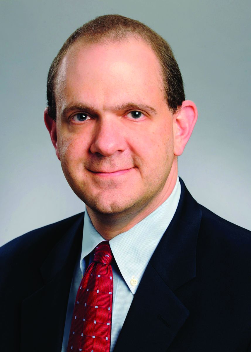
What has CHEST been up to during the first few months of my tenure? January saw us hold our first Board of Regents meeting for 2022, as well as the meeting of the CHEST Critical Care SEEK editorial board, where they worked to put together Volume 32, which will be out later this year. Watching some of the best and brightest medical minds from around the country discuss hot topics in critical care was a great experience (even if I didn’t have much to offer this august group), but the educational content was secondary to the interactions. Not only are these really smart folks teaching and learning from each other, but many of them are also clearly long-term colleagues, and watching this medical meeting was a lot like watching a reunion of friends who hadn’t seen each other in years. And, it struck me that what I’ve really been missing the most in the context of the social isolation that has accompanied the medical challenges of the pandemic is the pleasure of meeting in person with other folks to share stories, tell jokes, commiserate a bit, and catch up on the time that COVID-19 has stolen from us.
As we move further into 2022, I’m hoping that CHEST and our sister societies can help make up for this lost time by giving us the chance to meet in person once again. And to help build these experiences, we held an experiential design team along with our annual CHEST Program Committee meeting in February. Not only will the 2022 annual meeting in Nashville have the opportunity to hear from and network the best and brightest in pulmonary, critical care, and sleep medicine, but to celebrate our getting back together for the first time in years, we are also putting together some special surprises that CHEST has never done before. Keep an eye out for sneak peaks of these plans later in the spring and summer.
Another of our foci in 2022 is our ongoing push to help historically disenfranchised groups feel more engaged with CHEST. Many of you contributed to last year’s initiative to gather data on the kinds of things that we can do better, and I’ve just put together a presidential task force to develop final recommendations to further our goals of improving diversity, equity, and inclusion and to present to the Board of Regents for our April meeting.
Hopefully, many of you have seen some of the “Pardon the Introduction” series that CHEST has been featuring on its social media channels. We’ve put these together to showcase some of our leadership, their experiences, and opportunities for our members to get more involved with the College. Selfishly, I admit that they have also served as an excuse for me to catch up with some old friends and share our CHEST stories. We will be continuing to produce this series throughout the year; please let us know if there are specific folks you’d like us to feature!
Lastly, I wanted to thank the many of you who have reached out to me with questions, comments, and feedback. One of my main initiatives for the year is to make sure we are meeting the needs of as many of our members as possible, and this is something we can only do well if the lines of communication are wide open. Please continue to reach out to me, either by emailing me at [email protected] or messaging me on Twitter @ChestPrez.
New year, new CHEST President. Same as it has always been, except it has never been this way before. In past years, the transition of the CHEST Presidency occurred at our annual meeting, with a formal handover of leadership and a large reception. While there’s no Presidential football to hand over or secret codes to change for the incoming administration, there are usually several pending issues related to ongoing endeavors that need to be discussed between the outgoing and incoming leadership, in addition to some pearls of wisdom and the figurative “keys to the car.”
Now that CHEST has changed its President’s year to transition alongside the calendar year, there are few associated formalities. I awakened on New Year’s Day with my new title and the associated responsibility. Past President Steve Simpson, the mensch that he is, sent along with my colluding spouse a lovely and inspirational message for me to peruse, full of thoughtful advice and reflections on his year as President. I don’t know if this has ever been done before, but it is a tradition that I fully intend on continuing at the end of my term.
What has CHEST been up to during the first few months of my tenure? January saw us hold our first Board of Regents meeting for 2022, as well as the meeting of the CHEST Critical Care SEEK editorial board, where they worked to put together Volume 32, which will be out later this year. Watching some of the best and brightest medical minds from around the country discuss hot topics in critical care was a great experience (even if I didn’t have much to offer this august group), but the educational content was secondary to the interactions. Not only are these really smart folks teaching and learning from each other, but many of them are also clearly long-term colleagues, and watching this medical meeting was a lot like watching a reunion of friends who hadn’t seen each other in years. And, it struck me that what I’ve really been missing the most in the context of the social isolation that has accompanied the medical challenges of the pandemic is the pleasure of meeting in person with other folks to share stories, tell jokes, commiserate a bit, and catch up on the time that COVID-19 has stolen from us.
As we move further into 2022, I’m hoping that CHEST and our sister societies can help make up for this lost time by giving us the chance to meet in person once again. And to help build these experiences, we held an experiential design team along with our annual CHEST Program Committee meeting in February. Not only will the 2022 annual meeting in Nashville have the opportunity to hear from and network the best and brightest in pulmonary, critical care, and sleep medicine, but to celebrate our getting back together for the first time in years, we are also putting together some special surprises that CHEST has never done before. Keep an eye out for sneak peaks of these plans later in the spring and summer.
Another of our foci in 2022 is our ongoing push to help historically disenfranchised groups feel more engaged with CHEST. Many of you contributed to last year’s initiative to gather data on the kinds of things that we can do better, and I’ve just put together a presidential task force to develop final recommendations to further our goals of improving diversity, equity, and inclusion and to present to the Board of Regents for our April meeting.
Hopefully, many of you have seen some of the “Pardon the Introduction” series that CHEST has been featuring on its social media channels. We’ve put these together to showcase some of our leadership, their experiences, and opportunities for our members to get more involved with the College. Selfishly, I admit that they have also served as an excuse for me to catch up with some old friends and share our CHEST stories. We will be continuing to produce this series throughout the year; please let us know if there are specific folks you’d like us to feature!
Lastly, I wanted to thank the many of you who have reached out to me with questions, comments, and feedback. One of my main initiatives for the year is to make sure we are meeting the needs of as many of our members as possible, and this is something we can only do well if the lines of communication are wide open. Please continue to reach out to me, either by emailing me at [email protected] or messaging me on Twitter @ChestPrez.
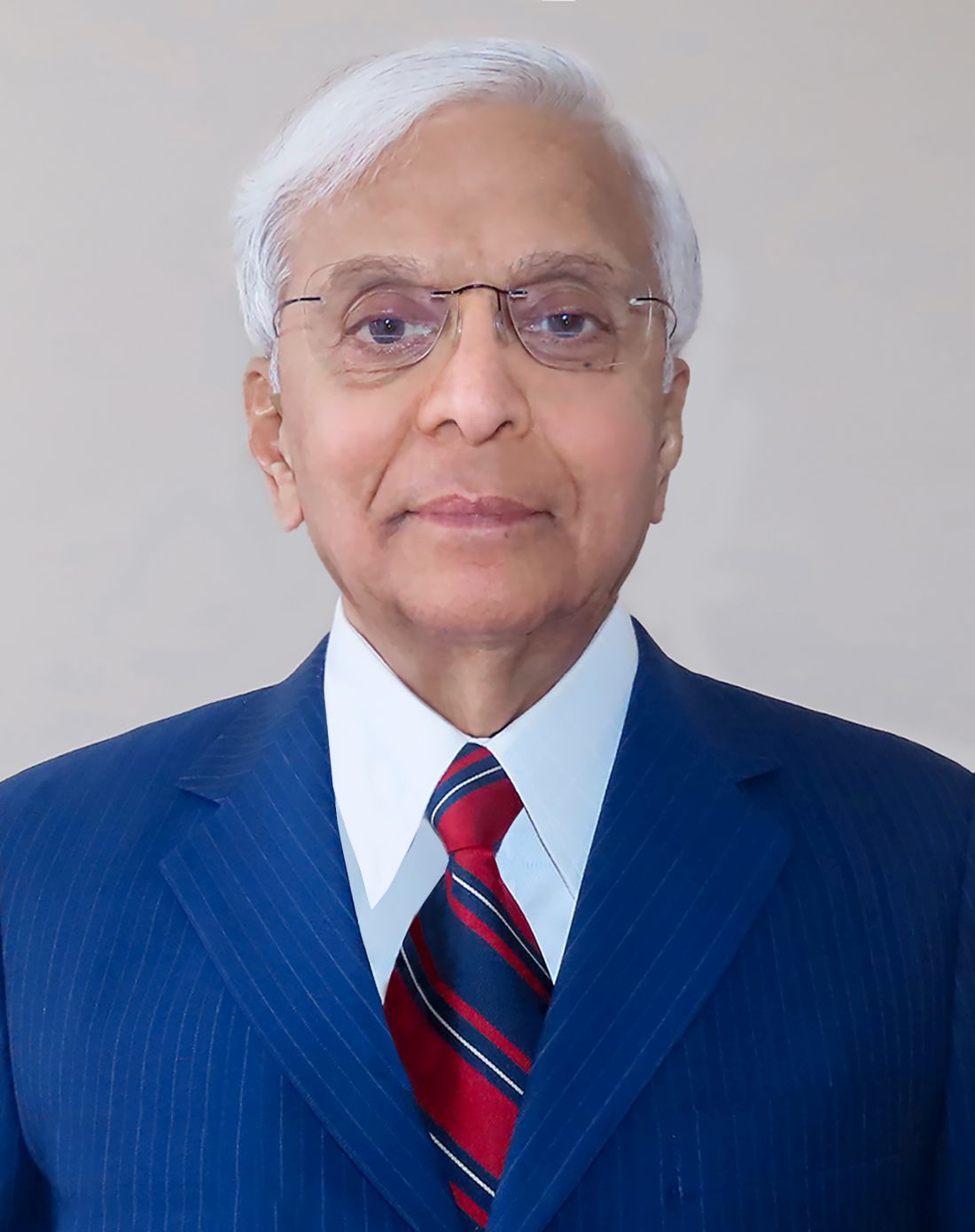
Meet our new CHEST President-Designate
We are happy to introduce John (Jack) D. Buckley, MD, MPH, FCCP, who will serve his term as CHEST President in 2024. A pulmonologist and critical care physician with an extensive background in education, he currently serves as the division leader of Pulmonary and Critical Care Medicine for the Henry Ford Medical Group and Health System.
Dr. Buckley received his undergraduate degree from Kalamazoo College and medical degree from Wayne State University. His residency in internal medicine and fellowships in pulmonary and critical care and health services research were completed at Indiana University. He additionally earned a Master of Public Health degree, also from Indiana University.

His impressive academic record includes authoring or co-authoring more than 40 publications and book chapters, as well as presenting over 100 lectures at international conferences. As a former fellowship program training director at two institutions, Dr. Buckley has long been committed to championing and furthering pulmonary and critical care medical education. In a collaboration between the Chinese Thoracic Society and CHEST, he served 6 years on a steering committee to help establish pulmonary and critical care medicine as a subspecialty in China. He and the group assisted with the development of fellowship training programs and presented at several Chinese Thoracic Society annual meetings and board review events.
For Dr. Buckley’s dedication to advancing PCCM education and faculty development, he received the CHEST Master Clinician Educator Award in 2016 and has been an annual Distinguished CHEST Educator (DCE) recipient since the award’s inception in 2017.
Dr. Buckley has been an active and engaged member of CHEST since 1997. He has served in leadership roles across many domains of the organization, including CHEST 2022 Congress Italy, Chair of the CHEST 2013 Scientific Program Committee, Board of Regents, Compensation Committee, Governance Committee, Training and Transitions Committee (Chair, 2011-2012) and Bylaws Committee (Chair, 2010-2012), Honor Lecture and Awards Committee, and the Affiliate NetWork. He currently serves on the CHEST SEEKTM Pulmonary Medicine Editorial Board and is the Chair of the Pulmonary Medicine Board Review.
We look forward to welcoming Dr. Buckley as CHEST President in 2024.
We are happy to introduce John (Jack) D. Buckley, MD, MPH, FCCP, who will serve his term as CHEST President in 2024. A pulmonologist and critical care physician with an extensive background in education, he currently serves as the division leader of Pulmonary and Critical Care Medicine for the Henry Ford Medical Group and Health System.
Dr. Buckley received his undergraduate degree from Kalamazoo College and medical degree from Wayne State University. His residency in internal medicine and fellowships in pulmonary and critical care and health services research were completed at Indiana University. He additionally earned a Master of Public Health degree, also from Indiana University.
His impressive academic record includes authoring or co-authoring more than 40 publications and book chapters, as well as presenting over 100 lectures at international conferences. As a former fellowship program training director at two institutions, Dr. Buckley has long been committed to championing and furthering pulmonary and critical care medical education. In a collaboration between the Chinese Thoracic Society and CHEST, he served 6 years on a steering committee to help establish pulmonary and critical care medicine as a subspecialty in China. He and the group assisted with the development of fellowship training programs and presented at several Chinese Thoracic Society annual meetings and board review events.
For Dr. Buckley’s dedication to advancing PCCM education and faculty development, he received the CHEST Master Clinician Educator Award in 2016 and has been an annual Distinguished CHEST Educator (DCE) recipient since the award’s inception in 2017.
Dr. Buckley has been an active and engaged member of CHEST since 1997. He has served in leadership roles across many domains of the organization, including CHEST 2022 Congress Italy, Chair of the CHEST 2013 Scientific Program Committee, Board of Regents, Compensation Committee, Governance Committee, Training and Transitions Committee (Chair, 2011-2012) and Bylaws Committee (Chair, 2010-2012), Honor Lecture and Awards Committee, and the Affiliate NetWork. He currently serves on the CHEST SEEKTM Pulmonary Medicine Editorial Board and is the Chair of the Pulmonary Medicine Board Review.
We look forward to welcoming Dr. Buckley as CHEST President in 2024.
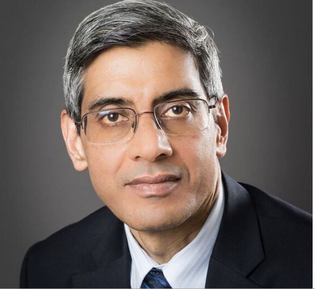
A mentor in medicine ... a mentor in life
Edward Carl Rosenow III, MD, Master FCCP
November 2, 1934 - December 21, 2021
We remember our close friend and colleague
On Monday, January 3, 1972, in Rochester, Minnesota, the weather was as expected - a high of 20 F and a low of -50 F. At 7:30 that morning, a group of three young physicians, residents in internal medicine on a month-long rotation in the inpatient pulmonary ward in one of the Mayo Clinic hospitals, was awaiting the arrival of the staff consultant to begin the rounds. It was the very first day of his first-year residency at Mayo for one of the residents. He had applied for residency training to begin in July but, instead, accepted Mayo’s offer to join the residency program 6 months earlier, in January – in Minnesota. That new resident was me. The pulmonary consultant had a friendly and disarming demeanor and gentle visage. He introduced himself to me with a smile, saying, “Welcome to Mayo. I am Ed Rosenow. Let me know if I can be of help in your training.”
Thus began my almost half-century’ relationship with Ed. During my residency in internal medicine and fellowship in pulmonary and critical care medicine, he was a constant and dependable fount of wisdom and knowledge. His daily lectures with chest x-rays after the morning rounds were legendary. By one estimate, he had collected over 4,500 chest x-rays to teach. These were hard copies and heavy to carry around. Ed lugged them under his arms daily for the lectures (there were no digital radiology or CT or MRI scanners then).
Ed was voted the “teacher of the year” every year for countless years. He was my first teacher in bronchoscopy and esophagoscopy. At that time, the division of pulmonary diseases was known as the division of thoracic diseases, and consultants in thoracic diseases performed bronchoscopy and rigid esophagoscopy.
Ed exemplified the best in compassion, amicability, collegiality, thoughtfulness, and a caring personality. In addition to possessing superb clinical acumen, he volunteered in local medical clinics for the less fortunate. In my mind, it is not too farfetched to describe Ed as “A man for all seasons.”*
Among Ed’s many professional accomplishments, his dedication and loyalty to the American College of Chest Physicians (CHEST) and the CHEST Foundation remain unsurpassed. In the years before and after he became the President of CHEST, Ed spent countless hours rewriting the ‘constitution’ of the organization, its bylaws. Many of the current committee structures, rules, and regulations are based on Ed’s work. During a meeting of the regents, one regent exclaimed, ”ACCP is Rosenow and Rosenow is ACCP!”
Ed was a founding member of the CHEST Foundation, the philanthropic arm of the College. Ed’s constant encouragement of young pulmonologists to participate in CHEST surely resulted in a significant increase in their membership. He was the ceaseless force behind my work and deep involvement with CHEST and the CHEST Foundation.
Describing my long association with Ed transcends this note. Suffice it to say that my being named the first Edward W. and Betty Knight Scripps Professor of Medicine in Honor of Edward C. Rosenow III, MD at Mayo Medical School is the greatest honor that I fondly cherish. I like to think that Ed strived for nearly a half-century to help me be a good person and a good doctor. I often question myself if I have met his goal. This question will linger in my mind for the rest of my life.
Udaya B. S. Prakash, MD, Master FCCP, Rochester, MN
Past President, American College of Chest Physicians (2002-2003)
*A man for all seasons: A man who is ready to cope with any contingency and whose behavior is always appropriate to every occasion. The English grammarian Robert Whittington (1480-1553) applied this description to the English statesman and scholar Sir Thomas More (1478-1535), and Robert Bolt used it as the title of his 1960 play about More.
My association with Dr. Rosenow dates back to 2002. I was attending the CHEST annual meeting, and I saw Dr. Rosenow walking toward me. He came up and said, “Hi. My name is Ed Rosenow. What is yours?” “Suhail Raoof,” I answered. Taking me aside, we spent almost a half-hour discussing my family, where I work, my career goals, and how the College could help me achieve some of those goals. His unassuming nature, humility, and sincere desire to help rang out loud and clear.
That day proved to be a turning point in my life. For the next almost 17 years, Ed and I set up monthly calls to connect. Each time, he was eager to listen and know what was happening in my life—the good, the bad, the important, and the mundane. My problems would become his problems; solutions to my problems would become joint solutions. He guided me on my path to leadership with CHEST, culminating in my presidency.
Ed taught thousands of his colleagues the true meaning and power of mentorship. Early in his career, he realized that true happiness comes from helping and guiding others. Dr. Rosenow experienced a sense of genuine happiness and pride in witnessing the accomplishments of his friends and trainees. His gentle ways, unparalleled kindness, and modesty made him the quintessential role model, who one and all tried to emulate.
Aptly stated by Dr. John Studdard, FCCP, one of his students at Mayo Clinic, “Ed Rosenow was the finest doctor I have ever known, but an even better person. He balanced a great intellect and curiosity with great humility, was an incredible teacher, educator, and mentor, and all of this with a special sense of humor.”
Among his many positive traits, Ed possessed two qualities that played very important roles in his life— inquisitiveness and perseverance. In the early 1960s, there was a paucity of information on drug-induced lung disease. He embraced this gap in medical knowledge, resolved to fill it, and, for the next almost 50 years, Dr. Rosenow extensively researched, published, and lectured on drug-related lung injury.
Ed felt strongly that a good pulmonologist had to be a skilled chest radiologist. He was instrumental in introducing “Chest Imaging for the Pulmonologist” sessions at the annual CHEST meetings. With his enduring cataloging of consultation cases and collection of teaching file chest x-rays, it is no wonder that he amassed one of the best teaching resources, including some of the most rare lung conditions.
Ed was a visionary. He envisioned CHEST to be a closely knit “family of professionals,” united in the desire to provide compassionate care of the highest order to patients and help each colleague accomplish their goals and aspirations. During his CHEST presidency, he and Dr. Bart Chernow realized the importance of having a philanthropic arm of the College that would support worthy projects through fundraising. Soon after the establishment of the CHEST Foundation, he became its President and then Chair.
Dr. Rosenow’s hard work, dedication, and unwavering commitment to professional and social organizations he served earned him the highest of accolades and honors. He had the rare distinction of being recognized as a Master by two professional organizations—CHEST and the American College of Physicians. Awards of the highest order were showered upon him by the Mayo Clinic, including establishing the Mayo Fellows Hall of Fame of Outstanding Teachers. Several endowed professorships and honors are named after him. The award he cherished most was the Karis Award (karis meaning “to care” in Greek). When I asked him how he felt to be the recipient of so many awards, he blushed and said, “Gee, there were plenty others who deserved them more than me. I was just doing my job.”
To everyone he met, Ed emphasized the importance of the “culture of caring and giving.” He taught his students that medicine is not a profession; it is a way of life. It is as much an art as it is a science. He reiterated his platinum rule to “Take care of every patient like you would want a member of your family cared for.”
Today, thousands of his students, including me, are deeply indebted to Dr. Rosenow for the impactful and profound role he played in our lives and for teaching us the core values that really matter. He left an indelible mark on our profession and our outlook. He redefined our responsibilities to our patients and colleagues. In his own quiet and effective way, Ed nurtured and inspired us to dream, think, persevere, and accomplish. His legacy will live on as we try to emulate his teachings, exceptional qualities, and humanistic approach. He will be missed greatly.
“His life was gentle, and the elements mixed so well in him that Nature might stand up and say to all the world,” ‘This was a man’.” Shakespeare
Suhail Raoof, MD, Master FCCP, New York, NY
Past President, American College of Chest Physicians (2011-2012)
Edward C. Rosenow III, MD, Master FCCP/Master Endowment - Master Teacher Endowment Lecture
The legacy and impact of Ed Rosenow, MD, Master FCCP, will never be forgotten. CHEST nominates and recognizes those physicians who embody that passion and commitment of Dr. Rosenow with an awarded lecture fully funded by the Ed Rosenow, MD, Master FCCP Endowment through the CHEST Foundation. Be a part of Dr. Rosenow’s legacy and make a donation in his memory to the Rosenow Endowment today at chestfoundation.org.
The legacy and impact of Ed Rosenow, MD, Master FCCP, will never be forgotten. CHEST nominates and recognizes those physicians who embody that passion and commitment of Dr. Rosenow with an awarded lecture fully funded by the Ed Rosenow, MD, Master FCCP Endowment through the CHEST Foundation. Be a part of Dr. Rosenow’s legacy and make a donation in his memory to the Rosenow Endowment today at chestfoundation.org.
The legacy and impact of Ed Rosenow, MD, Master FCCP, will never be forgotten. CHEST nominates and recognizes those physicians who embody that passion and commitment of Dr. Rosenow with an awarded lecture fully funded by the Ed Rosenow, MD, Master FCCP Endowment through the CHEST Foundation. Be a part of Dr. Rosenow’s legacy and make a donation in his memory to the Rosenow Endowment today at chestfoundation.org.
Partnership news from the CHEST Foundation: New grant concentrated on diversity, equity, and inclusion
American College of Chest Physicians, American Thoracic Society, and American Lung Association partner to support historically marginalized physician-scientists targeting lung disease
The American College of Chest Physicians (CHEST), the American Thoracic Society, and the American Lung Association are pleased to announce that they are partnering to sponsor a scholar in pulmonary and critical care medicine in the prestigious Harold Amos Medical Faculty Development Program (AMFDP), a Robert Wood Johnson Foundation initiative.
Developed to increase the pool of applicants from historically marginalized backgrounds pursuing careers in medicine, dentistry, or nursing, the AMFDP invites applicants to apply each year to help shape medicine into a more equitable, more accessible practice.
Together, CHEST, ATS, and American Lung Association will provide funding for awards of $420,000 over 4 years to support pulmonary/critical care medicine scholars.
“I am immensely proud to be leading an initiative that has continued to help shape the careers of so many physician-scientists in such a meaningful way,” said David Wilkes, MD, National Director of the Harold Amos Medical Faculty Development Program for the Robert Wood Johnson Foundation, and a member of ATS and CHEST. “That these three highly respected respiratory societies are joining efforts to help fulfill the AMFDP’s mission speaks volumes about their commitment as allies and influencers in the quest to eliminate lung health disparities. This is a model for other specialty societies to collaborate on addressing disparities.”
“In the context of an increasingly diverse population, it is more important than ever that our patients have confidence and trust in those who care for them, something that will be easier to develop as we diversify our workforce,” said CHEST President David Schulman, MD, MPH, FCCP. “CHEST is incredibly excited to be working with the American Lung Association, the American Thoracic Society, and the Harold Amos Medical Faculty Development Program to fund training for individuals who have been traditionally underrepresented in medicine as they pursue careers in pulmonary and critical care medicine.”
“Health equity is woven into the fabric of the ATS,” said ATS President Lynn Schnapp, MD, ATSF. “And, partnering with our peers in the pulmonary and critical care space is a wonderful opportunity to advance our shared goal of cultivating the next generation of leaders in health access and equity.”
“The American Lung Association has historically funded researchers at the beginning of their careers, helping to build the foundation for the next great group of leaders,” said Albert Rizzo, MD, Chief Medical Officer for the Lung Association. “It is critical to the advancement of lung health and the care of patients to have physicians and scientists from diverse backgrounds, so we are honored to provide support for these individuals and increase diversity in pulmonary medicine.”
The call for application is now open. To learn more and to apply go to rwjf.org.
American College of Chest Physicians, American Thoracic Society, and American Lung Association partner to support historically marginalized physician-scientists targeting lung disease
American College of Chest Physicians, American Thoracic Society, and American Lung Association partner to support historically marginalized physician-scientists targeting lung disease
The American College of Chest Physicians (CHEST), the American Thoracic Society, and the American Lung Association are pleased to announce that they are partnering to sponsor a scholar in pulmonary and critical care medicine in the prestigious Harold Amos Medical Faculty Development Program (AMFDP), a Robert Wood Johnson Foundation initiative.
Developed to increase the pool of applicants from historically marginalized backgrounds pursuing careers in medicine, dentistry, or nursing, the AMFDP invites applicants to apply each year to help shape medicine into a more equitable, more accessible practice.
Together, CHEST, ATS, and American Lung Association will provide funding for awards of $420,000 over 4 years to support pulmonary/critical care medicine scholars.
“I am immensely proud to be leading an initiative that has continued to help shape the careers of so many physician-scientists in such a meaningful way,” said David Wilkes, MD, National Director of the Harold Amos Medical Faculty Development Program for the Robert Wood Johnson Foundation, and a member of ATS and CHEST. “That these three highly respected respiratory societies are joining efforts to help fulfill the AMFDP’s mission speaks volumes about their commitment as allies and influencers in the quest to eliminate lung health disparities. This is a model for other specialty societies to collaborate on addressing disparities.”
“In the context of an increasingly diverse population, it is more important than ever that our patients have confidence and trust in those who care for them, something that will be easier to develop as we diversify our workforce,” said CHEST President David Schulman, MD, MPH, FCCP. “CHEST is incredibly excited to be working with the American Lung Association, the American Thoracic Society, and the Harold Amos Medical Faculty Development Program to fund training for individuals who have been traditionally underrepresented in medicine as they pursue careers in pulmonary and critical care medicine.”
“Health equity is woven into the fabric of the ATS,” said ATS President Lynn Schnapp, MD, ATSF. “And, partnering with our peers in the pulmonary and critical care space is a wonderful opportunity to advance our shared goal of cultivating the next generation of leaders in health access and equity.”
“The American Lung Association has historically funded researchers at the beginning of their careers, helping to build the foundation for the next great group of leaders,” said Albert Rizzo, MD, Chief Medical Officer for the Lung Association. “It is critical to the advancement of lung health and the care of patients to have physicians and scientists from diverse backgrounds, so we are honored to provide support for these individuals and increase diversity in pulmonary medicine.”
The call for application is now open. To learn more and to apply go to rwjf.org.
This month in the journal CHEST®
Editor’s picks
Bronchial thermoplasty in severe asthmatics at 5 years: The PAS2 study. By Dr. G. Chupp, et al.
Significant spirometric transitions and preserved ratio-impaired spirometry (PRISm) among ever-smokers. By Dr. E. Wan, et al.
Addressing advance care planning in patients with COPD. By Dr. E. Rose, et al.
The cost of ARDS: A systematic review. By Dr. P. Boucher, et al.
Acute management of high- and intermediate-risk pulmonary embolism in children: A review. By Dr. C. Ross, et al.
Surgical outcomes for early-stage non-small cell lung cancer at facilities with stereotactic body radiation therapy programs. By Dr. Y. Syed, et al.
Editor’s picks
Editor’s picks
Bronchial thermoplasty in severe asthmatics at 5 years: The PAS2 study. By Dr. G. Chupp, et al.
Significant spirometric transitions and preserved ratio-impaired spirometry (PRISm) among ever-smokers. By Dr. E. Wan, et al.
Addressing advance care planning in patients with COPD. By Dr. E. Rose, et al.
The cost of ARDS: A systematic review. By Dr. P. Boucher, et al.
Acute management of high- and intermediate-risk pulmonary embolism in children: A review. By Dr. C. Ross, et al.
Surgical outcomes for early-stage non-small cell lung cancer at facilities with stereotactic body radiation therapy programs. By Dr. Y. Syed, et al.
Antiseptic as good as antibiotics for preventing recurrent UTI
The antiseptic methenamine hippurate (MH) is known to sterilize urine and has been suggested to be of use in preventing urinary tract infections (UTIs), but firm evidence has so far been lacking. Now researchers led by clinicians and scientists from Newcastle-upon-Tyne, England, have provided the ALTAR trial (Alternative to Prophylactic Antibiotics for the Treatment of Recurrent UTIs in Women).
Daily low-dose antibiotics as recommended by current guidelines for prophylactic treatment of recurrent UTI have been linked to antibiotic resistance. Using MH as an alternative could play an important role in helping to tackle the global problem of increasing antibiotic resistance, the team said.
Study details
They recruited 240 women aged 18 or over with recurrent UTIs requiring prophylactic treatment from eight secondary care urology and urogynecology centers in the United Kingdom from June 2016 to June 2018. Women were randomized to receive MH or daily low-dose antibiotics for 12 months, with follow up for a further 6 months beyond that.
Before trial entry the women had experienced an average of more than six UTI episodes per year. During the 12-month treatment period, in the modified intention-to-treat population, there were 90 symptomatic, antibiotic-treated UTI episodes reported over 101 person-years of follow-up in the antibiotic group, and 141 episodes over 102 person-years in the MH group.
This yielded a UTI rate of 0.89 episodes per person-year in the antibiotic group, compared with 1.38 in the MH group, an absolute difference of 0.49 episodes per person-year. In the 6-month posttreatment follow-up period, the UTI incidence rate was 1.19 episodes per person-year in the antibiotic prophylaxis group versus 1.72 in the MH group, an absolute difference of 0.53.
Before the trial, a patient and public involvement group had predefined the noninferiority margin as one episode of UTI per person-year. The small difference between the two groups was less than this, confirming noninferiority of MH to antibiotic prophylaxis in this setting. This finding was consistent across the modified intention-to-treat, strict intention-to-treat, per protocol, and modified per protocol (post hoc) analyses.
Thus the ALTAR results showed that MH was no worse than antibiotics at preventing UTIs, and MH was also associated with reduced antibiotic consumption.
The vast majority of participants were over 90% adherent with the allocated treatment. Patient satisfaction was generally high and rates of adverse events and adverse reactions generally low, and both were comparable between treatment groups. Adverse reactions were reported by 34/142 (24%) in the antibiotic group and 35/127 (28%) in the MH group, and most reactions were mild. In the antibiotic group there were two serious adverse reactions (severe abdominal pain and raised alanine transaminase), whereas six participants in the MH group reported an episode of febrile UTI and four were admitted to hospital because of UTI.
Substantial global health care problem
At least 50% and up to 80% of all women have at least one acute UTI in their lifetime, most often uncomplicated acute cystitis. About a quarter of them go on to suffer recurrent infection, defined as three or more repeat infections in the past year, or two infections in the preceding 6 months. Frequent recurrences thus represent “a substantial global health care problem,” the authors say.
Guidelines from the United Kingdom, Europe, and the United States acknowledge the need for preventive strategies and strongly recommend the use of daily, low-dose antibiotics as standard prophylactic treatment. However, the United Kingdom’s antimicrobial resistance strategy recommends a “strong focus on infection prevention,” and aims to reduce antimicrobial use in humans by 15% before 2024.
“To achieve that, exploration of nonantibiotic preventive treatments in common conditions such as UTI is essential,” the team said.
MH is one such nonantibiotic treatment. It is bactericidal and works by denaturing bacterial proteins and nucleic acids. Although previous Cochrane systematic reviews had concluded that it could be effective for preventing UTI, further large trials were needed.
“This trial adds to the evidence base for the use of MH for prophylactic treatment in adult women with recurrent UTI. Although the MH group had a 55% higher rate of UTI episodes than the antibiotics group, the absolute difference was just 0.49 UTI episodes per year, which has limited clinical consequence,” the team concluded.
Results could ‘support a change in practice’
In older patients, particularly, the risks of long-term antibiotic prophylaxis might outweigh the benefits, and the authors said that their results “could support a change in practice in terms of preventive treatments for recurrent UTI and provide patients and clinicians with a credible alternative to daily antibiotics, giving them the confidence to pursue strategies that avoid long-term antibiotic use.”
They acknowledged limitations of the study, including that treatment allocation was not masked, crossover between arms was allowed, and differences in antibiotics prescribed may have affected the results. In addition, data regarding long-term safety of MH are scarce.
However, they said that the trial accurately represented the broad range of women with recurrent UTI, and that its results “might encourage patients and clinicians to consider MH as a first line treatment for UTI prevention in women.”
In a linked editorial, scientists from the Institute for Evidence-Based Healthcare at Bond University in Queensland, Australia, commented: “Although the results need cautious interpretation, they align with others, and this new research increases the confidence with which MH can be offered as an option to women needing prophylaxis against recurrent urinary tract infection.”
References
Harding C et al. Alternative to prophylactic antibiotics for the treatment of recurrent urinary tract infections in women: multicentre, open label, randomised, noninferiority trial. BMJ 2022 Mar 9;376:e068229.
Hoffmann TC et al. Methenamine hippurate for recurrent urinary tract infections. BMJ 2022 Mar 9;376:o533.
A version of this article first appeared on Medscape.co.uk.
Can a tool help overcome barriers to diabetes medication cost?
The resource, “Having Healthcare Cost Conversations to Improve Patient Outcomes: A Practical Guide,” was jointly developed by the Association of Diabetes Care & Education Specialists and Beyond Type 1, the nonprofit patient advocacy organization.
Indeed, the guide appeared as President Biden discussed his proposal to cap insulin costs at $35 per insulin vial during the State of the Union address, during which he introduced a young boy with type 1 diabetes in the guest box, as reported by this news organization. On March 3, Civica, a nonprofit coalition of health systems and philanthropies, announced it plans to manufacture generic insulin at a deeply discounted price, as reported by this news organization.
“Just to see diabetes front and center at the State of the Union followed by these announcements is certainly reflective of our own advocacy effort to make sure that people have affordable options for insulin, diabetes medications, services,” Kate Thomas, ADCES chief advocacy and external affairs officer, said in an interview. She added that ADCES has also pushed for legislation in Congress that would expand access to diabetes self-management training under the Medicare program.
The guide includes advice about overcoming barriers to discussing treatment costs with patients, suggested questions to ask patients about specific costs, and determinants of health and conversational approaches. Links are provided to resources for obtaining affordable insulin, other diabetes medications, and continuous glucose monitoring and insulin pump equipment.
“We know that, especially during primary care visits, there is limited time along with numbers of issues to talk about, so I think our challenge is how do we prioritize these conversations with something that can lead to action, not just saying you should do this but how do you actually do it,” Ms. Thomas said.
The introduction summarizes results from a 2021 Beyond Type 1 survey confirming prior findings reported by this news organization that cost is a frequent barrier for many individuals living with diabetes. “Especially right now where we are in terms of the impact of the pandemic and with peoples’ job statuses changing, I think it’s worthwhile to raise this in patient encounters,” Ms. Thomas said.
Overcoming conversational barriers
The first of three tables in the guide provides a list of “barriers to having a cost conversation” in the first column and “suggested solutions” in the second. For example, for the barrier, “You have insufficient time and/or knowledge about cost,” the suggestion is, “request and share available faculty and resources, including benefits coordinators, social workers, and community-based organizations. Work with the pharmacists and other members of the diabetes care team to identify resources that lower cost of medications.”
And for another barrier, “patients are often embarrassed or ashamed to initiate discussions of affordability,” the suggested solution is: “Normalize the issue of cost of care barriers for patients.”
A second table offers specific questions to ask patients about costs of medications and care, determinants of health, and financial barriers. These include: “What are some challenges you’ve had to accessing your medications or taking them as prescribed? What are some out-of-pocket health care costs you need help with? What challenges do you have accessing healthy food for you and your family?”
A link to a screening tool for social determinants of health is also included.
Language suggestions include talking about “cost of care” rather than “money,” asking patients if they’ve understood everything correctly by repeating back what they’ve said, and asking for confirmation and discussing follow-up.
Overall, the tool is designed to be a “broad conversation starter,” and not just about medications, Ms. Thomas said. “This is for all audiences and it’s meant to be something that the provider can tailor depending on who they’re speaking to. ... It’s about medications, but also the entire cost of care, including services and devices, transportation to appointments, access to food. ... Diabetes care isn’t just taking medication. It’s so many more factors.”
Ms. Thomas reported no relevant financial relationships.
A version of this article first appeared on Medscape.com.
Handheld ECGs ease AFib screening in the very elderly
The use of handheld, single-lead electrocardiograms (ECGs) did not increase diagnoses of AFib overall in patients aged 65 and older, but it did in patients 85 and up, researchers reported in Circulation.
“Incorporating single-lead ECGs into routine medical assessments as a new vital sign was widely feasible. Over 90% of people who were offered screening agreed to it and underwent screening,” said Steven Lubitz, MD, of the Cardiac Arrhythmia Service and Cardiovascular Research Center at Massachusetts General Hospital, Boston, who led the study.
Because advanced age is associated with a substantially increased risk of both AFib and stroke, point-of-care screening might be an efficient use of handheld ECGs, Dr. Lubitz said.
“The technology simply requires patients to place their fingers on the device to record an electrocardiogram and can be easily embedded in the routine clinical practice of primary care physicians,” he said in an interview.
The typical person has a 30% lifetime risk of developing AFib, and the chances of experiencing a stroke associated with the arrhythmia can be reduced significantly with anticoagulants, Dr. Lubitz said.
Professional organizations are split about the utility of screening for AFib. The European Society of Cardiology recommends opportunistic screening with either pulse palpation or ECG rhythm strip at clinic visits for patients 65 and older. The National Heart Foundation of Australia and the Cardiac Society of Australia and New Zealand have issued similar guidelines.
However, screening for AFib is not considered standard of care in the United States – although Dr. Lubitz predicted that that would change.
“I think the guidelines in the United States will evolve in the next few years, because I think we’re getting closer to understanding who we should be screening for atrial fibrillation and how we should be screening,” Dr. Lubitz told this news organization.
‘Very reassuring’ results
The randomized controlled trial found that for patients 85 and older, use of handheld ECGs led to a nearly 2% increase in new diagnoses of AFib in the screening group compared to conventional care.
The researchers also demonstrated an increased likelihood of diagnosing AFib during the patient’s primary-care encounter than at other sites, such as the emergency department or inpatient settings that might be more costly and resource-intensive. Moreover, the study reported that point-of-care screening was associated with high rates of oral anticoagulation prescriptions written for patients with newly diagnosed AFib, a finding Dr. Lubitz called “very reassuring.”
The Mass General researchers used single-lead devices attached to a tablet computer to screen more than 35,000 men and women from 16 primary care sites affiliated with the hospital’s practice-based research network.
Half the sites were randomly selected to include the screening intervention, where medical assistants used handheld ECGs at the start of the visit while checking routine vital signs.
The 1-year study screened 91% of eligible patients, demonstrating that single-lead rhythm assessment is feasible as part of routine primary care practice, Dr. Lubitz said. This finding supports other studies suggesting that handheld devices can enable rapid and scalable mass screening.
“We demonstrated that integration into routine practice by clinical personnel – in this case, medical assistants – is feasible. No study has measured and demonstrated such a high integration with routine care, reflecting both patient interest in screening and feasibility of incorporating screening into busy clinical practices,” Dr. Lubitz said.
Mobile ECGs with the handheld device take about 30 seconds to perform. In contrast, standard ECGs used in outpatient practices are bulky, and recording the ECG can take roughly 10 minutes.
Anthony Leazzo, DO, chairman of family practice at Northwestern Medicine Delnor Hospital, in Geneva, Ill., noted that smartwatches provide an alternative technology for detecting AFib.
But “a handheld, one-lead device would be more beneficial and should be more sensitive by measuring electrical activity similar to a normal ECG,” he said.
However, Dr. Leazzo said using such technology would need to be cost-effective because the patients at highest risk for AFib usually are on fixed incomes. Consumer versions of the devices can cost under $100. Dr. Lubitz said the actual cost for devices and a software platform used for a medical enterprise may differ.
Handheld ECGs are gradually being integrated into clinical practices, a trend driven by the rapid growth of telemedicine to remotely assess patients, Dr. Lubitz said.
“Our work affirmed that single-lead devices generate information for the physician that is actionable, though the proportion of newly detected AFib cases using a point-of-care ECG screening approach is likely to be very small,” Dr. Lubitz said in an interview. “For that reason, we think handheld devices are best deployed for people at the highest risk of AFib and stroke, and age is an excellent surrogate for that determination.”
The study was funded by Bristol-Myers Squibb–Pfizer Alliance.
A version of this article first appeared on Medscape.com.
FROM CIRCULATION
COVID-19 often more severe with congenital heart defects
Adults with a congenital heart defect (CHD) are at increased risk for serious illness and death when hospitalized with COVID-19, making vaccination and other preventive measures even important in this population, say researchers with the Centers for Disease Control and Prevention.
“We found that hospitalized patients with heart defects are up to twice as likely to have critical outcomes of COVID-19 illness (admission to the intensive care unit, use of a ventilator to help with breathing, or death) compared to hospitalized COVID-19 patients without heart defects,” Karrie Downing, MPH, epidemiologist, with the CDC’s National Center on Birth Defects and Developmental Disabilities, said in an interview.
“Additionally, we learned that people with hearts defects who were older or who also had other conditions like heart failure, pulmonary hypertension, Down syndrome, diabetes, or obesity were the most likely to have critical COVID-19 illness, but children and adults with heart defects without these other conditions were still at increased risk,” Ms. Downing said.
The message for health care providers is clear: “Encourage your patients with heart defects to get vaccinated and discuss with your patients the need for other preventive measures to avoid infection that may progress to severe COVID-19 illness,” Ms. Downing added.
The study was published online March 7, 2022, in Circulation.
The researchers analyzed data on 235,638 patients hospitalized with COVID-19 between March 2020 and January 2021, including 421 (0.2%) with CHD. Most CHD patients were older than 30 years (73%) and 61% were men, with 55% non-Hispanic white, 19% Hispanic and 16% non-Hispanic Black.
Overall, 68% of CHD patients had at least one comorbidity, as did 59% of patients without CHD.
Rates of ICU admission were higher in the CHD group (54% vs. 43%), as were rates of invasive mechanical ventilation (24% vs. 15%) and in-hospital death (11% vs. 7%).
After accounting for patient characteristics, ICU admission, invasive mechanical ventilation and death were more prevalent among COVID-19 patients with rather than without CHD, with adjusted prevalence ratios of 1.4, 1.8 and 2.0, respectively.
When stratified by high-risk characteristics, prevalence estimates for ICU admission, invasive mechanical ventilation and death remained higher among patients with COVID-19 and CHD across nearly all strata, including younger age groups and those without heart failure, pulmonary hypertension, Down syndrome, diabetes, or obesity, the researchers reported.
Ms. Downing said more work is needed to identify why the clinical course of COVID-19 disease results in admission to the ICU, the need for a ventilator, or death for some hospitalized patients with CHD and not for others.
“There could be a number of social, environmental, economic, medical, and genetic factors playing a role. But staying up to date with COVID-19 vaccines and following preventive measures for COVID-19 are effective ways to reduce the risk of severe illness from COVID-19,” Ms. Downing said.
The study had no specific funding. The authors reported no relevant disclosures.
A version of this article first appeared on Medscape.com.
FROM CIRCULATION