User login
Bimekizumab Outperforms Ustekinumab for PsA in a Matching-Adjusted Indirect Comparison
Key clinical point: A dose of 160 mg bimekizumab every 4 weeks demonstrated greater long-term efficacy than 45 or 90 mg ustekinumab every 12 weeks in patients with psoriatic arthritis (PsA) who were biologic-naïve or showed inadequate response to tumor necrosis factor inhibitors (TNFi-IR).
Major finding: At week 52, both biologic-naive (adjusted odds ratio [aOR] 3.33; P < .001) and TNFi-IR (aOR 9.85; P < .001) patients receiving bimekizumab vs 45 mg ustekinumab were more likely to achieve ≥70% improvement in the American College of Rheumatology response, with similar effect observed for bimekizumab vs 90 mg ustekinumab.
Study details: This was matching-adjusted indirect comparison of data from several phase 3 trials of bimekizumab (BE OPTIMAL, BE COMPLETE, and BE VITAL) and ustekinumab (PSUMMIT1 and PSUMMIT2). The trials involved patients with PsA who received bimekizumab (n = 698) or ustekinumab (45 mg: n = 265; 90 mg: n = 262).
Disclosures: This study was sponsored by UCB Pharma and supported by the NIHR Manchester Biomedical Research Centre, UK. Three authors declared being employees and shareholders of UCB Pharma. Several authors declared having ties with various sources, including UCB Pharma.
Source: Mease PJ, Warren RB, Nash P, et al. Comparative effectiveness of bimekizumab and ustekinumab in patients with psoriatic arthritis at 52 weeks assessed using a matching-adjusted indirect comparison. Rheumatol Ther. 2024 (Aug 9). Doi: 10.1007/s40744-024-00705-x Source
Sparing Effect of First-Line Targeted Therapy in PsA
Key clinical point: First-line targeted therapy, particularly use of tumor necrosis factor inhibitors (TNFi), reduced the use of symptomatic treatments, methotrexate, mood disorder treatments, hospitalizations, and sick leave in patients with psoriatic arthritis (PsA).
Major finding: First-line targeted therapy significantly reduced the use of non-steroidal anti-inflammatory drugs (NSAID; −15%), prednisone (−9%), methotrexate (−15%), mood disorder treatments (−2%), and rate of hospitalizations (−12%) and sick leave (−4%; all P < 10-4). TNFi showed greater reductions in NSAID (adjusted odds ratio [aOR] 1.04; 95% CI 1.01-1.07) and prednisone use (aOR 1.04; 95% CI 1.02-1.06) compared with interleukin 17 inhibitors (IL17i), with similar outcomes for IL12/23i.
Study details: This cohort study included 9793 patients with PsA age ≥18 years who had initiated targeted therapies for at least 9 months.
Disclosures: The authors did not declare any specific funding. Two authors declared receiving a subsidy to attend a congress or receiving consulting fees and serving as investigators for various sources.
Source: Pina Vegas L, Iggui S, Sbidian E, Claudepierre P. Impact of initiation of targeted therapy on the use of psoriatic arthritis-related treatments and healthcare consumption: A cohort study of 9793 patients from the French health insurance database (SNDS). RMD Open. 2024;10:e004631 (Aug 7). Doi: 10.1136/rmdopen-2024-004631 Source
Achieving Disease Control Linked to Better Quality of Life in PsA
Key clinical point: Patients with psoriatic arthritis (PsA) who achieved disease control despite having an inadequate response to conventional synthetic or biological disease-modifying antirheumatic drugs (cs/bDMARD) showed improved patient-reported outcomes (PRO).
Major finding: At week 104, patients who did vs did not achieve minimal disease activity had significant improvements in the Health Assessment Questionnaire–Disability Index (least squares mean change from baseline [Δ] −0.82 vs −0.17; P ≤ .0001), pain (Δ −4.75 vs −1.77; P ≤ .0001), and other investigated PRO.
Study details: This post hoc analysis of two phase 3 trials, SELECT-PsA 1 and SELECT-PsA 2, included 1069 and 317 patients with PsA and inadequate response to ≥1 csDMARD or bDMARD, respectively, who were randomly assigned to receive upadacitinib, placebo with crossover to upadacitinib, or adalimumab.
Disclosures: This study was funded by AbbVie, and AbbVie participated in the design of the trial and the publication of its results. Seven authors declared being employees of AbbVie and may own its stock or stock options. Several authors declared having ties with AbbVie and other sources.
Source: Kavanaugh A, Mease P, Gossec L, et al. Association between achievement of clinical disease control and improvement in patient-reported outcomes and quality of life in patients with psoriatic arthritis in the phase 3 SELECT-PsA 1 and 2 randomized controlled trials. ACR Open Rheumatol. 2024 (Aug 1). Doi: 10.1002/acr2.11714 Source
IL-23 and IL-12/23 Inhibitors Show Comparable Safety in Preventing PsA in Psoriasis
Key clinical point: Patients who received interleukin-23 inhibitors (IL-23i) and interleukin-12/23 inhibitors (IL-12/23i) for the management of psoriasis had a comparable risk for incident psoriatic arthritis (PsA).
Major finding: Patients treated with IL-23i vs IL-12/23i demonstrated no significant difference in the risk for PsA (hazard ratio 0.96; P = .812) and cumulative incidence of PsA (P = .812).
Study details: This retrospective cohort study included the propensity score–matched data of patients with psoriasis age 18 years or older from the TriNetX database who were treated with either IL-23i (n = 2142) or IL-12/23i (n = 2142).
Disclosures: This study did not receive any specific funding. The authors declared no conflicts of interest.
Source: Tsai SHL, Yang C-Y, Huo A-P, Wei JC-C. Interleukin 23 versus interleukin 12/23 inhibitors on preventing incidental psoriatic arthritis in patients with psoriasis? A real-world comparison from the TriNetX Global Collaborative Network. J Am Acad Dermatol. 2024 (Jul 27). Doi: 0.1016/j.jaad.2024.07.1473 Source
Bimekizumab Shows Promising Outcomes in PsA, With or Without Methotrexate
Key clinical point: Bimekizumab demonstrated sustained efficacy and was well tolerated for 52 weeks, regardless of concomitant methotrexate use, in patients with psoriatic arthritis (PsA) who were biologic-naive or intolerant to tumor necrosis factor inhibitors (TNFi-IR).
Major finding: Through week 52, nearly half of the patients receiving bimekizumab with or without methotrexate achieved a ≥50% improvement in American College of Rheumatology response (biologic-naive ~55%; TNFi-IR ~48-56%) and minimal disease activity (biologic-naive ~55%; TNFi-IR ~47%). The rates of experiencing at least one treatment emergent adverse event were similar across the subgroups.
Study details: This post hoc analysis of phase 3 trials (BE OPTIMAL, BE COMPLETE, and BE VITAL) included biologic-naive (n = 852) or TNFi-IR (n = 400) patients with PsA who received bimekizumab, placebo with crossover to bimekizumab at week 16, or adalimumab, with or without methotrexate.
Disclosures: This study was funded by UCB Pharma and supported by the NIHR Manchester Biomedical Research Centre, UK. Two authors declared being employees of or holding stocks in UCB. Several authors declared having other ties with UCB and other sources.
Source: McInnes IB, Mease PJ, Tanaka Y, et al. Efficacy and safety of bimekizumab in patients with psoriatic arthritis with or without methotrexate: 52-week results from two phase 3 studies. ACR Open Rheumatol. 2024 (Jul 30). Doi: 10.1002/acr2.11727 Source
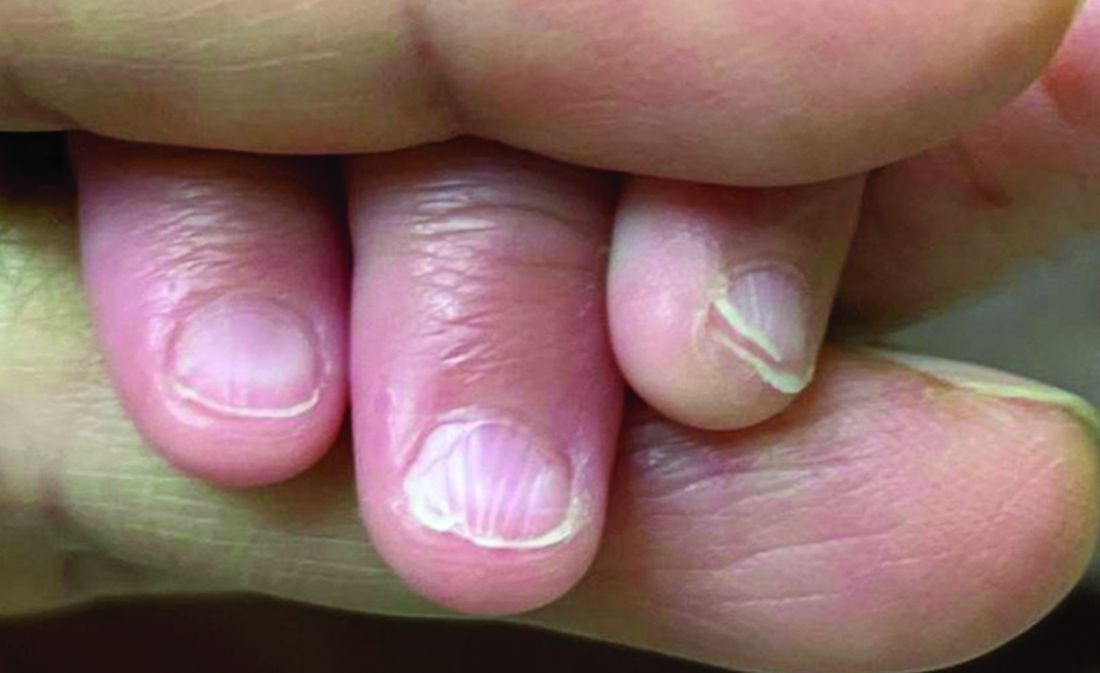
A 7-Month-Old Female Presented With Nail Changes
Given the clinical presentation and the absence of other systemic or dermatological findings, the diagnosis of chevron nails was made.
Discussion
The condition is characterized by transverse ridges on the nails that converge towards the center, forming a V or chevron shape. This condition was first described by Perry et al. and later by Shuster et al., who explained that the condition might result from axial growth of the nail with synchronous growth occurring from a chevron-shaped growing edge of the nail root. Alternatively, Shuster suggested that sequential growth, with localized variation in the nail production rate, could propagate a wave from the center of the nail to the edge.
The etiology of chevron nails is not well understood, but it is believed to result from temporary disruptions in the nail matrix, possibly related to minor illness or physiological stress during infancy.
In the case of our 7-month-old patient, the history of mild upper respiratory infections might have contributed to the development of chevron nails. However, the lack of other significant illness, skin involvement, or systemic findings supports the benign and self-limiting nature of this condition. Parents were reassured that chevron nails typically resolve on their own as the child grows and that no specific treatment is necessary.
Differential Diagnosis
The differential diagnosis of transverse nail changes in children includes other conditions such as trachyonychia, lichen planus, Darier disease, and pachyonychia congenita.
Trachyonychia, also known as “sandpaper nails,” trachyonychia is characterized by the roughening of the nail surface, giving it a dull and ridged appearance. The condition may affect all 20 nails and is often associated with underlying dermatological conditions such as lichen planus or alopecia areata. Unlike chevron nails, trachyonychia presents with more diffuse nail changes and does not typically feature the distinct V-shaped ridging seen in this patient.
Lichen planus is an inflammatory condition that can affect the skin, mucous membranes, and nails. Nail involvement in lichen planus can lead to longitudinal ridging, thinning, and sometimes even complete nail loss. The absence of other characteristic features of lichen planus, such as violaceous papules on the skin or white lacy patterns on mucous membranes (Wickham striae), makes this diagnosis less likely in our patient.
Darier disease, also known as keratosis follicularis, is a genetic disorder characterized by greasy, warty papules primarily on seborrheic areas of the skin, nail abnormalities, and sometimes mucosal involvement. Nail changes in Darier disease include longitudinal red and white streaks, V-shaped notching at the free edge of the nails, and subungual hyperkeratosis. These nail changes are more severe and distinct than the simple transverse ridging seen in chevron nails. The absence of other clinical signs of Darier disease, such as skin papules or characteristic nail notching, makes this diagnosis unlikely in our patient.
Pachyonychia congenita is a rare genetic disorder characterized by thickened nails (pachyonychia), painful plantar keratoderma, and sometimes oral leukokeratosis. The condition typically presents with significant nail thickening and other systemic findings, which were absent in our patient. The distinct pattern of V-shaped ridging observed in chevron nails does not align with the typical presentation of pachyonychia congenita.
Next Steps
No specific treatment is required for chevron nails. The condition is typically self-resolving, and the nails usually return to a normal appearance as the child continues to grow. Parents were advised to monitor the nails for any changes or new symptoms and were reassured about the benign nature of the findings. Follow-up was scheduled to ensure the resolution of the condition as the child develops.

Conclusion
Chevron nails are an important consideration in the differential diagnosis of transverse nail ridging in infants and young children. While the condition is benign and self-limiting, it is crucial to differentiate it from other nail dystrophies, such as trachyonychia, lichen planus, Darier disease, and pachyonychia congenita, which may require further investigation or intervention. Awareness of chevron nails can help prevent unnecessary worry and provide reassurance to parents and caregivers.
Dr. Matiz is a pediatric dermatologist at Southern California Permanente Medical Group, San Diego.
Suggested Reading
Delano S, Belazarian L. Chevron nails: A normal variant in the pediatric population. Pediatr Dermatol. 2014 Jan-Feb;31(1):e24-5. doi: 10.1111/pde.12193.
John JM et al. Chevron nail — An under-recognised normal variant of nail development. Arch Dis Child. 2024 Jul 18;109(8):648. doi: 10.1136/archdischild-2024-326975.
Shuster S. The significance of chevron nails. Br J Dermatol. 1996;135:151–152. doi: 10.1046/j.1365-2133.1996.d01-961.x.
Starace M et al. Nail disorders in children. Skin Appendage Disord. 2018 Oct;4(4):217-229. doi: 10.1159/000486020.
Given the clinical presentation and the absence of other systemic or dermatological findings, the diagnosis of chevron nails was made.
Discussion
The condition is characterized by transverse ridges on the nails that converge towards the center, forming a V or chevron shape. This condition was first described by Perry et al. and later by Shuster et al., who explained that the condition might result from axial growth of the nail with synchronous growth occurring from a chevron-shaped growing edge of the nail root. Alternatively, Shuster suggested that sequential growth, with localized variation in the nail production rate, could propagate a wave from the center of the nail to the edge.
The etiology of chevron nails is not well understood, but it is believed to result from temporary disruptions in the nail matrix, possibly related to minor illness or physiological stress during infancy.
In the case of our 7-month-old patient, the history of mild upper respiratory infections might have contributed to the development of chevron nails. However, the lack of other significant illness, skin involvement, or systemic findings supports the benign and self-limiting nature of this condition. Parents were reassured that chevron nails typically resolve on their own as the child grows and that no specific treatment is necessary.
Differential Diagnosis
The differential diagnosis of transverse nail changes in children includes other conditions such as trachyonychia, lichen planus, Darier disease, and pachyonychia congenita.
Trachyonychia, also known as “sandpaper nails,” trachyonychia is characterized by the roughening of the nail surface, giving it a dull and ridged appearance. The condition may affect all 20 nails and is often associated with underlying dermatological conditions such as lichen planus or alopecia areata. Unlike chevron nails, trachyonychia presents with more diffuse nail changes and does not typically feature the distinct V-shaped ridging seen in this patient.
Lichen planus is an inflammatory condition that can affect the skin, mucous membranes, and nails. Nail involvement in lichen planus can lead to longitudinal ridging, thinning, and sometimes even complete nail loss. The absence of other characteristic features of lichen planus, such as violaceous papules on the skin or white lacy patterns on mucous membranes (Wickham striae), makes this diagnosis less likely in our patient.
Darier disease, also known as keratosis follicularis, is a genetic disorder characterized by greasy, warty papules primarily on seborrheic areas of the skin, nail abnormalities, and sometimes mucosal involvement. Nail changes in Darier disease include longitudinal red and white streaks, V-shaped notching at the free edge of the nails, and subungual hyperkeratosis. These nail changes are more severe and distinct than the simple transverse ridging seen in chevron nails. The absence of other clinical signs of Darier disease, such as skin papules or characteristic nail notching, makes this diagnosis unlikely in our patient.
Pachyonychia congenita is a rare genetic disorder characterized by thickened nails (pachyonychia), painful plantar keratoderma, and sometimes oral leukokeratosis. The condition typically presents with significant nail thickening and other systemic findings, which were absent in our patient. The distinct pattern of V-shaped ridging observed in chevron nails does not align with the typical presentation of pachyonychia congenita.
Next Steps
No specific treatment is required for chevron nails. The condition is typically self-resolving, and the nails usually return to a normal appearance as the child continues to grow. Parents were advised to monitor the nails for any changes or new symptoms and were reassured about the benign nature of the findings. Follow-up was scheduled to ensure the resolution of the condition as the child develops.
Conclusion
Chevron nails are an important consideration in the differential diagnosis of transverse nail ridging in infants and young children. While the condition is benign and self-limiting, it is crucial to differentiate it from other nail dystrophies, such as trachyonychia, lichen planus, Darier disease, and pachyonychia congenita, which may require further investigation or intervention. Awareness of chevron nails can help prevent unnecessary worry and provide reassurance to parents and caregivers.
Dr. Matiz is a pediatric dermatologist at Southern California Permanente Medical Group, San Diego.
Suggested Reading
Delano S, Belazarian L. Chevron nails: A normal variant in the pediatric population. Pediatr Dermatol. 2014 Jan-Feb;31(1):e24-5. doi: 10.1111/pde.12193.
John JM et al. Chevron nail — An under-recognised normal variant of nail development. Arch Dis Child. 2024 Jul 18;109(8):648. doi: 10.1136/archdischild-2024-326975.
Shuster S. The significance of chevron nails. Br J Dermatol. 1996;135:151–152. doi: 10.1046/j.1365-2133.1996.d01-961.x.
Starace M et al. Nail disorders in children. Skin Appendage Disord. 2018 Oct;4(4):217-229. doi: 10.1159/000486020.
There was no family history of similar nail findings and no relatives had a history of chronic skin conditions or congenital nail disorders.
On physical examination, several of the child’s fingernails exhibited distinct longitudinal ridges, with a characteristic pattern where the ridges converged at the center of the nail, forming a V-shape. There were no other concerning dermatologic findings, such as rashes, plaques, or erosions, and the skin and hair appeared otherwise normal. The rest of the physical exam was unremarkable.
After Rapid Weight Loss, Monitor Antiobesity Drug Dosing
A patient who developed atrial fibrillation resulting from the failure to adjust the levothyroxine dose after rapid, significant weight loss while on the antiobesity drug tirzepatide (Zepbound) serves as a key reminder in managing patients experiencing rapid weight loss, either from antiobesity medications or any other means: Patients taking medications with weight-based dosing need to have their doses closely monitored.
“Failing to monitor and adjust dosing of these [and other] medications during a period of rapid weight loss may lead to supratherapeutic — even toxic — levels, as was seen in this [case],” underscore the authors of an editorial regarding the Teachable Moment case, published in JAMA Internal Medicine.
Toxicities from excessive doses can have a range of detrimental effects. In terms of thyroid medicine, the failure to adjust levothyroxine treatment for hypothyroidism in cases of rapid weight loss can lead to thyrotoxicosis, and in older patients in particular, a resulting thyrotropin level < 0.1 mIU/L is associated with as much as a threefold increased risk for atrial fibrillation, as observed in the report.
Case Demonstrates Risks
The case involved a 62-year-old man with obesity, hypothyroidism, and type 1 diabetes who presented to the emergency department with palpitations, excessive sweating, confusion, fever, and hand tremors. Upon being diagnosed with atrial fibrillation, the patient was immediately treated.
His medical history revealed the underlying culprit: Six months earlier, the patient had started treatment with the gastric inhibitory polypeptide (GIP)/glucagon-like peptide (GLP) 1 dual agonist tirzepatide. As is typical with the drug, the patient’s weight quickly plummeted, dropping from a starting body mass index of 44.4 down to 31.2 after 6 months and a decrease in body weight from 132 kg to 93 kg (a loss of 39 kg [approximately 86 lb]).
When he was prescribed tirzepatide, 2.5 mg weekly, for obesity, the patient had been recommended to increase the dose every 4 weeks as tolerated and, importantly, to have a follow-up visit in a month. But because he lived in different states seasonally, the follow-up never occurred.
Upon his emergency department visit, the patient’s thyrotropin level had dropped from 1.9 mIU/L at the first visit 6 months earlier to 0.001 mIU/L (well within the atrial fibrillation risk range), and his free thyroxine level (fT4) was 7.26 ng/ dL — substantially outside of the normal range of about 0.9-1.7 ng/dL for adults.
“The patient had 4-times higher fT4 levels of the upper limit,” first author Kagan E. Karakus, MD, of the Barbara Davis Center for Diabetes, University of Colorado Anschutz Medical Campus, Aurora, told this news organization. “That is why he had experienced the adverse event of atrial fibrillation.”
Thyrotoxicosis Symptoms Can Be ‘Insidious,’ Levothyroxine Should Be Monitored
Although tirzepatide has not been approved by the US Food and Drug Administration for the treatment of type 1 diabetes, obesity is on the rise among patients with this disorder and recent research has shown a more than 10% reduction in body weight in 6 months and significant reductions in A1c with various doses.
Of note, in the current case, although the patient’s levothyroxine dose was not adjusted, his insulin dose was gradually self-decreased during his tirzepatide treatment to prevent hypoglycemia.
“If insulin treatment is excessive in diabetes, it causes hypoglycemia, [and] people with type 1 diabetes will recognize the signs of hypoglycemia related to excessive insulin earlier,” Dr. Karakus said.
If symptoms appear, patients can reduce their insulin doses on their own; however, the symptoms of thyrotoxicosis caused by excessive levothyroxine can be more insidious compared with hypoglycemia, he explained.
“Although patients can change their insulin doses, they cannot change the levothyroxine doses since it requires a blood test [thyroid-stimulating hormone; TSH] and a new prescription of the new dose.”
The key lesson is that “following levothyroxine treatment initiation or dose adjustment, 4-6 weeks is the optimal duration to recheck [the] thyrotropin level and adjust the dose as needed,” Dr. Karakus said.
Key Medications to Monitor
Other common outpatient medications that should be closely monitored in patients experiencing rapid weight loss, by any method, range from anticoagulants, anticonvulsants, and antituberculosis drugs to antibiotics and antifungals, the authors note.
Of note, medications with a narrow therapeutic index include phenytoin, warfarin, lithium carbonate, digoxin theophylline, tacrolimus, valproic acid, carbamazepine, and cyclosporine.
The failure to make necessary dose adjustments “is seen more often since the newer antiobesity drugs reduce a great amount of weight within months, almost as rapidly as bariatric surgery,” Dr. Karakus said.
“It is very important for physicians to be aware of the weight-based medications and narrow therapeutic index medications since their doses should be adjusted carefully, especially during weight loss,” he added.
Furthermore, “the patient should also know that weight reduction medication may cause adverse effects like nausea, vomiting and also may affect metabolism of other medications such that some medication doses should be adjusted regularly.”
In the editorial published with the study, Tyrone A. Johnson, MD, of the Department of Medicine, University of California, San Francisco, and colleagues note that the need for close monitoring is particularly important with older patients, who, in addition to having a higher likelihood of comorbidities, commonly have polypharmacy that could increase the potential for adverse effects.
Another key area concern is the emergence of direct-to-consumer avenues for GLP-1/GIP agonists for the many who either cannot afford or do not have access to the drugs, providing further opportunities for treatment without appropriate clinical oversight, they add.
Overall, the case “highlights the potential dangers underlying under-supervised prescribing of GLP-1/GIP receptor agonists and affirms the need for strong partnerships between patients and their clinicians during their use,” they wrote.
“These medications are best used in collaboration with continuity care teams, in context of a patient’s entire health, and in comprehensive risk-benefit assessment throughout the entire duration of treatment.”
A Caveat: Subclinical Levothyroxine Dosing
Commenting on the study, Matthew Ettleson, MD, a clinical instructor of medicine in the Section of Endocrinology, Diabetes, & Metabolism, University of Chicago, noted the important caveat that patients with hypothyroidism are commonly on subclinical doses, with varying dose adjustment needs.
“The patient in the case was clearly on a replacement level dose. However, many patients are on low doses of levothyroxine (75 µg or lower) for subclinical hypothyroidism, and, in general, I think the risks are lower with patients with subclinical hypothyroidism on lower doses of levothyroxine,” he told this news organization.
Because of that, “frequent TSH monitoring may be excessive in this population,” he said. “I would hesitate to empirically lower the dose with weight loss, unless it was clear that the patient was unlikely to follow up.
“Checking TSH at a more frequent interval and adjusting the dose accordingly should be adequate to prevent situations like this case.”
Dr. Karakus, Dr. Ettleson, and the editorial authors had no relevant disclosures to report.
A version of this article appeared on Medscape.com.
Could Dry Fasting Aid in Metabolic Disorders, Diabetes?
Dry fasting, the practice of going without food and water, has enthusiastic advocates on TikTok, X, YouTube, and other social media platforms. Devotees claim a wide range of health effects, but medical professionals advise caution to ensure that the practice does more good than harm, especially for individuals with diabetes.
Purported benefits and risks vary, depending on who is following the regimen and how long they abstain from food and water. Advocates on social media assert that dry fasting makes “intuition skyrocket” and puts autophagy on “overdrive.” Although such statements may rev up followers, there is little evidence to support these and many other dry-fasting claims. In fact, several physicians warned about unintended consequences.
“I had one patient who followed this fasting method often, and over time she developed kidney stones that led to a severe infection,” said Deena Adimoolam, MD, an endocrinologist in private practice in New York City and New Jersey. “Lack of both water and food can fuel hunger and increase the likelihood of overeating or binge eating once the fast is completed, which does not lead to weight loss. Untreated dehydration can lead to loss of consciousness.”
“For individuals with type 2 diabetes, dehydration can exacerbate hyperglycemia and increase the risk of complications such as diabetic ketoacidosis (DKA),” said Abeer Bader, lead clinical nutrition specialist at the Massachusetts General Hospital Weight Center in Boston. “Research also consistently shows that adequate hydration is crucial for maintaining physical and cognitive performance.”
, Ms. Bader noted. “Prolonged dry fasting can result in nutrient deficiencies. For individuals with diabetes, maintaining adequate nutrition is crucial to manage blood sugar levels and overall health. The lack of both food and water can exacerbate deficiencies.”
Joanne Bruno, MD, an endocrinologist at NYU Langone Health, added, “Certain medications used for the management of type 2 diabetes, such as SGLT2 inhibitors, can cause dehydration. It is critical that patients stay well hydrated while on these medications to avoid serious side effects such as euglycemic DKA.”
What Exactly Is Dry Fasting?
Defining dry fasting, like any kind of fasting, has remained a challenge, according to authors of the first international consensus on fasting terminology, published on July 25 in Cell Metabolism. The clinical terminology “has remained heterogeneous and often confusing, with similar terms being used to define different fasting regimens ... reflecting the manifold contexts in which fasting is practiced.”
Indeed, dry fasting was among the most discussed terms by the consensus panel and went through several rounds before the panelists came to agreement. A few experts were critical of the practice, whereas those familiar with religious fasting traditions, such as during Ramadan, were clear about the importance of including this term in the consensus process.
“The dissent was resolved by the clarification that this form of fasting has historical and geographical extensions and that the present consensus process did not aim at evaluating therapeutic effectiveness or safety for any term defined,” the authors wrote.
The panel concluded that dry fasting is not the same as total or complete fasting because the latter can include water (such as water-only fasting). Their final definition of dry fasting is ‘’a fasting regimen during which a voluntary abstinence from all foods and beverages, including water, is practiced for a certain period of time.’’
Different types of fasting regimens, such as intermittent fasting, may include dry fasting, in which case it is referred to as “intermittent dry fasting.” This is defined in the consensus as intermittent fasting regimens that involve abstaining from food and fluid intake during the fasting interval, which typically lasts 9-20 hours.
Most dry fasts, including religious ones, are maintained for a specific interval and are followed by a refeeding period. These fasts are not starvation, defined as no food or water intake for days.
What the Evidence Says
All that said, dry fasting by any other name remains dry fasting. “Abundant” evidence from animal studies suggests the potential of various types of fasting for disease prevention and treatment in humans, noted the authors of the consensus report, Along with the risks described above, small studies have explored short-term effects in people, all of which have yet to be established by larger and longer-term studies.
In a recent small study, researchers at Baylor College of Medicine, Houston, Texas, reported that dawn-to-dusk dry fasting for 30 days reduced levels of inflammatory cytokines in the 13 participants with a high body mass index. Earlier work by the group showed that dawn-to-dusk dry fasting for 30 days induced “anti-atherosclerotic, anti-inflammatory, and anti-tumorigenic proteome” in peripheral blood mononuclear cells of 14 individuals with metabolic syndrome (The researchers declined to comment for this article.)
Importantly, the health effects can vary among individuals for unknown reasons, found a recent cross-sectional study of fasting blood glucose (FBG) changes in 181 patients with type 2 diabetes during Ramadan intermittent fasting (RIF), which involves dry fasting during daylight hours for 1 month. The researchers classified participants into three groups: reduced average FBG levels (44%), no change in FBG levels (24%), and increased FBG levels (32%). The authors wrote that further studies are needed to identify factors associated with the differences and to identify “those who are great candidates for RIF.”
In contrast to some of the concerns expressed by clinicians, an exploratory study of daytime dry fasting among 34 healthy Baha’i volunteers in Germany concluded that the 19-day regimen “is safe, has no negative effects on hydration, can improve fat metabolism and can cause transient phase shifts of circadian rhythms.” The authors acknowledge that a larger number and more diverse participants are needed to validate the findings and assess the impact on long-term health.
What to Advise Patients
For patients who want to fast as part of their weight loss regimen or to help manage diabetes, clinicians can consider suggesting “alternate ways of eating that might achieve similar goals,” Ms. Bader said. One is intermittent fasting without dry fasting: the 16:8 method (16 hours of fasting, 8 hours of eating) or the 5:2 method (normal eating for 5 days, reduced calorie intake for 2 days), which can support improved insulin sensitivity and metabolic health.
Caloric restriction can also work if the patient maintains a balanced diet that includes all essential nutrients, she said. A low-carbohydrate diet that focuses on limiting carbohydrate intake while increasing consumption of lean proteins and healthy fats has been shown to lower blood sugar levels and improve insulin sensitivity.
Other healthy strategies for patients include the Mediterranean diet, which emphasizes whole grains, fruits, vegetables, nuts, seeds, olive oil, and lean proteins such as fish, or a similar plant-based diet with less animal protein. Ms. Bader advises cultivating mindful eating, which involves paying attention to hunger and fullness cues, making thoughtful food choices, and focusing on being present during meals.
“Each of these dietary strategies offers potential benefits for managing type 2 diabetes and improving overall health,” Ms. Bader said. “I have not had any patients who have tried dry fasting specifically. However, I have encountered scenarios where individuals abstained from food and beverages due to religious practices. In those cases, we focused on ensuring that they maintained proper hydration and balanced nutrition during their eating periods to manage their diabetes effectively and prevent complications.”
Overall, Dr. Adimoolam suggests that clinicians help patients find a weight-loss plan that works best for them based on understanding the calories in the foods they like and don’t like. For fasting regimens, patients can be encouraged to choose one with fluids when possible, as well as intervals of time to fast and eat that work best for their lifestyle.
Ms. Bader, Dr. Bruno, and Dr. Adimoolam report no relevant conflicts.
A version of this article appeared on Medscape.com.
1 in 4 Unresponsive Coma Patients May Retain Some Awareness
“We found that at least 1 in 4 patients who are unresponsive to commands might actually be quite present and highly cognitive,” said study investigator Nicholas D. Schiff, MD, Feil Family Brain & Mind Research Institute and Department of Neurology, Weill Cornell Medicine, Rockefeller University Hospital, New York.
“In other words, if you go to the bedside and carefully examine someone with a severe brain injury and find no evidence of responsiveness, no one has been able to give you an a priori number to say how likely you are to be wrong in thinking this person is actually unaware, not processing language, and not capable of high-level cognitive work. And the answer to that now is at least 1 in 4 times.”
The findings were published online in The New England Journal of Medicine.
Clinical Implications?
Cognitive motor dissociation (CMD) is a condition whereby patients with a severe brain injury who are unresponsive to commands at the bedside show brain activity on functional MRI (fMRI) or electroencephalography (EEG) when presented with selective motor imagery commands, such as “imagine playing tennis,” or “ imagine opening and closing your hand.”
Previous research shows that CMD is present in 10%-20% of people with a disorder of consciousness, a rate similar to that in patients with acute or chronic brain injury.
Understanding that a patient who appears unconscious has signs of cognitive processing could change the way clinicians and family interact with such individuals. Unresponsive patients who are aware may eventually be able to harness emerging communication technologies such as brain-computer interfaces.
In addition, knowing an individual’s CMD status could aid in prognosis. “We know from one study that there’s a four times increased likelihood that patients will be independent in a year in their function if they have cognitive motor dissociation,” said Dr. Schiff.
Unlike most previous studies of CMD, which were conducted at single sites and had relatively small cohorts, this new study included 353 adults with a disorder of consciousness (mean age, 37.9 years; 64% male) at six multinational sites.
Participants were recruited using a variety of methods, including consecutive enrollment of critically ill patients in the intensive care unit and enrollment of those with chronic illness or injury who were in the postacute phase of brain injury.
Response to Commands
Study participants were at different stages of recovery from an acute brain injury that had occurred an average of 8 months before the study started.
To determine the presence or absence of an observable response to commands among participants, trained staff used the Coma Recovery Scale–Revised (CRS-R); scores on this instrument range from 0 to 23, and higher scores indicate better neurobehavioral function.
About 40% of individuals were diagnosed with coma or vegetative state, 29% with minimally conscious state–minus, and 22% with minimally conscious state–plus. In all, 10% had emerged from a minimally conscious state.
Researchers assessed response to timed and repeated commands using fMRI or EEG in participants without an observable response to verbal commands, including those with a behavioral diagnosis of coma, vegetative state, or minimally conscious state–minus, and in participants with an observable response to verbal commands.
Of the 353 study participants, 61% underwent at least one fMRI assessment and 74% at least one EEG assessment. Both fMRI and EEG were performed in 35% of participants.
Dr. Schiff explained the two assessment types provide slightly different information, in that they measuring different types of brain signals. He also noted that although “every medical center in the world” has EEG, many do not have fMRI.
The brain imaging assessments captured brain activity within the motor area of the frontal cortex when tasked with motor imagery.
Of the 241 participants deemed to be in a coma or vegetative state or minimally conscious state–minus on the basis of CRS-R score, 60 (25%) had a response to commands on task-based fMRI, task-based EEG, or both.
The percentage of participants with CMD varied across study sites, from 2% to 45%, but Dr. Schiff said the reason for this is unclear.
The proportion of participants with CMD may have been even higher if all individuals had been assessed with both imaging techniques, he said.
Higher Rate of Awareness Than in Previous Research
The investigators noted that the percentage of participants with CMD in their study was up to 10 percentage points higher than in previous studies. This may be due to the multimodal approach that classified participants undergoing assessment with both fMRI and EEG on the basis of responses on either technique, they said.
The median age was lower among participants with CMD than those without CMD (30.5 years vs 45.3 years).
Compared with participants without CMD, a higher percentage of those with such dissociation had brain trauma as an etiologic factor (65% vs 38%) and a diagnosis of minimally conscious state–minus on the CRS-R (53% vs 38%).
Among people with CMD, 18% were assessed with fMRI only, 22% with EEG only, and 60% with both fMRI and EEG.
Dr. Schiff noted that the use of both fMRI and EEG appears to be more sensitive in detecting brain activity during tasks compared with use of one of these techniques alone.
Of the 112 participants with a diagnosis of minimally conscious state–plus or who had emerged from the minimally conscious state, 38% had a response to commands on task-based fMRI, task-based EEG, or both. Among these participants, 23% were assessed with fMRI only, 19% with EEG only, and 58% with both fMRI and EEG.
Research shows “it’s very clear that people with severe brain injury continue to get better over time,” noted Dr. Schiff. “Every month and week matters, and so it probably is the case that a lot of these patients are picking up the level of recovery, and the later we go out to measure them, the more likely we are to find people who are CMD than not.”
These new results should prompt further study to explore whether detection of CMD can lead to improved outcomes, the investigators noted. “In addition, the standardization, validation, and simplification of task-based fMRI and EEG methods that are used to detect cognitive motor dissociation are needed to prompt widespread clinical integration of these techniques and investigation of the bioethical implications of the findings.”
All study participants with chronic brain injury had survived their initial illness or injury and had access to a research facility with advanced fMRI and EEG capabilities. “This survival bias may reflect greater cognitive reserve and resilience over time among the participants. As such, the results of our study may not be generalizable to the overall population of patients with cognitive motor dissociation,” the investigators wrote.
Another study limitation was that participating sites used heterogeneous strategies to acquire, analyze, and interpret data, which led to differences in the number, type, and ordering of the cognitive tasks assessed on fMRI and EEG.
“These differences, along with variations in recruitment strategies and participant characteristics, may have contributed to the unequal percentage of participants with cognitive motor dissociation observed at each site. Our findings may therefore not be generalizable across all centers,” the researchers wrote.
Only a few academic medical centers have the specially trained personnel and techniques needed to assess patients for CMD — which, the researchers noted, limits the feasibility of performing these assessments in general practice.
Challenging Research
Commenting on the research, Aarti Sarwal, MD, professor of neurology and section chief, Neurocritical Care, Virginia Commonwealth University, Richmond, Virginia, noted that this was a “very challenging” study to perform, given that only a few academic centers are equipped to perform both fMRI and quantitative EEG analysis.
“In general, finding patients this far out, who have access to clinical, radiological, and electrophysiological testing and were provided good care enough to receive these, is a mammoth task in itself.”
Dr. Sarwal said the study builds on efforts of the Curing Coma campaign , a clinical, scientific, and public health effort of the Neurocritical Care Society to tackle the concept of coma as a treatable medical entity.
“It continues to highlight the challenges of prognostication in acute brain injured patients by showing a higher presence of cognitive function than previously perceived,” she said.
Dr. Sarwal believes that the study’s largest impact is underscoring the need for more research into understanding the degree and quality of cognitive processing in patients with a disorder of consciousness. But it also underlines the need for a “healthy debate” on the cost/benefit analysis of pursuing such research, given the limited number of patients with access to resources.
“This debate needs to include the caregivers and families outside the traditional realms of stakeholders overseeing the science.”
Although communication with comatose patients is still “a ways away,” this research is “a step in the right direction,” said Dr. Sarwal.
The study was funded by the James S. McDonnell Foundation and others. Dr. Schiff and Dr. Sarwal report no relevant financial disclosures.
A version of this article first appeared on Medscape.com.
FROM THE NEW ENGLAND JOURNAL OF MEDICINE
Chronic Back Pain in Patients With Psoriasis, Uveitis, or Colitis: How Often Is It Axial Spondyloarthritis?
TOPLINE:
Patients with psoriasis, uveitis, or colitis who present with undiagnosed chronic back pain should be referred to a rheumatologist for the assessment of axial spondyloarthritis (axSpA), with MRI being a more accurate diagnostic method than clinical features.
METHODOLOGY:
- Researchers assessed the prevalence of axSpA according to the extra-articular presentation and human leukocyte antigen B27 (HLA-B27) status in two Canadian cohorts (SASPIC 1 and 2).
- Overall, 363 adult patients aged ≤ 45 years with psoriasis, uveitis, or colitis who presented with chronic undiagnosed back and/or buttock pain lasting 3 months or more were included.
- Participants were referred to rheumatologists with expertise in axSpA for structured diagnostic evaluations, including history, physical exam, levels of C-reactive protein, HLA-B27 status, and imaging studies.
- An MRI of the sacroiliac joints was conducted in all patients in the SASPIC-2 cohort and in 62.3% of those in the SASPIC-1 cohort.
- The primary outcome was the proportion of patients diagnosed with axSpA after final global evaluation, and the secondary outcome was the impact of MRI on diagnosis and classification.
TAKEAWAY:
- AxSpA diagnoses were made in 46.7% with psoriasis, 61.6% with uveitis, and 46.8% with colitis in the SASPIC-1 cohort and in 23.5%, 57.9%, and 23.3%, respectively, in the SASPIC-2 cohort.
- Being positive for HLA-B27 was linked to the presence of axSpA in 56%-88% of those in both the cohorts.
- Musculoskeletal clinical features were not helpful in differentiating between patients with and without axSpA.
- In both the cohorts, the MRI of the sacroiliac joints was indicative of axSpA in a significantly greater number of patients with psoriasis, uveitis, or colitis who were diagnosed with axSpA than in those not diagnosed with axSpA (P < .05 for all).
IN PRACTICE:
“Our data supports the benefit of recent referral recommendations that advocate referral to a rheumatologist of patients with chronic back pain and extra-articular features related to axSpA,” the authors wrote.
SOURCE:
The study was led by Walter P. Maksymowych, MB ChB, University of Alberta, Edmonton, Alberta, Canada. It was published online in Arthritis & Rheumatology.
LIMITATIONS:
MRI readers had to rely on their own expertise to decide if an MRI was indeed positive and thus indicative of axSpA. This study included only patients with undiagnosed back pain, and a longer follow-up duration could have led to a higher number of patients being diagnosed with axial inflammation. In SASPIC-1, local rheumatologists conducted MRI evaluations of the spinal lesions only when necessary, while in SASPIC-2, MRI of only the sacroiliac joints was required.
DISCLOSURES:
SASPIC-1 was supported by AbbVie Canada and Janssen Canada, and SASPIC-2 was supported by AbbVie Canada. The authors disclosed receiving grants, consulting fees, speaking fees, and/or honoraria and having other ties with AbbVie and several other pharmaceutical companies.
This article was created using several editorial tools, including AI, as part of the process. Human editors reviewed this content before publication. A version of this article first appeared on Medscape.com.