User login
Intermittent fasting plus exercise a good option for fatty liver
However, the combined approach did not give significantly added benefit, compared with fasting alone, the researchers report.
Eighty patients with NAFLD were randomized to one of four lifestyle strategies (alternate-day fasting, aerobic exercise, both, or neither) for 3 months.
The primary outcome was change in intrahepatic triglyceride (IHTG) content from baseline to study end, measured by magnetic resonance imaging proton density fat fraction.
The results suggest that “combining intermittent fasting with exercise is effective for reducing hepatic steatosis [fatty liver] in patients with NAFLD but may offer no additional benefit versus fasting alone,” Mark Ezpeleta, PhD, formerly at the University of Illinois, Chicago, and now at the University of Colorado Anschutz Medical Campus, and colleagues conclude.
“Our findings also indicate that the combination intervention was effective for reducing body weight, fat mass, waist circumference, [the liver enzyme alanine transaminase (ALT)], fasting insulin, [and] insulin resistance and increasing insulin sensitivity, among patients with obesity and NAFLD versus controls,” the group reports.
“When we compared the results of our study groups, we saw clearly that the most improved patients were in the group that followed the alternate-day fasting diet and exercised 5 days a week,” senior author Krista A. Varady, PhD, professor of nutrition, University of Illinois, said in a press release from the university.
“The people who only dieted or only exercised did not see the same improvements,” she added, “which reinforces the importance of these two relatively inexpensive lifestyle modifications on overall health and on combating chronic diseases like fatty liver disease.”
Moreover, “alternate-day fasting and exercise interventions can be difficult for people to stick to, and in prior studies we have seen significant dropout,” she noted. “It was very interesting to see that in this trial we had very high adherence to the interventions.”
The study was recently published in Cell Metabolism.
An estimated 65% of people with obesity have NAFLD, or fat in the liver that is not the result of excessive alcohol consumption, which is strongly related to the development of insulin resistance and type 2 diabetes, the group writes.
Thiazolidinediones such as pioglitazone reduce hepatic steatosis, but there is mounting concern about the weight-gaining effect of these compounds.
Recent attention has focused on lifestyle interventions to resolve hepatic steatosis, and previous trials showed that alternate-day fasting was effective for certain outcomes in NAFLD, but those studies did not measure changes in IHTG content or include an exercise intervention.
The researchers enrolled 80 adults with obesity and NAFLD and randomized them to one of four groups for 3 months:
- Alternate day fasting group: Participants were instructed to consume 600 kcal at dinner between 5 PM and 8 PM on a fasting day alternating with food as desired on a feasting day.
- Exercise group: A 60-minute moderate-intensity aerobic exercise session 5 times a week.
- Fasting plus exercise group.
- Control group (no intervention).
Participants were age 23-65 (mean age, 44) and 81% were women.
Half were Hispanic, and the rest were Black (30%), White (11%), or Asian (9%).
They had a mean weight of 99 kg (218 lb) and a mean body mass index of 36 kg/m2.
Dropout rates were minimal in the combination group (0%) and fasting groups (5%) and moderately high in the exercise group (25%).
IHTG content was reduced by a significantly greater amount in the combination group (–5.48%) than in the exercise alone group (–1.30%; P = .02) or in the control group (–0.17%; P < .01) and by a greater amount than in the fasting alone group, although this was not significant (–2.25%; P = .05).
Lean mass, aspartate transaminase (AST), A1c, blood pressure, plasma lipids, liver fibrosis score, and hepatokines (fetuin-A, FGF-21, and selenoprotein P) did not differ between groups.
Researchers acknowledge that although the combination intervention resulted in improved NAFLD parameters, IHTG and ALT did not reach the normal range.
Participants likely had early stage NAFLD (their baseline IHTG was in the 16% to 18% range, where 5% to 33% is mild steatosis) and they were likely highly motivated (indicated by the low dropout rate), so the findings may not be generalizable.
The study was funded by the National Institute of Diabetes and Digestive and Kidney Diseases, National Institutes of Health. Dr. Varady has reported receiving author fees from the Hachette Book Group for the book entitled “The Every Other Day Diet.” The other authors have reported no relevant financial relationships.
A version of this article first appeared on Medscape.com.
FROM CELL METABOLISM
Dietitian-led weight loss program improves difficult-to-treat asthma in obese patients
In a proof-of-concept feasibility study among adults with difficult-to-treat asthma and body mass index ≥ 30kg/m2, an evidence-based, dietitian-led program resulted in clinically important improvements in asthma control and quality of life over 16 weeks compared to usual care.
The Counterweight-Plus weight management program (CWP) used in the study includes 12 weeks of total diet replacement (TDR), stepwise food reintroduction in weeks 13-18, and weight loss maintenance up to 1 year, according to a report by Varun Sharma, MBChB, and fellow researchers at University of Glasgow.
Difficult-to-treat asthma, found among about 17% of asthma-affected patients, may be attributed to factors such as poor inhaler technique, treatment nonadherence, and comorbidities such as obesity. Obesity is frequently associated with difficult-to-treat, uncontrolled asthma and increased morbidity and mortality. Among multifactorial effects of obesity on asthma are direct ones on thoracic wall mechanics, increased airway closure, airway hyper-responsiveness and airway inflammation. Prior research showing that weight loss may improve asthma outcomes has been conducted among heterogeneous asthma populations, with no clear consensus regarding optimal methods of weight management, according to the authors.
They tested whether use of the CWP compared to usual care (1:1) would improve asthma control and quality of life in this population of patients with obesity. The TDR phase comprised a low-energy liquid diet consisting of 825-853 kcal/day (approximately 59% carbohydrate, 13% fat, 26% protein, 2% fiber), with meals supplied dried in sachets by the dietitian team and reconstituted with water by the participants. A review by the dietitian team at 1 week was followed by reviews every other week.
The primary outcome was difference in change in Asthma Control Questionnaire (ACQ6) from baseline (visit 1) to 16 weeks (visit 2), between CWP and usual care.
The single-center trial included 33 evaluable adult patients (75 years or younger; mean age 53 years; 63% women) with asthma (as per Global Initiative for Asthma guidelines) that was difficult to treat (as per Scottish Intercollegiate Guidelines Network/British Thoracic Society guidelines). The study population consisted of patients with frequent exacerbations with uncontrolled disease as reflected by the median interquartile range (IQR) for oral corticosteroid courses in the previous 12 months of 3 (2 to 5) and mean ACQ6 of 2.8 (2.4 to 3.1). Mean overall Asthma Quality of Life Questionnaire (AQLQ) was 3.8 (3.4 to 4.2). Median weight was 101.7 (91.4 to 118.7) kg, with a median BMI of 37.5 (35.0 to 42.3) kg/m2. Recruitment was discontinued before the target of 40 patients because the CWP dropout rate (n = 2) was lower than expected.
The researchers reported that the mean change in ACQ6 over 16 weeks was –0.45 for CWP and 0.23 for usual care with a mean difference of –0.69 (P = .048) between groups. The secondary outcome of mean change in overall AQLQ was 0.81 for CWP and 0.08 for usual care with a mean difference of 0.76 (P = .013) between groups.
No unexpected serious adverse events or intervention-related adverse events were observed during the trial.
“In this pragmatic open label, randomized, controlled trial we showed that delivery of a supported low-calorie total diet replacement program (Counterweight-Plus) to patients with difficult-to treat asthma and obesity, was safe and led to significant improvements in asthma control and quality of life compared to usual care over 16 weeks,” the authors wrote.
“Findings from the study are a welcome addition to this field of study,” Diego J. Maselli, MD, associate professor of medicine and interim chief, division of pulmonary diseases and critical care, UT Health at San Antonio, said in an interview. “ Obesity is an important comorbid condition because, although by itself it may have an effect on asthma patients, it is also associated with other comorbidities such as gastroesophageal reflux disease, obstructive sleep apnea, anxiety, depression, and others that in turn can affect asthmatics. Also, obesity may influence pulmonary physiology and it’s considered a proinflammatory state by many, and this can favor uncontrolled disease.”
While underscoring the clinically relevant weight loss and improvements in ACQ6 and AQLQ, Dr. Maselli said that the study did not follow the patients long enough to determine if weight loss was associated with a reduction in exacerbations and other long-term outcomes in asthma such as resource utilization and changes in maintenance medications, which may be explored in future studies.
“It remains to be seen if the weight loss of these types of programs can be sustained over longer periods of time, given the considerable caloric restriction in the initial stages of the weight reduction program. Interestingly, the majority of the patients in the study did not exhibit features of type 2 inflammation and had low-T2 endotype with low eosinophil count and low FeNO [fractional exhaled nitric oxide],” Dr. Maselli added. “Although obesity has been linked to this phenotype, the vast majority of [people with asthma], about 80%, have high T2 phenotype. Future studies are still need with larger and more representative samples and with longer follow-up times to determine the effects of weight loss on asthma outcomes, especially in severe asthma,” he concluded.
The trial was funded by an NHS Greater Glasgow and Clyde Endowment Fund grant. Several of the authors reported having received travel awards to attend conferences and funding from Cambridge Weight Plan and one author is an employee of and another a medical adviser for Counterweight Ltd., the developer of the program used. Other authors reported receiving funding from a variety of pharmaceutical companies. Dr. Maselli reported no relevant conflicts.
FROM CHEST
Autoantibodies signal reduced cancer risk in dermatomyositis
Adults with the inflammatory autoimmune myopathy dermatomyositis are at increased for concurrent cancers, but new research suggests that certain autoantibodies in patients with a specific dermatomyositis subtype may actually protect against cancer.
A study of cohorts of patients with dermatomyositis, other rheumatic diseases, and those without disease showed that among patients with dermatomyositis positive for antitranscriptional intermediary factor 1 (anti–TIF1-gamma) autoantibodies – a disease subtype associated with increased cancer risk – the presence of autoantibodies directed against cell division cycle and apoptosis regulator 1 (CCAR1) was associated with reduced cancer risk “to a level comparable to that seen in the general population,” Christopher A. Mecoli, MD, MHS, of Johns Hopkins University, Baltimore, and colleagues reported.
“Our prior data suggest that there are autoantigens that, when targeted simultaneously with CCAR1, provide additional cancer protection. Although these autoantigens are less frequently targeted, it is likely that additional, more prevalent ‘autoantigen hubs’ remain undiscovered,” they wrote in Arthritis & Rheumatology.
Identification of other autoantibodies both in the anti–TIF1-gamma–positive and other dermatomyositis subgroups may help with cancer risk stratification in patients with the disease and may ultimately improve cancer screening for adults with dermatomyositis, the investigators said.
Toward precision medicine
“I think this is a step toward precision medicine in patients with rheumatic disease, specifically myositis,” Dr. Mecoli said in an interview.
The study supports earlier work showing that dermatomyositis and related myopathies are heterogeneous, he said, noting that, “if you put 10 myositis patients in the same room, you wouldn’t get that they all have the same disease because they can look so different from one another.”
The association of dermatomyositis with concurrent cancers has been known for decades, but in recent years his team and other investigators have noted that the association holds true for only some patients with dermatomyositis, most notably those patients positive for anti–TIF1-gamma autoantibodies.
“And then, of course, once you really start studying just one gamma-positive dermatomyositis patient, you realize that even among that group it is heterogeneous in terms of their cancer risk, and that was the main focus of this study: to reconcile this clinical observation that I had a lot of patients with TIF1-gamma dermatomyositis who never get diagnosed with cancer,” Dr. Mecoli said.
Study details
Dr. Dr. Mecoli and colleagues previously reported that immune responses to CCAR1 and other autoantigens seen in patients with dermatomyositis were associated with lower probability of cancer occurrence.
In the current study, they focused on the disease specificity, clinical phenotype, and cancer risk for patients with dermatomyositis and anti-CCAR1 autoantibodies.
They looked at all patients aged 18 or older with a probable or definite finding of dermatomyositis, according to 2017 American College of Rheumatology/European Alliance of Associations for Rheumatology Idiopathic Inflammatory Myopathy criteria, who were seen at Stanford (Calif.) University Medical Center from August 2004 to April 2020 (101 patients), or the Johns Hopkins Myositis Center (141 patients) from January 2007 to December 2020.
Controls included 44 patients evaluated at the Johns Hopkins Myositis Center with immune-mediated necrotizing myopathy, 186 patients with anti–TIF1-gamma–negative dermatomyositis (defined as an enzyme-linked immunosorbent assay readout of less than seven units) evaluated at either Stanford or Johns Hopkins, 44 patients with inclusion body myositis evaluated at Johns Hopkins, and 46 patients with systemic lupus erythematosus from the Hopkins Lupus Cohort. The investigators also assayed serum from 32 healthy individuals.
They found that patients with anti–TIF1-gamma–positive dermatomyositis were significantly more likely than those with anti–TIF1-gamma–negative dermatomyositis to have anti-CCAR1 autoantibodies (32% vs. 8%; P < .001). Additionally, they noted that the anti-CCAR1 autoantibodies were not seen in serum from healthy controls and were found at only very low frequencies among patients with other rheumatic diseases.
When they looked at the incidence of cancer from the time of dermatomyositis onset (defined as the first patient-reported symptoms of rash, weakness, myalgia, or dyspnea) they found that the standardized incidence ratio in anti–TIF1-gamma–positive patients in both the Stanford and Hopkins cohorts was higher than expected, with SIRs of 3.49 and 4.54, respectively (P < .001 for each comparison).
However, among those patients who were both anti–TIF1-gamma positive and anti-CCAR1 positive, the SIRs were 1.78 in the Stanford cohort and 1.61 in the Hopkins cohort, and neither SIR was significantly higher than that of the general population.
Risk prediction
Their findings suggest that autoantibody profiles might be used for cancer risk stratification in patients with anti–TIF1-gamma–positive dermatomyositis, Dr. Mecoli said.
“Are we overscreening? What is the cost in terms of patient anxiety, in terms of radiation, and in terms of false positive results?” he asked. “If I had a patient in front of me with anti–TIF1-gamma dermatomyositis, I would probably manage them differently if I knew that they were CCAR1 positive, because the presence of that additional autoantibody attenuates their cancer risk relative to the general population.”
In an editorial accompanying the study, Manabu Fujimoto, MD, of the department of dermatology at Osaka (Japan) University, commented that it “is of clinical importance in that combination of autoantibodies can predict cancer risk with more accuracy. At the same time, this study will give an insight into the pathomechanisms of how antitumor activity may shape autoimmunity in dermatomyositis.”
It will be “intriguing” to discover whether anti-CCAR1 autoantibodies act only against tumors or might also have an impact on dermatomyositis itself, Dr. Fujimoto said.
The research was supported by grants from the National Institutes of Health; Huayi and Siuling Zhang Discovery Fund; Peter Buck, MD; and the Donald B. and Dorothy L. Stabler Foundation. The authors and Dr. Fujimoto reported no relevant financial relationships.
A version of this article first appeared on Medscape.com.
FROM ARTHRITIS & RHEUMATOLOGY
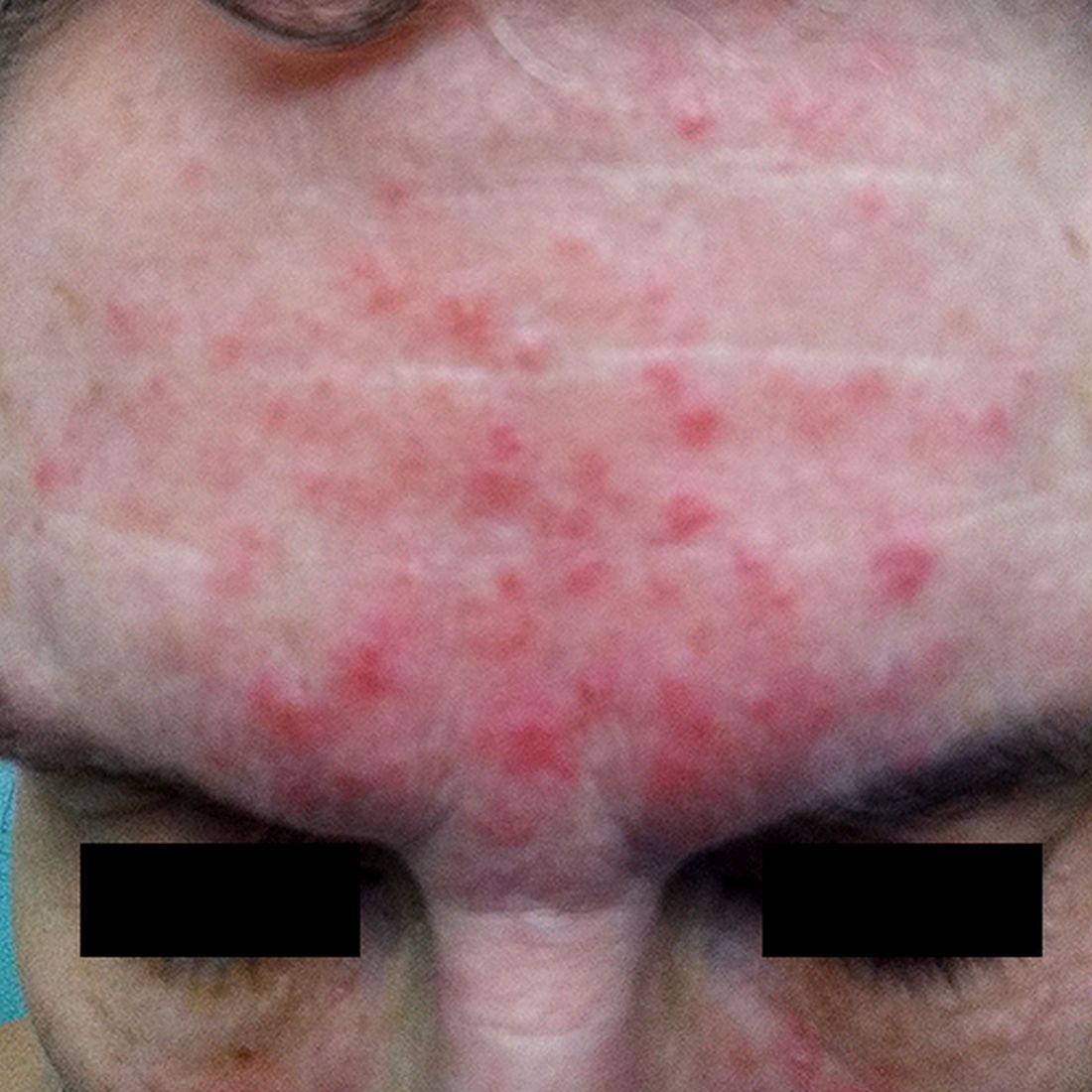
Treating rosacea: Combination therapy, benzoyl peroxide, and the ‘STOP’ mnemonic
HONOLULU – More often than not, patients with rosacea require a combination of treatments to optimize the management of the disease, according to Julie C. Harper, MD.
“We’ve been more comfortable with the idea of combination therapy for acne than we have been for rosacea,” Dr. Harper, who practices in Birmingham, Ala., said at the Hawaii Dermatology Seminar provided by MedscapeLIVE! “If patients are doing great on one treatment, then don’t change it. But if there’s room for improvement, think about combinations.”

Treatment options for papules and pustules include ivermectin, metronidazole, azelaic acid, sodium sulfacetamide/sulfur, modified release doxycycline, minocycline foam, and microencapsulated benzoyl peroxide, 5%. Options for persistent background erythema include brimonidine and oxymetazoline, as well as device-based treatments, which include the pulsed dye laser, the KTP laser, intense pulsed light, and electrosurgery.
Dr. Harper said that she has been especially surprised by the effectiveness of one of these options, microencapsulated benzoyl peroxide cream, 5% (Epsolay), which is approved by the Food and Drug Administration for treating inflammatory lesions of rosacea in adults. In two identical, phase 3 randomized clinical trials of patients with inflammatory rosacea lesions, those treated with microencapsulated benzoyl peroxide achieved a 68.8% reduction in inflammatory lesions at 12 weeks (including 42.5% at week 2), compared with 38%-46% of those on the vehicle, according to the April 2022 announcement of the approval from the manufacturers, Sol-Gel Technologies and Galderma.
“A common drug is playing a key role,” Dr. Harper said. “What’s the mechanism of action? I have no idea. I wonder if there may be a bacterial pathogen after all,” possibly Staphylococcus epidermidis, she added. However, she noted, “it does appear that benzoyl peroxide has an impact on Demodex, so maybe that’s the primary way it’s working.”
In her opinion, a key standout from the clinical trial data is the drug’s rapid onset of action, with a 42.5% reduction of lesions at week 2. “What makes this different is that the 5% microencapsulated benzoyl peroxide cream is wrapped up in a silica shell,” said Dr. Harper, a past president of the American Acne and Rosacea Society. “The silica shell kind of acts like a speed bump that slows the release of drug onto the skin. We think that’s what may be giving us this better tolerability.”
In an interview at the meeting, Linda Stein Gold, MD, director of clinical research and division head of dermatology at the Henry Ford Health System, Detroit, said that prior to the approval of Epsolay, benzoyl peroxide was never considered a first-line treatment for rosacea. “The problem is, the conventional formulation is irritating to the skin,” said Dr. Stein Gold, who was involved in clinical trials of Epsolay.

“The benzoyl peroxide encapsulated in the silica shell allows for a slow and steady delivery of medication to the skin in a very controlled manner. It is exceptionally good at getting rosacea under control. In the clinical trials, when we looked at the baseline irritation of the skin and followed those patients when they used the benzoyl 5% microencapsulated benzoyl peroxide cream, the irritation improved.”
‘STOP’ mnemonic
When treating her patients with rosacea, Dr. Harper incorporates the mnemonic “STOP” to these patient visits:
S: Identify signs and symptoms of rosacea.
T: Discuss triggers. “We cannot make this disease triggerless, so when you’re talking to your patients, you need to find out what’s triggering their rosacea,” she said.
O: Agree on a treatment outcome. “What is it that’s important to the patient?” she said. “They may tell you, ‘I want to be able to not be so red,’ or ‘I want to get rid of the bumps,’ or ‘I want my eyes to not feel so dry.’ ”
P: Develop a plan that helps achieve that desired outcome with patients.
Dr. Harper disclosed ties with Almirall, Cassiopeia, Cutera, Galderma, EPI, L’Oréal, Ortho Dermatologics, Sol Gel, Sun Pharmaceutical Industries, and Vyne.
Dr. Stein Gold disclosed ties with Almirall, Cutera, Dermata, Galderma, Novartis, Ortho Dermatologics, and Sun Pharmaceutical Industries.
Medscape and this news organization are owned by the same parent company.
HONOLULU – More often than not, patients with rosacea require a combination of treatments to optimize the management of the disease, according to Julie C. Harper, MD.
“We’ve been more comfortable with the idea of combination therapy for acne than we have been for rosacea,” Dr. Harper, who practices in Birmingham, Ala., said at the Hawaii Dermatology Seminar provided by MedscapeLIVE! “If patients are doing great on one treatment, then don’t change it. But if there’s room for improvement, think about combinations.”
Treatment options for papules and pustules include ivermectin, metronidazole, azelaic acid, sodium sulfacetamide/sulfur, modified release doxycycline, minocycline foam, and microencapsulated benzoyl peroxide, 5%. Options for persistent background erythema include brimonidine and oxymetazoline, as well as device-based treatments, which include the pulsed dye laser, the KTP laser, intense pulsed light, and electrosurgery.
Dr. Harper said that she has been especially surprised by the effectiveness of one of these options, microencapsulated benzoyl peroxide cream, 5% (Epsolay), which is approved by the Food and Drug Administration for treating inflammatory lesions of rosacea in adults. In two identical, phase 3 randomized clinical trials of patients with inflammatory rosacea lesions, those treated with microencapsulated benzoyl peroxide achieved a 68.8% reduction in inflammatory lesions at 12 weeks (including 42.5% at week 2), compared with 38%-46% of those on the vehicle, according to the April 2022 announcement of the approval from the manufacturers, Sol-Gel Technologies and Galderma.
“A common drug is playing a key role,” Dr. Harper said. “What’s the mechanism of action? I have no idea. I wonder if there may be a bacterial pathogen after all,” possibly Staphylococcus epidermidis, she added. However, she noted, “it does appear that benzoyl peroxide has an impact on Demodex, so maybe that’s the primary way it’s working.”
In her opinion, a key standout from the clinical trial data is the drug’s rapid onset of action, with a 42.5% reduction of lesions at week 2. “What makes this different is that the 5% microencapsulated benzoyl peroxide cream is wrapped up in a silica shell,” said Dr. Harper, a past president of the American Acne and Rosacea Society. “The silica shell kind of acts like a speed bump that slows the release of drug onto the skin. We think that’s what may be giving us this better tolerability.”
In an interview at the meeting, Linda Stein Gold, MD, director of clinical research and division head of dermatology at the Henry Ford Health System, Detroit, said that prior to the approval of Epsolay, benzoyl peroxide was never considered a first-line treatment for rosacea. “The problem is, the conventional formulation is irritating to the skin,” said Dr. Stein Gold, who was involved in clinical trials of Epsolay.
“The benzoyl peroxide encapsulated in the silica shell allows for a slow and steady delivery of medication to the skin in a very controlled manner. It is exceptionally good at getting rosacea under control. In the clinical trials, when we looked at the baseline irritation of the skin and followed those patients when they used the benzoyl 5% microencapsulated benzoyl peroxide cream, the irritation improved.”
‘STOP’ mnemonic
When treating her patients with rosacea, Dr. Harper incorporates the mnemonic “STOP” to these patient visits:
S: Identify signs and symptoms of rosacea.
T: Discuss triggers. “We cannot make this disease triggerless, so when you’re talking to your patients, you need to find out what’s triggering their rosacea,” she said.
O: Agree on a treatment outcome. “What is it that’s important to the patient?” she said. “They may tell you, ‘I want to be able to not be so red,’ or ‘I want to get rid of the bumps,’ or ‘I want my eyes to not feel so dry.’ ”
P: Develop a plan that helps achieve that desired outcome with patients.
Dr. Harper disclosed ties with Almirall, Cassiopeia, Cutera, Galderma, EPI, L’Oréal, Ortho Dermatologics, Sol Gel, Sun Pharmaceutical Industries, and Vyne.
Dr. Stein Gold disclosed ties with Almirall, Cutera, Dermata, Galderma, Novartis, Ortho Dermatologics, and Sun Pharmaceutical Industries.
Medscape and this news organization are owned by the same parent company.
AT THE MEDSCAPE LIVE! HAWAII DERMATOLOGY SEMINAR
Metformin linked to reductions in COVID-19 viral load
These findings add to a multitude of benefits the drug has been shown to have in COVID infection.
COVID-OUT did not meet its primary endpoint, but it did show important secondary outcomes including a 42% reduction in ED visits and in hospitalizations and/or deaths by day 14, and a 58% reduction in hospitalizations/death by day 28. A further subanalysis has shown a 42% reduction in long COVID, compared with placebo.
“In this phase 3 randomized controlled trial, metformin showed prevention of severe COVID, prevention of long COVID, and an antiviral effect, and this is consistent with other data,” said coauthor Carolyn Bramante, MD, University of Minnesota, Minneapolis, in presenting the findings at the Conference on Retroviruses & Opportunistic Infections.
Study details
For the new subanalysis, the authors further evaluated the effects of metformin treatment on SARS-CoV-2 viral load.
A total of 1,323 patients in the study, enrolled at six centers, were randomized to treatment either with metformin 1,000 mg per day on days 2-5 and 1,500 mg per day on days 6 to 14 (n = 187), or to ivermectin 390-470 mcg/kg per day for 3 days (n = 187), fluvoxamine 50 mg twice daily for 14 days, and/or an exact-matching placebo in a 2 x 3 factorial trial design.
The subanalysis on viral load included 483 patients from the trial who were treated with metformin versus 462 who received placebo, who were all enrolled within 3 days of a documented SARS-CoV-2 infection and less than 7 days after symptom onset.
The patients had a median age of 46 years, and all had either overweight or obesity. Only about 2% had diabetes, and only patients considered low-risk were excluded from the trial, including those under age 30 and those with a body mass index under 25.
About half of patients had received a primary vaccine and about 5% had received a vaccine booster. SARS-CoV-2 variants that were prominent during the study included Alpha, Delta, and Omicron.
The viral samples available on days 1, 5, and 10 showed a mean change in viral load from baseline to follow-up; the viral load was significantly lower with metformin versus placebo (–0.64 log10 copies/mL), representing a 4.4-fold greater decrease in viral load with metformin.
The mean rate of undetectable SARS-CoV-2 viral load at day 5 was 49.9% in the metformin group versus 54.6% in the placebo group (odds ratio, 1.235), and the undetectable rate at day 10 was 14.3% in the metformin group and 22.6% in the placebo group (OR, 1.663; P = .003).
An increased antiviral effect corresponded with increases in metformin dosing on days 6 through 14. Furthermore, the antiviral effect became stronger when metformin was started earlier in the course of infection.
Of note, the antiviral effect was more pronounced among those who were not vaccinated (mean, –0.95 log copies/mL), compared with the vaccinated (mean, –0.39 log copies/mL).
The antiviral effect with metformin was similar to that seen with nirmatrelvir at day 5 and was greater than nirmatrelvir at day 10.
No similar relationships in SARS-CoV-2 viral load were observed between ivermectin or fluvoxamine and placebo.
The findings are consistent with results of other recent observational studies, including research showing metformin to be associated with reductions in COVID-19 severity in patients with prediabetes, Dr. Bramante noted.
The authors’ previous analysis looking at long COVID in the COVID-OUT study showed that metformin treatment during acute COVID significantly reduced the risk for a diagnosis of long COVID versus placebo at 300 days following randomization, with a hazard ratio of 0.59 after adjustment for the study drug and vaccination at baseline.
Dr. Bramante noted that metformin’s potential antiviral properties have long been speculated, with some of the earliest research on the drug suggesting less severe outcomes in influenza, and more recently, RNA assays suggesting effects against other RNA viruses, including the Zika virus.
In terms of COVID, Dr. Bramante noted that the drug has plenty of potentially favorable benefits.
“Metformin is very safe and is known to have very few contraindications, so the next steps could be to consider looking at this in terms of a combination therapy,” she said.
‘Data from other studies are conflicting’
Commenting on the study, Diane V. Havlir, MD, cautioned that “metformin is currently not recommended in treatment guidelines, [and] data from other studies are conflicting; side effects can be an issue, and the study presented here was in a select population,” she said in an interview.
However, “what is both new and interesting in this presentation is the reduction of viral load, which [was observed] in the samples collected not only on days 1-5, but also days 6-14,” said Dr. Havlir, who is professor and associate chair of clinical research, department of medicine, and chief of the division of HIV, infectious diseases and global medicine and director of the AIDS Research Institute at the University of California, San Francisco.
Key questions the findings raise include whether the results correlate with clinical outcomes or transmission, and whether the findings are generalizable to other populations and settings, Dr. Havlir said.
Ultimately, “we need to continue to pursue all aspects of outpatient treatments for COVID to address questions like these for new and existing agents,” she added.
The trial received funding from the Parsemus Foundation, the Rainwater Charitable Foundation, Fast Grants, and the United Health Group. The authors and Dr. Havlir disclosed no relevant financial relationships.
A version of this article originally appeared on Medscape.com.
FROM CROI 2023
Zika virus still calls for preparedness and vaccine development
Warming U.S. temperatures, the resumption of travel, and new knowledge about Zika’s long-term effects on children signal that Zika prevention and vaccine development should be on public health officials’, doctors’, and communities’ radar, even when community infection is not occurring.
“Although we haven’t seen confirmed Zika virus circulation in the continental United States or its territories for several years, it’s still something that we are closely monitoring, particularly as we move into the summer months,” Erin Staples, MD, PhD, medical epidemiologist at the Arboviral Diseases Branch of the Centers for Disease Control and Prevention in Fort Collins, Colo., told this news organization.
“This is because cases are still being reported in other countries, particularly in South America. Travel to these places is increasing following the pandemic, leaving more potential for individuals who might have acquired the infection to come back and restart community transmission.”
How Zika might reemerge
The Aedes aegypti mosquito is the vector by which Zika spreads, and “during the COVID pandemic, these mosquitoes moved further north in the United States, into southern California, and were identified as far north as Washington, D.C.,” said Neil Silverman, MD, professor of clinical obstetrics and gynecology and director of the Infections in Pregnancy Program at UCLA Medical Center in Los Angeles.
“On a population level, Americans have essentially no immunity to Zika from prior infection, and there is no vaccine yet approved. If individuals infected with Zika came into a U.S. region where the Aedes aegypti mosquito was present, that population could be very susceptible to infection spread and even another outbreak. This would be a confluence of bad circumstances, but that’s exactly what infectious disease specialists continue to be watchful about, especially because Zika is so dangerous for fetuses,” said Dr. Silverman.
How the public can prepare
The CDC recommends that pregnant women or women who plan to become pregnant avoid traveling to regions where there are currently outbreaks of Zika, but this is not the only way that individuals can protect themselves.
“The message we want to deliver to people is that in the United States, people are at risk for several mosquito-borne diseases every summer beyond just Zika,” Dr. Staples said. “It’s really important that people are instructed to make a habit of wearing EPA [U.S. Environmental Protection Agency)–registered insect repellents when they go outside. Right now, that is the single best tool that we have to prevent mosquito-borne diseases in the U.S.
“From a community standpoint, there are several emerging mosquito control methods that are being evaluated right now, such as genetic modification and irradiation of mosquitoes. These methods are aimed at producing sterile mosquitoes that are released into the wild to mate with the local mosquito population, which will render them infertile. This leads, over time, to suppression of the overall Aedes aegypti mosquito population – the main vector of Zika transmission,” said Dr. Staples.
Monica Gandhi, MD, MPH, professor of medicine and associate chief of the division of HIV, infectious diseases, and global medicine at the University of California, San Francisco, encourages her patients to wear mosquito repellent but cautioned that “there’s no antiviral that you can take for Zika. Until we have a vaccine, the key to controlling/preventing Zika is controlling the mosquitoes that spread the virus.”
Vaccines
The National Institute of Allergy and Infectious Diseases (NIAID) is currently investigating a variety of Zika vaccines, including a DNA-based vaccine, (phase 2), a purified inactivated virus vaccine (phase 1), live attenuated vaccines (phase 2), and mRNA vaccines (phase 2).
“I’m most excited about mRNA vaccines because they help patients produce a lot of proteins. The protein from a typical protein-adjuvant vaccine will break down, and patients can only raise an immune response to whatever proteins are left. On the other hand, mRNA vaccines provide the body [with] a recipe to make the protein from the pathogen in high amounts, so that a strong immune response can be raised for protection,” noted Dr. Gandhi.
Moderna’s mRNA-1893 vaccine was recently studied in a randomized, observer-blind, controlled, phase 1 trial among 120 adults in the United States and Puerto Rico, the results of which were published online in The Lancet. “The vaccine was found to be generally well tolerated with no serious adverse events considered related to vaccine. Furthermore, the vaccine was able to generate a potent immune response that was capable of neutralizing the virus in vitro,” said Brett Leav, MD, executive director of clinical development for public health vaccines at Moderna.
“Our mRNA platform technology ... can be very helpful against emerging pandemic threats, as we saw in response to COVID-19. What is unique in our approach is that if the genetic sequence of the virus is known, we can quickly generate vaccines to test for their capability to generate a functional immune response. In the case of the mRNA-1893 trial, the vaccine was developed with antigens that were present in the strain of virus circulating in 2016, but we could easily match whatever strain reemerges,” said Dr. Leav.
A phase 2 trial to confirm the dose of mRNA-1893 in a larger study population is underway.
Although it’s been demonstrated that Moderna’s mRNA vaccine is safe and effective, moving from a phase 2 to a phase 3 study presents a challenge, given the fact that currently, the disease burden from Zika is low. If an outbreak were to occur in the future, these mRNA vaccines could potentially be given emergency approval, as occurred during the COVID pandemic, according to Dr. Silverman.
If approved, provisionally or through a traditional route, the vaccine would “accelerate the ability to tamp down any further outbreaks, because vaccine-based immunity could be made available to a large portion of the population who were pregnant or planning a pregnancy, not just in the U.S. but also in these endemic areas,” said Dr. Silverman.
Takeaways from the last Zika outbreak
Practical steps such as mosquito eradication and development of vaccines are not the only takeaway from the recent Zika epidemics inside and outside the United States. A clearer picture of the short- and long-term stakes of the disease has emerged.
According to the CDC, most people who become infected with Zika experience only mild symptoms, such as fever, rash, headache, and muscle pain, but babies conceived by mothers infected with Zika are at risk for stillbirth, miscarriage, and microcephaly and other brain defects.
Although a pregnant woman who tests positive for Zika is in a very high-risk situation, “data show that only about 30% of mothers with Zika have a baby with birth defects. If a pregnant woman contracted Zika, what would happen is we would just do very close screening by ultrasound of the fetus. If microcephaly in utero or fetal brain defects were observed, then a mother would be counseled on her options,” said Dr. Gandhi.
Dr. Silverman noted that “new data on children who were exposed in utero and had normal exams, including head measurements when they were born, have raised concerns. In recently published long-term follow-up studies, even when children born to mothers infected with Zika during pregnancy had normal head growth at least 3 years after birth, they were still at risk for neurodevelopmental delay and behavioral disorders, including impact on coordination and executive function.
“This is another good reason to keep the potential risks of Zika active in the public’s consciousness and in public health planning.”
Dr. Silverman, Dr. Gandhi, and Dr. Staples have disclosed no relevant financial relationships. Dr. Leav is an employee of Moderna and owns stock in the company.
A version of this article originally appeared on Medscape.com.
Psychosocial Burden of Hidradenitis Suppurativa
Hidradenitis suppurativa (HS) is a chronic inflammatory skin disease that typically presents as inflamed nodules, abscesses, and sinus tracts in the apocrine gland–bearing regions.
Chronic pain, fatigue, and social isolation put patients with HS at risk for psychiatric comorbidities, with the prevalence of depression estimated to be as high as 43%. Work productivity and sexual relationships may also be affected.
Early treatment of the condition, as well as screening for depression, may help alleviate the psychosocial burden of HS.
In this ReCAP, Dr Jennifer Hsiao from Keck School of Medicine, University of Southern California, Los Angeles, outlines mental health screening tools that dermatologists can use to help recognize at-risk patients and potentially refer them to mental health experts.
--
Jennifer Hsiao, MD, Associate Professor, Department of Dermatology, Keck School of Medicine, University of Southern California, Los Angeles, California
Jennifer Hsiao, MD, has disclosed the following relevant financial relationships:
Serve(d) as a director, officer, partner, employee, advisor, consultant, or trustee for: AbbVie; Boehringer Ingelheim; Novartis; UCB
Serve(d) as a speaker or a member of a speakers bureau for: AbbVie

Pfizer COVID vaccine effective in young children, study shows
A new study shows the Pfizer vaccine is safe and highly effective against COVID-19 in children as young as 6 months old.
A three-dose series of the Pfizer COVID-19 vaccine was 73% effective at preventing symptomatic COVID-19 in children aged 6 months to 4 years, the researchers found. They also found that an examination of reactions and safety results “did not suggest any concerns.”
The study, published in the New England Journal of Medicine, included 1,776 children aged 6 months to 2 years old, and 2,750 children aged 2-4 years. Children were randomly assigned to receive either the three-shot series of the Pfizer vaccine or placebo shots. Participants received the first dose of the vaccine by March 31, 2022, and lived in Brazil, Finland, Poland, Spain, or the United States.
The authors wrote that having safe and effective COVID vaccines for young children is important to protect them from hospitalization or death and because young children play a role in spreading highly transmissible variants of the virus. COVID hospitalizations for children under 5 years old peaked at a rate of 14.5 per 100,000 in January 2022, the authors wrote, noting that the Omicron virus variant appeared to affect young children more severely than the previous variant, Delta.
When the researchers evaluated vaccine effectiveness by age group, they found that it prevented symptomatic COVID in 75.8% of children aged 6 months to 2 years, and in 71.8% of children aged 2-4 years.
Less than 0.5% of participants reported severe reactions to the vaccine. The most common reactions reported were tenderness or pain. Reactions typically appeared within the first couple days following vaccine administration and resolved within 2 days. No cases of inflammation of the heart muscle or its lining were reported among participants.
Uptake of COVID vaccines for young children has been lower than other age groups in the United States. The Centers for Disease Control and Prevention says 10% of children younger than 5 have received at least one dose of a COVID-19 vaccine, and 5% have completed a primary vaccine series.
A version of this article originally appeared on WebMD.com.
FROM THE NEW ENGLAND JOURNAL OF MEDICINE
Obesity Workup
Are ‘Momi Pods’ the future of postnatal care?
Mindi Rosen met Seuli Brill, MD, at just the right time.
Ms. Rosen’s firstborn son was in the neointensive natal unit at The Ohio State University Wexner Medical Center in Columbus, and she didn’t have a pediatrician picked out yet for the baby. Nor did she have a primary care physician who could help her manage the gestational diabetes she developed during her pregnancy.
Dr. Brill, a clinical associate professor of internal medicine and pediatrics at Ohio State, suggested Ms. Rosen visit her at the new clinic she was piloting in Columbus. There, she provided pediatric care for newborns and primary care for mothers who had developed gestational diabetes.
“I looked at my husband, my husband looked at me, and I said: ‘Why not?’ “ Ms. Rosen, 38, recalled of that 2019 meeting. “I’m so glad she walked in at that moment.”
The mother of two is still part of the rapidly growing program at the medical facility that provides care for more than 200 mothers and babies.
Launched in 2018, the clinic – called the Multi-Modal Maternal Infant Perinatal Outpatient Delivery System, or “Momi Pods,” started with a focus on helping women with gestational diabetes, which occurs in up to 10% of pregnancies.
The program allows moms to book regular checkups for their baby, and then a follow-up appointment immediately after for themselves. Women are seen for the first 1,000 days (just under 3 years) after giving birth.
The idea was simple. Dr. Brill wanted to develop a more formalized program for the work she was already doing as a primary care physician and pediatrician. At the time, she was fielding referrals from specialists for young women who didn’t have a physician. She’d often develop a relationship with the patient over the years, go on to help oversee their care during pregnancy, then new mothers would select her as their newborn’s pediatrician.
“I would have a relationship with the mom when they did have the newborn, and then I would see the baby because I’m a pediatrician,” Dr. Brill said.
Dr. Brill was serving on the Ohio Gestational Diabetes Mellitus Collaborative, a state-backed program that aims to raise awareness about the condition and encourage more preventative care for patients. She presented her proposal to launch the program to the Ohio Department of Medicaid, which helped to fund the pilot.
The idea, she hoped, would improve postpartum follow-up care for mothers diagnosed with the condition.
Follow-up care is especially important for women who develop gestational diabetes because the condition raises their lifetime risk of developing type 2 diabetes up to 10-fold.
Yet most of those mothers don’t get the appropriate follow-up care during the crucial postpartum period, said Maya Subbalakshmi Venkataramani, MD, MPH, an assistant professor of medicine at Johns Hopkins University in Baltimore, who has researched parental care.
“Things get very busy after you have a child. There’s just the general logistics of a mom having to take care of a newborn and thinking about themselves,” Dr. Venkataramani, a primary care clinician and pediatrician, said. “A lot of parents in general may not put a lot of emphasis on their own health.”
Seeking care may be especially difficult for low-income mothers who might not have consistent health care coverage, she added.
In fact, only half of women who developed gestational diabetes received primary follow-up care, according to a study published in JAMA Network Open. The study, which examined more than 280,000 insurance claims between 2015 and 2018, found only 36% of women with gestational diabetes received the recommended blood glucose testing in the first 12 weeks of the postpartum period.
In the Momi Pods program, Dr. Brill checked in on Ms. Rosen’s gestational diabetes regularly during pediatric office visits for her newborn’s care. Ms. Rosen said whenever she brought her baby in for a visit during the postpartum period, Dr. Brill measured her blood sugar.
Dr. Brill and her team also asked how Ms. Rosen was doing physically and mentally during each visit. The screenings helped to catch a bout of postpartum depression Ms. Rosen experienced after the birth of her first son.
“I thought it was great, because honestly as a new mom I wouldn’t have followed up with myself so much,” Ms. Rosen said. “Every time you went into the doctor appointments, they’d ask you how you are doing. As a new mom, it’s so much easier to do it at the same time.”
Those who participate in the program are also more likely to complete postpartum visits with their ob.gyn. (95% vs. 58%, respectively; P < .001) than those who don’t participate, according to research Dr. Brill and colleagues published.
Dr. Brill began expanding the program’s reach nearly 2 years after its launch, targeting the services for women who are at risk for poor postpartum outcomes, including those with a history of depression, preterm labor, diabetes and congenital heart disease. Ob.gyns. in Ohio State’s network can refer their patients to the program, which now has 43 doctors trained to provide primary and pediatric care through Momi Pods. Soon-to-be moms can be referred to the program as early as the second trimester, Dr. Brill said.
Many of the mothers referred to the program don’t have a primary care clinician when they talk to Paola Beamon, RN, at Ohio State. Ms. Beamon reaches out to each referred patient over voicemail, a MyChart message, and even regular mail in hopes of helping them navigate the postpartum period. She also provides education on what a primary care clinician can offer new moms.
“Really, we’re pursuing these moms and doing everything we can so there’s less of a burden for them,” Ms. Beamon said. “A lot of them don’t even know what a primary care office does.”
One of the biggest perks to the program for new moms is that they don’t have to spend time and money traveling to a different doctor’s office, take time off work, or secure childcare in order to schedule a separate appointment for themselves, she said.
The program, which receives funding from the university and the state, even helps women get bus passes to a doctor’s appointment if needed.
Dyad programs targeting women with substance abuse disorders or mental health conditions have existed for many years. But catering to women with gestational diabetes or other medical conditions appears to be new. In part, Dr. Venkataramani said, because scheduling and space can be big hurdles to launch such a program, as well as finding doctors who can care for both baby and mother.
“There are logistical challenges to even doing this that makes it less common,” she said.
Dr. Brill said she is not aware of any other programs that are structured like the tandem care clinic at Ohio State. She hopes, however, that the program can be a model for other hospital systems to consider, and she is working to expand the program regionally. Her team is collecting data – including on the best way to schedule patients – to help other clinics develop something similar.
“We really want to leverage that expertise to make it easier for moms to get care with their infants and remove barriers to care,” she said.
A version of this article first appeared on Medscape.com.