User login
RT use on decline in early HL despite survival benefit
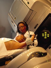
Credit: Rhoda Baer
SAN FRANCISCO—Results of a large study suggest that consolidation radiation therapy (RT) can improve survival in patients with stage I and II Hodgkin lymphoma (HL), but the use of RT in these patients may be on the decline.
In this study of more than 40,000 patients, the 10-year survival rate was 84% among those who received RT and 76% among those who did not.
Despite this benefit, the use of RT declined during the period studied, from 56% in 1998 to 41% in 2011.
These data were presented at the American Society for Radiation Oncology’s 56th Annual Meeting (abstract 1042).
“Multiple prospective, randomized trials have shown a significant improvement in disease control with the addition of RT,” said lead study author Rahul R. Parikh, MD, of the Mount Sinai Health System in New York.
“However, previous trials were limited by low patient numbers and limited follow-up and, thus, were unable to demonstrate an overall survival benefit. This is the largest dataset in this patient population to demonstrate a survival benefit with the addition of RT.”
Dr Parikh and his colleagues studied 41,502 patients who were diagnosed with stage I and II HL from 1998 to 2011. They were included in the National Cancer Data Base, which consists of cases from 1500 sites and represents more than 75% of all cancers diagnosed in the US.
The average patient age was 37 years (range, 18 to 90). The median follow-up was 7.5 years. Ninety-six percent of patients (n=39,842) received multi-agent chemotherapy, and 49% (n=20,441) received a median RT dose of 30.6 Gy.
The 10-year overall survival of the entire group was 80.8%. Patients receiving RT had significantly better overall survival than those who did not (84.4% vs 76.4%; P<0.00001).
When adjusting for age, stage, comorbidity, transplant, chemotherapy use, and socioeconomic status, RT use was still associated with significantly improved overall survival (hazard ratio=0.51; P<0.00001).
The study also showed that omitting RT was related to higher rates of salvage transplant procedures, a surrogate for persistent/relapsed disease (P=0.04).
Nevertheless, RT use decreased at the study sites from 56% to 41% between 1998 and 2011.
In 88.4% of patients who did not receive RT, the physician-reported reason was that RT was not part of the planned initial treatment strategy.
The research also indicated that RT use was more likely among younger patients (40 years or younger), those in a higher socioeconomic status, those who had access to health insurance, and those who received treatment at comprehensive cancer centers (all P<0.0001).
“[W]e have highlighted ongoing disparities in Hodgkin’s disease treatment, and it is important that we recognize these findings as potential barriers to care,” Dr Parikh said.
“Given the survival benefit demonstrated in this study, radiotherapy should be included in the combined modality approach of multi-agent chemotherapy followed by consolidation RT in order to maintain high overall survival rates for this curable disease.” ![]()
AB blood type linked to cognitive impairment

Credit: Graham Colm
Individuals with type AB blood may be more likely than those with other blood types to develop memory loss in later years, according to a study published in Neurology.
Investigators found that people with AB blood were 82% more likely to develop cognitive impairment, which can lead to dementia.
Previous studies have shown that individuals with type O blood have a lower risk of heart disease and stroke, factors that can increase the risk of memory loss and dementia.
The new research was part of a larger study—the REasons for Geographic And Racial Differences in Stroke (REGARDS) Study—of more than 30,000 subjects who were followed for an average of 3.4 years.
“Our study looks at blood type and risk of cognitive impairment, but several studies have shown that factors such as high blood pressure, high cholesterol, and diabetes increase the risk of cognitive impairment and dementia,” said Mary Cushman, MD, of the University of Vermont College of Medicine in Burlington.
“Blood type is also related to other vascular conditions like stroke, so the findings highlight the connections between vascular issues and brain health. More research is needed to confirm these results.”
Dr Cushman and her colleagues had set out to assess the relationship between ABO group, factor VIII (FVIII), and incident cognitive impairment in a large, prospective cohort of black and white adults in the US.
The team used cognitive domain tests to assess cognitive impairment. They identified 495 subjects who had no cognitive impairment at baseline but became impaired during follow-up. The investigators then compared these cases with 587 control subjects.
It turned out that subjects with AB blood made up 6% of the group that developed cognitive impairment, which is higher than the 4% of AB individuals found in the general population.
Multivariate analysis—adjusted for age, race, region, and sex—suggested that subjects with AB blood and those with higher FVIII had an increased risk of cognitive impairment. The odds ratios were 1.82 and 1.24, respectively.
Subjects with AB blood had a higher average level of FVIII than subjects with other blood types. The mean level of FVIII was 142 IU/dL among AB subjects and 104 IU/dL among subjects with type O blood.
However, the investigators also found that FVIII mediated only 18% of the association between AB blood type and cognitive impairment. ![]()
NPs and PAs in Hospital Medicine
Nurse practitioners (NPs) and physician assistants (PAs) have been caring for patients since the mid‐1960s.[1] Although both roles grew out of a need for more primary care providers, more recently there has been an increase in the utilization of NPs and PAs in acute care roles. This meteoric rise of advanced practice providers in the inpatient setting has been driven by stressors from residency work‐hour reforms and from growing financial pressures in healthcare systems, where NPs and PAs are seen as less expensive alternatives.[2, 3] Inadequate physician supply to meet the needs of growing healthcare service is also a driving factor. Despite increasing numbers of enrollees and increasing numbers of medical schools, many sources estimate a physician shortage of 50,000 providers by year 2025.[4] To address this growing shortage, the number of NP and PA providers in acute care continues to grow as Kartha and colleagues[5] clearly demonstrate in their study, published in this issue of Journal of Hospital Medicine. Their research shows that within hospitals in the Veterans Health Administration (VHA)the largest coordinated healthcare association in the United Statesfully half of all inpatient medical teams are utilizing NPs and PAs in some capacity, most commonly in staffing models working directly with attending physicians or on teams with housestaff.[5]
Many different practice models exist that incorporate NPs and PAs into acute care settings, including models in general medicine and intensive care settings, as well as in specialty care populations such as patients with diabetes or congestive heart failure.[1, 6] Few studies, however, delineate specific roles for NPs or PAs in inpatient acute care or provide outcomes‐based evidence in support of the proposed models. This is in contrast to research available regarding NP and PA staffing models in the outpatient setting.[7, 8] In the current study, Kartha et al.[5] shed light on the use of NPs and PAs in inpatient medical units at the VHA. Their findings show that the majority of NPs and PAs on the inpatient team function mostly autonomously and perform tasks including performing histories and physicals, writing progress notes, placing orders, and communicating with primary care providers and consultants. Almost half also serve on hospital committees and participate in quality improvement activities. Interestingly, although the training and regulation of NPs and PAs differ considerably,[1] Kartha et al. found that the scope of practice of these providers is generally the same. PAs are more likely to perform procedures and teach nonphysician students but otherwise function similarly to NPs. The clinical workload for NPs and PAs also does not differ, with an average of 6.5 patients seen per day. This information is crucial when analyzing the cost‐effectiveness of these providers, especially in light of evidence suggesting that hospitalist physicians typically care for approximately twice as many patients.[9]
Although Kartha et al.[5] focus primarily on describing the scope of NPs and PAs in hospital medicine, they also report on outcomes. Their findings show that presence of NPs and PAs on inpatient teams did not alter patient or nurse satisfaction nor were there any consistent improvements in the perception of care coordination. Of note, assessment of care coordination was based on survey responses from nurse managers and chiefs of medicine, individuals who are not necessarily direct members of the inpatient team, thus questioning the validity of this measure. Other studies on NP/PA models have also focused on patient‐centered outcomes. A study by Roy et al.[10] found that an inpatient PA‐run service supervised by hospitalists was comparable with a traditional resident‐run service, with no significant differences in risk‐adjusted length of stay (LOS), mortality, intensive care unit (ICU) transfers, or hospital readmissions. Although total costs were lower on the PA service, this difference was minimal. Gershengorn et al.[11] examined the impact of nonphysician staffing in an ICU setting and again found equivalent care. In this study, an ICU team staffed by NPs and PAs had similar hospital mortality and LOS as compared with a standard housestaff ICU service. Both these studies have limitations in that they are retrospective analyses rather than randomized controlled trials, and they were conducted at academic medical centers, thus narrowing their generalizability. Moreover, purity of data is difficult to achieve, as few systems exist where NPs and PAs are the sole providers managing patients without interaction or coverage from physician colleagues.
Given the considerable presence of NPs and PAs in acute care hospitals as documented by Kartha et al.,[5] providing appropriate training in hospital medicine to these clinicians is important. A study by Dhuper and Choksi[12] evaluated a 2‐year PA postgraduate training program in hospital medicine. PAs spent 40 hours per week on direct patient care while rotating on general medical floors and ICUs, along with 16 hours per week in didactic instruction. When compared with a traditional 3‐year medical residency at the same institution, the PA training program had similar outcomes on patient care including similar number of adverse events, readmissions, and patient satisfaction scores. A more formal postgraduate training program for PAs has been established at the Mayo Clinic Arizona.[13] This 12‐month program, based on the Society of Hospital Medicine's (SHM) Core Competencies, consists of general medicine and inpatient medical subspecialty rotations, didactic instruction, and self‐directed teaching modules to learn systems‐based practices. The Adult Hospital Medicine Boot Camp, sponsored by the SHM and the American Academy of Physician Assistants, is another training opportunity for both NPs and PAs who currently work in or are planning to practice hospital medicine.[14] Finally, in accordance with the move to provide standardized training for providers who practice in acute care settings, professional nursing organizations have developed the Consensus Model for Advanced Practice Registered Nurse Regulation that contains recommendations ensuring similar education and licensure requirements for those who practice in acute care.[15]
Although the optimal utilization of NPs and PAs in hospital medicine is still unknown, the reality is that the number of NPs and PAs actually working in this capacity is significant, as Kartha and his colleagues report.[5] A study of academic medical centers also found that among the institutions that responded to a survey, 31% and 42% used PAs and NPs, respectively, in hospitalist roles.[16] Current evidence suggests that NP‐ and PA‐based care with physician collaboration in an inpatient setting can result in comparable outcomes with physician‐only care models. However, much of this evidence is of poor quality or cannot be generalized to all settings. Kartha et al.[5] have provided a good first step in describing the role of NPs and PAs within hospital medicine. Though their education and training backgrounds are different, the ultimate scope of practice for these 2 groups of providers is very similar. Future research should focus on defining the best practice model for utilization of NPs and PAs in hospital medicine with emphasis on measurable goals. These can include standard outcomes such as LOS but also specific measures of quality and safety such as days of urinary catheter use or percentage of patients receiving venous thromboprophylaxis.[17] By understanding the scope of NP and PA practice, collecting more robust data regarding outcomes, and emphasizing training for NPs and PAs within hospital medicine, there is opportunity to impact the quality and efficiency of care of hospitalized patients.
- , , . Nurse practitioners and physician assistants in the intensive care unit: an evidence‐based review. Crit Care Med. 2008;36:2888–2897.
- , , . Quality and financial impact of adding nurse practitioners to inpatient care teams. J Nurs Adm. 2014;44:87–96.
- , , , et al. The effect of a multidisciplinary hospitalist/physician and advanced practice nurse collaboration on hospital costs. J Nurs Adm. 2006;36(2):79–85.
- , . Physician assistants in American medicine: the half‐century mark. Am J Manag Care. 2013;19:e333–e341.
- , , , et al. Nurse practitioner and physician assistant scope of practice in 118 acute care hospitals. J Hosp Med. 2014;9(10):615–620.
- , , , , , . Care directed by a specialty‐trained nurse practitioner or physician assistant can overcome clinical inertia in management of inpatient diabetes. Endocr Pract. 2014;20:112–119.
- , , , et al. Advanced practice nurse outcomes 1990–2008: a systematic review. Nurse Econ. 2011;29:230–250.
- , , , et al. The contribution of physician assistants in primary care: a systematic review. BMC Health Serv Res. 2013;13:223.
- , , , , . Effect of hospitalist workload on the quality and efficiency of care. JAMA Intern Med. 2014;174:786–793.
- , , , et al. Implementation of a physician assistant/hospitalist service in an academic medical center: impact on efficiency and patient outcomes. J Hosp Med. 2008;3:361–368.
- , , , et al. Impact of nonphysician staffing on outcomes in a medical ICU. Chest. 2011;139:1347–1353.
- , . Replacing an academic internal medicine residency program with a physician assistant‐hospitalist model. Am J Med Qual. 2009;24:132–139.
- , , , , . A hospitalist postgraduate training program for physician assistants. J Hosp Med. 2010;5:94–98.
- American Association of Physician Assistants. Adult hospital medicine boot camp. Available at: http://www.aapa.org/bootcamp. Accessed July 3 2014.
- , , , . Defining NP scope of practice and associated regulations: focus on acute care. J Am Acad Nurse Pract. 2012;24:11–18.
- , , , . Physician assistant and nurse practitioner utilization in academic medical centers. Am J Med Qual. 2011;26:452–460.
- , . Developing nurse practitioner associated metrics for outcomes assessment. J Am Assoc Nurse Pract. 2013;25:289–296.
Nurse practitioners (NPs) and physician assistants (PAs) have been caring for patients since the mid‐1960s.[1] Although both roles grew out of a need for more primary care providers, more recently there has been an increase in the utilization of NPs and PAs in acute care roles. This meteoric rise of advanced practice providers in the inpatient setting has been driven by stressors from residency work‐hour reforms and from growing financial pressures in healthcare systems, where NPs and PAs are seen as less expensive alternatives.[2, 3] Inadequate physician supply to meet the needs of growing healthcare service is also a driving factor. Despite increasing numbers of enrollees and increasing numbers of medical schools, many sources estimate a physician shortage of 50,000 providers by year 2025.[4] To address this growing shortage, the number of NP and PA providers in acute care continues to grow as Kartha and colleagues[5] clearly demonstrate in their study, published in this issue of Journal of Hospital Medicine. Their research shows that within hospitals in the Veterans Health Administration (VHA)the largest coordinated healthcare association in the United Statesfully half of all inpatient medical teams are utilizing NPs and PAs in some capacity, most commonly in staffing models working directly with attending physicians or on teams with housestaff.[5]
Many different practice models exist that incorporate NPs and PAs into acute care settings, including models in general medicine and intensive care settings, as well as in specialty care populations such as patients with diabetes or congestive heart failure.[1, 6] Few studies, however, delineate specific roles for NPs or PAs in inpatient acute care or provide outcomes‐based evidence in support of the proposed models. This is in contrast to research available regarding NP and PA staffing models in the outpatient setting.[7, 8] In the current study, Kartha et al.[5] shed light on the use of NPs and PAs in inpatient medical units at the VHA. Their findings show that the majority of NPs and PAs on the inpatient team function mostly autonomously and perform tasks including performing histories and physicals, writing progress notes, placing orders, and communicating with primary care providers and consultants. Almost half also serve on hospital committees and participate in quality improvement activities. Interestingly, although the training and regulation of NPs and PAs differ considerably,[1] Kartha et al. found that the scope of practice of these providers is generally the same. PAs are more likely to perform procedures and teach nonphysician students but otherwise function similarly to NPs. The clinical workload for NPs and PAs also does not differ, with an average of 6.5 patients seen per day. This information is crucial when analyzing the cost‐effectiveness of these providers, especially in light of evidence suggesting that hospitalist physicians typically care for approximately twice as many patients.[9]
Although Kartha et al.[5] focus primarily on describing the scope of NPs and PAs in hospital medicine, they also report on outcomes. Their findings show that presence of NPs and PAs on inpatient teams did not alter patient or nurse satisfaction nor were there any consistent improvements in the perception of care coordination. Of note, assessment of care coordination was based on survey responses from nurse managers and chiefs of medicine, individuals who are not necessarily direct members of the inpatient team, thus questioning the validity of this measure. Other studies on NP/PA models have also focused on patient‐centered outcomes. A study by Roy et al.[10] found that an inpatient PA‐run service supervised by hospitalists was comparable with a traditional resident‐run service, with no significant differences in risk‐adjusted length of stay (LOS), mortality, intensive care unit (ICU) transfers, or hospital readmissions. Although total costs were lower on the PA service, this difference was minimal. Gershengorn et al.[11] examined the impact of nonphysician staffing in an ICU setting and again found equivalent care. In this study, an ICU team staffed by NPs and PAs had similar hospital mortality and LOS as compared with a standard housestaff ICU service. Both these studies have limitations in that they are retrospective analyses rather than randomized controlled trials, and they were conducted at academic medical centers, thus narrowing their generalizability. Moreover, purity of data is difficult to achieve, as few systems exist where NPs and PAs are the sole providers managing patients without interaction or coverage from physician colleagues.
Given the considerable presence of NPs and PAs in acute care hospitals as documented by Kartha et al.,[5] providing appropriate training in hospital medicine to these clinicians is important. A study by Dhuper and Choksi[12] evaluated a 2‐year PA postgraduate training program in hospital medicine. PAs spent 40 hours per week on direct patient care while rotating on general medical floors and ICUs, along with 16 hours per week in didactic instruction. When compared with a traditional 3‐year medical residency at the same institution, the PA training program had similar outcomes on patient care including similar number of adverse events, readmissions, and patient satisfaction scores. A more formal postgraduate training program for PAs has been established at the Mayo Clinic Arizona.[13] This 12‐month program, based on the Society of Hospital Medicine's (SHM) Core Competencies, consists of general medicine and inpatient medical subspecialty rotations, didactic instruction, and self‐directed teaching modules to learn systems‐based practices. The Adult Hospital Medicine Boot Camp, sponsored by the SHM and the American Academy of Physician Assistants, is another training opportunity for both NPs and PAs who currently work in or are planning to practice hospital medicine.[14] Finally, in accordance with the move to provide standardized training for providers who practice in acute care settings, professional nursing organizations have developed the Consensus Model for Advanced Practice Registered Nurse Regulation that contains recommendations ensuring similar education and licensure requirements for those who practice in acute care.[15]
Although the optimal utilization of NPs and PAs in hospital medicine is still unknown, the reality is that the number of NPs and PAs actually working in this capacity is significant, as Kartha and his colleagues report.[5] A study of academic medical centers also found that among the institutions that responded to a survey, 31% and 42% used PAs and NPs, respectively, in hospitalist roles.[16] Current evidence suggests that NP‐ and PA‐based care with physician collaboration in an inpatient setting can result in comparable outcomes with physician‐only care models. However, much of this evidence is of poor quality or cannot be generalized to all settings. Kartha et al.[5] have provided a good first step in describing the role of NPs and PAs within hospital medicine. Though their education and training backgrounds are different, the ultimate scope of practice for these 2 groups of providers is very similar. Future research should focus on defining the best practice model for utilization of NPs and PAs in hospital medicine with emphasis on measurable goals. These can include standard outcomes such as LOS but also specific measures of quality and safety such as days of urinary catheter use or percentage of patients receiving venous thromboprophylaxis.[17] By understanding the scope of NP and PA practice, collecting more robust data regarding outcomes, and emphasizing training for NPs and PAs within hospital medicine, there is opportunity to impact the quality and efficiency of care of hospitalized patients.
- , , . Nurse practitioners and physician assistants in the intensive care unit: an evidence‐based review. Crit Care Med. 2008;36:2888–2897.
- , , . Quality and financial impact of adding nurse practitioners to inpatient care teams. J Nurs Adm. 2014;44:87–96.
- , , , et al. The effect of a multidisciplinary hospitalist/physician and advanced practice nurse collaboration on hospital costs. J Nurs Adm. 2006;36(2):79–85.
- , . Physician assistants in American medicine: the half‐century mark. Am J Manag Care. 2013;19:e333–e341.
- , , , et al. Nurse practitioner and physician assistant scope of practice in 118 acute care hospitals. J Hosp Med. 2014;9(10):615–620.
- , , , , , . Care directed by a specialty‐trained nurse practitioner or physician assistant can overcome clinical inertia in management of inpatient diabetes. Endocr Pract. 2014;20:112–119.
- , , , et al. Advanced practice nurse outcomes 1990–2008: a systematic review. Nurse Econ. 2011;29:230–250.
- , , , et al. The contribution of physician assistants in primary care: a systematic review. BMC Health Serv Res. 2013;13:223.
- , , , , . Effect of hospitalist workload on the quality and efficiency of care. JAMA Intern Med. 2014;174:786–793.
- , , , et al. Implementation of a physician assistant/hospitalist service in an academic medical center: impact on efficiency and patient outcomes. J Hosp Med. 2008;3:361–368.
- , , , et al. Impact of nonphysician staffing on outcomes in a medical ICU. Chest. 2011;139:1347–1353.
- , . Replacing an academic internal medicine residency program with a physician assistant‐hospitalist model. Am J Med Qual. 2009;24:132–139.
- , , , , . A hospitalist postgraduate training program for physician assistants. J Hosp Med. 2010;5:94–98.
- American Association of Physician Assistants. Adult hospital medicine boot camp. Available at: http://www.aapa.org/bootcamp. Accessed July 3 2014.
- , , , . Defining NP scope of practice and associated regulations: focus on acute care. J Am Acad Nurse Pract. 2012;24:11–18.
- , , , . Physician assistant and nurse practitioner utilization in academic medical centers. Am J Med Qual. 2011;26:452–460.
- , . Developing nurse practitioner associated metrics for outcomes assessment. J Am Assoc Nurse Pract. 2013;25:289–296.
NP and PA Scope of Practice
Nurse practitioners (NPs) and physician assistants (PAs) provide healthcare in numerous environments internationally and in the United States.[1, 2] However, their role in the inpatient medicine setting is not well described.[2] In the United States, there are more than 157,000 NPs and 85,000 PAs with projected increases.[3, 4] Although both professions provide direct medical care, there are key differences.[1, 3, 4, 5] NPs typically complete a master's or doctoral degree with advanced clinical training beyond nursing. PAs complete at least 2 years of college courses similar to premedical school requirements. PA programs use a medical school‐based curriculum and train for about 2 years before awarding a master's degree. NPs are regulated through state nursing boards, whereas PAs are regulated through state licensing or medical boards. NPs and PAs have different, yet overlapping scopes of practice. A key difference is that PAs can only practice collaborating with a physician.[5, 6] Overall, both have been shown to provide healthcare that is similar in quality to physicians in specific primary care and surgical settings.[2]
NPs and PAs, often referred to as advanced practice providers (APPs), are employed primarily in outpatient clinic settings providing direct patient care. Most APP studies have focused on the outpatient setting, despite nearly a third of US healthcare expenditure for hospital care.[2, 7] Little is known about APP involvement, specific roles, or impact on outcomes in inpatient medicine settings where they are often referred to as NP or PA hospitalists.[2, 8, 9, 10]
The Veterans Health Administration (VHA) is 1 of the largest employers of APPs, with 3.6% of all NPs and 2.1% of all PAs reported to practice in the VHA.[11, 12, 13] As the largest fully integrated healthcare system in the US, the VHA had 8.8 million veterans enrolled and 703,500 inpatient admissions in 2012.[14] Although this makes the VHA an ideal environment to study the role of APPs, few studies have done so.[13, 15, 16, 17, 18, 19] Although studies have compared NPs and PAs to physicians, very little is known about how NPs differ from PAs when practicing in the same environment.
Our objective was to describe the scope of practice, defined as activities that an individual healthcare practitioner is licensed to perform, of NPs and PAs in the inpatient medicine setting and in the VHA. A secondary objective was to explore important outcomes that could potentially be affected by the presence of NPs and PAs on inpatient medicine.
METHODS
The Organizational Factors and Inpatient Medical Care Quality and Efficiency (OFIM) study provides a basis for this study with detail published elsewhere.[20] The OFIM study was conducted between 2010 and 2011 to evaluate quality of care in VHA inpatient medicine surveying chiefs of medicine (COM), inpatient medicine nurse managers (NM), attending physicians, and extant VHA survey data. The COM is the senior attending physician in charge of departments of medicine that include most medical subspecialties within the VHA medical centers. We used the subset of questions specific to NPs and PAs from the COM and NM surveys. Both COMs and NMs answered identical questions for NPs and PAs in 2 separate sections to avoid overlap of responses. NM survey responses were only used for the coordination of care regression model. Surveys were conducted by e‐mail with up to 4 reminders and a subsequent paper mailing. The inpatient medicine service included adult general internal medicine, medical subspecialties, and critical care. The study was approved by the institutional review boards of the VA Boston Healthcare System, the University of Iowa, and the Iowa City VA Healthcare System.
Measurements
To create our primary variable of interestNP and PA employmentwe used the COM survey. Respondents indicated the number and full‐time employee equivalent (FTEE) values for APPs on inpatient medicine. Based on responses, we created a categorical variable with 4 options: (1) facilities with NPs only, (2) facilities with PAs only, (3) facilities with both NPs and PAs, and (4) facilities with neither NPs nor PAs. We selected 3 outcomes that could potentially be affected by the presence of NPs and PAs on inpatient medicine: patient satisfaction, registered nurse (RN) satisfaction, and coordination of care. Patient satisfaction has been shown to improve with NPs and PAs in prior studies, and improving coordination of care has been a stated goal of medical centers in hiring NPs and PAs.[2, 9] Based on our personal experience and previous studies that have shown that nurses report better communication with NPs than physicians,[21] and that NPs retain a visible nursing component in their NP role,[22] we hypothesized that nurse satisfaction on inpatient medicine would improve with the presence of NPs and PAs.
Patient satisfaction was obtained from the 2010 VHA Survey of Healthcare Experiences of Patients (SHEP).[23] The average response rate was 45%. Approximately half the questions on the SHEP are identical to the Hospital Consumer Assessment of Healthcare Providers and Systems survey (HCAHPS).[24] We examined 2 items: an overall rating and willingness to recommend the facility. For the overall rating, patients rated their hospitalization on a scale from 0 (worst hospital possible) to 10 (best hospital possible). Following HCAHPS guidelines, responses of either 9 or 10 were coded as positive and all other nonmissing responses were coded 0. For willingness to recommend, patients were asked Would you recommend this hospital to your friends and family? using a 4‐point response scale. Responses of definitely and probably no were coded as 0, and probably and definitely yes were coded as 1.
Nurse satisfaction was obtained from the 2011 Veterans Administration Nursing Outcomes Database, an annual survey of VHA nurses that includes demographic, work environment and satisfaction data.[25] The survey, a modified version of the Practice Environment Scale,[26] had a response rate of 52.9% (out of 51,870). For this analysis, we selected only inpatient medicine RNs. We used 2 measures: overall job satisfaction and collegial RN/MD (physician) relations. The former was assessed using the item Compared to what you think it should be, what is your current overall level of satisfaction with your job? The RN/MD relations scale had 3 items, including Physicians and nurses have good working relationships. Both items were evaluated on a similar 5‐point response scale.
Coordination of care was assessed from COM and NM surveys. Overall coordination was evaluated from the COM survey using 1 of 8 items in a question about care coordination, In the past month, how would you rate the following aspects of coordination of patient care inpatient coordination overall. Overall coordination was also evaluated from the NM survey using a similar item. Discharge coordination was evaluated only from the NM survey using 1 of 8 items, Thinking about your experiences during the past month, how would you rate the following aspects of the coordination of patient care related to the discharge process on your inpatient medicine unit discharge coordination overall. When a service had more than 1 response from the NM survey, we took an average of responses to represent the mean score. Responses for all questions ranged from 1 for poor to 5 for excellent (for all of the questions see Supporting Information, Appendix 1, in the online version of this article).
Last, we modeled for several contextual features that could influence outcomes: geographic region as a 4‐item categorical variable; teaching affiliation as a dichotomous variable based on whether the hospital was a member of the Council of Teaching Hospitals, urban or rural status, and facility size as a continuous variable using the number of inpatient medicine service beds.
Statistical Analysis
Descriptive bivariate analyses used t tests, 2, or 2‐tailed Fisher tests when appropriate to compare NP and PA autonomy, tasks, location of care, work schedule, clinical workload, organizational characteristics (ie, academic, urban, facility complexity, inpatient medicine team structure), and performance evaluations.
Next, we examined whether any of the contextual characteristics were associated with use of NPs or PAs using inferential statistics. For patient satisfaction, we developed a hierarchical linear model (HLM) that nested patients within facilities. We controlled for patient age, sex, health status, and length of stay. For nurse satisfaction, individual responses of RNs also were analyzed using the HLM. We controlled for whether the nurse had a leadership position, worked during the daily shift, and job tenure. Ordinary least squares regression was used to examine the 3 measures of coordination from the COM and NM surveys. All analyses were performed using Stata version 12 (StataCorp, College Station, TX) and SAS version 9.2 (SAS Institute Inc., Cary, NC).
RESULTS
Of 123 inpatient medicine services that we surveyed, we included responses from the COMs of 118 services (response rate 95.2%); 5 responses were incomplete. Across 123 inpatient medicine services, we surveyed 264 nurse managers and received 198 responses (75.0%) from 114 inpatient medicine services. In the only model using NM responsesthe care coordination model104 inpatient medicine services had responses from both COM and NM surveys.
Of 118 VHA inpatient medicine services, 56 (47.5%) had APPs, of which 27 (48.2%) had NPs only, 15 (26.8%) had PAs only, and 14 (25.0%) had both NPs and PAs. FTEEs for NPs ranged from 0.5 to 7 (mean=2.22) and for PAs from 1 to 9 (mean=2.23) on the inpatient medicine service per hospital.
There were no significant differences on use of NPs and PAs by teaching affiliation, urban or rural setting, and geography. A significant difference was observed based on bed size (F[3,109]=5.13, P<0.001); facilities with both NPs and PAs had, on average, a larger number of inpatient beds (mean=79.0, standard deviation [SD]=32.3) compared to those without NPs or PAs (mean=50.1, SD=29.4) or with PAs only (mean=44.2, SD=20.5) using Tukey post hoc analysis.
The most common staffing model used staff (attending) physicians only working directly with APPs (N=29, 24.6%). Next most common was an academic model with staff physicians, housestaff, and APPs working together in teams (N=16, 13.4%). For performance evaluations, COMs contributed for both NPs (60.2%) and PAs (56.4%); in fewer cases, COMs completed evaluations of NPs (12.9%) and of PAs (29.0%) without input from other service managers (P=0.02).
Table 1 shows the differences reported by COMs between NPs and PAs scope of practice. Overall, 58.9% of NPs and 65.4% of PAs functioned somewhat or completely autonomously; 23.1% of NPs and 30.8% of PAs worked in a role closer to a ward assistant (eg, work directly with a physician, cowriting orders, and making care decisions with physician oversight). Tasks frequently performed by the majority of NPs and PAs included writing orders (87.9%), coordinating discharge plans (86.7%), communicating with consultants (83.1%), performing history and physicals (82.5%), writing daily progress notes (80.7%), communicating with primary care providers (73.5%), and working directly with hospitalists (72.8%). Less common tasks included serving on committees (46.4%), championing quality improvement activities (40.6%), and research (2.9%). There were no statistically significant differences between tasks, except for a higher proportion of services reporting PAs rather than NPs performing procedures (50.0% vs 22.0%, P=0.02) and teaching nonphysicians (50.0% vs 24.4%, P=0.04).
| Services With NPs, | Services With PAs, | P Value | |
|---|---|---|---|
| |||
| How do NPs and PAs function in conjunction with inpatient medicine staff (attending) physicians in the day‐to‐day care of patients (ie, scope of practice)? | N=39 (%)* | N=26 (%)* | |
| Autonomously, in a manner similar to physicians | 10 (25.6%) | 5 (19.2%) | 0.77 |
| Somewhat autonomously, but with limitations | 13 (33.3%) | 12 (46.2%) | 0.31 |
| In a role closer to a ward assistant | 9 (23.1%) | 8 (30.8%) | 0.57 |
| Administrative | 2 (5.1%) | 0 (0.0%) | 0.51 |
| Other | 6 (15.4%) | 1 (3.8%) | 0.23 |
| What types of tasks do NPs and PAs perform? | N=41 (%)* | N=28 (%)* | |
| Write orders | 34 (82.9%) | 26 (92.9%) | 0.29 |
| Coordinate discharge plans | 33 (80.5%) | 26 (92.9%) | 0.18 |
| Communicate with consultants | 33 (80.5%) | 24 (85.7%) | 0.75 |
| History and physicals | 31 (75.6%) | 25 (89.3%) | 0.22 |
| Daily progress notes | 31 (75.6%) | 24 (85.7%) | 0.37 |
| Communicate with primary care providers | 31 (75.6%) | 20 (71.4% | 0.78 |
| Work directly with hospitalists | 26 (63.4%) | 23 (82.1%) | 0.18 |
| Committees | 16 (39.0%) | 16 (57.1%) | 0.15 |
| Champion quality improvement activities | 14 (34.1%) | 14 (50.0%) | 0.22 |
| Teach nonphysician students | 10 (24.4%) | 14 (50.0%) | 0.04 |
| Perform procedures | 9 (22.0%) | 14 (50.0%) | 0.02 |
| Research | 1 (2.4%) | 1 (3.6%) | 1.00 |
| Other | 6 (14.6%) | 0 (0.0%) | 0.04 |
Table 2 reports location of practice in the hospital and workload. There were no significant differences in locations where NPs and PAs provided care. Overall, 81.9% of APPs worked in inpatient wards, 23.1% in step‐down units, 18.6% in intensive care units, 13.8% in skilled care units, and 4.9% in other locations. In addition, 97.4% of NPs and 89.3% of PAs worked weekdays, whereas only 7.9% of NPs and 17.9% of PAs worked nights. More PAs than NPs worked federal holidays (32.1% vs 7.9%, P=0.02) and weekends (32.1% vs 13.2%, P=0.08). Most NPs and PAs handled a caseload of 4 to 10 patients with a mean of 6.5, with no difference between the 2. The minority, 27.0% of NPs and 23.1% of PAs, were not assigned specific patients.
| Services With NPs | Services With PAs | P Value | |
|---|---|---|---|
| |||
| Where do NPs and PAs provide care? | N=38 (%)* | N=28 (%)* | |
| Wards | 31 (81.6%) | 23 (82.1%) | 1.00 |
| Step‐down unit | 8 (21.1%) | 7 (25.0%) | 0.77 |
| Intensive care unit | 6 (15.8%) | 6 (21.4%) | 0.75 |
| Skilled care units | 5 (13.2%) | 4 (14.3%) | 1.00 |
| Other | 1 (2.6%) | 2 (7.1%) | 0.57 |
| What are NPs and PAs tours of duty? | N=38 (%)* | N=28 (%)* | |
| Weekdays | 37 (97.4%) | 25 (89.3%) | 0.30 |
| Weekends | 5 (13.2%) | 9 (32.1%) | 0.08 |
| Nights | 3 (7.9%) | 5 (17.9%) | 0.27 |
| Federal holidays | 3 (7.9%) | 9 (32.1%) | 0.02 |
| Other | 2 (5.3%) | 1 (3.6%) | 1.00 |
| What is the average clinical workload for NPs and PAs? | N=37 (%)* | N=26 (%)* | |
| Mean no. of patients | 6.81 | 6.18 | 0.45 |
| N/A | 10 (27.0%) | 6 (23.1%) | 0.56 |
| Other | 1 (2.7%) | 0 (0.0%) | |
In multivariable adjusted analyses evaluating the association between patient satisfaction and use of APPs (Table 3), no significant differences were observed for patients' rating of the hospital (F[3,95]=0.19; P=0.90) or willingness to recommend the hospital (F[3,95]=0.54; P=0.65). Similarly, no significant differences were observed based on use of APPs for nurse overall job satisfaction (F[3,101]=1.85; P=0.14) or collegial relations with physicians (F[3,101]=0.96; P=0.41).
| Patient Satisfaction | Nurse Satisfaction | Coordination of Care | |||||
|---|---|---|---|---|---|---|---|
| Overall Rating | Willingness to Recommend | RN Overall Job Satisfaction | RN/MD Relations | Chief of Medicine: Inpatient Coordination | Nurse Manager: Inpatient Coordination | Nurse Manager: Discharge Coordination | |
| |||||||
| Intercept | 0.67 (0.14) | 10.20 (0.15) | 30.41 (0.13) | 20.89 (0.07) | 30.78 (0.26) | 30.67 (0.24) | 30.23 (0.26) |
| Facilities with NPs only | 0.06 (0.10) | 0.12 (0.09) | 0.14 (0.09) | 0.02 (0.05) | 10.63 (0.91) | 0.00 (0.19) | 0.42 (0.20)* |
| Facilities with PAs only | 0.06 (0.09) | 0.10 (0.11) | 0.10 (0.10) | 0.06 (0.05) | 10.08 (0.87) | 0.41 (0.22) | 0.36 (0.25) |
| Facilities with both NPs and PAs | 0.02 (0.12) | 0.11 (0.130 | 0.17 (0.11) | 0.00 (0.00) | 0.31 (0.92) | 0.03 (0.27) | 0.21 (0.30) |
| Facilities with neither NPs nor PAs | |||||||
COM ratings of overall inpatient coordination were also nonsignificant (F[3, 100]=2.01; P=0.12), but their ratings of coordination were higher in facilities with NPs only than in those without either NPs or PAs (=1.63, P=0.08). Nurse manager ratings of overall inpatient coordination were not associated with APP use (F[3,91]=1.24; P=0.30), but were marginally lower with facilities using only PAs (=1.48; P=0.06). Nurse manager ratings of discharge coordination showed a significant effect for APP use (F[3,90]=3.30; P=0.02) with facilities having NPs only significantly higher than places without either NPs or PAs (=1.84, P=0.04).
DISCUSSION
Little evidence exists regarding the role of APPs in the inpatient medicine setting,[2] and important deficit concerns in medical knowledge, technical skills, and clinical experience have been raised.[27, 28] These concerns have called into question the appropriateness of involving APPs in the care of medical inpatients with extensive differential diagnoses and complex care requirements.[27, 28] In spite of these concerns, we found widespread use of APPs with almost half of the VHA inpatient medicine services reporting use, which stands in contrast to prior research.[9, 10, 22, 29, 30, 31, 32, 33, 34, 35] APPs practice in a variety of acute and subacute inpatient medicine settings including academic, community, rural, and urban settings without many discernable differences. The spectrum of activities performed by APPs in the VHA is similar to those reported in these inpatient medicine studies, although their scope of practice appears to be much broader than in these few small single academic center studies.[10, 22, 29, 30, 31, 32, 33, 34, 35, 36] For example, only 11% of hospitalist PAs did procedures in a 2006 Society of Hospital Medicine survey, whereas 50% did in our study.[36]
Interestingly, we found that VHA NPs and PAs perform very similar tasks with similar caseloads despite differences in their background, training, regulation, reimbursement, and the longstanding observation that nurse practitioners are not physician assistants.[1, 3, 4, 5] These findings may reflect that APP scope can be more extensive in the VHA. For example, PAs in the VHA practice under federal jurisdiction and can bypass state legislation of scope of practice.[13] It also may reflect ongoing expansion of the role of APPs in the healthcare system since prior studies.[33, 36]
We did, however, note a few significant differences in NP and PA scope. PAs are twice as likely to perform procedures as NPs in inpatient medicine. It is unclear why PAs may do more procedures, as acute care NPs also are commonly taught and perform similar procedures.[33] We also found that PAs teach nonphysician students twice as often as NPs. This may reflect the deep commitment shown by the VHA to PA education dating back to the 1960s.[13] Finally, we found that PAs were significantly more likely to work weekends and federal holidays, a finding that may have implications for inpatient medicine services hiring APPs. Although not statistically significant, PAs, in general, performed more clinically oriented tasks like history and physicals and more often worked directly with hospitalists.
We found no difference in patient satisfaction or nurse satisfaction related to the presence of APPs, consistent with prior studies, where higher levels of satisfaction with APPs are observed in primary care but not hospital settings.[2, 10] However, it is surprising that no differences were observed for nurse satisfaction. NPs traditionally have a nursing focus, which might foster better relationships with nurses.[22] Expecting changes in either patient or nurse satisfaction with just the addition of APPs in the inpatient medicine setting without addressing other factors may be unrealistic. Patient satisfaction is a complex amalgam of various factors including patient expectations, sociodemographics, emotional and physical state, quality of care, and physician communication.[24] Similarly, nurse satisfaction depends on many factors including job stress, nursephysician collaboration, autonomy, staffing, and support.[37]
Finally, we found higher perception of both overall coordination of inpatient care and discharge coordination on services with NPs. A primary reason stated by medical centers to hire APPs is to improve continuity of care.[9] Prior research has shown better communication and collaboration between nurses, physicians, and NPs on inpatient medicine services.[21] NPs may feel that coordination of care is a major focus for their profession and may spend more time than physicians on care coordination activities.[38] Moreover, their background in both nursing and medicine may better lend itself to coordinating care between disciplines.[39] However, we were surprised to find that services with PAs had lower ratings of overall coordination by nurse managers given that care coordination also is a core competency of PA practice and a primary reason for medical centers to employ them.[9] The lack of a nursing background for PAs and potentially less overall medical experience than NPs possibly may contribute to this finding. However, our study does not suggest a direct explanation for this finding, and we had no measure of prior clinical experience, and thus it should be an area for further research.
There are a number of limitations to our study. First, findings from the VHA may not be generalizable to other healthcare systems.[39] However, VHA inpatient medicine services are, in general, structured similarly to non‐VHA settings and are often affiliated with academic medical centers. Further, this is the largest study to our knowledge to look at the specific roles and perceptions of care provided by both NPs and PAs in inpatient medicine. Second, we did not measure other outcomes of care that may be affected by the use of APPs, such as clinical outcomes, process of care measures, or cost‐effectiveness, some of which have been shown in small studies to be impacted by APPs in inpatient medicine.[10, 22, 29, 30, 31, 32, 33, 34, 35] Third, we are unable to attribute causality to our findings and may not have accounted for all the differences between services. Ideally, a randomized controlled trial of APPs in inpatient medicine would be helpful to address these concerns, but no such trials have been conducted. Finally, we did not survey APPs directly, but surveyed the chiefs of their service instead. The chiefs, however, are directly responsible for the scope of practice of all providers on their service and were directly involved in performance evaluations of most of these practitioners.
In conclusion, we found that NPs and PAs, functioning as APP hospitalists are more widely used and have a broader scope of practice on inpatient medicine than previously known or appreciated, at least in the VHA. In spite of their different backgrounds, training, regulations, and reimbursements, they appear to have a similar scope of practice with few differences in roles or perceived impact. Their impact on inpatient healthcare should be a subject of future research. In the meantime, inpatient medicine services should factor these findings into their decision making as they rapidly expand the use of APPs to provide better care to their patients and to address challenges in healthcare reform.[3, 27, 28, 40]
Acknowledgments
Disclosures: The work reported here was supported by the Department of Veterans Affairs, Veterans Health Administration, Health Services Research and Development Service (IIR 08067) and the Comprehensive Access & Delivery Research and Evaluation (CADRE) Center at the Iowa City VAMC (CIN 13412), and the Center for Healthcare Organization and Implementation Research (CHOIR) at the Boston VA Healthcare System (HFP 04145). The funders did not play any role in the design and conduct of the study; in the collection, analysis, and interpretation of data; and in preparation, review, and approval of the manuscript. The authors do not have any conflicts of interest or financial relationships related to the content of this manuscript. The authors had full access to and take full responsibility for the integrity of the data and the accuracy of the data analysis. The views expressed in this article are those of the authors and do not necessarily represent the views of the Department of Veterans Affairs.
- . Advanced nurse practitioners and physician assistants: what is the difference? Comparing the USA and UK. Hosp Med. 2001;62:169–171.
- , , , , , . The impact of nonphysician clinicians: do they improve the quality and cost‐effectiveness of health care services? Med Care Res Rev. 2009;66(6 suppl):36S–89S.
- . Will the NP workforce grow in the future? New forecasts and implications for healthcare delivery. Med Care. 2012;50(7):606–610.
- , , . The certified physician assistant iin the United States: a 2011 snapshot. JAAPA. 2012;25(4):58.
- , , . The use of nonphysician providers in adult intensive care units. Am J Respir Crit Care Med. 2012;185(6):600–605.
- American Academy of Physician Assistants. State law issues: supervision of PAs: access and excellence in patient care. October 2011. Available at: http://www.aapa.org/WorkArea/DownloadAsset.aspx?id=632. Accessed on June 22, 2014.
- Centers for Medicare 5(2):99–102.
- , , , . Physician assistant and nurse practitioner utilization in academic medical centers. Am J Med Qual. 2011;26(6):452–460.
- , , , et al. Implementation of a physician assistant/hospitalist service in an academic medical center: impact on efficiency and patient outcomes. J Hosp Med. 2008;3(5):361–368.
- American Academy of Physician Assistants. 2010 AAPA Physician Assistant Census. Alexandria, VA, 2011. Available at: http://www.aapa.org/WorkArea/DownloadAsset.aspx?id=838. Accessed on June 22, 2014.
- . 2009–2010 AANP national nurse practitioner sample survey: an overview. J Am Acad Nurse Pract. 2011;23(5):266–268.
- , . Physician assistants working in the Department of Veterans Affairs. JAAPA 2010;23(11):41–44.
- National Center for Veterans Analysis and Statistics. Selected Veterans Health Administration Characteristics: FY2002 to FY2012. 2013; http://www.va.gov/vetdata/docs/Utilization/VHAStats.xls. Accessed January 7, 2014.
- , , , . The physician assistant profession and military veterans. Mil Med. 2011;176(2):197–203.
- , , , . Veterans' perceptions of care by nurse practitioners, physician assistants, and physicians: a comparison from satisfaction surveys. J Am Acad Nurse Pract. 2010;22(3):170–176.
- , , , . Nurse practitioners as primary care providers within the VA. Mil Med. 2011;176(7):791–797.
- . Federally employed physician assistants. Mil Med. 2008;173(9):895–899.
- , , , , . Variations in nurse practitioner use in Veterans Affairs primary care practices. Health Serv Res. 2004;39(4 pt 1):887–904.
- , , , , . The association of hospital characteristics and quality improvement activities in inpatient medical services. J Gen Intern Med. 2014;29(5):715–722.
- , , , . Effect of a multidisciplinary intervention on communication and collaboration among physicians and nurses. Am J Crit Care. 2005;14(1):71–77.
- , , , , . Utilization‐focused evaluation of acute care nurse practitioner role. Outcomes Manag Nurs Pract. 1998;2(4):152–160; quiz 160–151.
- , , , , . Factors affecting the use of patient survey data for quality improvement in the Veterans Health Administration. BMC Health Serv Res. 2011;11:334.
- , , , . Patients' perception of hospital care in the United States. N Engl J Med. 2008;359(18):1921–1931.
- , , , et al. Nurse staffing and patient outcomes in Veterans Affairs hospitals. J Nurs Adm. 2005;35(10):459–466.
- . Development of the practice environment scale of the Nursing Work Index. Res Nurs Health. 2002;25(3):176–188.
- , , , . Broadening the scope of nursing practice. N Engl J Med. 2011;364(3):193–196.
- . Expanding the role of advanced nurse practitioners—risks and rewards. N Engl J Med. 2013;368(20):1935–1941.
- , , , et al. The effect of a multidisciplinary hospitalist/physician and advanced practice nurse collaboration on hospital costs. J Nurs Adm. 2006;36(2):79–85.
- , , , . Description of a nurse practitioner inpatient service in a public teaching hospital. J Gen Intern Med. 1993;8(1):29–30.
- , . Acute care nurse practitioners: creating and implementing a model of care for an inpatient general medical service. Am J Crit Care. 2002;11(5):448–458.
- , , , , . Improving resource utilization in a teaching hospital: development of a nonteaching service for chest pain admissions. Acad Med. 2006;81(5):432–435.
- , , , et al. Care activities and outcomes of patients cared for by acute care nurse practitioners, physician assistants, and resident physicians: a comparison. Am J Crit Care. 1998;7(4):267–281.
- , , , et al. Impact of localizing general medical teams to a single nursing unit. J Hosp Med. 2012;7(7):551–556.
- , , . Resource use by physician assistant services versus teaching services. JAAPA 2002;15(1):33–38, 40, 42.
- . Physician assistants in hospital medicine. In: Ballweg R, Sullivan EM, Brown D, Vetrosky D, eds. Physician Assistant: A Guide to Clinical Practice. 5th ed. Philadelphia, PA: W.B. Saunders; 2013:450–455.
- , , . Factors contributing to nurse job satisfaction in the acute hospital setting: a review of recent literature. J Nurs Manage. 2010;18(7):804–814.
- , , , , . Outcomes of care managed by an acute care nurse practitioner/attending physician team in a subacute medical intensive care unit. Am J Crit Care. 2005;14(2):121–130; quiz 131–132.
- , . The organizational and performance effects of nurse practitioner roles. J Adv Nurs. 2004;47(6):672–681.
- , , . Gaps in the supply of physicians, advance practice nurses, and physician assistants. J Am Coll Surg. 2011;212(6):991–999.
Nurse practitioners (NPs) and physician assistants (PAs) provide healthcare in numerous environments internationally and in the United States.[1, 2] However, their role in the inpatient medicine setting is not well described.[2] In the United States, there are more than 157,000 NPs and 85,000 PAs with projected increases.[3, 4] Although both professions provide direct medical care, there are key differences.[1, 3, 4, 5] NPs typically complete a master's or doctoral degree with advanced clinical training beyond nursing. PAs complete at least 2 years of college courses similar to premedical school requirements. PA programs use a medical school‐based curriculum and train for about 2 years before awarding a master's degree. NPs are regulated through state nursing boards, whereas PAs are regulated through state licensing or medical boards. NPs and PAs have different, yet overlapping scopes of practice. A key difference is that PAs can only practice collaborating with a physician.[5, 6] Overall, both have been shown to provide healthcare that is similar in quality to physicians in specific primary care and surgical settings.[2]
NPs and PAs, often referred to as advanced practice providers (APPs), are employed primarily in outpatient clinic settings providing direct patient care. Most APP studies have focused on the outpatient setting, despite nearly a third of US healthcare expenditure for hospital care.[2, 7] Little is known about APP involvement, specific roles, or impact on outcomes in inpatient medicine settings where they are often referred to as NP or PA hospitalists.[2, 8, 9, 10]
The Veterans Health Administration (VHA) is 1 of the largest employers of APPs, with 3.6% of all NPs and 2.1% of all PAs reported to practice in the VHA.[11, 12, 13] As the largest fully integrated healthcare system in the US, the VHA had 8.8 million veterans enrolled and 703,500 inpatient admissions in 2012.[14] Although this makes the VHA an ideal environment to study the role of APPs, few studies have done so.[13, 15, 16, 17, 18, 19] Although studies have compared NPs and PAs to physicians, very little is known about how NPs differ from PAs when practicing in the same environment.
Our objective was to describe the scope of practice, defined as activities that an individual healthcare practitioner is licensed to perform, of NPs and PAs in the inpatient medicine setting and in the VHA. A secondary objective was to explore important outcomes that could potentially be affected by the presence of NPs and PAs on inpatient medicine.
METHODS
The Organizational Factors and Inpatient Medical Care Quality and Efficiency (OFIM) study provides a basis for this study with detail published elsewhere.[20] The OFIM study was conducted between 2010 and 2011 to evaluate quality of care in VHA inpatient medicine surveying chiefs of medicine (COM), inpatient medicine nurse managers (NM), attending physicians, and extant VHA survey data. The COM is the senior attending physician in charge of departments of medicine that include most medical subspecialties within the VHA medical centers. We used the subset of questions specific to NPs and PAs from the COM and NM surveys. Both COMs and NMs answered identical questions for NPs and PAs in 2 separate sections to avoid overlap of responses. NM survey responses were only used for the coordination of care regression model. Surveys were conducted by e‐mail with up to 4 reminders and a subsequent paper mailing. The inpatient medicine service included adult general internal medicine, medical subspecialties, and critical care. The study was approved by the institutional review boards of the VA Boston Healthcare System, the University of Iowa, and the Iowa City VA Healthcare System.
Measurements
To create our primary variable of interestNP and PA employmentwe used the COM survey. Respondents indicated the number and full‐time employee equivalent (FTEE) values for APPs on inpatient medicine. Based on responses, we created a categorical variable with 4 options: (1) facilities with NPs only, (2) facilities with PAs only, (3) facilities with both NPs and PAs, and (4) facilities with neither NPs nor PAs. We selected 3 outcomes that could potentially be affected by the presence of NPs and PAs on inpatient medicine: patient satisfaction, registered nurse (RN) satisfaction, and coordination of care. Patient satisfaction has been shown to improve with NPs and PAs in prior studies, and improving coordination of care has been a stated goal of medical centers in hiring NPs and PAs.[2, 9] Based on our personal experience and previous studies that have shown that nurses report better communication with NPs than physicians,[21] and that NPs retain a visible nursing component in their NP role,[22] we hypothesized that nurse satisfaction on inpatient medicine would improve with the presence of NPs and PAs.
Patient satisfaction was obtained from the 2010 VHA Survey of Healthcare Experiences of Patients (SHEP).[23] The average response rate was 45%. Approximately half the questions on the SHEP are identical to the Hospital Consumer Assessment of Healthcare Providers and Systems survey (HCAHPS).[24] We examined 2 items: an overall rating and willingness to recommend the facility. For the overall rating, patients rated their hospitalization on a scale from 0 (worst hospital possible) to 10 (best hospital possible). Following HCAHPS guidelines, responses of either 9 or 10 were coded as positive and all other nonmissing responses were coded 0. For willingness to recommend, patients were asked Would you recommend this hospital to your friends and family? using a 4‐point response scale. Responses of definitely and probably no were coded as 0, and probably and definitely yes were coded as 1.
Nurse satisfaction was obtained from the 2011 Veterans Administration Nursing Outcomes Database, an annual survey of VHA nurses that includes demographic, work environment and satisfaction data.[25] The survey, a modified version of the Practice Environment Scale,[26] had a response rate of 52.9% (out of 51,870). For this analysis, we selected only inpatient medicine RNs. We used 2 measures: overall job satisfaction and collegial RN/MD (physician) relations. The former was assessed using the item Compared to what you think it should be, what is your current overall level of satisfaction with your job? The RN/MD relations scale had 3 items, including Physicians and nurses have good working relationships. Both items were evaluated on a similar 5‐point response scale.
Coordination of care was assessed from COM and NM surveys. Overall coordination was evaluated from the COM survey using 1 of 8 items in a question about care coordination, In the past month, how would you rate the following aspects of coordination of patient care inpatient coordination overall. Overall coordination was also evaluated from the NM survey using a similar item. Discharge coordination was evaluated only from the NM survey using 1 of 8 items, Thinking about your experiences during the past month, how would you rate the following aspects of the coordination of patient care related to the discharge process on your inpatient medicine unit discharge coordination overall. When a service had more than 1 response from the NM survey, we took an average of responses to represent the mean score. Responses for all questions ranged from 1 for poor to 5 for excellent (for all of the questions see Supporting Information, Appendix 1, in the online version of this article).
Last, we modeled for several contextual features that could influence outcomes: geographic region as a 4‐item categorical variable; teaching affiliation as a dichotomous variable based on whether the hospital was a member of the Council of Teaching Hospitals, urban or rural status, and facility size as a continuous variable using the number of inpatient medicine service beds.
Statistical Analysis
Descriptive bivariate analyses used t tests, 2, or 2‐tailed Fisher tests when appropriate to compare NP and PA autonomy, tasks, location of care, work schedule, clinical workload, organizational characteristics (ie, academic, urban, facility complexity, inpatient medicine team structure), and performance evaluations.
Next, we examined whether any of the contextual characteristics were associated with use of NPs or PAs using inferential statistics. For patient satisfaction, we developed a hierarchical linear model (HLM) that nested patients within facilities. We controlled for patient age, sex, health status, and length of stay. For nurse satisfaction, individual responses of RNs also were analyzed using the HLM. We controlled for whether the nurse had a leadership position, worked during the daily shift, and job tenure. Ordinary least squares regression was used to examine the 3 measures of coordination from the COM and NM surveys. All analyses were performed using Stata version 12 (StataCorp, College Station, TX) and SAS version 9.2 (SAS Institute Inc., Cary, NC).
RESULTS
Of 123 inpatient medicine services that we surveyed, we included responses from the COMs of 118 services (response rate 95.2%); 5 responses were incomplete. Across 123 inpatient medicine services, we surveyed 264 nurse managers and received 198 responses (75.0%) from 114 inpatient medicine services. In the only model using NM responsesthe care coordination model104 inpatient medicine services had responses from both COM and NM surveys.
Of 118 VHA inpatient medicine services, 56 (47.5%) had APPs, of which 27 (48.2%) had NPs only, 15 (26.8%) had PAs only, and 14 (25.0%) had both NPs and PAs. FTEEs for NPs ranged from 0.5 to 7 (mean=2.22) and for PAs from 1 to 9 (mean=2.23) on the inpatient medicine service per hospital.
There were no significant differences on use of NPs and PAs by teaching affiliation, urban or rural setting, and geography. A significant difference was observed based on bed size (F[3,109]=5.13, P<0.001); facilities with both NPs and PAs had, on average, a larger number of inpatient beds (mean=79.0, standard deviation [SD]=32.3) compared to those without NPs or PAs (mean=50.1, SD=29.4) or with PAs only (mean=44.2, SD=20.5) using Tukey post hoc analysis.
The most common staffing model used staff (attending) physicians only working directly with APPs (N=29, 24.6%). Next most common was an academic model with staff physicians, housestaff, and APPs working together in teams (N=16, 13.4%). For performance evaluations, COMs contributed for both NPs (60.2%) and PAs (56.4%); in fewer cases, COMs completed evaluations of NPs (12.9%) and of PAs (29.0%) without input from other service managers (P=0.02).
Table 1 shows the differences reported by COMs between NPs and PAs scope of practice. Overall, 58.9% of NPs and 65.4% of PAs functioned somewhat or completely autonomously; 23.1% of NPs and 30.8% of PAs worked in a role closer to a ward assistant (eg, work directly with a physician, cowriting orders, and making care decisions with physician oversight). Tasks frequently performed by the majority of NPs and PAs included writing orders (87.9%), coordinating discharge plans (86.7%), communicating with consultants (83.1%), performing history and physicals (82.5%), writing daily progress notes (80.7%), communicating with primary care providers (73.5%), and working directly with hospitalists (72.8%). Less common tasks included serving on committees (46.4%), championing quality improvement activities (40.6%), and research (2.9%). There were no statistically significant differences between tasks, except for a higher proportion of services reporting PAs rather than NPs performing procedures (50.0% vs 22.0%, P=0.02) and teaching nonphysicians (50.0% vs 24.4%, P=0.04).
| Services With NPs, | Services With PAs, | P Value | |
|---|---|---|---|
| |||
| How do NPs and PAs function in conjunction with inpatient medicine staff (attending) physicians in the day‐to‐day care of patients (ie, scope of practice)? | N=39 (%)* | N=26 (%)* | |
| Autonomously, in a manner similar to physicians | 10 (25.6%) | 5 (19.2%) | 0.77 |
| Somewhat autonomously, but with limitations | 13 (33.3%) | 12 (46.2%) | 0.31 |
| In a role closer to a ward assistant | 9 (23.1%) | 8 (30.8%) | 0.57 |
| Administrative | 2 (5.1%) | 0 (0.0%) | 0.51 |
| Other | 6 (15.4%) | 1 (3.8%) | 0.23 |
| What types of tasks do NPs and PAs perform? | N=41 (%)* | N=28 (%)* | |
| Write orders | 34 (82.9%) | 26 (92.9%) | 0.29 |
| Coordinate discharge plans | 33 (80.5%) | 26 (92.9%) | 0.18 |
| Communicate with consultants | 33 (80.5%) | 24 (85.7%) | 0.75 |
| History and physicals | 31 (75.6%) | 25 (89.3%) | 0.22 |
| Daily progress notes | 31 (75.6%) | 24 (85.7%) | 0.37 |
| Communicate with primary care providers | 31 (75.6%) | 20 (71.4% | 0.78 |
| Work directly with hospitalists | 26 (63.4%) | 23 (82.1%) | 0.18 |
| Committees | 16 (39.0%) | 16 (57.1%) | 0.15 |
| Champion quality improvement activities | 14 (34.1%) | 14 (50.0%) | 0.22 |
| Teach nonphysician students | 10 (24.4%) | 14 (50.0%) | 0.04 |
| Perform procedures | 9 (22.0%) | 14 (50.0%) | 0.02 |
| Research | 1 (2.4%) | 1 (3.6%) | 1.00 |
| Other | 6 (14.6%) | 0 (0.0%) | 0.04 |
Table 2 reports location of practice in the hospital and workload. There were no significant differences in locations where NPs and PAs provided care. Overall, 81.9% of APPs worked in inpatient wards, 23.1% in step‐down units, 18.6% in intensive care units, 13.8% in skilled care units, and 4.9% in other locations. In addition, 97.4% of NPs and 89.3% of PAs worked weekdays, whereas only 7.9% of NPs and 17.9% of PAs worked nights. More PAs than NPs worked federal holidays (32.1% vs 7.9%, P=0.02) and weekends (32.1% vs 13.2%, P=0.08). Most NPs and PAs handled a caseload of 4 to 10 patients with a mean of 6.5, with no difference between the 2. The minority, 27.0% of NPs and 23.1% of PAs, were not assigned specific patients.
| Services With NPs | Services With PAs | P Value | |
|---|---|---|---|
| |||
| Where do NPs and PAs provide care? | N=38 (%)* | N=28 (%)* | |
| Wards | 31 (81.6%) | 23 (82.1%) | 1.00 |
| Step‐down unit | 8 (21.1%) | 7 (25.0%) | 0.77 |
| Intensive care unit | 6 (15.8%) | 6 (21.4%) | 0.75 |
| Skilled care units | 5 (13.2%) | 4 (14.3%) | 1.00 |
| Other | 1 (2.6%) | 2 (7.1%) | 0.57 |
| What are NPs and PAs tours of duty? | N=38 (%)* | N=28 (%)* | |
| Weekdays | 37 (97.4%) | 25 (89.3%) | 0.30 |
| Weekends | 5 (13.2%) | 9 (32.1%) | 0.08 |
| Nights | 3 (7.9%) | 5 (17.9%) | 0.27 |
| Federal holidays | 3 (7.9%) | 9 (32.1%) | 0.02 |
| Other | 2 (5.3%) | 1 (3.6%) | 1.00 |
| What is the average clinical workload for NPs and PAs? | N=37 (%)* | N=26 (%)* | |
| Mean no. of patients | 6.81 | 6.18 | 0.45 |
| N/A | 10 (27.0%) | 6 (23.1%) | 0.56 |
| Other | 1 (2.7%) | 0 (0.0%) | |
In multivariable adjusted analyses evaluating the association between patient satisfaction and use of APPs (Table 3), no significant differences were observed for patients' rating of the hospital (F[3,95]=0.19; P=0.90) or willingness to recommend the hospital (F[3,95]=0.54; P=0.65). Similarly, no significant differences were observed based on use of APPs for nurse overall job satisfaction (F[3,101]=1.85; P=0.14) or collegial relations with physicians (F[3,101]=0.96; P=0.41).
| Patient Satisfaction | Nurse Satisfaction | Coordination of Care | |||||
|---|---|---|---|---|---|---|---|
| Overall Rating | Willingness to Recommend | RN Overall Job Satisfaction | RN/MD Relations | Chief of Medicine: Inpatient Coordination | Nurse Manager: Inpatient Coordination | Nurse Manager: Discharge Coordination | |
| |||||||
| Intercept | 0.67 (0.14) | 10.20 (0.15) | 30.41 (0.13) | 20.89 (0.07) | 30.78 (0.26) | 30.67 (0.24) | 30.23 (0.26) |
| Facilities with NPs only | 0.06 (0.10) | 0.12 (0.09) | 0.14 (0.09) | 0.02 (0.05) | 10.63 (0.91) | 0.00 (0.19) | 0.42 (0.20)* |
| Facilities with PAs only | 0.06 (0.09) | 0.10 (0.11) | 0.10 (0.10) | 0.06 (0.05) | 10.08 (0.87) | 0.41 (0.22) | 0.36 (0.25) |
| Facilities with both NPs and PAs | 0.02 (0.12) | 0.11 (0.130 | 0.17 (0.11) | 0.00 (0.00) | 0.31 (0.92) | 0.03 (0.27) | 0.21 (0.30) |
| Facilities with neither NPs nor PAs | |||||||
COM ratings of overall inpatient coordination were also nonsignificant (F[3, 100]=2.01; P=0.12), but their ratings of coordination were higher in facilities with NPs only than in those without either NPs or PAs (=1.63, P=0.08). Nurse manager ratings of overall inpatient coordination were not associated with APP use (F[3,91]=1.24; P=0.30), but were marginally lower with facilities using only PAs (=1.48; P=0.06). Nurse manager ratings of discharge coordination showed a significant effect for APP use (F[3,90]=3.30; P=0.02) with facilities having NPs only significantly higher than places without either NPs or PAs (=1.84, P=0.04).
DISCUSSION
Little evidence exists regarding the role of APPs in the inpatient medicine setting,[2] and important deficit concerns in medical knowledge, technical skills, and clinical experience have been raised.[27, 28] These concerns have called into question the appropriateness of involving APPs in the care of medical inpatients with extensive differential diagnoses and complex care requirements.[27, 28] In spite of these concerns, we found widespread use of APPs with almost half of the VHA inpatient medicine services reporting use, which stands in contrast to prior research.[9, 10, 22, 29, 30, 31, 32, 33, 34, 35] APPs practice in a variety of acute and subacute inpatient medicine settings including academic, community, rural, and urban settings without many discernable differences. The spectrum of activities performed by APPs in the VHA is similar to those reported in these inpatient medicine studies, although their scope of practice appears to be much broader than in these few small single academic center studies.[10, 22, 29, 30, 31, 32, 33, 34, 35, 36] For example, only 11% of hospitalist PAs did procedures in a 2006 Society of Hospital Medicine survey, whereas 50% did in our study.[36]
Interestingly, we found that VHA NPs and PAs perform very similar tasks with similar caseloads despite differences in their background, training, regulation, reimbursement, and the longstanding observation that nurse practitioners are not physician assistants.[1, 3, 4, 5] These findings may reflect that APP scope can be more extensive in the VHA. For example, PAs in the VHA practice under federal jurisdiction and can bypass state legislation of scope of practice.[13] It also may reflect ongoing expansion of the role of APPs in the healthcare system since prior studies.[33, 36]
We did, however, note a few significant differences in NP and PA scope. PAs are twice as likely to perform procedures as NPs in inpatient medicine. It is unclear why PAs may do more procedures, as acute care NPs also are commonly taught and perform similar procedures.[33] We also found that PAs teach nonphysician students twice as often as NPs. This may reflect the deep commitment shown by the VHA to PA education dating back to the 1960s.[13] Finally, we found that PAs were significantly more likely to work weekends and federal holidays, a finding that may have implications for inpatient medicine services hiring APPs. Although not statistically significant, PAs, in general, performed more clinically oriented tasks like history and physicals and more often worked directly with hospitalists.
We found no difference in patient satisfaction or nurse satisfaction related to the presence of APPs, consistent with prior studies, where higher levels of satisfaction with APPs are observed in primary care but not hospital settings.[2, 10] However, it is surprising that no differences were observed for nurse satisfaction. NPs traditionally have a nursing focus, which might foster better relationships with nurses.[22] Expecting changes in either patient or nurse satisfaction with just the addition of APPs in the inpatient medicine setting without addressing other factors may be unrealistic. Patient satisfaction is a complex amalgam of various factors including patient expectations, sociodemographics, emotional and physical state, quality of care, and physician communication.[24] Similarly, nurse satisfaction depends on many factors including job stress, nursephysician collaboration, autonomy, staffing, and support.[37]
Finally, we found higher perception of both overall coordination of inpatient care and discharge coordination on services with NPs. A primary reason stated by medical centers to hire APPs is to improve continuity of care.[9] Prior research has shown better communication and collaboration between nurses, physicians, and NPs on inpatient medicine services.[21] NPs may feel that coordination of care is a major focus for their profession and may spend more time than physicians on care coordination activities.[38] Moreover, their background in both nursing and medicine may better lend itself to coordinating care between disciplines.[39] However, we were surprised to find that services with PAs had lower ratings of overall coordination by nurse managers given that care coordination also is a core competency of PA practice and a primary reason for medical centers to employ them.[9] The lack of a nursing background for PAs and potentially less overall medical experience than NPs possibly may contribute to this finding. However, our study does not suggest a direct explanation for this finding, and we had no measure of prior clinical experience, and thus it should be an area for further research.
There are a number of limitations to our study. First, findings from the VHA may not be generalizable to other healthcare systems.[39] However, VHA inpatient medicine services are, in general, structured similarly to non‐VHA settings and are often affiliated with academic medical centers. Further, this is the largest study to our knowledge to look at the specific roles and perceptions of care provided by both NPs and PAs in inpatient medicine. Second, we did not measure other outcomes of care that may be affected by the use of APPs, such as clinical outcomes, process of care measures, or cost‐effectiveness, some of which have been shown in small studies to be impacted by APPs in inpatient medicine.[10, 22, 29, 30, 31, 32, 33, 34, 35] Third, we are unable to attribute causality to our findings and may not have accounted for all the differences between services. Ideally, a randomized controlled trial of APPs in inpatient medicine would be helpful to address these concerns, but no such trials have been conducted. Finally, we did not survey APPs directly, but surveyed the chiefs of their service instead. The chiefs, however, are directly responsible for the scope of practice of all providers on their service and were directly involved in performance evaluations of most of these practitioners.
In conclusion, we found that NPs and PAs, functioning as APP hospitalists are more widely used and have a broader scope of practice on inpatient medicine than previously known or appreciated, at least in the VHA. In spite of their different backgrounds, training, regulations, and reimbursements, they appear to have a similar scope of practice with few differences in roles or perceived impact. Their impact on inpatient healthcare should be a subject of future research. In the meantime, inpatient medicine services should factor these findings into their decision making as they rapidly expand the use of APPs to provide better care to their patients and to address challenges in healthcare reform.[3, 27, 28, 40]
Acknowledgments
Disclosures: The work reported here was supported by the Department of Veterans Affairs, Veterans Health Administration, Health Services Research and Development Service (IIR 08067) and the Comprehensive Access & Delivery Research and Evaluation (CADRE) Center at the Iowa City VAMC (CIN 13412), and the Center for Healthcare Organization and Implementation Research (CHOIR) at the Boston VA Healthcare System (HFP 04145). The funders did not play any role in the design and conduct of the study; in the collection, analysis, and interpretation of data; and in preparation, review, and approval of the manuscript. The authors do not have any conflicts of interest or financial relationships related to the content of this manuscript. The authors had full access to and take full responsibility for the integrity of the data and the accuracy of the data analysis. The views expressed in this article are those of the authors and do not necessarily represent the views of the Department of Veterans Affairs.
- . Advanced nurse practitioners and physician assistants: what is the difference? Comparing the USA and UK. Hosp Med. 2001;62:169–171.
- , , , , , . The impact of nonphysician clinicians: do they improve the quality and cost‐effectiveness of health care services? Med Care Res Rev. 2009;66(6 suppl):36S–89S.
- . Will the NP workforce grow in the future? New forecasts and implications for healthcare delivery. Med Care. 2012;50(7):606–610.
- , , . The certified physician assistant iin the United States: a 2011 snapshot. JAAPA. 2012;25(4):58.
- , , . The use of nonphysician providers in adult intensive care units. Am J Respir Crit Care Med. 2012;185(6):600–605.
- American Academy of Physician Assistants. State law issues: supervision of PAs: access and excellence in patient care. October 2011. Available at: http://www.aapa.org/WorkArea/DownloadAsset.aspx?id=632. Accessed on June 22, 2014.
- Centers for Medicare 5(2):99–102.
- , , , . Physician assistant and nurse practitioner utilization in academic medical centers. Am J Med Qual. 2011;26(6):452–460.
- , , , et al. Implementation of a physician assistant/hospitalist service in an academic medical center: impact on efficiency and patient outcomes. J Hosp Med. 2008;3(5):361–368.
- American Academy of Physician Assistants. 2010 AAPA Physician Assistant Census. Alexandria, VA, 2011. Available at: http://www.aapa.org/WorkArea/DownloadAsset.aspx?id=838. Accessed on June 22, 2014.
- . 2009–2010 AANP national nurse practitioner sample survey: an overview. J Am Acad Nurse Pract. 2011;23(5):266–268.
- , . Physician assistants working in the Department of Veterans Affairs. JAAPA 2010;23(11):41–44.
- National Center for Veterans Analysis and Statistics. Selected Veterans Health Administration Characteristics: FY2002 to FY2012. 2013; http://www.va.gov/vetdata/docs/Utilization/VHAStats.xls. Accessed January 7, 2014.
- , , , . The physician assistant profession and military veterans. Mil Med. 2011;176(2):197–203.
- , , , . Veterans' perceptions of care by nurse practitioners, physician assistants, and physicians: a comparison from satisfaction surveys. J Am Acad Nurse Pract. 2010;22(3):170–176.
- , , , . Nurse practitioners as primary care providers within the VA. Mil Med. 2011;176(7):791–797.
- . Federally employed physician assistants. Mil Med. 2008;173(9):895–899.
- , , , , . Variations in nurse practitioner use in Veterans Affairs primary care practices. Health Serv Res. 2004;39(4 pt 1):887–904.
- , , , , . The association of hospital characteristics and quality improvement activities in inpatient medical services. J Gen Intern Med. 2014;29(5):715–722.
- , , , . Effect of a multidisciplinary intervention on communication and collaboration among physicians and nurses. Am J Crit Care. 2005;14(1):71–77.
- , , , , . Utilization‐focused evaluation of acute care nurse practitioner role. Outcomes Manag Nurs Pract. 1998;2(4):152–160; quiz 160–151.
- , , , , . Factors affecting the use of patient survey data for quality improvement in the Veterans Health Administration. BMC Health Serv Res. 2011;11:334.
- , , , . Patients' perception of hospital care in the United States. N Engl J Med. 2008;359(18):1921–1931.
- , , , et al. Nurse staffing and patient outcomes in Veterans Affairs hospitals. J Nurs Adm. 2005;35(10):459–466.
- . Development of the practice environment scale of the Nursing Work Index. Res Nurs Health. 2002;25(3):176–188.
- , , , . Broadening the scope of nursing practice. N Engl J Med. 2011;364(3):193–196.
- . Expanding the role of advanced nurse practitioners—risks and rewards. N Engl J Med. 2013;368(20):1935–1941.
- , , , et al. The effect of a multidisciplinary hospitalist/physician and advanced practice nurse collaboration on hospital costs. J Nurs Adm. 2006;36(2):79–85.
- , , , . Description of a nurse practitioner inpatient service in a public teaching hospital. J Gen Intern Med. 1993;8(1):29–30.
- , . Acute care nurse practitioners: creating and implementing a model of care for an inpatient general medical service. Am J Crit Care. 2002;11(5):448–458.
- , , , , . Improving resource utilization in a teaching hospital: development of a nonteaching service for chest pain admissions. Acad Med. 2006;81(5):432–435.
- , , , et al. Care activities and outcomes of patients cared for by acute care nurse practitioners, physician assistants, and resident physicians: a comparison. Am J Crit Care. 1998;7(4):267–281.
- , , , et al. Impact of localizing general medical teams to a single nursing unit. J Hosp Med. 2012;7(7):551–556.
- , , . Resource use by physician assistant services versus teaching services. JAAPA 2002;15(1):33–38, 40, 42.
- . Physician assistants in hospital medicine. In: Ballweg R, Sullivan EM, Brown D, Vetrosky D, eds. Physician Assistant: A Guide to Clinical Practice. 5th ed. Philadelphia, PA: W.B. Saunders; 2013:450–455.
- , , . Factors contributing to nurse job satisfaction in the acute hospital setting: a review of recent literature. J Nurs Manage. 2010;18(7):804–814.
- , , , , . Outcomes of care managed by an acute care nurse practitioner/attending physician team in a subacute medical intensive care unit. Am J Crit Care. 2005;14(2):121–130; quiz 131–132.
- , . The organizational and performance effects of nurse practitioner roles. J Adv Nurs. 2004;47(6):672–681.
- , , . Gaps in the supply of physicians, advance practice nurses, and physician assistants. J Am Coll Surg. 2011;212(6):991–999.
© 2014 Society of Hospital Medicine
Spleen-like device could solve problems in treating sepsis
Credit: Wyss Institute
A device inspired by the human spleen could change the way we treat sepsis, researchers say.
This “biospleen” was able to cleanse human blood in lab tests and increase survival in animals with infected blood.
Experiments showed that, in a matter of hours, the biospleen can filter live and dead pathogens from the blood, as well as dangerous toxins released from the pathogens.
The researchers detailed these experiments in Nature Medicine.
“Sepsis is a major medical threat, which is increasing because of antibiotic resistance,” said study author Donald Ingber, MD, PhD, of the Wyss Institute for Biologically Inspired Engineering in Boston, Massachusetts.
“We’re excited by the biospleen because it potentially provides a way to treat patients quickly without having to wait days to identify the source of infection, and it works equally well with antibiotic-resistant organisms. We hope to move this toward human testing [by] advancing to large animal studies as quickly as possible.”
The biospleen is a microfluidic device that works outside the body like a dialysis machine and removes living and dead microbes of all varieties, as well as toxins.
It consists of 2 adjacent, hollow channels that are connected to each other by a series of slits. One channel contains flowing blood, and the other has a saline solution that collects and removes the pathogens that travel through the slits.
Key to the success of the device are nanometer-sized magnetic beads coated with a genetically engineered version of the protein mannose binding lectin (MBL).
In its innate state, MBL has a branch-like “head” and a stick-like “tail.” The head binds to specific sugars on the surfaces of all sorts of bacteria, fungi, viruses, protozoa, and toxins, and the tail cues the immune system to destroy them.
However, other immune system proteins sometimes bind to the MBL tail and activate clotting and organ damage. So Dr Ingber and his colleagues used genetic engineering tools to lop off the tail and graft on a similar one from an antibody protein that does not cause these problems.
The team then attached the hybrid proteins to magnetic beads measuring 128 nanometers in diameter. These novel beads could be added to infected blood to bind to the pathogens and toxins without having to first identify the type of infectious agent.
The biospleen has a magnet that pulls the pathogen-coated magnetic beads through the channels to cleanse the blood flowing through the device, which can then be returned to the patient.
The researchers first tested the biospleen using human blood spiked with pathogens. They were able to filter blood faster than ever before, and the magnets efficiently pulled the beads—coated with pathogens—out of the blood.
More than 90% of key sepsis pathogens were bound and removed when the blood flowed through a single device at a rate of about 0.5 L to 1 L per hour. Many devices can be linked together to obtain levels required for human blood cleansing at dialysis-like rates.
Next, the researchers tested the device using rats infected with E coli, S aureus, and toxins—mimicking many of the bloodstream infections human sepsis patients experience. After 5 hours of filtering, about 90% of the bacteria and toxins were removed from the rats’ bloodstreams.
“We didn’t have to kill the pathogens,” said Michael Super, PhD, also of the Wyss Institute. “We just captured and removed them.”
What’s more, 90% of the treated animals survived, compared to 14% of the controls. And the modified MBL prevented the activation of complement factors and coagulation. ![]()
FDA approves new treatment for PI
Credit: Baxter
The US Food and Drug Administration (FDA) has approved a subcutaneous immune globulin product for use in adults with primary immunodeficiency (PI).
The product, HyQvia, is an immune globulin with a recombinant human hyaluronidase. It requires a single infusion every 3 to 4 weeks and 1 injection site per infusion to deliver a full therapeutic dose of immune globulin.
Current therapies require weekly or bi-weekly treatment with multiple infusion sites per treatment.
Baxter International Inc. expects to launch HyQvia in the US in the coming weeks. The product has been FDA-approved with a black-box warning detailing the risk of thrombosis associated with immune globulin products.
The immune globulin component of HyQvia is a 10% solution prepared from large pools of human plasma consisting of at least 98% IgG. The recombinant human hyaluronidase increases the dispersion and absorption of the immune globulin.
In a phase 3 trial, HyQvia compared well with intravenous human immune globulin 10% (IVIG).
Researchers compared the treatments at different time periods in a cohort of PI patients with a median age of 35 (range, 4-78 years). All 87 patients studied received IVIG, and 83 of the patients received at least 1 dose of HyQvia.
Patients received HyQvia for a median of 366 days and IVIG for a median of 91 days. The median ratio (HyQvia:IVIG) for the IgG dosage administered was 1.088 (range, 0.986–1.382).
Trough IgG concentrations, the incidence of infection, and rates of adverse events were generally similar during the HyQvia treatment period and the IVIG treatment period.
For patients aged 12 years and older, the median IgG Ctrough values with HyQvia were approximately the same as with IVIG. The median trough ratio (HyQvia:IVIG) was 0.985.
For patients younger than 12 (n=11), the median IgG Ctrough values were 10.0 and 9.6 g/L after HyQvia and IVIG, respectively, with a median trough ratio of 1.038.
The overall infection rates were 2.97 per patient-year with HyQvia and 4.51 per patient-year with IVIG.
During the HyQvia treatment period, the rate of acute serious bacterial infection (SBI) was 0.025 per patient-year. The rate of acute SBIs occurring during IVIG treatment was not reported.
In patients age 18 and older (n=59), the rate of acute SBIs was 0.00 per patient-year, and the overall infection rate was 3.20 per patient-year.
For this same patient group, the local adverse reaction rate was 0.286 per infusion.
The rate of systemic adverse events temporally related to an infusion was 0.20 per infusion with HyQvia and 0.33 per infusion with IVIG. There were no serious adverse events reported in these patients with either treatment.
HyQvia was approved in Europe in 2013 for adults with PI syndromes and myeloma or chronic lymphocytic leukemia with severe secondary hypogammaglobulinemia and recurrent infections.
For more details on HyQvia, see the prescribing information. ![]()
Palliative Care for Patients With Head and Neck Cancer
Mark Klein, MD, and his team of fellow researchers came to the 2014 AVAHO Meeting to present their poster presentation, Incorporation of Palliative Care with Chemotherapy and Radiation in Patients Treated for Head and Neck Cancer.
To hear Dr. Klein discuss his research, watch the video below.
Mark Klein, MD, and his team of fellow researchers came to the 2014 AVAHO Meeting to present their poster presentation, Incorporation of Palliative Care with Chemotherapy and Radiation in Patients Treated for Head and Neck Cancer.
To hear Dr. Klein discuss his research, watch the video below.
Mark Klein, MD, and his team of fellow researchers came to the 2014 AVAHO Meeting to present their poster presentation, Incorporation of Palliative Care with Chemotherapy and Radiation in Patients Treated for Head and Neck Cancer.
To hear Dr. Klein discuss his research, watch the video below.

Using Data to Improve Lung Cancer Screening
Michael Kelley, MD, presented a session on how an improved understanding of lung cancer screening in the VA Health Care System can help physicians better identify the inherent biases associated with uncontrolled trials. According to Kelley—who works as the national program director of oncology for the Durham VAMC in North Carolina—patients with lung cancer can often face biases such as the healthy volunteer bias, the lead-time bias, and the overdiagnosis bias. These can often lead to a misidentification and failure to send the right candidates for screening.
Dr. Kelley’s presentation discussed a limited pilot study being conducted by the VA known as the National Lung Screening Trial. This aim of the trial is to collect data to help identify the right candidates for screening and to determine if additional tests, such as CT scans, can help identify barriers in lung cancer treatment. Dr. Kelley hopes that the inclusion of this data during lung cancer screening can reduce and eventually eliminate lung cancer-associated mortality in the VA.
Test allows for rapid diagnosis of anemia

Credit: Gary Meek
A simple device could provide more rapid diagnosis of anemia and allow for inexpensive at-home monitoring, according to a paper published in The Journal of Clinical Investigation.
The disposable device analyzes a single droplet of blood using a chemical reagent that produces visible color changes corresponding to different levels of anemia.
The test produces results in about 60 seconds, and a smartphone application can correlate the visual results to specific hemoglobin levels.
“Our goal is to get this device into patients’ hands so they can diagnose and monitor anemia themselves,” said Wilbur Lam, MD, PhD, of the Georgia Institute of Technology and Emory University in Atlanta.
“Patients could use this device in a way that’s very similar to how diabetics use glucose-monitoring devices, but this will be even simpler because this is a visual-based test that doesn’t require an additional electrical device to analyze the results.”
The device was developed through a collaboration between Emory University, Children’s Healthcare of Atlanta, and Georgia Tech. It grew out of a 2011 undergraduate senior design project in the Wallace H. Coulter Department of Biomedical Engineering at Georgia Tech and Emory University.
Using a 2-piece prototype device, the test works this way: A patient sticks a finger with a lance to produce a droplet of blood. The device’s cap, a small vial, is touched to the droplet, drawing in a precise amount of blood using capillary action.
The cap containing the blood sample is then placed onto the body of the clear plastic test kit, which contains the chemical reagent. After the cap is closed, the device is briefly shaken to mix the blood and reagent.
“When the capillary is filled, we have a very precise volume of blood, about 5 microliters, which is less than a droplet—much less than what is required by other anemia tests,” explained Erika Tyburski, the paper’s first author and leader of the team that developed the device.
Hemoglobin then serves as a catalyst for a reduction-oxidation reaction that takes place in the device. After about 45 seconds, the reaction is complete, and the user sees a color ranging from green-blue to red, indicating the degree of anemia.
A label on the device helps with interpretation of the color, or patients could use a smartphone app that automatically correlates the color to a specific hemoglobin level.
To evaluate sensitivity and specificity of the device, Tyburski studied blood taken from 238 pediatric and adult patients. Each blood sample was tested 4 times using the device, and the results were compared to reports provided by conventional hematology analyzers.
The results of the 1-minute test were consistent with those of the conventional analyses. The smartphone app produced the best results for measuring severe anemia.
“The test doesn’t require a skilled technician or a draw of venous blood, and you see the results immediately,” Dr Lam said. “We think this is an empowering system, both for the general public and for our patients.”
Tyburski and Dr Lam have teamed up with 2 other partners to launch a startup company called Sanguina to commercialize the test, which will be known as AnemoCheck™.
The test will require approval from the US Food and Drug Administration, but the researchers believe the device could be on pharmacy shelves sometime in 2016.
The team also plans to study how the test may be applied to sickle cell anemia and other diseases. ![]()
Heparin lot recalled due to particulate
Hospira Inc. has initiated a US-wide recall of 1 lot of heparin sodium, following a customer report of particulate in a single unit.
The recall affects lot 41-046-JT of 1000 USP Heparin Units/500 mL (2 USP Heparin Units/mL), in 0.9% Sodium Chloride Injection, 500 mL (NDC 0409-7620-03; expiration date November 1, 2015).
The foreign particle found in a unit from this lot was a human hair sealed between the tube and the film at the round seal of the unused administrative port on the non-print side of the container.
To date, Hospira has not received reports of any adverse events associated with this issue. The root cause has not been determined and is under investigation.
Heparin sodium injection is indicated as an anticoagulant to maintain catheter patency. In the event that a particulate breaks and pieces are able to pass through the intravenous catheter, injected particulate material may result in local inflammation, phlebitis, and/or low-level allergic response.
Capillaries may become occluded. Patients with a pre-existing condition of trauma or another medical condition that adversely affects the microvascular blood supply are at an increased risk.
The lot of heparin affected by this recall was distributed nationwide between June 2014 and August 2014 to wholesalers/distributors, hospitals, and pharmacies.
Anyone with existing inventory should stop use and distribution and quarantine the product immediately. Customers should also inform potential users of this product about the recall.
Hospira will be notifying its direct distributors/customers via a recall letter and will arrange for the impacted product to be returned to Stericycle. For additional assistance, call Stericycle at 1-855-201-4337 between the hours of 8 am and 5pm ET, Monday through Friday.
For clinical inquiries, contact Hospira. To report adverse events or product complaints, call 1-800-441-4100 (8 am to 5 pm CT, Monday through Friday) or email [email protected].
For medical inquiries, call 1-800-615-0187 (available 24 hours a day, 7 days per week) or email [email protected].
Adverse reactions or quality problems associated with this product can be reported to the US Food and Drug Administration’s MedWatch Adverse Event Reporting Program. ![]()