User login
Small victories add up to paradigm shifts for hard-to-treat tumors
Click on the PDF icon at the top of this introduction to read the full article.
Click on the PDF icon at the top of this introduction to read the full article.
Click on the PDF icon at the top of this introduction to read the full article.
2013 ACC/AHA guideline on the assessment of cardiovascular risk: a report of the American College of Cardiology/American Heart Association Task Force on Practice Guidelines.
Recommendations
Each recommendation has been mapped from the NHLBI grading format to the American College of Cardiology/American Heart Association Class of Recommendation/Level of Evidence (ACC/AHA COR/LOE) construct and is expressed in both formats. Because of the inherent differences in grading systems and the clinical questions driving the recommendations, alignment between the NHLBI and ACC/AHA formats is in some cases imperfect. Definitions for the NHLBI strength of recommendation (A-E, N) and quality of evidence (High, Moderate, Low) and the ACC/AHA levels of the evidence (LOE: A-C) and classes of recommendations (COR: I-III) are provided at the end of the "Major Recommendations" field.
Summary of Recommendations for Risk Assessment
Assessment of 10-Year Risk of a First Hard Atherosclerotic Cardiovascular Disease (ASCVD) Event
- The race- and sex-specific Pooled Cohort Equations* to predict 10-year risk of a first hard ASCVD event should be used in non-Hispanic African Americans and non-Hispanic whites, 40–79 years of age. NHLBI Grade: B (Moderate); ACC/AHA COR: I; ACC/AHA LOE: B (Dawber, Kannel, & Lyell, 1963; Fried et al., 1991; Kannel et al., 1979; "The Atherosclerosis Risk in Communities (ARIC) Study," 1989)
- Use of the sex-specific Pooled Cohort Equations for non-Hispanic whites may be considered for estimation of risk in patients from populations other than African Americans and non-Hispanic whites. NHLBI Grade: E (Expert Opinion); ACC/AHA COR: IIb; ACC/AHA LOE: C
Critical Question (CQ) 1: Use of Newer Risk Markers after Quantitative Risk Assessment
- If, after quantitative risk assessment, a risk-based treatment decision is uncertain, assessment of ≥1 of the following–family history, high-sensitivity C-reactive protein (hs-CRP), coronary artery calcium (CAC) score, or ankle-brachial index (ABI)–may be considered to inform treatment decision making. NHLBI Grade: E (Expert Opinion); ACC/AHA COR: IIb†; ACC/AHA LOE: B (Buckley et al., 2009; Empana et al., 2011; Ankle Brachial Index Collaboration et al., 2008; Helfand et al., 2009; Emerging Risk Factors Collaboration et al., 2010; Kashani et al., 2013; U.S. Preventive Services Task Force (USPSTF), 2013; Peters et al., 2012; Schnell-Inderst et al., 2010)
- Routine measurement of carotid intima-media thickness (CIMT) is not recommended in clinical practice for risk assessment for a first ASCVD event. NHLBI Grade: N (No recommendation for or against); ACC/AHA COR: III: No Benefit†; ACC/AHA LOE: B (Helfand et al., 2009; Peters et al., 2012; Den Ruijter et al., 2012)
- The contribution of apolipoprotein B (ApoB), chronic kidney disease (CKD), albuminuria, and cardiorespiratory fitness to risk assessment for a first ASCVD event is uncertain at present. NHLBI Grade: N (No recommendation for or against)
CQ2: Long-Term Risk Assessment
- It is reasonable to assess traditional ASCVD risk factors‡ every 4 to 6 years in adults 20 to 79 years of age who are free from ASCVD and to estimate 10-year ASCVD risk every 4 to 6 years in adults 40 to 79 years of age who are free from ASCVD. NHLBI Grade: B (Moderate); ACC/AHA COR: IIa; ACC/AHA LOE: B (Karp et al., 2004; Pencina et al., 2009)
- Assessment of 30-year or lifetime ASCVD risk on the basis of traditional risk factors‡ may be considered in adults 20 to 59 years of age who are free from ASCVD and are not at high short-term risk. NHLBI Grade: C (Weak); ACC/AHA COR: IIb; ACC/AHA LOE: C (Pencina et al., 2009; Lloyd-Jones et al., 2006; Lloyd-Jones et al., 2004)
A downloadable spreadsheet enabling estimation of 10-year and lifetime risk of ASCVD and a Web-based calculator is available from the American Heart Association Web site ![]()
![]()
*Derived from the ARIC (Atherosclerosis Risk in Communities) study (1989), Cardiovascular Health Study (Fried et al., 1991), CARDIA (Coronary Artery Risk Development in Young Adults) study (Friedman et al., 1988), and Framingham original and offspring cohorts (Dawber, Kannel, & Lyell, 1963; Kannel et al., 1979).
†Based on new evidence reviewed during ACC/AHA update of evidence.
‡Age, sex, total cholesterol, high-density lipoprotein cholesterol, systolic BP, use of antihypertensive therapy, diabetes, and current smoking.
Definitions:
NHLBI Grading of the Strength of Recommendations
Note: Each recommendation has been mapped from the National Heart, Lung and Blood Institute (NHLBI) grading format below to the American College of Cardiology/American Heart Association (ACC/AHA) Classification of Recommendation/Level of Evidence (COR/LOE) construct (see the "Rating Scheme for the Strength of the Evidence" field) and is expressed in both formats.
NHLBI Grading of the Strength of Recommendations
| Grade | Strength of Recommendation* |
|---|---|
| A | Strong recommendation There is high certainty based on evidence that the net benefit† is substantial. |
| B | Moderate recommendation There is moderate certainty based on evidence that the net benefit is moderate to substantial, or there is high certainty that the net benefit is moderate. |
| C | Weak recommendation There is at least moderate certainty based on evidence that there is a small net benefit. |
| D | Recommendation against There is at least moderate certainty based on evidence that there is no net benefit or that risks/harms outweigh benefits. |
| E | Expert opinion ("There is insufficient evidence or evidence is unclear or conflicting, but this is what the Work Group recommends.") Net benefit is unclear. Balance of benefits and harms cannot be determined because of no evidence, insufficient evidence, unclear evidence, or conflicting evidence, but the Work Group thought it was important to provide clinical guidance and make a recommendation. Further research is recommended in this area. |
| N | No recommendation for or against ("There is insufficient evidence or evidence is unclear or conflicting.") Net benefit is unclear. Balance of benefits and harms cannot be determined because of no evidence, insufficient evidence, unclear evidence, or conflicting evidence, and the Work Group thought no recommendation should be made. Further research is recommended in this area. |
*In most cases, the strength of the recommendation should be closely aligned with the quality of the evidence; however, under some circumstances, there may be valid reasons for making recommendations that are not closely aligned with the quality of the evidence (e.g., strong recommendation when the evidence quality is moderate, such as smoking cessation to reduce cardiovascular disease [CVD] risk or ordering an electrocardiogram [ECG] as part of the initial diagnostic work-up for a patient presenting with possible myocardial infarction [MI]). Those situations should be limited and the rationale explained clearly by the Work Group.
†Net benefit is defined as benefits minus risks/harms of the service/intervention.
NHLBI Quality Rating of the Strength of Evidence
| Type of Evidence | Quality Rating* |
|---|---|
| High |
| Moderate |
| Low |
*In some cases, other evidence, such as large all-or-none case series (e.g., jumping from airplanes or tall structures), can represent high- or moderate-quality evidence. In such cases, the rationale for the evidence rating exception should be explained by the Work Group and clearly justified.
†"Well-designed, well-executed" refers to studies that directly address the question; use adequate randomization, blinding, and allocation concealment; are adequately powered; use intention-to-treat analyses; and have high follow-up rates.
‡Limitations include concerns with the design and execution of a study that result in decreased confidence in the true estimate of the effect. Examples of such limitations include but are not limited to: inadequate randomization, lack of blinding of study participants or outcome assessors, inadequate power, outcomes of interest that are not prespecified for the primary outcomes, low follow-up rates, and findings based on subgroup analyses. Whether the limitations are considered minor or major is based on the number and severity of flaws in design or execution. Rules for determining whether the limitations are considered minor or major and how they will affect rating of the individual studies will be developed collaboratively with the methodology team.
§Nonrandomized controlled studies refer to intervention studies where assignment to intervention and comparison groups is not random (e.g., quasi-experimental study design).
¶Observational studies include prospective and retrospective cohort, case-control, and cross-sectional studies.
Applying Classification of Recommendations and Level of Evidence
| Size of Treatment Effect | |||||||
|---|---|---|---|---|---|---|---|
| CLASS I Benefit >>> Risk Procedure/Treatment SHOULD be performed/ administered | CLASS IIa Benefit >> Risk Additional studies with focused objectives needed IT IS REASONABLE to perform procedure/administer treatment | CLASS IIb Benefit ≥ Risk Additional studies with broad objectives needed; additional registry data would be helpful Procedure/Treatment MAY BE CONSIDERED | CLASS III No Benefit or Class III Harm | ||||
| Procedure/Test | Treatment | ||||||
| COR III: No Benefit | Not helpful | No proven benefit | |||||
| COR III: Harm | Excess cost without benefit or harmful | Harmful to patients | |||||
| Estimate of Certainty (Precision) of Treatment Effect | LEVEL A Multiple populations evaluated* Data derived from multiple randomized clinical trials or meta-analyses |
|
|
|
| ||
| LEVEL B Limited populations evaluated* Data derived from a single randomized trial or nonrandomized studies |
|
|
|
| |||
| LEVEL C Very limited populations evaluated* Only consensus opinion of experts, case studies, or standard of care |
|
|
|
| |||
A recommendation with Level of Evidence B or C does not imply that the recommendation is weak. Many important clinical questions addressed in the guidelines do not lend themselves to clinical trials. Although randomized trials are unavailable, there may be a very clear clinical consensus that a particular test or therapy is useful or effective.
*Data available from clinical trials or registries about the usefulness/efficacy in different subpopulations, such as sex, age, history of diabetes, history of prior myocardial infarction, history of heart failure, and prior aspirin use.
†For comparative-effectiveness recommendations (Class I and IIa; Level of Evidence A and B only), studies that support the use of comparator verbs should involve direct comparisons of the treatments or strategies being evaluated.
OBJECTIVE: To develop or recommend an approach to quantitative risk assessment that could be used to guide care. To use systematic review methodology to pose and address a small number of questions judged to be critical to refining and adopting risk assessment in clinical practice.
Guidelines are copyright © 2014 American College of Cardiology/American Heart Association. All rights reserved. The summary is provided by the Agency for Healthcare Research and Quality.
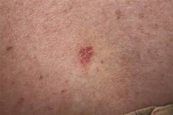
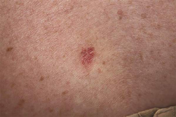
Ionizing radiation linked to BCC
SAN DIEGO – Patients treated with ionizing radiation were 2.65 to 3.78 times more likely to develop basal cell carcinoma than were controls, and exposure at younger ages or relatively high doses further increased that risk, according to a pooled analysis presented at the annual meeting of the American Society for Dermatologic Surgery.
“Concomitant exposure to ultraviolet radiation may potentiate this effect,” said Dr. Min Deng, a dermatology resident at the University of Chicago. “With newer treatment protocols and improved shielding, it would be interesting to see if the effect of ionizing radiation has changed, or whether we as dermatologists should more actively screen this at-risk population.”
Basal cell carcinoma (BCC) is the most common skin cancer worldwide, but relatively few dermatology papers have assessed the effects of ionizing radiation on the incidence of BCC, said Dr. Deng and coauthor Dr. Diana Bolotin, also of the University of Chicago.
To better understand the link, the researchers searched PubMed for controlled studies on the topic by using the terms “radiation,” “risk,” and “basal cell carcinoma.” They excluded case reports, animal studies, studies published in languages other than English, and trials of radiation as a treatment of BCC, they said. They also excluded studies of atomic bomb survivors, because exposure was uncontrolled and methods to estimate exposure in this group have changed over time, they noted.
In all, six studies published between 1991 and 2012 met the inclusion criteria, and all six showed a statistically significant relative risk or odds of BCC with ionizing radiation exposure, the investigators reported. Three analyses calculated the relative risk of BCC in patients who received radiation for tinea capitis or who underwent total-body irradiation before hematopoietic cell transplantation, they said. Two studies evaluated the odds of BCC in patients with a past history of radiation exposure, and one study assessed time to subsequent BCCs in patients with a history of BCC.
For the three studies that calculated relative risk, the researchers calculated a pooled RR of BCC after ionizing radiation treatment of 2.65 (95% confidence interval, 1.22-5.72) compared with controls. The study of total-body irradiation (TBI) did not report or control for the primary disease for which patients were treated, “which may have confounded the results,” they said. When they excluded that study from their calculation, the overall RR rose to 3.78 (95% CI, 2.62 to 5.44).
Notably, in the two studies of patients with tinea capitis, the relative risk of BCC fell by 12% to 16% for every additional year of age at which patients received radiation treatment, the researchers said. Similarly, risk of BCC dropped by 10.9% for every 1-year increase in age at total-body irradiation prior to cell transplantation.
The combined odds ratio for the next two studies was 4.28 (1.45-12.63). “Likewise, both studies found an elevated odds ratio with younger age at radiation exposure,” the researchers added. One study that stratified patients by type of medical condition detected a “markedly elevated” 8.7 odds of BCC after radiation treatment for acne (2.0 to 38.0), they noted.
The sixth study was a nested case-control analysis that found a statistically significant increase in the odds of BCC starting at a 1-Gy dose of ionizing radiation, and rising linearly up to doses of 35-63.3 Gy. “The risk for developing multiple BCCs also appears to be elevated in patients with a history of radiation therapy,” the researchers said. Patients who had been exposed to ionizing ratio were 2.3 times more likely to develop new BCCs compared with unexposed patients (1.7 to 3.1), they said.
The investigators reported no funding sources or conflicts of interest.
Key clinical point: Ionizing radiation therapy significantly increased risk of basal cell carcinoma, especially when patients were younger or were treated at relatively high doses.
Major finding: The pooled relative risk of BCC after ionizing radiation treatment was 2.65 (95% confidence interval, 1.22-5.72) compared with controls.
Data source: Pooled analysis of six studies of ionizing radiation exposure and risk or odds of basal cell carcinoma.
Disclosures: The researchers reported no funding sources or conflicts of interest.
Blinatumomab confirmed as treatment option in MRD+ ALL

SAN FRANCISCO—The first international, multicenter trial in acute lymphoblastic leukemia (ALL) using minimal residual disease (MRD) as a criterion for inclusion has confirmed clinical benefit for patients using a non-chemotherapeutic approach.
In the BLAST trial, blinatumomab, a bispecific T-cell engager antibody that directs cytotoxic T cells to CD19-positive cells, produced a complete MRD response in 80% of patients who were in complete hematologic remission but had quantifiable MRD at the time of treatment.
This echoes results of an earlier phase 2 study, in which blinatumomab produced an 80% MRD response rate in 20 patients with B-precursor ALL and persistent or relapsed MRD.
Nearly all patients with persistent or recurrent MRD relapse despite continued chemotherapy, according to Nicola Gökbuget, MD, PhD, of Goethe University Hospital in Frankfurt, Germany.
“The question,” she said, “is how to treat these patients.”
She presented one effective method, as shown by the BLAST trial, at the 2014 ASH Annual Meeting (abstract 379).
The trial enrolled 116 patients aged 18 or older with B-precursor ALL in complete hematologic remission but with MRD ≥ 10-3.
Patients could be in second or later remission, but they were excluded if they had a prior allogeneic stem cell transplant, circulating blasts or extra medullary ALL involvement, CNS pathology, prior chemotherapy within 2 weeks, or radiotherapy within 4 weeks.
Enrolled patients were a median age of 45 years (range, 18-76), and 34% were 65 or older. Most patients had high baseline MRD levels: 39% were ≥ 10-2 to < 10-1, and 45% were ≥ 10-3 to < 10-2.
Investigators evaluated MRD levels after each cycle. Analysis was performed in a central lab in Kiel, Germany, and was based on amplification of immunoglobulin and/or T-cell receptor gene rearrangements by PCR.
The primary endpoint was the proportion of patients achieving a complete MRD response after 1 cycle of blinatumomab.
Patients received 15 μg/m2 of blinatumomab per day by continuous intravenous infusion for 4 weeks, followed by a treatment-free period of 2 weeks. Responders could receive up to 4 cycles of therapy or receive a transplant any time after the first cycle.
Results
In the efficacy-evaluable population, 80% of 103 patients achieved a complete MRD response after 1 cycle, defined as MRD negative with no amplification in PCR with a minimum sensitivity of 10-4. And 85% achieved an incomplete MRD response of <10-4 with a minimum sensitivity of 10-4.
Results were similar in the full analysis set of 113 patients: 78% achieved complete MRD response after 1 cycle, and 85% achieved an incomplete MRD response.
Dr Gökbuget noted that 2 patients who initially achieved an incomplete response achieved a complete response during continued treatment in cycle 2.
The investigators analyzed the clinical characteristics of the patients and found that no factor—age, treatment interruptions, neurologic events, relapse history, nor gender—correlated with MRD response.
“I think that the good news is that it means that the compound was active in all of the patients,” Dr Gökbuget commented.
Adverse events
All patients experienced at least one adverse event (AE), and 2 were fatal—subdural hemorrhage and treatment-related pneumonitis. Serious treatment-related AEs that occurred in 3% or more of the
patients included tremor (7%), aphasia (5%), and encephalopathy (5%).
Thirty-one percent of patients interrupted their treatment because of AEs, which were primarily due to cytokine-related symptoms like pyrexia and neurologic events, including tremor, aphasia, and dizziness. Seventeen percent of patients permanently discontinued treatment due to an AE.
However, Dr Gökbuget pointed out that most AEs were grade 1 or 2, and the treatment interruption did not correlate with response.
She said the next step for this trial is to investigate whether high MRD response translates into long-term clinical benefit such as continued molecular remission and long-term survival.
“For me personally, this trial is very important because it is an up-to-date trial where we used PCR-based methods to identify patients with a high risk of relapse and treat them before the relapse occurs,” Dr Gökbuget said. “[W]e used a new endpoint, which is also MRD based, and . . . we used a new, non-chemotherapy treatment to eradicate this highly resistant, persistent ALL subclone.”
Blinatumomab received US Food and Drug Administration approval a few days before the start of the ASH Annual Meeting.
The BLAST trial was funded by Amgen Inc., the company developing blinatumomab. ![]()
FDA approves pathogen inactivation system for plasma
The US Food and Drug Administration (FDA) has approved the INTERCEPT Blood System for plasma, the first pathogen inactivation system for the preparation of plasma to reduce the risk of transfusion-transmitted infections.
The system inactivates pathogens through a photochemical process involving controlled exposure to ultraviolet light and the chemical amotosalen. The plasma is then purified to remove the chemical and its byproducts.
The INTERCEPT Blood System for plasma can be used to reduce pathogens in plasma derived from whole blood and plasma obtained by apheresis.
The system has proven effective in reducing a broad spectrum of viruses, bacteria, spirochetes, and parasites. However, there is no pathogen inactivation process that has been shown to eliminate all pathogens. Certain viruses (eg, human parvovirus B19) and spores formed by certain bacteria are known to be resistant to the INTERCEPT process.
“The approval of devices like the INTERCEPT Blood System allows blood establishments to prepare plasma that carries a lower risk of transmitting infectious pathogens through transfusion,” said Karen Midthun, MD, director of the FDA’s Center for Biologics Evaluation and Research.
The INTERCEPT Blood System for plasma, which is marketed by Cerus Corporation, has been approved in Europe since 2006 and was recently made available in the US under an Investigational Device Exemption study. The system is being used to prepare Ebola convalescent plasma for transfusion into acutely infected patients.
Clinical studies
Researchers investigated the safety and effectiveness of plasma prepared with the INTERCEPT Blood System in 6 clinical studies and 2 post-marketing studies.
The research showed no significant difference in coagulation factor activity between INTERCEPT-processed plasma and unprocessed fresh-frozen plasma (FFP) in healthy subjects.
There was no significant difference in prothrombin time or activated partial thromboplastin time between INTERCEPT-processed plasma and FFP in patients with coagulation factor deficiencies or in patients with liver disease.
In patients who underwent liver transplant, there was no significant difference in plasma use, hepatic artery thrombosis, or mortality whether patients received INTERCEPT-process plasma or FFP.
In a prospective, phase 3 trial and a retrospective study of patients with thrombotic thrombocytopenic purpura (TTP), there was no significant difference in the percentage of patients who achieved remission with INTERCEPT-treated plasma or FFP.
Adverse events
In a post-marketing study*, researchers analyzed acute transfusion reactions (ATRs) after 57,171 INTERCEPT-processed plasma components were transfused to 9669 patients. Thirty-two subjects (0.3%) experienced an ATR after 41 transfusion episodes (0.2%), including 5 subjects (0.05%) who experienced an ATR after more than 1 transfusion.
Six ATRs were considered serious and possibly or probably related to transfusion. There were 3 instances of allergic reaction or symptoms of allergic reaction (eg, rash, tachycardia, hypotension, respiratory symptoms, and chills), 2 cases of fluid overload, and 1 report of respiratory distress.
In another hemovigilance study, researchers compared the frequency of adverse events due to any of the 4 types of FFP prepared and delivered by the French Blood Establishment over a 10-year period—methylene blue, amotosalen, quarantine, and solvent-detergent.
The study showed that all types of FFP were associated with a low rate of adverse events. Per 10,000 deliveries, there were 7.14 events in the quarantine group, 4.86 in the solvent-detergent group, 4.16 in the amotosalen group, and 1.05 in the methylene blue group.
The package insert of the INTERCEPT Blood System for plasma warns that amotosalen-treated plasma has been associated with cardiac events.
In the aforementioned prospective trial of the system in patients with TTP, 5 patients treated with INTERCEPT-processed plasma and none with conventional plasma had adverse events in the cardiac system organ class.
This included angina pectoris (n=3), cardiac arrest (n=1), bradycardia (n=1), tachycardia (n=1), and sinus arrhythmia (n=1). None of the events resulted in documented myocardial infarction or death. ![]()
*Results have been updated since publication.
Newborn screening test for SCID gets FDA approval

Credit: Vera Kratochvil
The US Food and Drug Administration (FDA) has approved marketing of the EnLite Neonatal TREC Kit, the first screening test for severe combined immunodeficiency (SCID) in newborns.
Using a few drops of blood from the newborn’s heel, which is dried on filter paper, the EnLite Neonatal TREC Kit can determine whether T-cell receptor excision circles (TREC) DNA is low or missing from the newborn’s blood.
Newborns with SCID typically have no or low amounts of TREC DNA compared to healthy infants.
Additional testing is required to obtain a SCID diagnosis once the EnLite Neonatal TREC Kit has suggested a child may have SCID.
“SCID is a fatal disease that can be treated with early intervention, including screening,” said Alberto Gutierrez, PhD, director of the Office of In Vitro Diagnostics and Radiological Health in the FDA’s Center for Devices and Radiological Health.
“For the first time, the FDA is allowing the marketing of a newborn screening test that will enable states to incorporate an FDA-reviewed SCID test into their standard newborn screening panels and allow earlier identification for affected individuals.”
The FDA reviewed the EnLite Neonatal TREC Kit through its de novo classification process, a regulatory pathway for some novel, low- to moderate-risk medical devices that are not substantially equivalent to an already legally marketed device.
The FDA’s review included a clinical study of approximately 6400 blood spot specimens from routine screening of newborns, 17 of which had a confirmed SCID diagnosis. The EnLite Neonatal TREC Kit correctly identified all 17 SCID cases.
The agency also evaluated the test’s ability to accurately distinguish low TREC DNA numbers that would be observed in newborns with SCID from high TREC DNA numbers that would be present in healthy newborns. The FDA found the EnLite Neonatal TREC Kit could adequately detect very low TREC DNA values that are associated with SCID.
The EnLite Neonatal TREC Kit is not intended for use as a diagnostic test or to screen for SCID-like syndromes, such as DiGeorge syndrome or Omenn syndrome. Likewise, it is not intended to screen for less acute SCID syndromes, such as leaky SCID or variant SCID.
The EnLite Neonatal TREC Kit is manufactured by Wallac Oy, a subsidiary of PerkinElmer, at its facility in Turku, Finland. PerkinElmer is based in Waltham, Massachusetts. ![]()
Study reveals superior antiviral drug for lymphoma

A study of lymphoma patients receiving R-CHOP chemotherapy showed that use of the antiviral drug entecavir resulted in a lower incidence of hepatitis B virus (HBV)-related hepatitis and HBV reactivation, when compared with the drug lamivudine.
HBV reactivation is a well-documented complication of chemotherapy that can result in life-threatening liver failure, as well as delays in chemotherapy or
premature termination of treatment.
However, researchers have not identified an optimal approach to prevent HBV reactivation.
So He Huang, MD, of the Sun Yat-sen University Cancer Center in Guangzhou, China, and colleagues set out to do that. They reported their findings in JAMA alongside a related editorial.
The team enrolled 121 patients with untreated diffuse large B-cell lymphoma who were receiving chemotherapy with rituximab, cyclophosphamide, doxorubicin, vincristine, and prednisone (R-CHOP) and were seropositive for the hepatitis B surface antigen.
Patients were randomized to receive entecavir (n=61) or lamivudine (n=60). They began taking these drugs 1 week before the initiation of R-CHOP and continued until 6 months after they completed chemotherapy.
The study was conducted from February 2008 through December 2012 at 10 medical centers in China. This trial was a substudy of a parent study designed to compare a 3-week R-CHOP regimen with a 2-week R-CHOP regimen.
Results showed that, compared to the lamivudine group, patients in the entecavir group had significantly lower rates of hepatitis (8.2% vs 23.3%, P=0.02), HBV-related hepatitis (0% vs 13.3%, P=0.003), HBV reactivation (6.6% vs 30%, P=0.001), delayed hepatitis B (0% vs 8.3%, P=0.03), and chemotherapy disruption (1.6% vs 18.3%, P=0.002).
The rate of treatment-related adverse events was not significantly different between the groups. Such events occurred in 24.6% of patients in the entecavir group and 30.0% of patients in the lamivudine group (P=0.50).
The researchers noted that entecavir is more expensive than lamivudine, so additional studies are needed to determine whether all patients who are seropositive for the hepatitis B surface antigen and are receiving rituximab-based immunosuppressive therapy should be given entecavir.
Additional studies could also help pinpoint which patients will benefit most from entecavir prophylaxis. However, if these findings are replicated, they support the use of entecavir in these patients. ![]()
Evaluating the impact of FDA’s pregnancy and lactation labeling rule
Since 1979, obstetric and other health care providers who treat pregnant or potentially pregnant and breastfeeding women have relied heavily on the Food and Drug Administration’s pregnancy labeling categories for pharmaceuticals – the familiar A, B, C, D, X. However, as early as 1997, a public hearing was held that challenged the value of these labels as typically used in clinical practice by both providers and patients.
Now, 17 years later, in December 2014, the FDA has released the “Pregnancy and Lactation Labeling Rule” (also known as PLLR or final rule). In brief, the revised label will require that:
• Contact information be prominently listed for a pregnancy exposure registry for the drug, when one is available.
• Narrative sections be presented that include a risk summary, clinical considerations, and the supporting data.

• A lactation subsection be included that provides information about using the drug while breastfeeding, such as the amount of drug in breast milk and potential effects on the breastfed infant.
• A subsection on females and males of reproductive potential be included, when necessary, with information about the need for pregnancy testing, contraception recommendations, and information about infertility as it relates to the drug.
The labeling changes go into effect on June 30, 2015. Prescription drugs and biologic products submitted for FDA review after that date will use the new format immediately, while labeling for prescription drugs approved on or after June 30, 2001, will be phased in gradually.
Why are these changes needed, and what impact will they likely have for clinical practice?
First a bit of history – the pregnancy label A, B, C, D, X categories were initially introduced in the 1970s, following the recognition that thalidomide was a human teratogen. The intention was to help the clinician deal in a more standardized way with the increasing amount of experimental animal data and human reports generated for drugs that might be used by women of reproductive potential.
However, the letter categories, and their accompanying standard text statements, were frequently misinterpreted in oversimplistic and inaccurate ways (Birth Defects Res. Part A 2007,79:627-30). Clinicians and patients often believe that risk increases as you move across the letter categories; for example, that a category C drug is worse than a category B drug for a given patient.
Clinicians and patients also commonly think that drugs in the same category have the same level of risk, or that there is a similar quantity and quality of information to support that risk category. Frequently cited examples of misinterpretation include those drugs assigned a category X, for example, label text indicating that the drug is “contraindicated in women who are or may become pregnant.” In reality, in some cases, the X category has been applied to drugs with known human teratogenic potential (such as isotretinoin or thalidomide). However, in other cases, the X has been assigned to drugs for which there were no or very limited human data indicating risk (such as ribavirin or leflunomide) or for which the treatment for the underlying condition would not be necessary or advisable in pregnancy (such as statins or some weight loss drugs).
There is no differentiation made in the category X label for varying risks specifically related to dose and timing of exposure in gestation. In each of these situations where there are no clear-cut human data, inadvertent exposure to the drug in an unplanned pregnancy can easily lead to the misunderstanding that the drug is known to cause birth defects in humans.
The immediate impact of the PLLR label revision will be to require narrative sections that describe the actual data, provide a summary of risks, and also present clinical considerations that may include the risk of undertreatment or no treatment with the drug. The new format is intended to provide the clinician (and the patient) with more comprehensive information on which to base decisions.
The downside of the label revision is that clinicians will have to learn to interpret more comprehensive information and deal with the unknowns, which are many.
The other major impact of the label revision will be to highlight the clear lack of sufficient human data for most drugs currently marketed in the United States. A recent review of drugs (both prescription and over the counter) approved by the FDA between 2000 and 2010 found that 73.3% had no human data available that was relevant to pregnancy safety (Am. J. Med. Genet. C. Semin. Med. Genet. 157C:175-82).
In the short term, the learning curve for label writers and the end users of such labels will be steep. However, clinicians and patients can contribute to the compilation of data for most drugs by more proactively engaging in pregnancy and lactation safety studies. Information about the existence of any pregnancy registries in drug labeling has been recommended in the past, but will now be required. Acting on that information by enrolling patients in these studies can ensure that labels can more quickly be populated with evidence-based data that can better inform patient care.
Dr. Chambers is a professor of pediatrics and director of clinical research at Rady Children’s Hospital, and associate director of the Clinical and Translational Research Institute at the University of California, San Diego. She is director of MotherToBaby California, a past president of the Organization of Teratology Information Specialists, and past president of the Teratology Society. She has no relevant financial disclosures. To comment, e-mail her at [email protected].
Since 1979, obstetric and other health care providers who treat pregnant or potentially pregnant and breastfeeding women have relied heavily on the Food and Drug Administration’s pregnancy labeling categories for pharmaceuticals – the familiar A, B, C, D, X. However, as early as 1997, a public hearing was held that challenged the value of these labels as typically used in clinical practice by both providers and patients.
Now, 17 years later, in December 2014, the FDA has released the “Pregnancy and Lactation Labeling Rule” (also known as PLLR or final rule). In brief, the revised label will require that:
• Contact information be prominently listed for a pregnancy exposure registry for the drug, when one is available.
• Narrative sections be presented that include a risk summary, clinical considerations, and the supporting data.
• A lactation subsection be included that provides information about using the drug while breastfeeding, such as the amount of drug in breast milk and potential effects on the breastfed infant.
• A subsection on females and males of reproductive potential be included, when necessary, with information about the need for pregnancy testing, contraception recommendations, and information about infertility as it relates to the drug.
The labeling changes go into effect on June 30, 2015. Prescription drugs and biologic products submitted for FDA review after that date will use the new format immediately, while labeling for prescription drugs approved on or after June 30, 2001, will be phased in gradually.
Why are these changes needed, and what impact will they likely have for clinical practice?
First a bit of history – the pregnancy label A, B, C, D, X categories were initially introduced in the 1970s, following the recognition that thalidomide was a human teratogen. The intention was to help the clinician deal in a more standardized way with the increasing amount of experimental animal data and human reports generated for drugs that might be used by women of reproductive potential.
However, the letter categories, and their accompanying standard text statements, were frequently misinterpreted in oversimplistic and inaccurate ways (Birth Defects Res. Part A 2007,79:627-30). Clinicians and patients often believe that risk increases as you move across the letter categories; for example, that a category C drug is worse than a category B drug for a given patient.
Clinicians and patients also commonly think that drugs in the same category have the same level of risk, or that there is a similar quantity and quality of information to support that risk category. Frequently cited examples of misinterpretation include those drugs assigned a category X, for example, label text indicating that the drug is “contraindicated in women who are or may become pregnant.” In reality, in some cases, the X category has been applied to drugs with known human teratogenic potential (such as isotretinoin or thalidomide). However, in other cases, the X has been assigned to drugs for which there were no or very limited human data indicating risk (such as ribavirin or leflunomide) or for which the treatment for the underlying condition would not be necessary or advisable in pregnancy (such as statins or some weight loss drugs).
There is no differentiation made in the category X label for varying risks specifically related to dose and timing of exposure in gestation. In each of these situations where there are no clear-cut human data, inadvertent exposure to the drug in an unplanned pregnancy can easily lead to the misunderstanding that the drug is known to cause birth defects in humans.
The immediate impact of the PLLR label revision will be to require narrative sections that describe the actual data, provide a summary of risks, and also present clinical considerations that may include the risk of undertreatment or no treatment with the drug. The new format is intended to provide the clinician (and the patient) with more comprehensive information on which to base decisions.
The downside of the label revision is that clinicians will have to learn to interpret more comprehensive information and deal with the unknowns, which are many.
The other major impact of the label revision will be to highlight the clear lack of sufficient human data for most drugs currently marketed in the United States. A recent review of drugs (both prescription and over the counter) approved by the FDA between 2000 and 2010 found that 73.3% had no human data available that was relevant to pregnancy safety (Am. J. Med. Genet. C. Semin. Med. Genet. 157C:175-82).
In the short term, the learning curve for label writers and the end users of such labels will be steep. However, clinicians and patients can contribute to the compilation of data for most drugs by more proactively engaging in pregnancy and lactation safety studies. Information about the existence of any pregnancy registries in drug labeling has been recommended in the past, but will now be required. Acting on that information by enrolling patients in these studies can ensure that labels can more quickly be populated with evidence-based data that can better inform patient care.
Dr. Chambers is a professor of pediatrics and director of clinical research at Rady Children’s Hospital, and associate director of the Clinical and Translational Research Institute at the University of California, San Diego. She is director of MotherToBaby California, a past president of the Organization of Teratology Information Specialists, and past president of the Teratology Society. She has no relevant financial disclosures. To comment, e-mail her at [email protected].

Death penalty opponents rally to protest execution date for mentally ill prisoner
According to the last Gallup poll, most Americans still favor the death penalty. Nevertheless, over the years, the number of capital-eligible defendants has been gradually whittled down because of challenges over the execution of juveniles, the intellectually disabled, and for defendants guilty of crimes other than murder. The latest challenge came up again in December, when lawyers for Texas death row inmate Scott Panetti sought a reprievealleging that he was incompetent to understand the reason for his execution.
Scott Panetti had a longstanding history of schizoaffective disorder dating back to the age of 20 years and had been hospitalized for treatment of his hallucinations and delusions at least 14 times. He once was convinced that the devil had possessed his home and had buried several valuables next to the house to “cleanse” it. His wife sought emergency intervention once when he threatened to kill her. In 1992, dressed in camouflage, he broke into the home of his estranged wife’s parents and killed them in front of her. He took his wife and young daughter hostage overnight but eventually surrendered to police.
At trial he wanted to represent himself, so the court ordered an evaluation of his competence to waive counsel. He was found competent and proceeded to present his own insanity defense to a jury. By the time of trial, he had been off medication for 2 months, and his behavior during the hearing was later described as bizarre and confusing at best. He came to court dressed in a cowboy suit, referred to himself as his alter-ego “Sarge,” and attempted to subpoena the Pope. He was convicted, in part, because jurors feared his behavior enough that they wanted to ensure he would never be released. He was sentenced to death.
Panetti received appointed counsel for his appeal after he was found incompetent to waive representation. His execution date was set in 2003, but the defense filed a motion alleging that he was incompetent to be put to death. The motion was accompanied by supporting documentation from a psychologist that Panetti did not understand the reasons for his punishment. The court appointed two experts who disagreed, stating that Panetti knew that he was about to be put to death and that he had the ability to understand the reason for the execution. They also implied that Panetti’s bizarre behavior in court was a calculated plan to present himself as insane.
Without a hearing, the court found Panetti competent. Panetti appealed to the Supreme Court, stating that under an earlier Supreme Court case, Ford v. Wainwright, he had a right to have a hearing on the competency issue, which would allow him to challenge the state’s experts and to present contrary medical evidence. The question at appeal was not whether execution of the mentally ill per se was cruel and unusual, but what standard should be applied to determine competency, and whether Texas followed the proper procedure to determine it.
Under Ford v. Wainwright, the bar was set extremely low. In order to be competent for execution, a defendant had only to know that he was being put to death, and that the death sentence was a punishment for a crime he had committed. This was a standard that required no more than intellectual or factual knowledge. Panetti was found competent to be executed because he could recite these facts. However, he disagreed with the facts: He believed that he was being executed by the state to prevent him from preaching the gospel. The Court of Appeals held that delusions alone would not necessarily mean that someone was incompetent. In 2007, the Supreme Court held in Panetti v. Quartermanthat competency requires a prisoner to have a rational as well as a factual understanding of the execution.
The American Psychiatric Association, the American Psychological Association, and the National Alliance on Mental Illness all joined to file an amicus brief on the case. In the brief, all the organizations agreed that “a prisoner is not competent to be executed if he ‘has a mental disorder or disability that significantly impairs his or her capacity to understand the nature and purpose of the punishment, or to appreciate the reason for its imposition in the prisoner’s own case.’ ” In other words, punishment is futile if the person in question doesn’t have the ability to understand that he is being punished.
The case was sent back for a formal hearing, and Panetti was again found competent. This time, the Supreme Court refused to hear his appeal. An execution date was set for Dec. 3 but was stayed 7 hours before he was to be put to death. Another competency assessment is probably on the horizon, given that 7 years have passed since the last one. The outcome this time will probably be the same, since Panetti reportedly has been on no medications for most of the last 20 years, and he has not been diagnosed with a psychotic illness in the prison system by any of the 14 mental health staff who have evaluated him.
Of course, none of this litigation to date settles the question of whether our judiciary should be executing anyone, with or without a mental illness. But until that question is settled, psychiatry will struggle along with the courts to decide exactly how ill is too ill to die.
Dr. Hanson is a forensic psychiatrist and coauthor of “Shrink Rap: Three Psychiatrists Explain Their Work.” The opinions expressed are those of the author only, and do not represent those of any of Dr. Hanson’s employers or consultees, including the Maryland Department of Health and Mental Hygiene or the Maryland Division of Correction.

Using, and not using, antipsychotic medications
Introduction
Both the medical and lay press have directed a lot of attention lately to the treatment of children and adolescents with antipsychotic medications. The literature is clear that the number of children taking this class of medications has risen sharply since their release (Arch. Gen. Psychiatry 2006;63:679-85). What is much less clear is the degree to which this increase represents a reasonable intervention for patients in significant need versus an overuse when other strategies are more appropriate.
Case Summary
Cody is a 6-year-old boy who lives with his younger sister and single mother. The family struggles financially, and the father, who has never had much contact with his son, is currently incarcerated. Since he was a toddler, Cody has been prone to high levels of aggressive behavior and frequent, intense angry outbursts. He was asked to leave his preschool due to his behavior and now is commonly disruptive at school. His pediatrician diagnosed him with attention-deficit/hyperactivity disorder a year ago and began a trial of a psychostimulant, which made him even more irritable, and was discontinued. Cody and his mother now present with concerns that there is “something more” affecting his behavior. The pediatrician now considers whether or not treatment with an antipsychotic medication is reasonable at this point.

Discussion
The above clinical scenario represents a critical and often antagonizing moment in treatment for both the family and the treating physician, yet it is hardly uncommon. The situation often is made more complicated by the fact that what is often the first plan of action, namely referral or consultation with a child psychiatrist, can be very difficult to access.
The American Academy of Child and Adolescent Psychiatry has published online guidelines for the use of antipsychotic medication in youth (http://bit.ly/1eat7e9). Key recommendations and points from this 27-page document and 19 recommendations include the following:
• Patients being considered for treatment with an antipsychotic medication should receive a “meticulous diagnostic assessment” with any medication prescribed being part of a “multidisciplinary” treatment plan (Recommendation 1).
• Prescribers should “regularly check the current literature” regarding the scientific evidence for antipsychotic medication use (Recommendation 2).
• Antipsychotic medications are considered first-line medication treatment for bipolar disorder, schizophrenia, tics/Tourette’s, and autism. (Recommendation 2).
• Antipsychotic medications are not first-line treatment for several other diagnoses and behaviors, including disruptive behavior disorders such as ADHD, aggression, eating disorders, and post-traumatic stress disorder (PTSD). Their use should be considered only after other pharmacologic and nonpharmacologic interventions have failed (Recommendation 2).
• Antipsychotic medications are not advised for preschool-aged patients. (Recommendation 2).
• Dosing should be as low as possible and not exceed the maximum recommended dose for adults (Recommendation 4).
• Simultaneous treatment with multiple antipsychotic medications is not recommended (Recommendation 8).
• Patients should receive regular metabolic monitoring, including lab work, both before and during treatment (Recommendations 11-13).
These are rigorous guidelines that challenge even those who regularly assess and treat children with serious psychiatric disorders. The clinical and legal implications of prescribing antipsychotic medications without adhering to these guidelines will, and probably should, give many physicians pause. Further, the specific point about the need for a thorough psychiatric evaluation underlies the commonly heard recommendation that this class of medicines generally should be avoided by primary care physicians. At the same time, many pediatricians are acutely aware of how dire the clinical situation often is for these families. At this point, it can easily begin to feel very much like a “no-win” situation.
Here are some thoughts that may be useful to consider in these moments:
• Remember that many non-MD mental health professionals can offer a lot of help. Although they can’t do the prescribing themselves, referral to a psychologist or another type of therapist can be useful in getting information about a patient’s diagnosis and the degree to which nonpharmacologic options have been exhausted. If the patient is already seeing a therapist, it is certainly worthwhile to seek their advice as to whether or not antipsychotic medications are now reasonable to consider.
• Look for opportunities to talk “curbside” to a child psychiatrist. Most of us are keenly aware of how inadequate access is to child psychiatry and want to help. Indeed, many states now have specific brief consultation programs in place.
• Get the lab work. A recent study in Pediatrics reported that a baseline glucose was obtained in only 11% of youth receiving antipsychotic medication treatment (Pediatrics 2014;134:e1308-14). In addition to providing important information, this step signals to everyone involved that the decision to use these medications is not something to be taken lightly.
Case follow-up
Cody’s pediatrician decides to get a diagnostic evaluation from a psychologist, who confirms the ADHD diagnosis without associated conditions such as bipolar disorder. The psychologist recommends a course of therapy to build regulatory skills for Cody and provide the mother with some parent behavioral guidance about how to best manage Cody’s challenges and encourage health-promoting behaviors such as physical activity, reading, and a regular sleep routine. The pediatrician decides to try a second line ADHD medication, guanfacine, and the school also begins to institute an incentive plan to reinforce positive behavior. In combination, these efforts significantly reduce the level of aggression and dysregulated behavior.
Dr. Rettew is an associate professor of psychiatry and pediatrics at the University of Vermont, Burlington. Dr. Rettew said he has no relevant financial disclosures. Follow him on Twitter @pedipsych.
Introduction
Both the medical and lay press have directed a lot of attention lately to the treatment of children and adolescents with antipsychotic medications. The literature is clear that the number of children taking this class of medications has risen sharply since their release (Arch. Gen. Psychiatry 2006;63:679-85). What is much less clear is the degree to which this increase represents a reasonable intervention for patients in significant need versus an overuse when other strategies are more appropriate.
Case Summary
Cody is a 6-year-old boy who lives with his younger sister and single mother. The family struggles financially, and the father, who has never had much contact with his son, is currently incarcerated. Since he was a toddler, Cody has been prone to high levels of aggressive behavior and frequent, intense angry outbursts. He was asked to leave his preschool due to his behavior and now is commonly disruptive at school. His pediatrician diagnosed him with attention-deficit/hyperactivity disorder a year ago and began a trial of a psychostimulant, which made him even more irritable, and was discontinued. Cody and his mother now present with concerns that there is “something more” affecting his behavior. The pediatrician now considers whether or not treatment with an antipsychotic medication is reasonable at this point.
Discussion
The above clinical scenario represents a critical and often antagonizing moment in treatment for both the family and the treating physician, yet it is hardly uncommon. The situation often is made more complicated by the fact that what is often the first plan of action, namely referral or consultation with a child psychiatrist, can be very difficult to access.
The American Academy of Child and Adolescent Psychiatry has published online guidelines for the use of antipsychotic medication in youth (http://bit.ly/1eat7e9). Key recommendations and points from this 27-page document and 19 recommendations include the following:
• Patients being considered for treatment with an antipsychotic medication should receive a “meticulous diagnostic assessment” with any medication prescribed being part of a “multidisciplinary” treatment plan (Recommendation 1).
• Prescribers should “regularly check the current literature” regarding the scientific evidence for antipsychotic medication use (Recommendation 2).
• Antipsychotic medications are considered first-line medication treatment for bipolar disorder, schizophrenia, tics/Tourette’s, and autism. (Recommendation 2).
• Antipsychotic medications are not first-line treatment for several other diagnoses and behaviors, including disruptive behavior disorders such as ADHD, aggression, eating disorders, and post-traumatic stress disorder (PTSD). Their use should be considered only after other pharmacologic and nonpharmacologic interventions have failed (Recommendation 2).
• Antipsychotic medications are not advised for preschool-aged patients. (Recommendation 2).
• Dosing should be as low as possible and not exceed the maximum recommended dose for adults (Recommendation 4).
• Simultaneous treatment with multiple antipsychotic medications is not recommended (Recommendation 8).
• Patients should receive regular metabolic monitoring, including lab work, both before and during treatment (Recommendations 11-13).
These are rigorous guidelines that challenge even those who regularly assess and treat children with serious psychiatric disorders. The clinical and legal implications of prescribing antipsychotic medications without adhering to these guidelines will, and probably should, give many physicians pause. Further, the specific point about the need for a thorough psychiatric evaluation underlies the commonly heard recommendation that this class of medicines generally should be avoided by primary care physicians. At the same time, many pediatricians are acutely aware of how dire the clinical situation often is for these families. At this point, it can easily begin to feel very much like a “no-win” situation.
Here are some thoughts that may be useful to consider in these moments:
• Remember that many non-MD mental health professionals can offer a lot of help. Although they can’t do the prescribing themselves, referral to a psychologist or another type of therapist can be useful in getting information about a patient’s diagnosis and the degree to which nonpharmacologic options have been exhausted. If the patient is already seeing a therapist, it is certainly worthwhile to seek their advice as to whether or not antipsychotic medications are now reasonable to consider.
• Look for opportunities to talk “curbside” to a child psychiatrist. Most of us are keenly aware of how inadequate access is to child psychiatry and want to help. Indeed, many states now have specific brief consultation programs in place.
• Get the lab work. A recent study in Pediatrics reported that a baseline glucose was obtained in only 11% of youth receiving antipsychotic medication treatment (Pediatrics 2014;134:e1308-14). In addition to providing important information, this step signals to everyone involved that the decision to use these medications is not something to be taken lightly.
Case follow-up
Cody’s pediatrician decides to get a diagnostic evaluation from a psychologist, who confirms the ADHD diagnosis without associated conditions such as bipolar disorder. The psychologist recommends a course of therapy to build regulatory skills for Cody and provide the mother with some parent behavioral guidance about how to best manage Cody’s challenges and encourage health-promoting behaviors such as physical activity, reading, and a regular sleep routine. The pediatrician decides to try a second line ADHD medication, guanfacine, and the school also begins to institute an incentive plan to reinforce positive behavior. In combination, these efforts significantly reduce the level of aggression and dysregulated behavior.
Dr. Rettew is an associate professor of psychiatry and pediatrics at the University of Vermont, Burlington. Dr. Rettew said he has no relevant financial disclosures. Follow him on Twitter @pedipsych.
