User login
Experts debate venous sinus stenting for intracranial hypertension
SCOTTSDALE, ARIZ. – Venous sinus stenting remains a controversial treatment for headache associated with benign intracranial hypertension. Opponents highlight potentially serious adverse effects and a lack of rigorous studies on the procedure, while supporters describe it as safe and effective. Experts recently debated the procedure at a symposium sponsored by the American Headache Society.
Which came first?
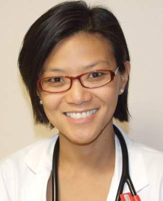
Stenting is based on the rationale that lowering venous sinus pressure might lower intracranial pressure, said Dr. Deborah Friedman, a neuro-ophthalmologist at the University of Texas Southwestern Medical Center in Dallas.

“Other surgical techniques create a fistula the body wants to close,” she said. “Stenting may provide a permanent solution.”
The crux of the debate on stenting is whether venous sinus stenosis is a cause or outcome of benign intracranial hypertension (BIH), said Dr. Felipe Albuquerque, an endovascular neurosurgeon at the Barrow Neurological Institute in Phoenix. The fact that 90% of affected patients have venous sinus stenosis on MRI suggests stenosis is a valid etiology the condition, he said.
But Dr. Friedman disagreed. In a retrospective study of 51 patients with BIH and transverse sinus stenosis, 71% had more than 50% stenosis, but the degree and location of stenosis did not predict clinical outcome. The analysis also found no link between cerebrospinal fluid pressure and the location, degree, or residual area of stenosis. Nonetheless, the procedure may be useful in patients who have persistent transverse sinus stenosis after undergoing a shunt procedure, she said.
The cons of stenting
“Stenting carries a high risk of morbidity and mortality, compared with currently used treatments, and should only be used as a last resort in patients who are losing vision,” Dr. Friedman asserted. Most patients do well, but others suffer serious complications, including life-threatening anaphylaxis, subdural hematomas, subarachnoid hemorrhage, and brain herniation, she said.
In contrast, optic nerve sheath fenestration for BIH has no associated fatalities, according to Dr. Friedman. And in a National Inpatient Sample database analysis of 1,224 shunts, mortality was only 0.9% for ventriculoperitoneal shunt and 0.3% for lumbar-peritoneal shunt. No deaths were recorded for shunts performed for BIH or pseudotumor cerebri, she added.
The literature on venous sinus stenting “is a mess,” Dr. Friedman continued. “All data are from retrospective case series, and the diagnosis of idiopathic intracranial hypertension is questionable in some cases,” she said. “The indication for stenting is not well defined.”
Studies on the safety and efficacy of stenting have lacked a common primary outcome variable and may reflect reporting bias, Dr. Friedman said. Studies have been heterogeneous with regard to disease duration and the presence or absence of papilledema, visual status, previous treatments, and definitions of treatment failure, she added. No randomized, controlled trials have been carried out with sham stenting to assess the possibility of placebo effect, she noted.
“I think stenting may have utility in patients who have failed conventional therapy and have had another procedure that did not work. I think it very likely that there is a strong placebo effect in this group,” Dr. Friedman said.
“It’s not like our existing procedures are wonderful,” she added. “I wish we had something better to offer our patients.”
The pros
Venous sinus stenting “is usually effective in ameliorating both subjective headache and objective papilledema symptoms,” Dr. Albuquerque said. “One could argue that both ventriculoperitoneal shunt and lumbar-peritoneal shunt, the most commonly performed surgical interventions for benign intracranial hypertension, are substantially more invasive than stenting is and associated with far more severe complications.”
Dr. Albuquerque described a prospective study he conducted with his associates on 15 patients with BIH who underwent venous sinus stenting. In all, 80% of patients said their headaches improved, and 60% reported at least a 50% decrease in headache pain. Rates of patency and technical success were 100%, and no patients had permanent complications from the procedure, although one patient developed acute retroperitoneal hematoma, he said.
Dr. Albuquerque also reported his long-term follow-up of 27 patients who underwent venous sinus stenting to treat pseudotumors. All patients had more than 50% stenosis confirmed by retrograde venogram and a transstenotic pressure gradient that was greater than 12 mm Hg. Fully 70% of patients improved symptomatically, but five underwent a shunt procedure after they failed to improve. Patients experienced no permanent complications, although one required a stent for femoral artery pseudoaneurysm. Angiographies performed an average of 23 months later showed that all stents remained patent, although four patients had mild (less than 25%) stenosis. Five patients had narrowing of the sinus proximal to the stent.
Patients need dual antiplatelet therapy after venous sinus stenting, and the rate of chronic patency after the procedure is unknown, Dr. Albuquerque noted. Patients can develop scalp pain over the stented segment, he added.
“I think if you limit this procedure to a very select group of patients, I think its efficacy is tremendous,” Dr. Albuquerque concluded.
Dr. Albuquerque declared no conflicts of interest. Dr. Friedman reported serving on the speakers bureau of Allergan, receiving research grants from the National Eye Institute, Merck & Co., and ElectroCore, and having served as an expert witness (for the plaintiff and defense) on idiopathic intracranial hypertension.
SCOTTSDALE, ARIZ. – Venous sinus stenting remains a controversial treatment for headache associated with benign intracranial hypertension. Opponents highlight potentially serious adverse effects and a lack of rigorous studies on the procedure, while supporters describe it as safe and effective. Experts recently debated the procedure at a symposium sponsored by the American Headache Society.
Which came first?
Stenting is based on the rationale that lowering venous sinus pressure might lower intracranial pressure, said Dr. Deborah Friedman, a neuro-ophthalmologist at the University of Texas Southwestern Medical Center in Dallas.
“Other surgical techniques create a fistula the body wants to close,” she said. “Stenting may provide a permanent solution.”
The crux of the debate on stenting is whether venous sinus stenosis is a cause or outcome of benign intracranial hypertension (BIH), said Dr. Felipe Albuquerque, an endovascular neurosurgeon at the Barrow Neurological Institute in Phoenix. The fact that 90% of affected patients have venous sinus stenosis on MRI suggests stenosis is a valid etiology the condition, he said.
But Dr. Friedman disagreed. In a retrospective study of 51 patients with BIH and transverse sinus stenosis, 71% had more than 50% stenosis, but the degree and location of stenosis did not predict clinical outcome. The analysis also found no link between cerebrospinal fluid pressure and the location, degree, or residual area of stenosis. Nonetheless, the procedure may be useful in patients who have persistent transverse sinus stenosis after undergoing a shunt procedure, she said.
The cons of stenting
“Stenting carries a high risk of morbidity and mortality, compared with currently used treatments, and should only be used as a last resort in patients who are losing vision,” Dr. Friedman asserted. Most patients do well, but others suffer serious complications, including life-threatening anaphylaxis, subdural hematomas, subarachnoid hemorrhage, and brain herniation, she said.
In contrast, optic nerve sheath fenestration for BIH has no associated fatalities, according to Dr. Friedman. And in a National Inpatient Sample database analysis of 1,224 shunts, mortality was only 0.9% for ventriculoperitoneal shunt and 0.3% for lumbar-peritoneal shunt. No deaths were recorded for shunts performed for BIH or pseudotumor cerebri, she added.
The literature on venous sinus stenting “is a mess,” Dr. Friedman continued. “All data are from retrospective case series, and the diagnosis of idiopathic intracranial hypertension is questionable in some cases,” she said. “The indication for stenting is not well defined.”
Studies on the safety and efficacy of stenting have lacked a common primary outcome variable and may reflect reporting bias, Dr. Friedman said. Studies have been heterogeneous with regard to disease duration and the presence or absence of papilledema, visual status, previous treatments, and definitions of treatment failure, she added. No randomized, controlled trials have been carried out with sham stenting to assess the possibility of placebo effect, she noted.
“I think stenting may have utility in patients who have failed conventional therapy and have had another procedure that did not work. I think it very likely that there is a strong placebo effect in this group,” Dr. Friedman said.
“It’s not like our existing procedures are wonderful,” she added. “I wish we had something better to offer our patients.”
The pros
Venous sinus stenting “is usually effective in ameliorating both subjective headache and objective papilledema symptoms,” Dr. Albuquerque said. “One could argue that both ventriculoperitoneal shunt and lumbar-peritoneal shunt, the most commonly performed surgical interventions for benign intracranial hypertension, are substantially more invasive than stenting is and associated with far more severe complications.”
Dr. Albuquerque described a prospective study he conducted with his associates on 15 patients with BIH who underwent venous sinus stenting. In all, 80% of patients said their headaches improved, and 60% reported at least a 50% decrease in headache pain. Rates of patency and technical success were 100%, and no patients had permanent complications from the procedure, although one patient developed acute retroperitoneal hematoma, he said.
Dr. Albuquerque also reported his long-term follow-up of 27 patients who underwent venous sinus stenting to treat pseudotumors. All patients had more than 50% stenosis confirmed by retrograde venogram and a transstenotic pressure gradient that was greater than 12 mm Hg. Fully 70% of patients improved symptomatically, but five underwent a shunt procedure after they failed to improve. Patients experienced no permanent complications, although one required a stent for femoral artery pseudoaneurysm. Angiographies performed an average of 23 months later showed that all stents remained patent, although four patients had mild (less than 25%) stenosis. Five patients had narrowing of the sinus proximal to the stent.
Patients need dual antiplatelet therapy after venous sinus stenting, and the rate of chronic patency after the procedure is unknown, Dr. Albuquerque noted. Patients can develop scalp pain over the stented segment, he added.
“I think if you limit this procedure to a very select group of patients, I think its efficacy is tremendous,” Dr. Albuquerque concluded.
Dr. Albuquerque declared no conflicts of interest. Dr. Friedman reported serving on the speakers bureau of Allergan, receiving research grants from the National Eye Institute, Merck & Co., and ElectroCore, and having served as an expert witness (for the plaintiff and defense) on idiopathic intracranial hypertension.
EXPERT ANALYSIS AT THE SCOTTSDALE HEADACHE SYMPOSIUM
At-home radiofrequency devices
The field of body contouring and tissue tightening has expanded over the years, with many new devices appearing on the market that utilize radiofrequency (RF) energy to tighten and rejuvenate the skin. What originally began with a single monopolar RF device has progressed to a world in which there are skin-tightening devices that use bipolar energy and tripolar energy, as well as monopolar, and newer machines that boast five and eight poles of RF energy.
In addition to in-office radiofrequency devices, at-home devices are now available.
Radiofrequency energy uses the tissue’s resistance within the various layers of the skin to transform the RF energy given to the skin into thermal energy. This process induces collagen remodeling and neocollagenesis, resulting in skin tightening. Since RF energy produces an electrical current instead of a light source like lasers, tissue damage can be minimized, and epidermal melanin is not targeted or typically damaged. Therefore, RF energies can be used for patients of all skin types and colors. Adverse events to RF therapy in general may include pain, erythema, swelling, and rare reports of burns or fat atrophy with first-generation devices.
Many at-home devices delivering RF energy have been developed and are now on the market for skin tightening and rejuvenation. These devices range in cost from about $30 to more than $1,000, and are marketed for skin tightening as well as body contouring. Most machines require multiple uses, daily or weekly, to achieve desired results, compared with in-office devices that are typically used once, or not more than once every 6 months. A recent study published in the Journal of Drugs in Dermatology of a newer at-home device that uses phase-controlled multisource radiofrequency technology found statistically significant improvement using a Fitzpatrick wrinkle and elastosis scale of 62 patients when pre- and post-photographs of 62 patients were evaluated by three independent board-certified dermatologists.
At-home devices do not deliver energies as high as in-office devices, and no head-to-head studies comparing in-office versus at-home RF devices are currently available. As even in-office radiofrequency device results can be subtle, or occur over 6 months, patient expectations should be managed, and clinicians should be realistic when counseling patients about the use of these devices. Patient selection is key for successful therapy. If skin laxity is severe enough that the patient warrants a face lift or surgical correction to achieve the desired results, then they may not be the best candidate for RF therapy alone.
Dr. Talakoub and Dr. Wesley are co-contributors to a monthly Aesthetic Dermatology column in Skin & Allergy News. Dr. Talakoub is in private practice in McLean, Va. Dr. Wesley practices dermatology in Beverly Hills, Calif. This month’s column is by Dr. Wesley.

Maintaining Adequate Third-Party Compensation
In a previous column I discussed the challenges inherent to incorporating the Patient Protection and Affordable Care Act’s health insurance exchanges into private practices.1 While it is important to pay close attention to newer third-party vehicles, do not ignore established payers or assume their compensation schedules are up-to-date.
Because traditional insurers and managed care organizations typically do not take it upon themselves to update their payment schedules for private practices on a regular basis, you should take a close look at your third-party plans; you may be surprised to find that you have unknowingly remained associated with an outdated plan with an inappropriate fee schedule or with few patients generating negligible remuneration for your practice when you could have replaced it with a young, aggressive, well-paying organization long ago.
As is usually the case, you will never know unless you look. The process is the sort of disagreeable task that smaller practices often postpone or ignore completely, but the effort is well worth it. First, ask your employees to assemble some data. Start with lists of the last 50 patients affiliated with each third-party contract; your electronic records should allow you to assemble these data easily. For each patient, note the diagnoses; the procedure codes billed; the amounts billed and paid for each code; and any problems encountered, especially payment delays and records requests. Also ask for any correspondence you have on file with claims departments and medical directors over the last year.
Next, send out a questionnaire to the provider relations department for each third-party payer. Tell them you are updating your managed care data. Include a list of your 25 most commonly used Current Procedural Terminology (CPT) codes and ask for their maximum allowable reimbursement on each code. Then ask some basic questions. There are 5 questions we routinely ask in my practice:
- Does your organization recognize the use of CPT modifier 25?
- If a diagnostic or surgical procedure and an evaluation and management encounter are performed during the same patient visit, does your organization reimburse them as separate (unbundled) services?
- If multiple diagnostic or surgical procedures are performed on the same day, how does your organization reimburse such procedures?
- What are your official criteria for coding consultations versus office visits?
- What is your average and maximum time for processing a clean claim?
Have a staffer follow-up with a telephone call 10 days later on each letter to make sure it was received and will be answered promptly.
Once these questions have been answered, schedule a meeting with your office manager and your insurance specialist. Put the telephones on service, ask someone else to cover emergencies, and otherwise make sure you will not be disturbed during this time. Armed with the answers received from each payer and the data you have collected, analyze each plan in detail during this meeting. How many of the payer’s patients are currently active in your practice? Is that number increasing or decreasing? How well does each one compensate you compared to other payers, Medicare, and your regular fees, and how promptly are you paid? What problems have you had with referral and claim forms? Are you permitted to bill patients for uncovered charges?
More specific issues also should be addressed. For instance, what services, precisely, are not covered? Which procedures are paid particularly well and which are paid poorly (or not at all) despite being ostensibly “covered”? Are there any unusual or unorthodox rules for certain surgical or diagnostic procedures? Do you get an inordinate number of requests for further information from the payer? Are you asked for the same information repeatedly? Are there problems with CPT modifiers 25 and 78, or other modifiers?
Then take a hard look at the numbers. What fraction of your accounts receivable is attributed to each plan at any particular moment? Is that number increasing? If so, is it because the number of patients in that plan is increasing, or is it because the plan is losing momentum in paying its bills? The latter is a red flag; either growth is outstripping efficiency or financial problems are looming.
It also is important to look at mechanics of each plan. How easy is it for patients to get a referral to your office? Do primary care practitioners dole out referrals as if they were diamonds? Be sure to review the referral requirements in each of your contracts. On those all-too-common occasions when patients show up for an appointment without a valid referral, how easy does the plan make it to get them one quickly?
Finally, talk to your insurance representatives, the staffers who deal with these plans on a daily basis. Their subjective impressions are just as important as any hard data. They will immediately separate the good plans from the bad, but it also is important to ask them some specific questions. Is your staff constantly cutting through red tape to get patients seen? Are claim forms confusing or hard to file? How hard is it to get a hold of provider relations representatives, and once contacted, are they helpful and courteous? Are provider relations representatives constantly calling your office with unnecessary or inappropriate questions?
After you collect all of this information, you will have your own up-to-the-minute managed care database, which you can consult immediately to determine which plans you will keep and which you should disengage. Repeat this exercise regularly—we now do it yearly in my practice—because the private insurance environment is evolving ever more rapidly due to the advent of the Patient Protection and Affordable Care Act and other factors.
Another important use for your managed care database is to renegotiate your fee schedule. Any payer with fees that are below your average remuneration should receive a letter informing them that the payments are below the level that is recognized as usual and customary in your area. Inform them that your office will be pleased to give them the opportunity to remain associated with your practice if their reimbursements are immediately increased. Although insurers are not always receptive to requests for increased compensation, they are usually willing to open a dialogue; if not, you will need to reconsider your practice’s continued association with that plan.
This exercise requires a lot of work, but your time and effort will be well spent. In addition to ensuring that your services are properly compensated, you will be putting your third-party payers on notice that you are paying attention and that your office will not tolerate unfair remuneration or inordinate delays in payment.
- Eastern J. Should you accept insurance exchange coverage? Cutis. 2014;94:75-77.
In a previous column I discussed the challenges inherent to incorporating the Patient Protection and Affordable Care Act’s health insurance exchanges into private practices.1 While it is important to pay close attention to newer third-party vehicles, do not ignore established payers or assume their compensation schedules are up-to-date.
Because traditional insurers and managed care organizations typically do not take it upon themselves to update their payment schedules for private practices on a regular basis, you should take a close look at your third-party plans; you may be surprised to find that you have unknowingly remained associated with an outdated plan with an inappropriate fee schedule or with few patients generating negligible remuneration for your practice when you could have replaced it with a young, aggressive, well-paying organization long ago.
As is usually the case, you will never know unless you look. The process is the sort of disagreeable task that smaller practices often postpone or ignore completely, but the effort is well worth it. First, ask your employees to assemble some data. Start with lists of the last 50 patients affiliated with each third-party contract; your electronic records should allow you to assemble these data easily. For each patient, note the diagnoses; the procedure codes billed; the amounts billed and paid for each code; and any problems encountered, especially payment delays and records requests. Also ask for any correspondence you have on file with claims departments and medical directors over the last year.
Next, send out a questionnaire to the provider relations department for each third-party payer. Tell them you are updating your managed care data. Include a list of your 25 most commonly used Current Procedural Terminology (CPT) codes and ask for their maximum allowable reimbursement on each code. Then ask some basic questions. There are 5 questions we routinely ask in my practice:
- Does your organization recognize the use of CPT modifier 25?
- If a diagnostic or surgical procedure and an evaluation and management encounter are performed during the same patient visit, does your organization reimburse them as separate (unbundled) services?
- If multiple diagnostic or surgical procedures are performed on the same day, how does your organization reimburse such procedures?
- What are your official criteria for coding consultations versus office visits?
- What is your average and maximum time for processing a clean claim?
Have a staffer follow-up with a telephone call 10 days later on each letter to make sure it was received and will be answered promptly.
Once these questions have been answered, schedule a meeting with your office manager and your insurance specialist. Put the telephones on service, ask someone else to cover emergencies, and otherwise make sure you will not be disturbed during this time. Armed with the answers received from each payer and the data you have collected, analyze each plan in detail during this meeting. How many of the payer’s patients are currently active in your practice? Is that number increasing or decreasing? How well does each one compensate you compared to other payers, Medicare, and your regular fees, and how promptly are you paid? What problems have you had with referral and claim forms? Are you permitted to bill patients for uncovered charges?
More specific issues also should be addressed. For instance, what services, precisely, are not covered? Which procedures are paid particularly well and which are paid poorly (or not at all) despite being ostensibly “covered”? Are there any unusual or unorthodox rules for certain surgical or diagnostic procedures? Do you get an inordinate number of requests for further information from the payer? Are you asked for the same information repeatedly? Are there problems with CPT modifiers 25 and 78, or other modifiers?
Then take a hard look at the numbers. What fraction of your accounts receivable is attributed to each plan at any particular moment? Is that number increasing? If so, is it because the number of patients in that plan is increasing, or is it because the plan is losing momentum in paying its bills? The latter is a red flag; either growth is outstripping efficiency or financial problems are looming.
It also is important to look at mechanics of each plan. How easy is it for patients to get a referral to your office? Do primary care practitioners dole out referrals as if they were diamonds? Be sure to review the referral requirements in each of your contracts. On those all-too-common occasions when patients show up for an appointment without a valid referral, how easy does the plan make it to get them one quickly?
Finally, talk to your insurance representatives, the staffers who deal with these plans on a daily basis. Their subjective impressions are just as important as any hard data. They will immediately separate the good plans from the bad, but it also is important to ask them some specific questions. Is your staff constantly cutting through red tape to get patients seen? Are claim forms confusing or hard to file? How hard is it to get a hold of provider relations representatives, and once contacted, are they helpful and courteous? Are provider relations representatives constantly calling your office with unnecessary or inappropriate questions?
After you collect all of this information, you will have your own up-to-the-minute managed care database, which you can consult immediately to determine which plans you will keep and which you should disengage. Repeat this exercise regularly—we now do it yearly in my practice—because the private insurance environment is evolving ever more rapidly due to the advent of the Patient Protection and Affordable Care Act and other factors.
Another important use for your managed care database is to renegotiate your fee schedule. Any payer with fees that are below your average remuneration should receive a letter informing them that the payments are below the level that is recognized as usual and customary in your area. Inform them that your office will be pleased to give them the opportunity to remain associated with your practice if their reimbursements are immediately increased. Although insurers are not always receptive to requests for increased compensation, they are usually willing to open a dialogue; if not, you will need to reconsider your practice’s continued association with that plan.
This exercise requires a lot of work, but your time and effort will be well spent. In addition to ensuring that your services are properly compensated, you will be putting your third-party payers on notice that you are paying attention and that your office will not tolerate unfair remuneration or inordinate delays in payment.
- Eastern J. Should you accept insurance exchange coverage? Cutis. 2014;94:75-77.
- Eastern J. Should you accept insurance exchange coverage? Cutis. 2014;94:75-77.
Practice Points
- Third-party payers do not typically update their compensation schedules on a regular basis; therefore, a regular review of all your third-party contracts is mandatory.
- Discarding outdated plans and regularly renegotiating fee schedules with payers who are retained is essential to the financial solvency of any private practice.

Autologous stem-cell transplant boosts survival in sequential transformed indolent lymphoma
Autologous stem-cell transplantation can be highly effective in securing better long-term results for patients undergoing rituximab-based chemotherapy for transformed indolent lymphoma with high-grade histologies, according to a retrospective study published online in Annals of Oncology.
ASCT improved outcomes in patients with sequential TIL (S-TIL), but not in those with composite/discordant TIL (CD-TIL). The benefits of ASCT were greater in patients who were rituximab-naive at transformation, wrote Dr. Carsten Madsen of Denmark’s Aarhus University Hospital, and his coauthors (Ann. Oncol. 2014 Nov. 18 [doi: 10.1093/annonc/mdu537]).
Patients with “CD-TIL had a better outcome than [did those with] S-TIL regardless of treatment strategy at transformation. With regard to ASCT in particular, we found that it had a beneficial influence on outcome limited to S-TIL,” the investigators concluded.
In a multicenter cohort study, Dr. Madsen and his associates used the National Danish Pathology Registry to identify patients aged 18-68 years with histologically verified TIL diagnosed between 1999 and 2012 at the Aarhus, Odense, and Aalborg university hospitals in Denmark. Researchers looked for TIL, defined as “a biopsy proven IL in addition to a DLBCL [diffuse large B-cell lymphoma] lesion that was either coexisting at primary diagnosis or histologically ascertained over time through a subsequent biopsy.” In total, 85 patients were selected for the study – 72 subjects with follicular lymphoma at histological grades between 1 and 3A, and 13 subjects with otherwise unspecified forms of indolent lymphoma (IL).
Data for all patients were used to calculate 5-year overall survival (OS) and progression-free survival (PFS) rates. Calculations were done in three cohorts: an “all TIL” whole cohort, a CD-TIL cohort of subjects with “coexisting evidence of both indolent and aggressive histology at diagnosis,” and an S-TIL cohort of subjects who transformed after having indolent lymphoma for a prolonged period of time.
Of the 85 subjects, 54 (64%) received ASCT consolidation and 31 (36%) did not.
In the “all TIL” cohort, the OS and PFS rates were higher in subjects who received rituximab-containing regimens and ASCT compared with those who received only the chemotherapy. The OS rates were 67% vs. 48% (P = .11); the PFS rates were 60% versus 30% (P = .02).
There was no evidence of an advantage in the CD-TIL cohort, the OS rates were 76% for the combined therapy versus 67% for those given rituximab-based chemo only (P = .66), and the PFS rates were 71% versus 36% (P = .54).
The sequential TIL cohort, however, saw improvements in both OS and PFS – 62% versus 36% (P = .07) and 53% versus 6% (P = .002), respectively – regardless of whether or not patients had previously received rituximab-based chemo.
Prospective clinical trials, specifically designed for TIL patients, should be encouraged to investigate the optimal treatment strategy for this still largely unmet clinical need, the researchers concluded.
The authors disclosed no conflicts of interest.
FROM ANNALS OF ONCOLOGY
Key clinical point: Autologous stem-cell transplantation (ASCT) plus chemo improved responses for patients with sequential indolent lymphoma transformed to high-grade histology (TIL), and was most effective in patients who were rituximab-naive at transformation.
Major finding: The sequential TIL cohort had better outcomes with ASCT plus chemo than did those with chemo alone: OS was 62% versus 36% (P = .07) and PFS was 53% versus 6% (P = .002), respectively.
Data source: A total of 85 patients selected from a registry of those with histologically verified TIL at the Aarhus, Odense, and Aalborg university hospitals in Denmark.
Disclosures: The authors disclosed no conflicts of interest.
From the Washington Office
Congress has returned for the lame duck session of the 113th Congress. As has been the case for as many years as many of your D.C. staff can remember, the repeal of the flawed Sustainable Growth Rate is a primary focus of our legislative efforts during this brief time that Congress has remaining before the end of the year.
As many will recall, in February 2014, Congress came to a bipartisan, bicameral agreement for repeal of the SGR formula and overhaul of the Medicare physician payment system. The SGR Repeal and Medicare Provider Payment Modernization Act of 2014 (S. 2000/H.R. 4015 – the SGR Repeal Act) was the product of a yearlong collaborative effort between Congress and key stakeholders, including the American College of Surgeons. In fact, ACS was the only physician group to testify before all three congressional committees of jurisdiction (House Ways and Means, House Energy and Commerce, and Senate Finance) during the process culminating in the legislation.

Congress was subsequently unable to agree on offsets to pay for the cost of the SGR Repeal Act. This is extremely unfortunate and, in sum, represents a classic example of partisanship trumping good policy. This is particularly significant when one considers the exemplary bipartisan and bicameral efforts that culminated in the legislation and the fact that the $170 billion cost of the 17 temporary “patches” Congress has utilized over the past 11 years far surpasses the estimated cost of the current agreed-upon policy.
In September, representatives from five major physician organizations, including ACS, made visits to offices of congressional leaders specifically to urge action on S. 2000/H.R. 4015 during the lame duck session. Subsequently, letters urging action on the SGR Repeal Act have been sent to House leaders by the Pennsylvania congressional delegation; the House Doctors’ Caucus; and 114 additional members who signed a bipartisan letter circulated by Rep. Reid Ribble (R-Wis.) and Rep. Kurt Schrader (D-Ore.). When the signatures on these three letters are combined with those from similar correspondence circulated by Rep. Bill Flores (R-Tex.) and Rep. Dan Maffei (D-N.Y.) and sent to House leaders in November 2013, a total of 287 of the 435 members of the House of Representatives have indicated their support for passage of H.R. 4015.
Fellows received an e-mail earlier in October requesting that they contact their individual member of Congress to urge action on the SGR Repeal Act. The message is simple: The physician community has united around a sound bicameral and bipartisan payment reform policy that will permanently repeal the flawed SGR formula and make sound reforms to modernize Medicare physician payment. It is now Congress’ job to develop bipartisan, bicameral offsets and pass the legislation. For Fellows who have not taken the opportunity to act, they can still do so by logging on to www.surgeonsvoice.org and following the links for “Take Action Now.”
There is certainly a compelling argument that action during this lame duck session represents a real opportunity to permanently address and resolve the SGR. All seem to agree that we are long past the time to address this chronic, festering issue. Lame duck sessions also present opportunities for legislators who will not be returning to proceed without the considerations of short-term political consequences. For those returning for the 114th Congress, an opportunity to address a recurrent problem, clean it off the plate, and start fresh with the new Congress in January is also very appealing. Finally, one can also argue, as was done by a prominent member of the House Doctors’ Caucus, that the lame duck session presents an opportunity to more palatably return to bipartisan cooperation on the issue – a sort of “Butch Cassidy and the Sundance Kid Theory” of jumping off the cliff together.
Without action, the latest short-term patch is scheduled to expire on March 31, 2015. At that time, another patch, the 18th, would be necessary to preclude the cuts to Medicare physician payment that all, even Medicare’s Board of Trustees, agree Congress is likely never to allow to take place for fear of the political repercussions following such cuts from seniors and the physician community. Short-term patches obviously do not solve the problem. It is reasonable to predict that any short-term patch put in place in March would be set to expire around the time of the next debt limit debate, currently predicted to be just before the August recess in the summer. That one would be the 19th. Thus, to quote a famous American philosopher and poet, “The road goes on forever and the party never ends.”
In closing, I urge all Fellows to contact the offices of their representatives and senators – whether by phone, e-mail, logging on to www.surgeonsvoice.org, or paying a personal visit to their local district office – and seize the opportunity to support favorable action on the SGR Repeal and Medicare Provider Payment Modernization Act of 2014 (S. 2000/H.R. 4015) during the lame duck session.
Until next month …
Dr. Bailey is a pediatric surgeon and Medical Director, Advocacy, for the Division of Advocacy and Health Policy, in the ACS offices in Washington, D.C.
Congress has returned for the lame duck session of the 113th Congress. As has been the case for as many years as many of your D.C. staff can remember, the repeal of the flawed Sustainable Growth Rate is a primary focus of our legislative efforts during this brief time that Congress has remaining before the end of the year.
As many will recall, in February 2014, Congress came to a bipartisan, bicameral agreement for repeal of the SGR formula and overhaul of the Medicare physician payment system. The SGR Repeal and Medicare Provider Payment Modernization Act of 2014 (S. 2000/H.R. 4015 – the SGR Repeal Act) was the product of a yearlong collaborative effort between Congress and key stakeholders, including the American College of Surgeons. In fact, ACS was the only physician group to testify before all three congressional committees of jurisdiction (House Ways and Means, House Energy and Commerce, and Senate Finance) during the process culminating in the legislation.
Congress was subsequently unable to agree on offsets to pay for the cost of the SGR Repeal Act. This is extremely unfortunate and, in sum, represents a classic example of partisanship trumping good policy. This is particularly significant when one considers the exemplary bipartisan and bicameral efforts that culminated in the legislation and the fact that the $170 billion cost of the 17 temporary “patches” Congress has utilized over the past 11 years far surpasses the estimated cost of the current agreed-upon policy.
In September, representatives from five major physician organizations, including ACS, made visits to offices of congressional leaders specifically to urge action on S. 2000/H.R. 4015 during the lame duck session. Subsequently, letters urging action on the SGR Repeal Act have been sent to House leaders by the Pennsylvania congressional delegation; the House Doctors’ Caucus; and 114 additional members who signed a bipartisan letter circulated by Rep. Reid Ribble (R-Wis.) and Rep. Kurt Schrader (D-Ore.). When the signatures on these three letters are combined with those from similar correspondence circulated by Rep. Bill Flores (R-Tex.) and Rep. Dan Maffei (D-N.Y.) and sent to House leaders in November 2013, a total of 287 of the 435 members of the House of Representatives have indicated their support for passage of H.R. 4015.
Fellows received an e-mail earlier in October requesting that they contact their individual member of Congress to urge action on the SGR Repeal Act. The message is simple: The physician community has united around a sound bicameral and bipartisan payment reform policy that will permanently repeal the flawed SGR formula and make sound reforms to modernize Medicare physician payment. It is now Congress’ job to develop bipartisan, bicameral offsets and pass the legislation. For Fellows who have not taken the opportunity to act, they can still do so by logging on to www.surgeonsvoice.org and following the links for “Take Action Now.”
There is certainly a compelling argument that action during this lame duck session represents a real opportunity to permanently address and resolve the SGR. All seem to agree that we are long past the time to address this chronic, festering issue. Lame duck sessions also present opportunities for legislators who will not be returning to proceed without the considerations of short-term political consequences. For those returning for the 114th Congress, an opportunity to address a recurrent problem, clean it off the plate, and start fresh with the new Congress in January is also very appealing. Finally, one can also argue, as was done by a prominent member of the House Doctors’ Caucus, that the lame duck session presents an opportunity to more palatably return to bipartisan cooperation on the issue – a sort of “Butch Cassidy and the Sundance Kid Theory” of jumping off the cliff together.
Without action, the latest short-term patch is scheduled to expire on March 31, 2015. At that time, another patch, the 18th, would be necessary to preclude the cuts to Medicare physician payment that all, even Medicare’s Board of Trustees, agree Congress is likely never to allow to take place for fear of the political repercussions following such cuts from seniors and the physician community. Short-term patches obviously do not solve the problem. It is reasonable to predict that any short-term patch put in place in March would be set to expire around the time of the next debt limit debate, currently predicted to be just before the August recess in the summer. That one would be the 19th. Thus, to quote a famous American philosopher and poet, “The road goes on forever and the party never ends.”
In closing, I urge all Fellows to contact the offices of their representatives and senators – whether by phone, e-mail, logging on to www.surgeonsvoice.org, or paying a personal visit to their local district office – and seize the opportunity to support favorable action on the SGR Repeal and Medicare Provider Payment Modernization Act of 2014 (S. 2000/H.R. 4015) during the lame duck session.
Until next month …
Dr. Bailey is a pediatric surgeon and Medical Director, Advocacy, for the Division of Advocacy and Health Policy, in the ACS offices in Washington, D.C.
From the Washington Office
This month, I would like to call Fellows’ attention to some pending changes in the quality measures landscape, which can impact Medicare payment. By the time this issue goes to press, all Fellows should have received e-mail from ACS leadership referencing the document, “How to Avoid Medicare Penalties” available at . This document highlights the penalties Fellows might face if they do not participate in the various Medicare quality incentive programs. It also summarizes an excellent article in the September issue of the ACS Bulletin (Vol. 99, No. 9, pp. 28-32), “The benefits of PQRS participation and what the College is doing on your behalf,” by Charles D. Mabry, MD, FACS, Chair, ACS Health Policy Advisory Council (HPAC) and Sana Z. Gokak, MPH, a member of the Quality Affairs team here in the Washington office.
As most are acutely aware, the last few years have seen a change in the quantity and types of data collected by the Centers for Medicare & Medicaid Services (CMS). As part of this change, a shift has been made from the collection of administrative data toward collection of clinical data. Medicare payment has begun to shift from pay for reporting with the Physician Quality Reporting System (PQRS) program to pay for performance with the Value-Based Payment Modifier (VM).

The current calendar year of 2014 is the last year in which physicians may earn PQRS and Electronic Health Record (EHR) Incentive Program bonus payments based on their participation in CMS programs. Based on their record from 2014 of either successful participation or nonparticipation in the PQRS, VM, and EHR programs, surgeons’ Medicare payment could be impacted by up to a –6% in 2016. Whether or not providers participate in PQRS will also be indicated on the PHYSICIAN COMPARE website. The deadline for submission of data to CMS for 2014 is Jan. 31, 2015.
Physicians and other providers must submit data to the PQRS program in order to have such indicated on the PHYSICIAN COMPARE website. In addition, the data submitted through PQRS will be utilized in future years to increase or decrease fee-for-service (FFS) payments based on the VM. The VM payment adjustment will begin in 2015, but the data utilized for the 2015 adjustment will be based on 2013 performance. Payment adjustment will apply to all physicians by 2017, based on their 2015 data. For most Medicare incentive programs, penalties will apply to the physician’s Medicare payments 2 years after their performance in the programs.
Providing data to PQRS is thus the key and may be submitted by physician group practices via the Group Practice Reporting Option (GPRO) or individually via claims, EHRs, or Qualified Registries, or Qualified Clinical Data Registries (QCDR). The ACS has two registries available to assist Fellows to be successful participants in the submission of data for the PQRS program. These are the Surgeon Specific Registry (SSR) Qualified Registry and the Metabolic and Bariatric Surgery Accreditation and Quality Improvement Program (MBSAQIP) QCDR.
Many surgeons are familiar with the SSR as the ACS Case Log system whereby data was collected for the American Board of Surgery Maintenance of Certification program. Through this online program, surgeons can report on either the Perioperative Measures Group or the General Surgery Measures Group. To be successful for 2014, and thus avoid a penalty applied to payment in 2016, reports on 20 majority Medicare patients for either of the two measures groups seen during calendar year 2014 should be submitted to the SSR by Jan, 31, 2015. The SSR will submit the PQRS data to CMS.
The SSR is available at no cost to ACS members. For those not familiar with the Case Log, they may register for same at https://www.facs.org/quality-programs/ssr. Surgeons must consent to and sign up for PQRS reporting through the SSR if they want their data submitted to CMS.
In sum, it is frequently said that the only constant is change. Though the administrative requirements are much different than they were in the past, the ACS is working hard to make resources available to assist members in being successful in the ever-changing health care environment. The following ACS staff members are available to answer questions and assist members in participating in the 2014 PQRS program, EHR Incentive Program, the VM and to facilitate enrollment in the SSR and MBSAQIP:
• General PQRS, EHR, and VM questions: Sana Gokak, ACS Division of Advocacy and Health Policy, 202/337-2701 or [email protected].
• Information on the SSR: Bianca Reyes, ACS Division of Research and Optimal Patient Care, 312/202-5000 or [email protected].
• Information on MBSAQIP: Rasa Krapikas, ACS Division of Research and Optimal Patient Care, 312/202-5000 or [email protected].
Until next month …
This month, I would like to call Fellows’ attention to some pending changes in the quality measures landscape, which can impact Medicare payment. By the time this issue goes to press, all Fellows should have received e-mail from ACS leadership referencing the document, “How to Avoid Medicare Penalties” available at . This document highlights the penalties Fellows might face if they do not participate in the various Medicare quality incentive programs. It also summarizes an excellent article in the September issue of the ACS Bulletin (Vol. 99, No. 9, pp. 28-32), “The benefits of PQRS participation and what the College is doing on your behalf,” by Charles D. Mabry, MD, FACS, Chair, ACS Health Policy Advisory Council (HPAC) and Sana Z. Gokak, MPH, a member of the Quality Affairs team here in the Washington office.
As most are acutely aware, the last few years have seen a change in the quantity and types of data collected by the Centers for Medicare & Medicaid Services (CMS). As part of this change, a shift has been made from the collection of administrative data toward collection of clinical data. Medicare payment has begun to shift from pay for reporting with the Physician Quality Reporting System (PQRS) program to pay for performance with the Value-Based Payment Modifier (VM).
The current calendar year of 2014 is the last year in which physicians may earn PQRS and Electronic Health Record (EHR) Incentive Program bonus payments based on their participation in CMS programs. Based on their record from 2014 of either successful participation or nonparticipation in the PQRS, VM, and EHR programs, surgeons’ Medicare payment could be impacted by up to a –6% in 2016. Whether or not providers participate in PQRS will also be indicated on the PHYSICIAN COMPARE website. The deadline for submission of data to CMS for 2014 is Jan. 31, 2015.
Physicians and other providers must submit data to the PQRS program in order to have such indicated on the PHYSICIAN COMPARE website. In addition, the data submitted through PQRS will be utilized in future years to increase or decrease fee-for-service (FFS) payments based on the VM. The VM payment adjustment will begin in 2015, but the data utilized for the 2015 adjustment will be based on 2013 performance. Payment adjustment will apply to all physicians by 2017, based on their 2015 data. For most Medicare incentive programs, penalties will apply to the physician’s Medicare payments 2 years after their performance in the programs.
Providing data to PQRS is thus the key and may be submitted by physician group practices via the Group Practice Reporting Option (GPRO) or individually via claims, EHRs, or Qualified Registries, or Qualified Clinical Data Registries (QCDR). The ACS has two registries available to assist Fellows to be successful participants in the submission of data for the PQRS program. These are the Surgeon Specific Registry (SSR) Qualified Registry and the Metabolic and Bariatric Surgery Accreditation and Quality Improvement Program (MBSAQIP) QCDR.
Many surgeons are familiar with the SSR as the ACS Case Log system whereby data was collected for the American Board of Surgery Maintenance of Certification program. Through this online program, surgeons can report on either the Perioperative Measures Group or the General Surgery Measures Group. To be successful for 2014, and thus avoid a penalty applied to payment in 2016, reports on 20 majority Medicare patients for either of the two measures groups seen during calendar year 2014 should be submitted to the SSR by Jan, 31, 2015. The SSR will submit the PQRS data to CMS.
The SSR is available at no cost to ACS members. For those not familiar with the Case Log, they may register for same at https://www.facs.org/quality-programs/ssr. Surgeons must consent to and sign up for PQRS reporting through the SSR if they want their data submitted to CMS.
In sum, it is frequently said that the only constant is change. Though the administrative requirements are much different than they were in the past, the ACS is working hard to make resources available to assist members in being successful in the ever-changing health care environment. The following ACS staff members are available to answer questions and assist members in participating in the 2014 PQRS program, EHR Incentive Program, the VM and to facilitate enrollment in the SSR and MBSAQIP:
• General PQRS, EHR, and VM questions: Sana Gokak, ACS Division of Advocacy and Health Policy, 202/337-2701 or [email protected].
• Information on the SSR: Bianca Reyes, ACS Division of Research and Optimal Patient Care, 312/202-5000 or [email protected].
• Information on MBSAQIP: Rasa Krapikas, ACS Division of Research and Optimal Patient Care, 312/202-5000 or [email protected].
Until next month …

From the Washington Office: Who we are and what we do
Senior staff of the Division of Advocacy and Health Policy was recently approached and asked to consider contributing a monthly column for ACS Surgery News. Unanimously, we agreed that it was imperative to do so and with this inaugural column, we embark on such an endeavor.
As many Fellows may be unfamiliar with the College’s Washington, DC, office and the Division of Advocacy and Health Policy, we thought it might be instructive with our first contribution to provide an overview of the office. However, being mindful of the proximity to the November elections, I will also refer Fellows to Election 2014: Issue-based considerations from a surgeon’s perspective in the Sept. 30th edition of the ACS Advocate for a quick refresher on some of the cogent issues expected to impact surgeons and their patients in the 114th Congress.
The ACS offices in Washington, DC, are located at 20 F St., NW, on the “Senate side” of Capitol Hill. The Division of Advocacy and Health Policy consists of 20 staff members working in the following areas: Legislative and Political Affairs, Quality Affairs, and Regulatory Affairs and State Affairs.
The Legislative and Political Affairs section currently consists of a Deputy Director, John Hedstrom, JD; as well as Manager of Political Affairs, Sara Morse; and lobbyists Matt Coffron and Heather Smith. Obviously, their focus is upon working toward the passage of legislation that furthers the College’s objectives of assuring access to quality surgical care. In the course of doing so, ACS staff routinely interface and work with not only individual members of Congress but also with congressional staff representing those individual members as well as the committee staff of the major committees of jurisdiction for our issues: 1) House Committee on Energy and Commerce; 2) House Ways and Means Committee; and 3) Senate Finance Committee. This section supports the College’s Legislative Committee.
ACSPA-SurgeonsPAC and the ACSPA-SurgeonsVoice platform of the Health Policy Advisory Council are managed by this section as well. The access to legislators facilitated by SurgeonsPAC enables us to have an opportunity to frame the argument on the issues affecting surgical patients and the practice of surgery. SurgeonsVoice provides resources for Fellows to develop relationships with their individual senators and representatives such that they too can actively participate in advocacy both at home via in-district meetings and here on Capitol Hill.
Though legislation and its attendant politics frequently garner more attention, it is the sections of Quality Affairs and Regulatory Affairs that concentrate on the effects of that successful legislation that becomes law and thus impacts surgeons and their patients. Staffing these sections are Manager of Regulatory Affairs, Vinita Ollapaly, JD; Manager of Quality Affairs, Jill Sage, MPH; as well as Sana Gokak, MPH; Neha Agrawal and Sarah Kurusz. Specific activities of this section include support of the General Surgery Coding and Reimbursement Committee and the annual ACS response to the Medicare Physician Fee Schedule proposed rule. This year that rule included proposals to eliminate 10- and 90-day global codes and to eliminate an exemption in the Open Payments (“Sunshine Act”) for physicians who serve as speakers at accredited CME programs. These areas of the ACS also assist Fellows in understanding and maintaining compliance with the Physician Quality Reporting System (PQRS). An excellent example of their work product can be found in the September issue of the ACS Bulletin entitled, “The benefits of PQRS participation and what the College is doing on your behalf (p. 28).
The State Affairs section moved to the Washington, DC, office from Chicago just over a year ago. Jon Sutton, who has been with ACS for 16 years, heads up this section and is assisted by Justin Rosen and Tara Leystra, MPH. Current active projects on which Jon and his team are engaged include obtaining coverage for bariatric surgery in the essential benefit packages of the various state exchanges, passage of the Uniform Emergency Volunteer Health Practitioners Act, as well as supporting the legislative and regulatory advocacy efforts of state chapters. The State Affairs section also supports the ACS delegation to the AMA House of Delegates and administers the State Lobby Day grant program.
The Director of the Division of Advocacy and Health Policy is Christian Shalgian, who has been with ACS for more than 16 years. Frank Opelka, MD, FACS (Medical Director for Quality and Health Policy) and I (Medical Director for Advocacy) are extremely pleased to have joined him this past June and look forward to assisting our Fellows and the College with challenges facing healthcare in the near and long-term future. We also look forward to contributing to ACS Surgery News as a means by which to communicate with you about our efforts relative to same.
Until next month …..
Dr. Bailey is a Pediatric Surgeon and Medical Director, Advocacy for the Division of Advocacy and Health Policy in the ACS offices in Washington, DC.

The Rural Surgeon: Critical staff ‘wearing many hats’
In my recent travels to ACS state chapter gatherings and in my meeting with rural surgeons in a variety of settings, I have heard on many occasions a particular theme of concern: the lack of resources, in particular, personnel resources available in rural hospitals. A shortage of nurses, assistants, and support staff of all kinds has emerged as a serious, continuing challenge for surgeons who work in small communities.
Rural surgery still “gets it done” in spite of resource challenges. Actually, rural surgeons perform more procedures per year with more variety in procedure types than did their urban peers, in spite of limited resources (J. Am. Coll. Surg. 2005;201:732-6).

Resource shortage for rural practices is a common subject on the rural surgery Listserv. Rural surgeons might exclaim, “We have done so much with so little for so long that we are now expected to do everything with nothing forever!” Nursing and support personnel are key resources that can be in short supply.
The rural surgery Listserv has hosted a contest inviting rural surgeons to complete the following statement: “You know you are a rural surgeon if … (YKYAARSI).” At a recent rural surgery dinner at a scientific meeting, we had a little fun amidst all of the serious topics. The winner surgeon read his winning entry: “You know you are a rural surgeon if your first OR assistant is also the OR director, the director of nursing, the DRG coordinator, the director of QA for the ER, the coordinator of emergency preparedness, the committee coordinator for the annual hospital Christmas party, the chairwoman of the committee for quality measures, the best enema nurse, and the best interpreter of the local jargon, such as ‘casophagus’ (esophagus), ‘whistle’ (penis), ‘physic’ (enema), ‘hepmotoma’ (hematoma ), and ‘toodinitis’ (vaginal infection).” Laughter and cheers followed.
Is this hyperbole? The winning YKYAARSI entry is likely closer to reality for many rural surgeons than might be supposed by those who practice in institutions with abundant personnel resources. In small community hospitals, one dedicated staff nurse can have many roles and many jobs.
Early in my career, I practiced with a true team. Individuals had specific roles. My physician assistants assisted me during operations and on rounds. The OR head nurse “ran the board.” The director of surgical services coordinated all functions of the OR from an office. The emergency room was under the management of a veteran emergency nurse. Patients received outstanding bedside nursing care on the surgical floors and in the ICU. IV teams developed and provided valuable services. Skilled lab techs drew blood. An administrative assistant coordinated social events and fund raising. Clearly, trained individuals focused on their areas of expertise.
Gradually, this structure changed. Individuals had to assume many different roles and fill positions that were otherwise unfamiliar to them. The nursing and support personnel resources available for surgery started to diminish significantly.
Today, the concept of team is reduced to single individuals “wearing many hats.” They have all accepted the additional roles to support their hospitals and then made sincere efforts to perform well. The added responsibilities have stressed these well-intentioned nurses, technicians, and assistants both physically and emotionally. Frequently circumstances prevented them from being resources for surgery, straining surgical performance and patient care. I have experienced such situations firsthand, and I have no doubt many readers have as well.
The YKYAARSI winning entry came to life at my critical access hospital. The OR charge nurse became the director for surgical services, ob.gyn., and the emergency department. Previously, this capable nurse would either circulate my cases or be the first assistant. She also took call. She had the greatest skill starting IVs. With the additional duties outside of the OR, this “resource” nearly disappeared, depriving the practice of her abilities and experience. Providing overall quality care became even more challenging. The new “director of almost everything” also led efforts to raise funds for the hospital building campaign. In addition, she developed and coordinated a half-marathon to lift spirits and get pledges for the hospital. In spite of being encouraged to the contrary by hospital administration, even this smart, determined nurse fell behind, and was no longer a resource for surgery.
Like other rural surgeons in similar circumstances, I persisted and adapted to working with less, but I recognize that this situation is not ideal, and perhaps in the long run, not sustainable.
The root of the problem of limited personnel resources for rural surgery is multifactorial and the topic for another column. If you have experienced a personnel shortage or a situation of critical staff “wearing many hats” and would like to contribute to this discussion, please feel free to e-mail me.
Dr. Caropreso is a general surgeon at Keokuk (Iowa) Area Hospital and Clinical Professor of Surgery at the University of Iowa Carver College of Medicine. He has practiced surgery in the rural communities of Mason City, Iowa; Keokuk, Iowa; and Carthage, Ill., for 37 years.
In my recent travels to ACS state chapter gatherings and in my meeting with rural surgeons in a variety of settings, I have heard on many occasions a particular theme of concern: the lack of resources, in particular, personnel resources available in rural hospitals. A shortage of nurses, assistants, and support staff of all kinds has emerged as a serious, continuing challenge for surgeons who work in small communities.
Rural surgery still “gets it done” in spite of resource challenges. Actually, rural surgeons perform more procedures per year with more variety in procedure types than did their urban peers, in spite of limited resources (J. Am. Coll. Surg. 2005;201:732-6).
Resource shortage for rural practices is a common subject on the rural surgery Listserv. Rural surgeons might exclaim, “We have done so much with so little for so long that we are now expected to do everything with nothing forever!” Nursing and support personnel are key resources that can be in short supply.
The rural surgery Listserv has hosted a contest inviting rural surgeons to complete the following statement: “You know you are a rural surgeon if … (YKYAARSI).” At a recent rural surgery dinner at a scientific meeting, we had a little fun amidst all of the serious topics. The winner surgeon read his winning entry: “You know you are a rural surgeon if your first OR assistant is also the OR director, the director of nursing, the DRG coordinator, the director of QA for the ER, the coordinator of emergency preparedness, the committee coordinator for the annual hospital Christmas party, the chairwoman of the committee for quality measures, the best enema nurse, and the best interpreter of the local jargon, such as ‘casophagus’ (esophagus), ‘whistle’ (penis), ‘physic’ (enema), ‘hepmotoma’ (hematoma ), and ‘toodinitis’ (vaginal infection).” Laughter and cheers followed.
Is this hyperbole? The winning YKYAARSI entry is likely closer to reality for many rural surgeons than might be supposed by those who practice in institutions with abundant personnel resources. In small community hospitals, one dedicated staff nurse can have many roles and many jobs.
Early in my career, I practiced with a true team. Individuals had specific roles. My physician assistants assisted me during operations and on rounds. The OR head nurse “ran the board.” The director of surgical services coordinated all functions of the OR from an office. The emergency room was under the management of a veteran emergency nurse. Patients received outstanding bedside nursing care on the surgical floors and in the ICU. IV teams developed and provided valuable services. Skilled lab techs drew blood. An administrative assistant coordinated social events and fund raising. Clearly, trained individuals focused on their areas of expertise.
Gradually, this structure changed. Individuals had to assume many different roles and fill positions that were otherwise unfamiliar to them. The nursing and support personnel resources available for surgery started to diminish significantly.
Today, the concept of team is reduced to single individuals “wearing many hats.” They have all accepted the additional roles to support their hospitals and then made sincere efforts to perform well. The added responsibilities have stressed these well-intentioned nurses, technicians, and assistants both physically and emotionally. Frequently circumstances prevented them from being resources for surgery, straining surgical performance and patient care. I have experienced such situations firsthand, and I have no doubt many readers have as well.
The YKYAARSI winning entry came to life at my critical access hospital. The OR charge nurse became the director for surgical services, ob.gyn., and the emergency department. Previously, this capable nurse would either circulate my cases or be the first assistant. She also took call. She had the greatest skill starting IVs. With the additional duties outside of the OR, this “resource” nearly disappeared, depriving the practice of her abilities and experience. Providing overall quality care became even more challenging. The new “director of almost everything” also led efforts to raise funds for the hospital building campaign. In addition, she developed and coordinated a half-marathon to lift spirits and get pledges for the hospital. In spite of being encouraged to the contrary by hospital administration, even this smart, determined nurse fell behind, and was no longer a resource for surgery.
Like other rural surgeons in similar circumstances, I persisted and adapted to working with less, but I recognize that this situation is not ideal, and perhaps in the long run, not sustainable.
The root of the problem of limited personnel resources for rural surgery is multifactorial and the topic for another column. If you have experienced a personnel shortage or a situation of critical staff “wearing many hats” and would like to contribute to this discussion, please feel free to e-mail me.
Dr. Caropreso is a general surgeon at Keokuk (Iowa) Area Hospital and Clinical Professor of Surgery at the University of Iowa Carver College of Medicine. He has practiced surgery in the rural communities of Mason City, Iowa; Keokuk, Iowa; and Carthage, Ill., for 37 years.

The Rural Surgeon: Thanksgiving
Thanksgiving will soon be here and celebrated joyfully with food, friends, and family in many homes around the country. Football, as well, has become a big part of this tradition on the fourth Thursday of November, an official holiday proclaimed by Abraham Lincoln in 1863. The first Thanksgiving was at Plymouth Plantation in 1621. It is unknown if any rural surgeons were present then. During this holiday, many rural surgeons will be giving thanks for more than food, friends, family, and football.
Twenty-five percent of the population is rural and fewer than 10% of surgeons practice in rural locations. This fact is complicated by the advanced age of the surgeons (~55 y/o) with more than 60% of them planning to retire in less than 10 years. The changes to all aspects of health care today, such as the Affordable Care Act, ACOs, and EMRs, affect the rural health care environment more profoundly and actually promote the deterioration of rural surgical practices. In recent years, rural surgeons have had few reasons to give thanks for the trajectory of their professional situation and fewer yet for the degree of acknowledgment of their challenges by the profession as a whole.

In the past 2 years, thanks to the tireless efforts of a few activists and enlightened leadership in the College, the ACS is now reaching out to rural surgeons, to recognize and to represent them. An important first step was the establishment of the Advisory Council on Rural Surgery, with its pillars for member services, education, quality, advocacy, as well as communication. The Council is chaired by Dr. Tyler Hughes.
Some critics might conclude that this council is just another group of surgeons more concerned about appearances and prominent positions than making an impact on the professional lives of rural surgeons. But such a conclusion would be wrong. Since its inception, the ACRS has worked relentlessly to identify and address the needs of rural surgeons. Efforts of all the pillars have involved education, along with local and national engagement of rural surgeons with emphasis on training, recruitment, and retention. The ACRS has stressed and promoted the value and quality found in rural surgical practices. Members of the Council have generously given their time and enthusiasm to advance these goals.
One of the major roles played by ACRS members has been contributing articles on the realities of a rural practice for the ACS Bulletin’s regular column, “Dispatches from rural surgeons.” Member services chair Dr. Mike Sarap wrote the initial article “The value of chapter membership: The rural surgeon’s perspective,” detailing chapter membership benefits. It concluded that patient care would benefit from a united, networked surgical community. Local ACS chapters can be a foundation of community and professional support among rural surgeons.
Dr. Mark Savarise, advocacy chair, wrote the feature article, “CPT 2012 brings with it new codes and code changes,” that provided invaluable information to rural surgeons, who otherwise would have struggled to secure it. Recently, Dr. Savarise wrote “ACS intervenes to resolve questions about the 96-hour rule” on an issue of great concern to rural surgeons. These efforts to raise awareness, articulate concerns, advocate for change, and inform members about issues are among the most important tasks of the ACRS.
The ACRS also emphasizes continuing education for rural surgeons. Rural surgery symposiums and skills courses over the years attest to that fact and acknowledge the ACRS connection with the Mithoefer Center, Cooperstown, NY, and the Nora Institute in Chicago, IL. ACRS council member, Dr. David Borgstrom, “Rural surgical practice requires a new training model, offers great opportunities” in another Bulletin rural surgery dispatch. The article describes the existing and emerging training programs, ranging from rural surgery rotations and dedicated rural surgery tracks to immersion and fellowship opportunities. The ACS has hosted a series of regional meetings such as the ACS Surgical Healthcare Quality Forum Iowa in June 2014 to engage the surgical community to share ideas on training and workforce needs, maintenance of quality care, and staff retention. In addition, ACS has introduced the Transition to Practice Program in General Surgery, which has been established in several institutions to help residents move into rural surgical practice. This program offers clinical training but also practice management training tailored to rural surgery.
ACRS quality chair, Dr. Don Nakayama, professor and chair at West Virginia University, Morgantown, has collected standards criteria for rural surgery centers of excellence and is composing a verification document for rural surgical practices. This document will assist rural surgeons and be instrumental in maintaining resources and quality in their practices. Once implemented by the ACS, the verification process for rural surgery, like the well-established program for trauma, will enable the highest standards required for the inspiring quality and for better outcomes. This new verification process and its standards give the rural surgeon a basis for the best practice.
The College has stepped up its advocacy efforts to have an impact on policy and legislation to support all surgeons. The ACS Washington office is vital and Dr. Pat Bailey, ACS medical director of advocacy, has worked with the ACRS to make sure the concerns of rural surgeons are incorporated into the advocacy strategy.
One of the four pillars of the ACRS is communication. The rural surgeons listserv was developed in by the ACRS in 2012 and has been a great success. With more than 1,000 members, and several million emails exchanged, the listserv has become meeting place for the community of rural surgeons. During the numerous rural listserv discussions and threads, many of the emails expressed thanks and appreciation. One rural surgeon volunteered, “This is the most tangible, personally applicable arm of the College I have been exposed to in nearly 40 years as a Fellow.” The conversation continued with this sentiment, “I am terrifically gratified to find that the guilt I have suffered all these years (30+) is misplaced.” As the thread continued, the word thanks appeared multiple times. The communication mission of the ACRS will continue with the development of the ACS Communities platform.
The ACS support for rural surgeons came at an ideal time. And for many in the community, this support is a cause for thankfulness.
In participating in the listserv, rural surgeons identified many topics of interest and some actual concerns to them. Call and locum tenens coverage were the most prominent potential issues. The ACS leadership is aware of both subjects and is currently investigating each one. These matters are vital to rural surgeons, and this publication, ACS Surgery News, invites rural surgeons (or any reader) to respond with any additional information. A productive dialogue could follow and be useful to the ACS. Regardless, the ACS will continue without interruption the process of support rural surgeons.
By its actions, the ACS gives living proof to its motto – “inspiring quality, highest standards, better outcomes” – which now have improved chances for surgeons to continue their work in rural communities. Rural surgeons have experienced the true meaning of ACS Fellowship. Indeed, during this Thanksgiving, rural surgeons will be giving thanks for food, friends, family, and the calling to the surgical profession. Rural surgeons will also be grateful for their College and its personal support of their profession and their practices. Thank you and Happy Thanksgiving to everyone.
Dr. Caropreso is a general surgeon at Keokuk (Iowa) Area Hospital and Clinical Professor of Surgery at the University of Iowa Carver College of Medicine. He has practiced surgery in the rural communities of Mason City, Iowa; Keokuk, Iowa; and Carthage, Ill., for 37 years.
Thanksgiving will soon be here and celebrated joyfully with food, friends, and family in many homes around the country. Football, as well, has become a big part of this tradition on the fourth Thursday of November, an official holiday proclaimed by Abraham Lincoln in 1863. The first Thanksgiving was at Plymouth Plantation in 1621. It is unknown if any rural surgeons were present then. During this holiday, many rural surgeons will be giving thanks for more than food, friends, family, and football.
Twenty-five percent of the population is rural and fewer than 10% of surgeons practice in rural locations. This fact is complicated by the advanced age of the surgeons (~55 y/o) with more than 60% of them planning to retire in less than 10 years. The changes to all aspects of health care today, such as the Affordable Care Act, ACOs, and EMRs, affect the rural health care environment more profoundly and actually promote the deterioration of rural surgical practices. In recent years, rural surgeons have had few reasons to give thanks for the trajectory of their professional situation and fewer yet for the degree of acknowledgment of their challenges by the profession as a whole.
In the past 2 years, thanks to the tireless efforts of a few activists and enlightened leadership in the College, the ACS is now reaching out to rural surgeons, to recognize and to represent them. An important first step was the establishment of the Advisory Council on Rural Surgery, with its pillars for member services, education, quality, advocacy, as well as communication. The Council is chaired by Dr. Tyler Hughes.
Some critics might conclude that this council is just another group of surgeons more concerned about appearances and prominent positions than making an impact on the professional lives of rural surgeons. But such a conclusion would be wrong. Since its inception, the ACRS has worked relentlessly to identify and address the needs of rural surgeons. Efforts of all the pillars have involved education, along with local and national engagement of rural surgeons with emphasis on training, recruitment, and retention. The ACRS has stressed and promoted the value and quality found in rural surgical practices. Members of the Council have generously given their time and enthusiasm to advance these goals.
One of the major roles played by ACRS members has been contributing articles on the realities of a rural practice for the ACS Bulletin’s regular column, “Dispatches from rural surgeons.” Member services chair Dr. Mike Sarap wrote the initial article “The value of chapter membership: The rural surgeon’s perspective,” detailing chapter membership benefits. It concluded that patient care would benefit from a united, networked surgical community. Local ACS chapters can be a foundation of community and professional support among rural surgeons.
Dr. Mark Savarise, advocacy chair, wrote the feature article, “CPT 2012 brings with it new codes and code changes,” that provided invaluable information to rural surgeons, who otherwise would have struggled to secure it. Recently, Dr. Savarise wrote “ACS intervenes to resolve questions about the 96-hour rule” on an issue of great concern to rural surgeons. These efforts to raise awareness, articulate concerns, advocate for change, and inform members about issues are among the most important tasks of the ACRS.
The ACRS also emphasizes continuing education for rural surgeons. Rural surgery symposiums and skills courses over the years attest to that fact and acknowledge the ACRS connection with the Mithoefer Center, Cooperstown, NY, and the Nora Institute in Chicago, IL. ACRS council member, Dr. David Borgstrom, “Rural surgical practice requires a new training model, offers great opportunities” in another Bulletin rural surgery dispatch. The article describes the existing and emerging training programs, ranging from rural surgery rotations and dedicated rural surgery tracks to immersion and fellowship opportunities. The ACS has hosted a series of regional meetings such as the ACS Surgical Healthcare Quality Forum Iowa in June 2014 to engage the surgical community to share ideas on training and workforce needs, maintenance of quality care, and staff retention. In addition, ACS has introduced the Transition to Practice Program in General Surgery, which has been established in several institutions to help residents move into rural surgical practice. This program offers clinical training but also practice management training tailored to rural surgery.
ACRS quality chair, Dr. Don Nakayama, professor and chair at West Virginia University, Morgantown, has collected standards criteria for rural surgery centers of excellence and is composing a verification document for rural surgical practices. This document will assist rural surgeons and be instrumental in maintaining resources and quality in their practices. Once implemented by the ACS, the verification process for rural surgery, like the well-established program for trauma, will enable the highest standards required for the inspiring quality and for better outcomes. This new verification process and its standards give the rural surgeon a basis for the best practice.
The College has stepped up its advocacy efforts to have an impact on policy and legislation to support all surgeons. The ACS Washington office is vital and Dr. Pat Bailey, ACS medical director of advocacy, has worked with the ACRS to make sure the concerns of rural surgeons are incorporated into the advocacy strategy.
One of the four pillars of the ACRS is communication. The rural surgeons listserv was developed in by the ACRS in 2012 and has been a great success. With more than 1,000 members, and several million emails exchanged, the listserv has become meeting place for the community of rural surgeons. During the numerous rural listserv discussions and threads, many of the emails expressed thanks and appreciation. One rural surgeon volunteered, “This is the most tangible, personally applicable arm of the College I have been exposed to in nearly 40 years as a Fellow.” The conversation continued with this sentiment, “I am terrifically gratified to find that the guilt I have suffered all these years (30+) is misplaced.” As the thread continued, the word thanks appeared multiple times. The communication mission of the ACRS will continue with the development of the ACS Communities platform.
The ACS support for rural surgeons came at an ideal time. And for many in the community, this support is a cause for thankfulness.
In participating in the listserv, rural surgeons identified many topics of interest and some actual concerns to them. Call and locum tenens coverage were the most prominent potential issues. The ACS leadership is aware of both subjects and is currently investigating each one. These matters are vital to rural surgeons, and this publication, ACS Surgery News, invites rural surgeons (or any reader) to respond with any additional information. A productive dialogue could follow and be useful to the ACS. Regardless, the ACS will continue without interruption the process of support rural surgeons.
By its actions, the ACS gives living proof to its motto – “inspiring quality, highest standards, better outcomes” – which now have improved chances for surgeons to continue their work in rural communities. Rural surgeons have experienced the true meaning of ACS Fellowship. Indeed, during this Thanksgiving, rural surgeons will be giving thanks for food, friends, family, and the calling to the surgical profession. Rural surgeons will also be grateful for their College and its personal support of their profession and their practices. Thank you and Happy Thanksgiving to everyone.
Dr. Caropreso is a general surgeon at Keokuk (Iowa) Area Hospital and Clinical Professor of Surgery at the University of Iowa Carver College of Medicine. He has practiced surgery in the rural communities of Mason City, Iowa; Keokuk, Iowa; and Carthage, Ill., for 37 years.

A post-Thanksgiving post
In the wake of the grand jury’s decision not to indict Officer Darren Wilson for the death of Michael Brown in Ferguson, Mo., an artist/comedian named Joe Veix made a brilliant fake mock-up of what the New York Times’ banner page would look like the following day. The fake headline says, “Everything’s ... Awful,” (expletive deleted) and it calls out the endlessly scatological nature of the opinion pages. It captured the bleakness of the moment. The world-on-fire sentiment is one that I often feel, particularly when calamities, man-made or otherwise, strike.
So the Thanksgiving holiday was a welcome break from the world. It was a time to retreat from the world.
Starting with the peaceful drive from Providence, R.I., to New York, we insulated ourselves briefly from the 24-hour news cycle, the bad news, the critics and thought pieces, and the criticisms of the thought pieces. We managed to tune out our phones, those instruments of endless mind-numbing connectivity, and enjoy each other’s company. We listened to music, told stories, spent time together in the kitchen making old family favorites. We talked about our anxieties and aspirations. We shared stories of our childhood and sibling rivalry, and the curious relationship of parents and children. We had dinner with friends of a friend, strangers who welcomed us into their home. We made new friends, got well fed, and drank plenty of hot apple cider and whiskey. We basked in love and affection, and went to bed that night content as pigs in a very warm blanket.
The holiday was enough, for the briefest of moments, to be at peace with the mess of the human condition.
This left me thinking of the ultimate big-level picture of the condition we are in. In 1990, the spacecraft Voyager 1 left our atmosphere, and when it was about 4 billion miles away, it took a snapshot of Earth, seen here.
It is this image that inspired Carl Sagan to write the following passage. Every reading of it leaves me with different measures but always the same combination of conflicting emotions: of sadness, and insignificance, of awe, and gratitude, and hope.
“From this distant vantage point, the Earth might not seem of any particular interest. But for us, it’s different. Consider again that dot. That’s here, that’s home, that’s us. On it everyone you love, everyone you know, everyone you ever heard of, every human being who ever was, lived out their lives. The aggregate of our joy and suffering, thousands of confident religions, ideologies, and economic doctrines, every hunter and forager, every hero and coward, every creator and destroyer of civilization, every king and peasant, every young couple in love, every mother and father, hopeful child, inventor and explorer, every teacher of morals, every corrupt politician, every “superstar,” every “supreme leader,” every saint and sinner in the history of our species lived there – on the mote of dust suspended in a sunbeam.”
Dr. Chan practices rheumatology in Pawtucket, R.I.