User login
Algorithm may predict intracranial pressure swings after TBI
LAKE BUENA VISTA, FLA. – An algorithm might predict whether patients with severe traumatic brain injury are recovering well or need interventions to preempt evolving intracranial hypertension.
“Valid predictive algorithms have the potential to revolutionize the care of patients with traumatic brain injury [TBI] and transform physiologic data from just a pure numeric value buried in a never-ending nursing flow sheet into a useful triage and decision-assist tool,” study author Dr. Brandon Bonds said at the annual scientific assembly of the Eastern Association for the Surgery of Trauma.

A minimum of 10 hours of continuous data on vital signs (intracranial pressure, heart rate, systolic blood pressure, shock index, and mean arterial pressure) were used to predict intracranial pressure (ICP) values for a retrospective cohort of 132 adults with severe TBI, 97% of which was the result of blunt trauma. Even relatively brief episodes of elevated ICP have been shown to be associated with poor outcomes in TBI patients, while marked elevation of ICP may lead to herniation and death, said Dr. Bonds of the R. Adams Cowley Shock Trauma Center, University of Maryland, Baltimore.
At the trauma center, vital signs are automatically collected every 6 seconds, 24 hours a day, on all TBI patients. This granularity of data was used to map patterns in the patients’ physiology. The approach used a nearest neighbor regression (NNR) method: A model was constructed that predicts future numerical values for an individual based on comparisons to data from historical subjects.
The same mathematical principal is used by a variety of industries to predict likely responses. NetFlix, for example, uses a system similar to the NNR method to predict future television and movie picks based on prior selections, Dr. Bonds explained.
About 20 minutes of continuously collected, automated vital sign data were then used to test the algorithm on a per-patient basis. The algorithm was used to predict future ICP values at 5 minutes to 2 hours from that time. The predictions are made on a rolling basis, with patient data updates every 5 minutes.
The NNR model was good at predicting actual ICP at 5 minutes, with a bias of 0.02 (± 2 standard deviations of 4 mm Hg). As expected, agreement was somewhat lessened at 2 hours (± 2 standard deviations of 10 mm Hg), “but this may still represent a clinically significant value,” Dr. Bonds said.
The next step is a prospective study of the algorithm’s utility.
Dr. Bonds said that NNR research really isn’t all that alien to medicine. Think about the experienced emergency physician who can look out into the wait room and “tell the nurse to bring back [a certain patient] because he didn’t look good,” Dr. Bonds said. Such a physician uses “the minimum amount of data he has and compares that patient to the historic data set of the thousands of patients that he’s seen previously to identify a patient that’s not going to do well. What we’re trying to do with this model is take this subjective skill and turn it into an objective tool.”
In an interview, session comoderator Dr. David A. Hampton, M.Eng., of Oregon Health and Science University in Portland, commented that he could definitely see the NNR method eventually having utility in severe TBI.
Future work will need to address outliers in the data because the standard deviation of 4 mm Hg “is pretty big for ICP swings” and to determine whether multiple libraries of data will need to be created based upon the different types of patients who come in, he said.
The study was funded by the United States Air Force. Dr. Bonds and his coauthors reported no financial disclosures.
LAKE BUENA VISTA, FLA. – An algorithm might predict whether patients with severe traumatic brain injury are recovering well or need interventions to preempt evolving intracranial hypertension.
“Valid predictive algorithms have the potential to revolutionize the care of patients with traumatic brain injury [TBI] and transform physiologic data from just a pure numeric value buried in a never-ending nursing flow sheet into a useful triage and decision-assist tool,” study author Dr. Brandon Bonds said at the annual scientific assembly of the Eastern Association for the Surgery of Trauma.
A minimum of 10 hours of continuous data on vital signs (intracranial pressure, heart rate, systolic blood pressure, shock index, and mean arterial pressure) were used to predict intracranial pressure (ICP) values for a retrospective cohort of 132 adults with severe TBI, 97% of which was the result of blunt trauma. Even relatively brief episodes of elevated ICP have been shown to be associated with poor outcomes in TBI patients, while marked elevation of ICP may lead to herniation and death, said Dr. Bonds of the R. Adams Cowley Shock Trauma Center, University of Maryland, Baltimore.
At the trauma center, vital signs are automatically collected every 6 seconds, 24 hours a day, on all TBI patients. This granularity of data was used to map patterns in the patients’ physiology. The approach used a nearest neighbor regression (NNR) method: A model was constructed that predicts future numerical values for an individual based on comparisons to data from historical subjects.
The same mathematical principal is used by a variety of industries to predict likely responses. NetFlix, for example, uses a system similar to the NNR method to predict future television and movie picks based on prior selections, Dr. Bonds explained.
About 20 minutes of continuously collected, automated vital sign data were then used to test the algorithm on a per-patient basis. The algorithm was used to predict future ICP values at 5 minutes to 2 hours from that time. The predictions are made on a rolling basis, with patient data updates every 5 minutes.
The NNR model was good at predicting actual ICP at 5 minutes, with a bias of 0.02 (± 2 standard deviations of 4 mm Hg). As expected, agreement was somewhat lessened at 2 hours (± 2 standard deviations of 10 mm Hg), “but this may still represent a clinically significant value,” Dr. Bonds said.
The next step is a prospective study of the algorithm’s utility.
Dr. Bonds said that NNR research really isn’t all that alien to medicine. Think about the experienced emergency physician who can look out into the wait room and “tell the nurse to bring back [a certain patient] because he didn’t look good,” Dr. Bonds said. Such a physician uses “the minimum amount of data he has and compares that patient to the historic data set of the thousands of patients that he’s seen previously to identify a patient that’s not going to do well. What we’re trying to do with this model is take this subjective skill and turn it into an objective tool.”
In an interview, session comoderator Dr. David A. Hampton, M.Eng., of Oregon Health and Science University in Portland, commented that he could definitely see the NNR method eventually having utility in severe TBI.
Future work will need to address outliers in the data because the standard deviation of 4 mm Hg “is pretty big for ICP swings” and to determine whether multiple libraries of data will need to be created based upon the different types of patients who come in, he said.
The study was funded by the United States Air Force. Dr. Bonds and his coauthors reported no financial disclosures.
AT THE EAST SCIENTIFIC ASSEMBLY
Key clinical point: A short duration of vital sign data might prove useful for forecasting secondary intracranial pressure swings after traumatic brain injury.
Major finding: A model based on data from severe TBI patients predicted fluctuations in ICP at 5 minutes with a standard deviation of 4 mm Hg.
Data source: Retrospective study in 132 patients.
Disclosures: The study was funded by the United States Air Force. Dr. Bonds and his coauthors reported no financial disclosures.
Product News: 03 2015
Avène Hydrating Sunscreen Lotions
Pierre Fabre Dermo-Cosmétique USA introduces 5 Avène Hydrating Sunscreen Lotions with sun protection factor 50+. The Hydrating Sunscreen Lotion contains avobenzone, homosalate, octisalate, and octocrylene for broad-spectrum UVA and UVB protection on the face and body with a smooth finish. The Ultra-Light Hydrating Sunscreen Lotion is suitable for the face with a matte finish. The Ultra-Light Hydrating Sunscreen Lotion Spray is fast absorbing with a sheer finish for application on the body. The Mineral Hydrating Sunscreen Lotions are available in light and ultra-light formulations. They offer sheer, nonwhitening, broad-spectrum UVA and UVB protection with both titanium dioxide and zinc oxide. The light formulation is suitable for the face and body, and the ultra-light formulation is suitable for the face. All of the sunscreens are water resistant for 80 minutes of water activity and are formulated with vitamin E (tocopheryl acetate) to provide antioxidant protection and glycerin for long-lasting hydration. These sunscreens will be available mid-March 2015. For more information, visit www.aveneusa.com.
cosmelan
Mesoestetic introduces cosmelan for the correction of pigmentary imperfections on the skin. This topical treatment eliminates dark spots by affecting melanocytes, which inhibits melanin production in hyperpigmented areas. As a result, spots disappear or are noticeably lightened. Its formula has antioxidant properties to enhance the skin’s radiance and even out skin tone. The cosmelan pack includes an intense depigmenting mask (cosmelan 1) and an oil-removing solution for skin cleansing and exfoliation, both for use in the physician’s office. It also contains a depigmenting maintenance cream (cosmelan 2) and hydra-vital factor k moisturizing cream, both for at-home use. The treatment consists of 2 phases: 1 or 2 sessions at the physician’s office with cosmelan 1, followed by at-home treatment (cosmelan 2) for 6 to 9 months. cosmelan is suitable for all skin types. For more information, visit www.mesoestetic.com.
Cresemba
Astellas Pharma US, Inc, announces approval of the investigational once-daily intravenous and oral broad-spectrum Cresemba (isavuconazonium) by the Anti-infective Drugs Advisory Committee of the US Food and Drug Administration. Cresemba is indicated for the treatment of invasive aspergillosis and invasive mucormycosis, both life-threatening fungal infections that predominately occur in immunocompromised patients. The review of the New Drug Application is expected to be completed in March 2015. For more information, visit www.astellas.us.
Glytone Lipid Recovery Cream
Pierre Fabre Dermo-Cosmétique USA introduces an enhanced Glytone Lipid Recovery Cream for use on inflamed and dry skin following cosmetic procedures such as chemical peels, lasers, microdermabrasion, and fillers. It contains camelina oil to preserve lipid barrier function; retinyl palmitate to repair damaged skin tissue; sodium hyaluronate to help skin cell proliferation and tissue repair; and shea butter, glycerin, and squalane to hydrate and moisturize. This postprocedure cream is now lighter in texture, allowing for faster absorption. Glytone Lipid Recovery Cream is physician dispensed. For more information, visit www.glytone-usa.com.
Opdivo
Bristol-Myers Squibb Company announces US Food and Drug Administration approval of Opdivo (nivolumab) for the treatment of patients with unresectable or metastatic melanoma and disease progression following ipilimumab and, if BRAF V600 mutation positive, a BRAF inhibitor. It works by inhibiting the PD-1 (programmed death receptor-1) protein on cells, which blocks the body’s immune system from attacking melanoma tumors. The indication represents accelerated approval based on tumor response rate and durability of response. For more information, visit www.opdivo.com.
Pazeo
Alcon, a Novartis company, receives US Food and Drug Administration approval of Pazeo (olopatadine hydrochloride ophthalmic solution) 0.7% for daily ocular itch relief associated with allergic conjunctivitis. It is administered one drop in each affected eye once daily and was approved with efficacy data at 24 hours following a dose. Pazeo is anticipated to be available by prescription in the United States in March 2015. For more information, visit www.myalcon.com/products/pharmaceutical/pazeo.
If you would like your product included in Product News, please e-mail a press release to the Editorial Office at [email protected].

Study seems to support liberal transfusion strategy
![]()
Photo courtesy of UAB Hospital
Results of a large, randomized trial suggest a liberal transfusion strategy may benefit patients undergoing cardiac surgery.
Patients who received blood transfusions when their hemoglobin (Hb) levels were below 9 g/dL fared better than patients who only received transfusions once their Hb levels were below 7.5 g/dL.
The “low” Hb group had a slightly higher incidence of serious complications and a significantly higher rate of 90-day mortality than the “high” Hb group.
However, the researchers noted that the latter finding, while important, is difficult to interpret because the trial was not primarily designed to compare the difference in the number of deaths.
“Although only a hypothesis, the suggestion that it might be better rather than worse to transfuse patients who are only mildly anemic goes against the evidence about when to transfuse in non-cardiac surgery settings,” said Barnaby Reeves, DPhil, of the University of Bristol in the UK.
“Transfusing more rather than fewer patients would create a challenge for hospitals. With an aging population and possibly an increase in heart disease, obesity, and diabetes, it can only become more difficult in the future to maintain the national blood supply in the UK and in other developed countries around the world. Our findings emphasize the importance of interventions to reduce blood loss in the first place.”
Dr Reeves and his colleagues reported their findings in NEJM.
The team conducted their randomized, controlled trial to determine whether transfusing cardiac surgery patients at a lower Hb level would be safer or more cost-effective, as has been shown in other patient groups.
Individuals older than 16 who were undergoing non-emergency cardiac surgery were recruited to the trial at 17 UK hospitals. Patients with an Hb level of less than 9 g/dL after their operations were randomized to have a transfusion either when they became substantially anemic—with an Hb level of less than 7.5 g/dL—or right away, when they were mildly anemic—with an Hb level of less than 9 g/dL.
To compare the two transfusion strategies, the researchers assessed the incidence of serious infection, ischemic event, heart attack, infarction of the gut, and acute kidney injury in the first 3 months after the operation.
The team analyzed data for 2003 patients. Nearly all of the patients in the high Hb group received a transfusion (92.2%), compared to just over half of patients in the low Hb group (53.4%).
Slightly more patients in the low Hb group than the high Hb group had one or more of the aforementioned serious complications—35.1% and 33%, respectively (P=0.30). And significantly more patients had died at 90 days in the low Hb group than the high group—4.2% and 2.6%, respectively (P=0.045).
The researchers found no significant differences between the high and low Hb groups with respect to other information measured to assess recovery, but some of the other findings in the trial showed a trend in the same direction.
In addition, healthcare costs up to 3 months after surgery were similar in the high and low Hb groups.
“Even though the high group were given more blood, it was interesting that this did not lead to them costing more once the costs of treating complications were added to the analysis,” said Sarah Wordsworth, PhD, of the University of Oxford in the UK.
Based on the overall pattern of findings, the researchers proposed that a high or liberal transfusion threshold may be better after cardiac surgery. This challenges most prevailing guidelines and current health policy.
“Existing national and international transfusion guidelines recommend that blood transfusions only be given to patients who develop very low hemoglobin concentrations,” said Gavin Murphy, FRCS, of the University of Leicester in the UK.
“We have shown that this strategy may increase the number of deaths in cardiac surgery. This was the largest randomized trial ever conducted in the UK in a surgical or cardiac surgery population. It was the largest trial ever conducted that has considered indications for transfusion in cardiac surgery, and recruited over twice the number of patients recruited in all the previous trials put together. It . . . recruited patients from the majority of [National Health Service] cardiac surgery centers in the UK and therefore reflects current UK practice and is relevant to UK patients.” ![]()
ZFNs can correct sickle cell mutation

and a normal one
Image by Betty Pace
Gene editing via zinc-finger nucleases (ZFNs) shows early promise for treating sickle cell disease (SCD), according to preclinical research published in Blood.
Investigators used ZFNs to correct the SCD mutation in CD34+ hematopoietic stem and progenitor cells (HSPCs) derived from the bone marrow of SCD patients.
And these modified cells were able to produce normal red blood cells in vitro.
“This is a very exciting result,” said study author Donald Kohn, MD, of the University of California, Los Angeles.
“It suggests the future direction for treating genetic diseases will be by correcting the specific mutation in a patient’s genetic code.”
For this research, Dr Kohn and his colleagues took ZFNs designed to flank the SCD mutation and delivered them with a homologous donor template (either an integrase-defective lentiviral vector [IDLV] or a DNA oligonucleotide).
The investigators first tested this gene-editing system in CD34+ HSPCs derived from healthy donors and observed “high levels” of gene modification at the beta-globin locus. They also discovered off-target cleavage in the delta-globin gene but noted that this gene is “functionally dispensable.”
Additional experiments revealed that the modified CD34+ HSPCs could differentiate into erythroid, myeloid, and lymphoid cell types in vitro and in NSG mice.
So the investigators tested ZFN messenger RNA and the IDLV donor in CD34+ cells derived from the bone marrow of SCD patients. The team cultured cells in erythroid expansion medium and, after expansion but before enucleation, harvested the cells.
Genomic analysis revealed correction of the SCD mutation in 18.4±6.7% of the reads, and with that came the production of wild-type hemoglobin.
The investigators said these results show that ZFNs can provide site-specific gene correction and lay the groundwork for a potential therapy to treat SCD. The next steps will be to make the gene correction process more efficient and conduct additional research to determine if the method is effective and safe enough to move to clinical trials.
“This is a promising first step in showing that gene correction has the potential to help patients with sickle cell disease,” said Megan Hoban, a graduate student at UCLA. “The study data provide the foundational evidence that the method is viable.”
Another research group has reported similarly promising early results using induced pluripotent stem cells and CRISPR/Cas9 to correct the SCD mutation. ![]()
Drug incompatible with certain devices, FDA warns
The US Food and Drug Administration (FDA) is warning healthcare professionals not to use Treanda (bendamustine hydrochloride) solution with closed-system transfer devices (CSTD), adapters, and syringes containing polycarbonate or acrylonitrile-butadiene-styrene (ABS).
Most marketed CSTDs contain either polycarbonate or ABS. And these materials dissolve when they come into contact with N, N-dimethylacetamide (DMA), an ingredient in Treanda solution.
This can lead to device failure, possible product contamination, and potential serious adverse health consequences, including skin reactions in healthcare professionals preparing and administering this product and the risk of small blood vessel blockage in patients.
Discovering the incompatibility
Treanda, which is manufactured by Teva, is used to treat patients with chronic lymphocytic leukemia and indolent B-cell non-Hodgkin lymphoma that has progressed during or within 6 months of treatment with rituximab or a rituximab-containing regimen.
Treanda is available as a solution—Treanda Injection (45 mg/0.5 mL or 180 mg/2 mL solution)—and a lyophilized powder—Treanda for Injection (25mg/vial or 100 mg/vial lyophilized powder).
The incompatibility of DMA with polycarbonate and ABS is only an issue with Treanda solution—not the lyophilized powder.
Since December 2014, Teva has received 40 complaints of the incompatibility issue, which was recently brought to the FDA’s attention. The agency also received a notification of device incompatibility with Treanda solution from a pharmacist.
These incompatibility issues included leaking of the CSTD, breaking or operational failure of the CSTD components, and a cloudy appearance or presence of particulate matter in the intravenous bag after dilution. To date, no adverse events have been reported related to the incompatibility.
FDA recommendations
The FDA has required label changes for both the solution and the powder formulations of Treanda to reflect the following safe preparation information.
The agency is recommending that healthcare professionals use Treanda solution only with polypropylene syringes containing a metal needle and a polypropylene hub. Polypropylene syringes are translucent in appearance.
Treanda solution should be inspected visually for particulate matter and discoloration prior to administration whenever the solution and container permit. The solution must be withdrawn and transferred for dilution in a biosafety cabinet or containment isolator.
If they aim to use a CSTD with Treanda solution, healthcare professionals should verify with the CSTD manufacturer or Teva US Medical Information (1-800-896-5855) that the CSTD is compatible with Treanda solution before preparing the drug.
Alternatively, healthcare professionals can use Treanda lyophilized powder with a CSTD. The solution and lyophilized powder formulations of Treanda should not be mixed.
For additional details on safe preparation of Treanda solution and lyophilized powder, see Teva’s Dear Health Care Provider letter.
Adverse events or quality problems associated with the use of Treanda products can be reported to the FDA’s MedWatch Adverse Event Reporting Program. ![]()
Clinical trial mandate not being followed

Photo by Esther Dyson
A study published in NEJM suggests that results from most trials on ClinicalTrials.gov are not posted on the site within a year of trial completion or termination, even though this violates the Food and Drug Administration Amendments Act (FDAAA) mandate.
Among all the trials analyzed, those funded by industry were the most likely to be publicly disclosed in a timely fashion, researchers found.
Even so, industry compliance with the FDAAA mandate was poor.
And compliance was worse for research funded by the National Institutes of Health (NIH) and other government and academic institutions.
Study authors said this lack of transparency has created a critical information gap about investigational drugs, devices, and biologic therapies that hampers progress and violates obligations to patients.
“Patients who participate in clinical research have the expectation that the risk of participation will be offset by the creation of generalizable knowledge and the advancement of science, and that is achieved through the availability of clinical trial results,” said study author Monique Anderson, MD, of the Duke Clinical Research Institute in Durham, North Carolina.
“Sponsors who lead clinical trials have an ethical and legal obligation to publically report their findings, whether the results are positive or negative.”
In 2000, the US Congress authorized the creation of the ClinicalTrials.gov registry to provide information about clinical trials. Seven years later, the FDAAA mandated that sponsors of most trials begin registering and reporting basic summary results on the registry so the American public could have access to the resulting data.
The requirement covers non-phase-1 trials of drugs, medical devices, or biologics that had at least 1 US research site. Trial results are to be reported by the sponsor within a year of completing data collection.
To gauge compliance with this mandate, Dr Anderson and her colleagues searched trials posted on ClinicalTrials.gov. They identified 13,327 trials that were likely to be subject to FDAAA provisions and were terminated or completed from January 1, 2008, through August 31, 2012.
Most trials were industry-funded (65.6%), 14.2% were funded by the NIH, and the remaining 20.2% were funded by other government or academic institutions.
In all, 13.4% of trials had results posted on ClinicalTrials.gov within 12 months of trial completion/termination, and 6.1% of trials did not have results reported but had a legally acceptable delay because of a certification or an exemption request.
A higher percentage of trials—38.3%—had results posted on ClinicalTrials.gov at any time during the 5-year study period.
By September 27, 2013, sponsors for 15.8% of all the trials analyzed had submitted a certification or extension request to delay reporting to ClinicalTrials.gov. Of these, 23.0% had results reported during the study period.
At 12 months, 17% of industry-funded trials had results posted on ClinicalTrials.gov, as did 8.1% of trials funded by the NIH, and 5.7% of trials funded by other government or academic institutions. At 5 years, those percentages had increased to 41.5%, 38.9%, and 27.7%, respectively.
“The law requiring public disclosure was enacted amid concerns that sponsors and investigators were selectively publishing trials that favored sponsors’ interests,” Dr Anderson said. “Industry sponsors in particular were criticized for selective reporting.”
“Since the law’s enactment, many companies have developed disclosure policies and have actively pursued expanded public disclosure of data, but there may be a lack of knowledge about the law in academia, or a lack of resources to ensure timely reporting.”
Dr Anderson said penalties for failing to submit data within the 1-year reporting period could be as high as $10,000 a day and/or the loss of NIH funding, but enforcement has not occurred, pending a rule approval. ![]()
Suicide prevention app for primary care providers expected to improve suicide screening
WASHINGTON – Primary care providers are being urged by a federal agency to heighten their sensitivities to patients at risk for suicide.
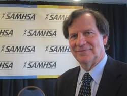
Citing data that nearly half of all persons who died by suicide had seen a primary care provider within the month prior to their death, Pamela S. Hyde, J.D., administrator of the Substance Abuse and Mental Health Services Administration, overseen by the U.S. Department of Health & Human Services, today announced the launch of the Suicide Safe app, a free risk-assessment tool for providers to screen patients at risk for suicide.
According to Ms. Hyde, who spoke during a SAMHSA news briefing, suicide is the leading cause of death in the United States among those aged 15-29 years, with one person dying of suicide every 13 minutes. Ms. Hyde also noted that suicide was seen to trend upward between the years 1999 and 2010 when the age-adjusted suicide rate for people aged 35-64 years increased 28%.

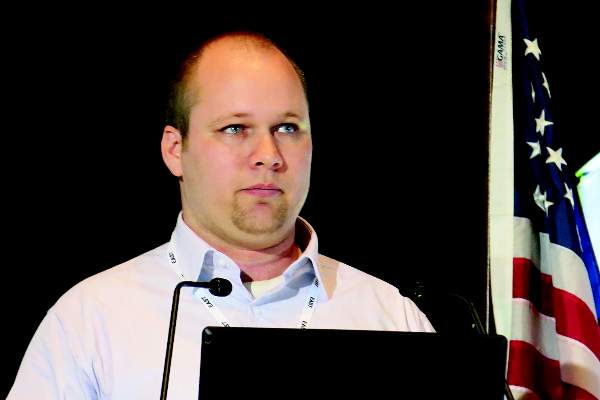
“Our offices are the key access points where we can start to identify these patients,” said Dr. Fabian Sandoval, a Washington-based researcher and clinical protocols consultant who spoke at the news briefing. “But we are not the experts, we are not the psychiatrists, so what are we going to do?”
Dr. Sandoval said the tool would support the generations of primary care providers, including advance practice nurses and physician assistants, who did not receive any formal mental or behavioral health care training, which he said was most of the “old guard” in medicine.
Based on the SAFE-T (Suicide Assessment Five-Step Evaluation and Triage) protocols developed by Dr. Douglas Jacobs of the department of psychiatry at Harvard Medical School, Boston, and the chair of the American Psychiatric Association Practice Guidelines on Suicide, the app helps identify at-risk patients and find local facilities where patients can be referred for emergent or other treatment.
“If I am a primary care provider who doesn’t feel adequately prepared to deal with a patient in the middle of a suicidal crisis, I can use the treatment locator to find a specialized behavioral health provider within a certain radius and immediately link to that person right off the app and get that patient in for an appointment or an emergency visit,” Robert Carrol, an advanced practice registered nurse and a member of the app’s research and development team, said in an interview.
“It’s a tool that can bridge that gap of fear that often prevents providers from even opening the conversation. That’s always the first challenge, providers not wanting to ‘go there’ because they don’t want to answer a question that they won’t know how to answer or respond to.”
Starting the conversation in an open-ended way is crucial to getting a patient to speak honestly about his or her condition, Dr. Mitra Ahadpour, a primary care physician from Germantown, Md., and a clinical communications consultant with SAMHSA, said in an interview. “The open-ended question prompts in the app are very important. We have research that shows closed-ended questions turn the patient off, questions such as ‘You’re not thinking of killing yourself, are you?’ Instead, keep it very simple. Ask, ‘How are you doing today? Then take a second to see what they say.”

Even if primary care physicians are willing to ask and respond to their patients in acute danger of suicide, there is still the need to ensure that even if patients are referred to the appropriate facility, that there is enough room to accommodate them, according to Dr. Sandoval. “We’ll be able to identify more patients who are in suicidal situations than before, but now what? What do we do with this patient? That is a question we have to [address] as well.”
Incorporating the app into clinical practice should not be difficult and could even help encourage a physician to treat the patient him- or herself if the risk for suicide is found to be low, according to Dr. Ahadpour. “There are many points in the system where the app can be integrated. The PHQ-9 (patient health questionnaire that screens for depression) is included in the app, and you can have your patients fill that out ahead of time,” Dr. Ahadpour, who was not on the news briefing panel, said in an interview. “That way the clinician will already know if there is a need to take things to the second step or [assess] for a suicidal risk. If the provider feels comfortable that there is no risk for suicide, then they can treat the patient for depression, for example.” If they do not feel comfortable treating the patient, then “at least they have assessed the patient,” she added. Validated evaluation tools for other mental health conditions, including one for general anxiety disorder are also included in the app, Dr. Ahadpour said. Depression and anxiety are two major risk factors for suicidal ideation.
When asked by a reporter about how primary care physicians should bill their time for using the app, Mr. Carroll said the app included IDC-9 and ICD-10 codes.
For more information about the app, visit SAMHSA’s website.
Dr. Jacobs said the app is freely available at any smart phone app store.
On Twitter @whitneymcknight
WASHINGTON – Primary care providers are being urged by a federal agency to heighten their sensitivities to patients at risk for suicide.
Citing data that nearly half of all persons who died by suicide had seen a primary care provider within the month prior to their death, Pamela S. Hyde, J.D., administrator of the Substance Abuse and Mental Health Services Administration, overseen by the U.S. Department of Health & Human Services, today announced the launch of the Suicide Safe app, a free risk-assessment tool for providers to screen patients at risk for suicide.
According to Ms. Hyde, who spoke during a SAMHSA news briefing, suicide is the leading cause of death in the United States among those aged 15-29 years, with one person dying of suicide every 13 minutes. Ms. Hyde also noted that suicide was seen to trend upward between the years 1999 and 2010 when the age-adjusted suicide rate for people aged 35-64 years increased 28%.
“Our offices are the key access points where we can start to identify these patients,” said Dr. Fabian Sandoval, a Washington-based researcher and clinical protocols consultant who spoke at the news briefing. “But we are not the experts, we are not the psychiatrists, so what are we going to do?”
Dr. Sandoval said the tool would support the generations of primary care providers, including advance practice nurses and physician assistants, who did not receive any formal mental or behavioral health care training, which he said was most of the “old guard” in medicine.
Based on the SAFE-T (Suicide Assessment Five-Step Evaluation and Triage) protocols developed by Dr. Douglas Jacobs of the department of psychiatry at Harvard Medical School, Boston, and the chair of the American Psychiatric Association Practice Guidelines on Suicide, the app helps identify at-risk patients and find local facilities where patients can be referred for emergent or other treatment.
“If I am a primary care provider who doesn’t feel adequately prepared to deal with a patient in the middle of a suicidal crisis, I can use the treatment locator to find a specialized behavioral health provider within a certain radius and immediately link to that person right off the app and get that patient in for an appointment or an emergency visit,” Robert Carrol, an advanced practice registered nurse and a member of the app’s research and development team, said in an interview.
“It’s a tool that can bridge that gap of fear that often prevents providers from even opening the conversation. That’s always the first challenge, providers not wanting to ‘go there’ because they don’t want to answer a question that they won’t know how to answer or respond to.”
Starting the conversation in an open-ended way is crucial to getting a patient to speak honestly about his or her condition, Dr. Mitra Ahadpour, a primary care physician from Germantown, Md., and a clinical communications consultant with SAMHSA, said in an interview. “The open-ended question prompts in the app are very important. We have research that shows closed-ended questions turn the patient off, questions such as ‘You’re not thinking of killing yourself, are you?’ Instead, keep it very simple. Ask, ‘How are you doing today? Then take a second to see what they say.”
Even if primary care physicians are willing to ask and respond to their patients in acute danger of suicide, there is still the need to ensure that even if patients are referred to the appropriate facility, that there is enough room to accommodate them, according to Dr. Sandoval. “We’ll be able to identify more patients who are in suicidal situations than before, but now what? What do we do with this patient? That is a question we have to [address] as well.”
Incorporating the app into clinical practice should not be difficult and could even help encourage a physician to treat the patient him- or herself if the risk for suicide is found to be low, according to Dr. Ahadpour. “There are many points in the system where the app can be integrated. The PHQ-9 (patient health questionnaire that screens for depression) is included in the app, and you can have your patients fill that out ahead of time,” Dr. Ahadpour, who was not on the news briefing panel, said in an interview. “That way the clinician will already know if there is a need to take things to the second step or [assess] for a suicidal risk. If the provider feels comfortable that there is no risk for suicide, then they can treat the patient for depression, for example.” If they do not feel comfortable treating the patient, then “at least they have assessed the patient,” she added. Validated evaluation tools for other mental health conditions, including one for general anxiety disorder are also included in the app, Dr. Ahadpour said. Depression and anxiety are two major risk factors for suicidal ideation.
When asked by a reporter about how primary care physicians should bill their time for using the app, Mr. Carroll said the app included IDC-9 and ICD-10 codes.
For more information about the app, visit SAMHSA’s website.
Dr. Jacobs said the app is freely available at any smart phone app store.
On Twitter @whitneymcknight
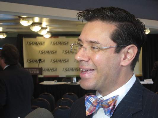
Dark circles under the eyes
How many times a week are we asked by our patients about “dark circles” under the eyes? The term “dark circles” is a catch-all term that refers to problems that have a vast range of genetic, environmental, and skin causes. However, it is a common frustrating problem with little structure in its definition and few foolproof treatments.
We propose a classification system for the definition of dark circles, and offer some clinical pearls in their treatment. Most patients, however, have dark circles with multifactorial causes that need to be addressed.
I. Infraorbital fat pad protrusion (“bags under my eyes”)
Blepharoplasty is the best solution and for now, the only solution for fat pad prominence. The fat may be removed in lower lid blepharoplasty or repositioned. Referral to a board certified plastic surgeon, oculoplastic surgeon, or dermatologic surgeon is recommended. If there is also significant tear trough deformity, fillers may be placed in the tear trough to help “camouflage” the appearance of the fat pad protrusion but it does not rid the patient of the fat pads.
II. Infraorbital edema (“puffiness”)
The infraorbital skin is very thin and highly sensitive to fluid compartmentalization. Seasonal allergies, sinus infections, crying, or water retention from high blood pressure or consumption of high sodium foods are some of the reasons the loose, thin epidermis becomes edematous. Recommendations for patients:
• Treat seasonal allergies with over-the-counter allergy medications, or see your doctor for prescription medications for resistant allergies or possible sinus infections.
• Switch your sleep position. Sleep position can be contributing to under-eye bags through gravity. Sleeping on your side or stomach can encourage fluids to collect under your eyes. If you’re a side sleeper, you may notice a heavier bag on the side you sleep on. Patients who wake up with puffy eyes can sleep on their backs and add an extra pillow under the head.
• Avoid rubbing eyes frequently, going to bed with makeup on, and harsh cleansers. Anything that irritates the eyes can cause fluids to pool. Sleeping in eye makeup can irritate eyes, causing undereye edema.
• Eye bags might be a sign of an underlying medical condition, if they appear suddenly and none of the above conditions apply. Thyroid, cardiovascular, or kidney problems can cause under-eye fluid retention and the patients need to see their primary care doctors for further evaluation.
• Place an ice pack, slices of cucumbers, chilled tea bags, or even a package of frozen peas on eyes. This can constrict leaky blood vessels and lessen the periorbital edema.
• A few topical eye creams have been developed, such as Neotensil, that temporarily reduce the appearance of lower eyelid puffiness. The product is a blend of polymers that provide compression, smoothing, and hydrating benefits to the skin. In addition, a makeup is often applied over it to reduce the appearance further.
III: Periorbital hyperpigmentation (“dark circles”)
Pigmentation of the periorbital skin is very common in skin of color because of the increased melanin content. Genetics, rubbing, and inflammatory skin diseases such as eczema may play a role in exacerbating the pigmentation of the thin under-eye skin. Recommendations for patients:
• Remind them to avoid rubbing the area – chronic rubbing and the development of lichen simplex chronicus can lead to dark, thickened under-eye skin.
• Retinoic acid creams can help slough the dark pigmented skin. However, it should be used in very small amounts with increasing use over several weeks to avoid severe irritation.
• Skin lightening creams with azaleic acid, kojic acid, and glycolic acid, can be found in varying strengths in dermatologist office preparations, over-the-counter creams, or prescriptions. Hydroquinone creams have demonstrated success in lightening under-eye hyperpigmentation. Strengths in over-the-counter preparations start at 1%-2% and in prescription strength can be compounded to higher than 4%.
• Chemical peels: Light chemical peels such as glycolic acid and Jessner’s peels will assist in lightening dark under-eye pigmentation. Dermatologists also can use peels with hydroquinone or retinoic acid for an added lightening benefit.
• Intense pulsed light (IPL) can help minimize under eye pigmentation, particularly UV-induced pigmentation.
IV: Infraorbital tear trough depression
Most often, dark circles aren’t about changes in the color of the skin at all. Instead, they’re created by a loss of volume in the area around the eye. This exposes the underlying blue veins and orbital bone, creating a hollow trough that shows up as a dark circle. These changes are often caused by genetics; however, significant weight loss and aging with resorption or displacement of the infraorbital fat pads can also expose under-eye tear trough depressions.
The best way to treat this problem is with a small amount of a hyaluronic acid filler placed by a dermatologist in the trough. Very small aliquots are needed in even the deepest trough but can give outstanding results. Caution however, must be taken as this is a highly specialized technique and injector dependent procedure. There are crucial vascular structures around the eye that need to be avoided, and overfilled troughs will give patients a puffy appearance that may pose a worse and more difficult problem to fix. Hyaluronic acid fillers are not approved by the Food and Drug Administration for treatment of under-eye depressions, so patients should be educated about the risks and benefits prior to undergoing these procedures.
V: Periorbital vascular prominence
With age, the skin around the eye becomes thinner, exposing the small capillaries and venules just below the thin epidermal layer. Vascular prominence can leave a bluish undertone to the infraorbital skin which can cast dark shadows and make the area appear dark or sallow.
• Eye creams that contain caffeine can constrict the underlying blood vessels and temporarily diminish small vessel prominence.
• For large blue veins, vascular lasers such as a long pulse Nd:Yag lasers can be recommended. But in darker skin types these lasers can cause hyperpigmented scars if not used with adequate skin cooling techniques. Proper eye protection should also be used.
VI: Periorbital static and dynamic rhytids
• Botulinum toxin placed in small aliquots around the orbital rim will reduce the dynamic rhytids in this area. Treatments spaced 3 months apart will ensure long-lasting benefits as botulinum toxin often wears off.
• Laser resurfacing with CO2, fractionated CO2, or erbium lasers may also be used to treat periorbital rhytides.
Additional tips for your patients:
• For most of the types of infraorbital issues, makeup can help conceal some skin imperfections. Patients should choose a concealer that matches or is slightly lighter than their skin tone. If the patient has mild discoloration, choose a liquid formula. For more prominent imperfections, a cream full-coverage concealer works best.
• Recommend that patients avoid smoking, which dehydrates the skin and causes premature aging and collagen degradation.
• Remind patients to apply a sunscreen around the eye area. Hyperpigmentation and tear troughs can accentuate with UV-induced skin pigmentation. Physical blocking sunscreens may be less irritating than chemical blockers for those with sensitive eyelid skin.
• Remind patients to apply a moisturizer to the eye area nightly to keep the skin from becoming dry, irritated, and dehydrated.
• Advise patients not to break the bank with over-the-counter creams that promise cures for under-eye circles. Most over-the-counter preparations provide temporary, mild benefits at most, and often do not provide any lasting benefit.
Dr. Talakoub and Dr. Wesley are co-contributors to a monthly Aesthetic Dermatology column in Dermatology News. Dr. Talakoub is in private practice in McLean, Va. Dr. Wesley practices dermatology in Beverly Hills, Calif. This month’s column is an update by Dr. Wesley of a previous column by Dr. Talakoub.

Patented knowledge
The other day I had a chat with a chiropractor I’ll call Stan. Stan was excited about a new technique he has used to build his practice, whose clients now number several celebrities (some of whom I’d even heard of). “I specialize in muscular work and performance enhancement,” he said. “Performers with strenuous routines and a lot of stress need to sustain peak performance.”
A colleague out West whom Stan described as “very brilliant” had developed this technique. “It’s patented,” he said.
Stan went on to describe how this method has given him a whole new sense of the body. “I can actually feel the small transverse muscles of the vertebrae respond under my fingers,” he said. “The results are amazing.”
“How did you learn this technique?” I asked him.
“I took courses with the inventor,” he said. “His courses are patented, and can only be taught by accredited instructors. That ensures that the method is being done right. There are nine levels of certification.”
“Nine?”
“Yes, you have to keep taking more courses, learning new things. It’s very exciting.”
“By the way,” I asked, “how do you know that this method works better than the older ones you learned when you went to school?”
“Oh, you can feel and see the difference,” he said. “If you’re asking if there are studies or things like that, I guess there aren’t. But there’s no question that it’s better.”
“Do they teach this technique in chiropractic school?”
“No. As I said, it’s patented.”
“In that case,” I said, “it seems your professional schools are teaching inferior treatment methods.”
That gave Stan some pause. While he was thinking, I continued.
“It’s interesting,” I said. “In my profession, if someone came up with a treatment that was better than what everyone else was doing, he would need to do studies that proved he was right. He would also feel ethically bound to let everyone else know about the method, so all patients could be treated that way.”
Stan’s blank look suggested that this line of analysis had not occurred to him.
“But you must have some special techniques you use in your practice,” he said.
“No,” I said, “actually I don’t. I just practice conventional dermatology. Nothing special or unique about what I do.”
“What about the teaching hospitals in Boston,” he went on. “Don’t they do things no one else does?”
“Possibly,” I said. “But if they come up with a new technique, they have to convince others in the profession – and insurance companies – that their innovation is better in some measurable way.”
I’d gone as far as I wanted to. “It’s exciting that your patients do so well,” I said. “By the way,” I said, “what are celebrities really like up close and personal?”
“Most of them are very nice people,” he said. “They work hard to be good at what they do.”
“Do they ever complain that your treatment didn’t work, didn’t give their performance the boost they were expecting?”
“No,” he said. “Not one. They’re all happy.”
“That is really amazing,” I said. Stan nodded in agreement, though I don’t think he knew what I found so remarkable about it.
Stan is no cynic. He truly believes that what he does is valid, and that it helps his patients more than other treatments do. His patients believe it too.
Nor is our own profession as selfless and sharing as I made it sound. Hospitals love to trumpet their cyberknife technology or state-of-the-art orthopedic techniques or comprehensive cancer care, implying that they do whatever they do better than anyone else can.
Patients love to read this. They want to believe they’re seeing the “top” doctor, the one with the best results. Boston magazine (and the equivalent in every other city) publishes a list of “Top Doctors” this time of year.
Patients sometimes say, “I came to you because you did such a great job clearing up my sister’s acne,” or “You cleared my older son’s wart when nobody else could – one freeze, and it was gone. You’re a miracle worker!”
Oh sure I am. Nobody sprays liquid nitrogen the way I do.
I didn’t patent it, though. Teddy Roosevelt banned patent medicines in this country in 1906. But I guess in some quarters, patented never has gone away.
Dr. Rockoff practices dermatology in Brookline, Mass., and is a longtime contributor to Dermatology News. He serves on the clinical faculty at Tufts University, Boston, and has taught senior medical students and other trainees for 30 years.
Heroin overdoses up dramatically since 2010
Drug-poisoning deaths involving heroin have soared since 2000, and most of the increase occurred since 2010, according to a report from the National Center for Health Statistics.
From 2010 to 2013, the rate of heroin overdose deaths increased 163% for men, from a rate of 1.6/100,000 population in 2010 to 4.2 in 2013. For women, the death rate increased by 200%, from 0.4/100,000 in 2010 to 1.2/100,000 in 2013. From 2000 to 2010, however, the rate of increase was much slower, with the death rate increasing from 1.1 to 1.6 for men and from 0.2 to 0.4 for women.

The overall rate for heroin overdose from 2000 to 2013 increased from 0.7 to 2.7/100,000. Most of this increase occurred from 2010 to 2013: From 2000 to 2010, the death rate increased to only 1/100,000, a growth rate of 6%, but after 2010, the rate grew by 37% per year, the NCHS reported.
In 2013, non-Hispanic whites aged 18-44 years had the highest heroin poisoning death rate among measured racial/ethnic groups at 7/100,000. In 2000, older, non-Hispanic blacks aged 45-64 years had the highest death rate among the reported racial/ethnic groups at 2/100,000. The death rate for whites aged 18-44 in 2000 was 1.2/100,000, meaning that the death rate increased by 483% from 2000 to 2013. For non-Hispanic blacks aged 45-64, the death rate in 2013 was 4.9, an increase of 145%.
The number of heroin-related overdose deaths climbed in every region of the country from 2000 through 2013. The largest change in heroin overdose by region occurred in the Midwest, where the death rate rose from 0.4/100,000 in 2000 to 4.3 in 2013, an increase of 975%, said the NCHS report, which used data collected by the National Vital Statistics System.
Drug-poisoning deaths involving heroin have soared since 2000, and most of the increase occurred since 2010, according to a report from the National Center for Health Statistics.
From 2010 to 2013, the rate of heroin overdose deaths increased 163% for men, from a rate of 1.6/100,000 population in 2010 to 4.2 in 2013. For women, the death rate increased by 200%, from 0.4/100,000 in 2010 to 1.2/100,000 in 2013. From 2000 to 2010, however, the rate of increase was much slower, with the death rate increasing from 1.1 to 1.6 for men and from 0.2 to 0.4 for women.
The overall rate for heroin overdose from 2000 to 2013 increased from 0.7 to 2.7/100,000. Most of this increase occurred from 2010 to 2013: From 2000 to 2010, the death rate increased to only 1/100,000, a growth rate of 6%, but after 2010, the rate grew by 37% per year, the NCHS reported.
In 2013, non-Hispanic whites aged 18-44 years had the highest heroin poisoning death rate among measured racial/ethnic groups at 7/100,000. In 2000, older, non-Hispanic blacks aged 45-64 years had the highest death rate among the reported racial/ethnic groups at 2/100,000. The death rate for whites aged 18-44 in 2000 was 1.2/100,000, meaning that the death rate increased by 483% from 2000 to 2013. For non-Hispanic blacks aged 45-64, the death rate in 2013 was 4.9, an increase of 145%.
The number of heroin-related overdose deaths climbed in every region of the country from 2000 through 2013. The largest change in heroin overdose by region occurred in the Midwest, where the death rate rose from 0.4/100,000 in 2000 to 4.3 in 2013, an increase of 975%, said the NCHS report, which used data collected by the National Vital Statistics System.