User login
VIDEO: Less tricuspid regurgitation seen with Sano shunt in Norwood procedures
SEATTLE – Sano shunts outperform Blalock-Taussig shunts for Norwood procedures in neonates with hypoplastic left heart syndrome, according to a research registry study of 322 patients at the Cleveland Clinic and elsewhere.
The 166 newborns who had Sano shunts were matched to 166 who had Blalock-Taussig (BT) shunts.
“For comparable neonates with HLHS [hypoplastic left heart syndrome] undergoing Norwood operations, Sano offers better late survival [more than 3 years], less late tricuspid regurgitation, and perhaps less late right ventricular dysfunction than BT,” the investigators concluded.
Even so, Dr. Richard Ohye, professor of cardiac surgery at the University of Michigan, Ann Arbor, said the jury is still out on which shunt is best. He explained why in an interview at the American Association for Thoracic Surgery annual meeting. He also shared tips on shunt selection and explained a novel technique he has developed for doing a Sano shunt with a smaller hole in the right ventricle.
The video associated with this article is no longer available on this site. Please view all of our videos on the MDedge YouTube channel
AT THE AATS ANNUAL MEETING
Reversing glucocorticoid resistance in ALL

Photo courtesy of St. Jude
Researchers say they have identified a mechanism that helps leukemia cells resist glucocorticoids.
They believe the mechanism is responsible for about a third of steroid resistance in children and adolescents with acute lymphoblastic leukemia (ALL).
However, additional research is needed to determine if the process is at work in adults with ALL, where steroid resistance is more common and long-term survival lags.
The researchers described the mechanism in Nature Genetics.
“Based on these findings, research has already begun to identify small molecules with the potential to reverse glucocorticoid resistance, leading to more effective treatment and increased survival,” said study author William Evans, PharmD, of St. Jude Children’s Research Hospital in Memphis, Tennessee.
Dr Evans and his colleagues analyzed samples from 444 newly diagnosed ALL patients being treated at St. Jude or in clinical trials sponsored by the Dutch Childhood Oncology Group and the German Cooperative Study Group for Childhood ALL.
The team also analyzed samples collected at diagnosis and relapse from 49 pediatric ALL patients enrolled in clinical trials organized by the Children’s Oncology Group.
The researchers found differences in gene expression that correlated with sensitivity to steroids. CASP1 and NLRP3 were among the genes with increased activity in steroid-resistant leukemia cells.
The team also identified a reason for the increased gene activity. Leukemia cells overexpressing CASP1 and NLRP3 had lower levels of methylation compared to cells with normal expression.
Previous research showed that steroid resistance was more common in young ALL patients who relapsed than in newly diagnosed patients. Dr Evans and his colleagues found that expression of CASP1 and NLRP3 was significantly higher in ALL patients who relapsed.
The researchers also discovered that CASP1 blocks glucocorticoids by splitting the receptor where the drug binds and therefore blocks its access to the nucleus.
“Cells that overexpress CASP1 are chewing up their glucocorticoid receptor,” Dr Evans said. “That means when steroids enter the cell, there is no receptor for the drugs to bind to or fulfill its therapeutic function.”
To confirm that CASP1 cleavage of the steroid receptor is pivotal to ALL steroid resistance, the researchers engineered a receptor that lacked the CASP1 cleavage site. When they introduced the genetically engineered receptors into ALL cells that expressed high levels of CASP1, the cells remained sensitive to steroids.
Using a variety of techniques, the researchers showed that steroid resistance rose or fell in leukemia cells based on CASP1 levels. Overexpression of CASP1 rendered ALL cells 5 to 15 times more resistant to the glucocorticoids dexamethasone and prednisolone.
However, reducing CASP1 using genetic, pharmacologic, and other methods restored steroid sensitivity in leukemia cells. ![]()
Defining the role of TAMs in DLBCL

pseudopodia to engulf particles
PHILADELPHIA—New research suggests the prognostic value of tumor-associated macrophages (TAMs) is disease-specific as well as treatment-specific.
Investigators set out to determine if TAMs have a negative prognostic impact in diffuse large B-cell lymphoma (DLBCL), as previous studies produced conflicting results.
The team found that higher TAM levels are associated with worse survival in DLBCL, but only in patients who do not receive rituximab. The drug can overcome the poor prognosis TAMs confer in DLBCL.
Eri Matsuki, MD, PhD, of Memorial Sloan-Kettering Cancer Center in New York, New York, and her colleagues presented these findings in a poster at the AACR Annual Meeting 2015 (abstract 2371*).
To ascertain the role of TAMs in DLBCL, the investigators analyzed specimens from 103 DLBCL patients, 61 of whom received rituximab, 33 who did not, and 9 whose rituximab status was unknown. The team first looked at the expression of CD163 as a marker of TAMs.
“CD163 is more specific to M2-type macrophages, which have pro-tumor effects, compared to a more pan-macrophage marker which is widely used—CD68,” Dr Matsuki noted.
She and her colleagues found that a high level of CD163-positive cells (more than 150) was significantly associated with advanced-stage disease (P=0.016), non-GCB DLBCL (P=0.0071), and higher expression of c-Myc (P=0.0022).
“Our interpretation of that at this point is that, the more aggressive the tumor, the more likely that it would induce macrophages into the tissue,” Dr Matsuki said.
The investigators then assessed the impact of rituximab use. Among patients who didn’t receive rituximab, having a high level of CD163-positive cells (more than 150) was associated with inferior overall survival (OS, P=0.022). However, if patients did receive rituximab, there was no significant difference in OS.
Dr Matsuki said this supports previous studies showing that the prognostic effect of TAMs diminishes with rituximab use, as well as the in vitro finding that M2 macrophages exhibit increased phagocytosis of rituximab-opsonized tumor cells.
“So overall, what we’re seeing is that . . . the negative influence of TAMs can be overcome with rituximab use,” she summarized.
Dr Matsuki and her colleagues also looked at the patients’ lymphocyte-to-monocyte-ratio (LMR) because TAMs partly arise from peripheral blood monocytes.
The team found that having an LMR higher than 2.77 was significantly associated with superior OS (P=0.03) in patients who did not receive rituximab. And there was a trend toward improved OS with a higher LMR in patients who did receive the drug (P=0.07).
The investigators believe the differences they observed in the prognostic value of CD163 and LMR could be explained by the fact that TAMs are derived from both circulating monocytes and resident macrophages.
Dr Matsuki said this research has improved her group’s understanding of DLBCL, but they are still working to identify additional biomarkers associated with prognosis in this disease. ![]()
*Information in the abstract differs from that presented at the meeting.
New model simulates human MM

Photo by Ben Skála
By growing human tumors on chicken embryos, scientists have created a new model system for screening drugs to treat multiple myeloma (MM).
Several MM drug candidates have shown promising activity in this system, according to the researchers.
Gerold Untergasser, PhD, of Innsbruck Medical University in Austria, and his colleagues described how they developed the system in the Journal of Visualized Experiments.
First, the researchers prepared cultures of the MM cell lines OPM2 and RPMI 8226, as well as human mesenchymal stem cells. They then transfected MM cells with enhanced green fluorescent protein, making the cells easy to observe through a fluorescence microscope.
Next, the team cultured MM cells with mesenchymal cells and collagen to create 3-dimensional cell spheres, thereby simulating the natural microenvironment of the tumor.
The researchers then transferred their cell spheres to the outer membrane of chicken embryos. This choriallantoid membrane provides a suitable base for growing miniature human tumors in culture.
The team has introduced anti-myeloma drugs, such as bortezomib, into this system and assessed the drugs’ ability to target MM cells and prevent tumor growth and angiogenesis. They were also able to evaluate toxicity.
“The chicken egg is much easier to handle and cheaper than mice, and it reduces the number of animal experiments,” Dr Untergasser said. “In our new video publication, we give detailed information on how our system works.”
“We provide an easy-to-repeat protocol for broad use within the research community. Our vision for the future is that a separate test is performed for each patient to determine which drug is best suitable.” ![]()
Inhibitor improves OS in poor-prognosis MDS

WASHINGTON, DC—A small-molecule inhibitor can improve overall survival (OS) in certain patients with previously treated myelodysplastic syndromes (MDS), results of a phase 3 trial suggest.
Overall, patients who received the dual PI3K/PLK pathway inhibitor rigosertib along with best supportive care (BSC) did not see a significant improvement in OS compared to patients who received BSC alone.
However, rigosertib did improve OS in patients with poor prognosis.
Guillermo Garcia-Manero, MD, of the MD Anderson Cancer Center in Houston, Texas, and his colleagues presented these results at the 13th International Symposium on Myelodysplastic Syndromes (abstract 112).
The trial, known as ONTIME, was sponsored by Onconova Therapeutics, Inc., the company developing rigosertib.
The trial included 299 higher-risk MDS patients with excess blasts (5% to 30% bone marrow blasts) who had failed to respond to (25%), progressed on (37%), or relapsed after (38%) treatment with hypomethylating agents (HMAs).
Patients were randomized 2:1 to receive rigosertib plus BSC or BSC alone. Patients treated with rigosertib received 1800 mg every 24 hours for 72 hours as a continuous, intravenous, ambulatory infusion, every 2 weeks for the first 16 weeks, then every 4 weeks.
The treatment arms were generally balanced in terms of baseline characteristics. The majority of patients were male (66%) and white (82%). The median age was 74 years. Most patients (85%) had an Eastern Cooperative Oncology Group score of 0 or 1.
The median duration of the last HMA therapy was 8.8 months for patients in the rigosertib arm and 10.3 months for patients in the BSC arm.
The researchers found no significant difference in OS between the treatment arms. The median OS was 8.4 months in the rigosertib arm and 5.9 months in the BSC arm, and the 12-month OS was 35% and 25%, respectively (hazard ratio[HR]=0.87, P=0.31).
On the other hand, certain patients did see a significant improvement in OS with rigosertib. Among patients with primary HMA failure (those who failed to respond to or progressed during HMA therapy), the median OS was 8.6 months in the rigosertib arm and 5.3 months in the BSC arm (HR=0.69, P=0.040).
For patients who received HMAs for less than 9 months, the median OS was 7.7 months in the rigosertib arm and 4.5 months in the BSC arm (HR=0.55, P=0.003). Among patients younger than 75 years of age, the median OS was 9.7 months in the rigosertib arm and 4.1 months in the BSC arm (HR=0.52, P=0.0004).
And for patients with very high-risk disease according to the Revised International Prognostic Scoring System, the median OS was 7.6 months in the rigosertib arm and 3.2 months in the BSC arm (HR=0.56, P=0.005).
The researchers said there were no obvious differences between the treatment arms with regard to overall adverse events (AEs) or grade 3 or higher AEs.
Overall, 99% of patients in the rigosertib arm and 85% in the BSC arm experienced treatment-emergent AEs. The incidence of grade 3 or higher AEs was 79% and 68%, respectively.
Treatment-emergent AEs of all grades—occurring in the rigosertib and BSC arms, respectively—included nausea (35% vs 18%), diarrhea (33% vs 20%), constipation (31% vs 11%), fatigue (30% vs 18%), pyrexia (27% vs 21%), anemia (23% vs 9%), peripheral edema (21% vs 16%), and thrombocytopenia (21% vs 8%).
Considering the study results together, Dr Garcia-Manero and his colleagues concluded that rigosertib is likely most effective in high-risk MDS patients with the worst prognosis, and these patients can safely receive the drug. ![]()
Society of Hospital Medicine (SHM)-American Academy of Family Physicians (AAFP) Joint Statement on Hospitalists Trained in Family Medicine
Hospitalists are physicians whose primary professional focus is the general medical care of hospitalized patients. Both the Society of Hospital Medicine (SHM) and the American Academy of Family Physicians (AAFP) hold that the opportunity to participate as a Hospitalist should be granted to all physicians commensurate with their documented training and/or experience, demonstrated abilities and current competencies.
During their training Family Physicians acquire the necessary attitudes, skills, and knowledge that enable them to provide continuing and comprehensive medical care across the spectrum of care settings, including the inpatient setting. Education in the primary management of hospitalized patients occurs during the required general inpatient ward and intensive care unit experiences. In addition, Family Physicians are required to train with general surgeons and surgical subspecialists, enhancing recognition and understanding of surgical disease states upon which Hospitalists are frequently asked to consult or co-manage. Family Medicine training also encompasses additional skills essential to the practice of Hospital Medicine, including participation in quality improvement, addressing the psychosocial needs of patients, coordinating across levels of care, and functioning as members of interdisciplinary teams.
Given this training, many Family Physicians effectively manage their patients in an inpatient setting after the completion of their residency.
Demand for Hospitalists continues to outweigh supply in the United States, including needs in underserved and rural areas. Hospitalists Trained in Family Medicine (HTFM) fulfill an important public health need by providing frontline inpatient services in a variety of geographic settings. In addition, while many HTFM focus exclusively on the care of adults, others are providing inpatient care across the spectrum of ages, as well as providing obstetric services. More than two-thirds of HTFM are also involved in the training of residents and medical students, enhancing the skills of our future physicians.
Recognition of achievement by HTFM from the SHM is available by meeting standards set for all Hospitalists, regardless of residency training, in the form of the designation of Fellow of Hospital Medicine. HTFM also have the opportunity to professionally qualify and sit for the Recognition of Focused Practice in Hospital Medicine board examination. This examination is administered and recognized jointly by the American Board of Family Medicine and the American Board of Internal Medicine.
In consideration of the above factors, both the Society of Hospital Medicine and the American Academy of Family Physicians endorse and encourage the growing contribution of Hospitalists Trained in Family Medicine.
Society of Hospital Medicine Welcomes New Fellows Class
Last month, more than 230 hospitalists were inducted as Fellows in Hospital Medicine (FHM), Senior Fellows in Hospital Medicine (SFHM), and Masters in Hospital Medicine (MHM) by SHM at the 2015 annual meeting at the Gaylord National Resort and Convention Center in National Harbor, Md.
This year represents the largest fellows class in history, with 175 FHM and 61 SFHM honorees.
“Through their commitment to the specialty, through education and self-improvement, hospitalists earning the Fellow and Senior Fellow designations represent the very best of the hospital medicine movement and its goal to improve the care of hospitalized patients,” says SHM President Bob Harrington, MD, SFHM. “I hope you will join me in congratulating them in this professional milestone.”
Fellows and Senior Fellows have earned the right to use the “FHM” and “SFHM” designation.
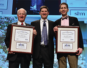
SHM also inducted two new Masters in Hospital Medicine, the highest honor from SHM: Bradley Flansbaum, DO, MPH, MHM, and Larry Wellikson, MD, MHM.
Dr. Flansbaum was a founding member of SHM and served as a board member and officer; today, he is a hospitalist at Lenox Hill Hospital in New York City and physician editor for SHM’s blog, The Hospital Leader.
Dr. Wellikson joined SHM in January 2000 and serves as SHM’s chief executive officer.
Drs. Flansbaum and Wellikson join 16 other leaders in the specialty, including co-founders Win Whitcomb, MD, MHM, and John Nelson, MD, MHM, along with Bob Wachter, MD, MHM, who published the seminal article for the hospitalist movement in a 1996 New England Journal of Medicine article.
Brendon Shank is SHM’s associate vice president of communications.
Last month, more than 230 hospitalists were inducted as Fellows in Hospital Medicine (FHM), Senior Fellows in Hospital Medicine (SFHM), and Masters in Hospital Medicine (MHM) by SHM at the 2015 annual meeting at the Gaylord National Resort and Convention Center in National Harbor, Md.
This year represents the largest fellows class in history, with 175 FHM and 61 SFHM honorees.
“Through their commitment to the specialty, through education and self-improvement, hospitalists earning the Fellow and Senior Fellow designations represent the very best of the hospital medicine movement and its goal to improve the care of hospitalized patients,” says SHM President Bob Harrington, MD, SFHM. “I hope you will join me in congratulating them in this professional milestone.”
Fellows and Senior Fellows have earned the right to use the “FHM” and “SFHM” designation.
SHM also inducted two new Masters in Hospital Medicine, the highest honor from SHM: Bradley Flansbaum, DO, MPH, MHM, and Larry Wellikson, MD, MHM.
Dr. Flansbaum was a founding member of SHM and served as a board member and officer; today, he is a hospitalist at Lenox Hill Hospital in New York City and physician editor for SHM’s blog, The Hospital Leader.
Dr. Wellikson joined SHM in January 2000 and serves as SHM’s chief executive officer.
Drs. Flansbaum and Wellikson join 16 other leaders in the specialty, including co-founders Win Whitcomb, MD, MHM, and John Nelson, MD, MHM, along with Bob Wachter, MD, MHM, who published the seminal article for the hospitalist movement in a 1996 New England Journal of Medicine article.
Brendon Shank is SHM’s associate vice president of communications.
Medicare Develops Next Generation Accountable Care Organization Model
The Centers for Medicare and Medicaid Services (CMS) Innovation Center recently announced the development of a new accountable care organization (ACO) model—the Next Generation ACO—that hopes to move closer to the goal of efficient, coordinated care for Medicare beneficiaries.
“This ACO model provides for greater engagement of beneficiaries, a more predictable, prospective financial model, and more tools to coordinate care for beneficiaries,” writes Patrick Conway, MD, MSc, chief medical officer and deputy administrator for innovation and quality at CMS, in a blog post announcing the Next Generation ACO.
ACOs align hospitals, physicians, nursing facilities, and other critical healthcare providers as a sort of one-stop shop for seamless patient care across settings and among providers. By bringing together the full range of services, ACOs aim to provide higher quality coordinated care while reducing costs for patients and Medicare.
Since the passage of the Affordable Care Act, CMS has overseen two distinct tracks for ACOs: the Medicare Shared Savings Program and the Pioneer ACO. The Shared Savings Program was a first step in moving toward streamlined healthcare delivery systems while incentivizing care coordination across settings. Pioneer ACOs, on the other hand, were designed as a test for more aggressive reforms that promised higher potential rewards in exchange for higher risk, while moving participants toward population-based payments.
The Next Generation ACO builds off of the Pioneer and Shared Savings Program ACO models to test whether the fundamental concepts behind an ACO—improving care and reducing costs—can be achieved using stronger financial incentives. Notably, the Next Generation ACO establishes stable, prospective targets for benchmarking expenditures and offers an array of payment mechanisms, including capitation.
ACO goals read like a laundry list of hospitalist goals and practice, such as reducing readmissions, maximizing efficiency, improving care transitions, and reducing length of stay.
Participants of the Next Generation ACO model will have new tools to help coordinate patient care, including expanded coverage for telehealth and home health services and increased access for skilled nursing facility coverage without prior hospitalizations. Because the Next Generation ACO model comes from the CMS Innovation Center, it’s specifically designed to help policymakers evaluate the impact of reimbursement and system changes with an eye toward scalability. The knowledge gained from this model could help structure the Medicare payment system of tomorrow.
Hospitalists have long been interested in the impact of ACOs on their practices, with good reason. Hospitals form an integral part of an ACO, and hospitalists serve critical roles within their hospitals. ACO goals read like a laundry list of hospitalist goals and practice, such as reducing readmissions, maximizing efficiency, improving care transitions, and reducing length of stay. The Next Generation ACO model offers the potential to further capitalize on the expertise of hospitalists as the healthcare system explores ways to move away from traditional fee-for-service payments.
The way in which Medicare pays providers is evolving rapidly as CMS seeks to reimburse for the quality rather than the quantity of services provided to beneficiaries. Over the next five years, CMS has set aggressive targets for transitioning fee-for-service payments into value-based payment systems; the Next Generation ACO is one tool for helping to push that goal onward.
Joshua Lapps is SHM’s manager of government relations.
Society of Hospital Medicine's HM15 Meeting Draws Thousands
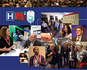
NATIONAL HARBOR, Md.—Cherry trees weren’t the only things that blossomed around Washington last month. SHM’s annual meeting, with roughly 2,500 attendees, featured 100 educational sessions, a day of Congressional lobbying, and plenaries from the “Checklist Doctor” and the Dean of Hospital Medicine. Pre-courses, the popular poster competition, and updates on everything from anticoagulants to VTE helped round out HM15, the specialty’s biggest annual event.
“I come to the meeting,” says new SHM President Robert Harrington, Jr., MD, SFHM, “and then for the next 362 days, this is enough to get me through the rest of the year ‘til I come back.”
Hospitalists Have Stake in Improving Quality, Patient Safety
NATIONAL HARBOR, Md.—Don Lee, MD, MPH, is building what one might call an analog quality improvement (QI) project focused on reducing readmissions. What the medical director for clinical integration at Columbia St. Mary’s in Milwaukee does is work with patient navigators to make follow-up phone calls after discharge to get ahead of potential issues.
What he wants to do is design a system that ensures that happens.
So, he came to HM15 for help.
“I’m very interested in continuous quality improvement. I wanted to work on how to not only get the project off the ground, but also to make sure what we are doing is good, and it’s doing what it’s supposed to be doing,” Dr. Lee says.
Well, he came to the right place. Quality and patient safety are hallmarks of the annual meeting; this year’s gathering was no exception. Plenaries provided advice from national thought leaders on improving safety by improving the patient experience; breakout sessions focused on how to build, maintain, and sustain QI projects; and SHM unveiled a new educational track dubbed the “Doctor-Patient Relationship.”
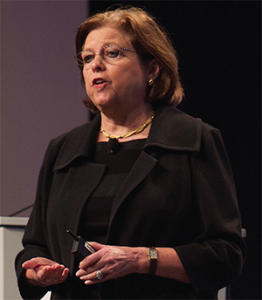
Hospital medicine, and healthcare in a broader sense, needs to be able to define safety better to attack it proactively, says Maureen Bisognano, president and CEO of the Institute for Healthcare Improvement (IHI). She compared medicine to NASA, which tracks its missions in a continuum of both successes and failures to understand what processes and protocols lay behind each.
Medicine has no such pathway laid out to date, though Bisognano said her task for the next year is to try to define one.
“We don’t know what the system of safety looks like,” she said. “We don’t know how many times we duplicate tests on admission because we haven’t connected with primary care. We don’t know how many times we send somebody home with inadequate social support, no food, and no way to pick up their prescription.
“We don’t have a sense of where our near-misses are, so we don’t have a vision of safety.”
Hospitalist Kedar Mate, MD, senior vice president for innovation at IHI, says that QI projects can seem daunting in the midst of daily censuses, hospital committee meetings, and a myriad of other responsibilities physicians face. But much of that fear is perception. A project can be simple or system-wide. The trick is just getting started in the face of perceived hurdles, he adds.
“Language around quality improvement tends to confuse and create mystery, and the jargon and so on creates interference,” says Dr. Mate, an assistant professor of medicine at Weill Cornell Medical College in New York City and a research fellow at Harvard Medical School’s Division of Global Health Equity. “It’s not that mysterious. It’s kind of a straightforward thing, actually, if you work through it logically and stepwise.”
And, as front-line providers, hospitalists are primed to lead healthcare systems in how to deliver care, he said.
“Formerly, physicians were iterant, right?” Dr. Mate adds. “They would come in and out of institutions and didn’t really have a stake in the game, on some level, of institutional quality. That’s totally different now.”
But, while the individual hospitalist has a responsibility to embrace safety initiatives, employers and industry groups have a duty to provide the proper resources to make that connection easier.
“The individual’s responsibility is to try to access that information to carry on in the face of busy schedules and busy lives,” Dr. Mate says. “SHM, IHI, and others have a responsibility to try to make those that are inclined able to continue and able to build and move their efforts forward in an even more productive way.”
Inclined docs like Dr. Lee, who know that their hospitals collect reams of data that can be useful for patient safety projects, many times have no idea how to extract said data. He has learned that partnering with “gatekeepers” is a way to help others help him.
“We are collecting data every second, every minute,” Dr. Lee says. “It’s amazing how much data we have, but to actually sift through it and make it meaningful is very difficult. You have to know what questions to ask and you have to get buy-in from the [gatekeepers], because they get thousands of requests for data extraction.”
Richard Quinn is a freelance writer in New Jersey.
NATIONAL HARBOR, Md.—Don Lee, MD, MPH, is building what one might call an analog quality improvement (QI) project focused on reducing readmissions. What the medical director for clinical integration at Columbia St. Mary’s in Milwaukee does is work with patient navigators to make follow-up phone calls after discharge to get ahead of potential issues.
What he wants to do is design a system that ensures that happens.
So, he came to HM15 for help.
“I’m very interested in continuous quality improvement. I wanted to work on how to not only get the project off the ground, but also to make sure what we are doing is good, and it’s doing what it’s supposed to be doing,” Dr. Lee says.
Well, he came to the right place. Quality and patient safety are hallmarks of the annual meeting; this year’s gathering was no exception. Plenaries provided advice from national thought leaders on improving safety by improving the patient experience; breakout sessions focused on how to build, maintain, and sustain QI projects; and SHM unveiled a new educational track dubbed the “Doctor-Patient Relationship.”
Hospital medicine, and healthcare in a broader sense, needs to be able to define safety better to attack it proactively, says Maureen Bisognano, president and CEO of the Institute for Healthcare Improvement (IHI). She compared medicine to NASA, which tracks its missions in a continuum of both successes and failures to understand what processes and protocols lay behind each.
Medicine has no such pathway laid out to date, though Bisognano said her task for the next year is to try to define one.
“We don’t know what the system of safety looks like,” she said. “We don’t know how many times we duplicate tests on admission because we haven’t connected with primary care. We don’t know how many times we send somebody home with inadequate social support, no food, and no way to pick up their prescription.
“We don’t have a sense of where our near-misses are, so we don’t have a vision of safety.”
Hospitalist Kedar Mate, MD, senior vice president for innovation at IHI, says that QI projects can seem daunting in the midst of daily censuses, hospital committee meetings, and a myriad of other responsibilities physicians face. But much of that fear is perception. A project can be simple or system-wide. The trick is just getting started in the face of perceived hurdles, he adds.
“Language around quality improvement tends to confuse and create mystery, and the jargon and so on creates interference,” says Dr. Mate, an assistant professor of medicine at Weill Cornell Medical College in New York City and a research fellow at Harvard Medical School’s Division of Global Health Equity. “It’s not that mysterious. It’s kind of a straightforward thing, actually, if you work through it logically and stepwise.”
And, as front-line providers, hospitalists are primed to lead healthcare systems in how to deliver care, he said.
“Formerly, physicians were iterant, right?” Dr. Mate adds. “They would come in and out of institutions and didn’t really have a stake in the game, on some level, of institutional quality. That’s totally different now.”
But, while the individual hospitalist has a responsibility to embrace safety initiatives, employers and industry groups have a duty to provide the proper resources to make that connection easier.
“The individual’s responsibility is to try to access that information to carry on in the face of busy schedules and busy lives,” Dr. Mate says. “SHM, IHI, and others have a responsibility to try to make those that are inclined able to continue and able to build and move their efforts forward in an even more productive way.”
Inclined docs like Dr. Lee, who know that their hospitals collect reams of data that can be useful for patient safety projects, many times have no idea how to extract said data. He has learned that partnering with “gatekeepers” is a way to help others help him.
“We are collecting data every second, every minute,” Dr. Lee says. “It’s amazing how much data we have, but to actually sift through it and make it meaningful is very difficult. You have to know what questions to ask and you have to get buy-in from the [gatekeepers], because they get thousands of requests for data extraction.”
Richard Quinn is a freelance writer in New Jersey.