User login
Open Surgical Repair Session to Highlight CLI
Attendees at Tuesday afternoon’s “Topics Related to Open Surgery and Aortic Coarctations” session will learn when open surgery is the best choice despite remarkable advances in endovascular treatment.
“There are still open procedures that every vascular surgeon needs to know how to do well,” said moderator Dr. Fred Weaver of the University of Southern California, Los Angeles.
Coral reef lesions, aortoesophageal fistulas, extensive aortobifemoral occlusions, and celiac axis compression syndromes are often just too complex and extensive for endovascular approaches, and endovascular procedures are less likely to work when patients have underlying problems such as Marfan syndrome and Takayasu arteritis. In addition, open fixes for renal artery disease better preserve kidney function, Dr. Weaver noted. They also make sense for young patients needing a durable, one-time fix. “We don’t have much long-term follow-up experience with endovascular procedures, but we have almost a half-century of experience with open vascular procedures. We know how durable they can be,” he said.
Issues surrounding open procedures and more will be addressed in the session. “Open surgical solutions are oftentimes deferred or not even offered for fear of bad outcomes,” but “they can be successful, and you have to have them in your playbook,” said co-moderator Dr. James Black of Johns Hopkins University.
Three presentations will tackle critical limb ischemia (CLI), a “hot topic that is still very controversial,” Dr. Weaver noted.
Dr. Julien Sfeir of Lebanese University, Beirut, will discuss open salvage after failed endovascular CLI intervention. Dr. R. Clement Darling of Albany Medical College in New York, and also a moderator, will discuss the role of open procedures in the endovascular era, and Dr. Carlo Setacci of the University of Siena, Italy, will address combining open and endovascular techniques for especially difficult cases.
BEST-CLI, an ongoing randomized controlled trial comparing endovascular therapy with open surgical bypass in CLI, will likely be discussed, which is “one of the reasons someone might want to come to this session. Nobody really knows what’s the best option in a lot of situations,” Dr. Weaver said.
Dr. Black will also address when open repair is best – and how to do it – when there’s a distal aortic failure after thoracic endovascular aortic repair (TEVAR). “Stents may palliate dissection in the chest, but dissection beyond the stent into the abdomen remains untreated, and there can life-threatening growth of the aortic diameter. Because the dissection anatomy can be so variable, open surgery is the most expeditious fix, especially in a patient with acute symptoms” he said.
One presentation will buck the theme: a new endovascular approach for aortic coarctation. With the recent approval of the NuMED Cheatham Platinum Stent System, “we now have a viable device solution to handle what otherwise would be a major surgery. It’s a very large diameter, balloon-expandable stent graft with significant radial force made to order for this problem. We’ve totally reversed the treatment paradigm, from 10 days in the hospital and 2 months recovery to 1 night in the hospital and you are driving in 2 days,” Dr. Black said.
Yet “whether it works over the long term is an area of considerable uncertainty. This is an area which is in flux,” Dr. Weaver noted. Dr. Elchanan Bruckheimer will speak about the COAST 1 and 2 trials that preceded the Food and Drug Administration approval of the Cheatham Platinum Stent System.
Session 16:
Topics Related to Open Surgery and Aortic Coarctations
Tuesday, 4:32 p.m. – 5:58 p.m.
Grand Ballroom West, 3rd Floor
Identifying the ‘No Option for Treatment’ Patient in Critical Limb Ischemia
Over the last decade, advanced techniques for extreme limb salvage have become available, but are not necessarily preferable for every patient. The focus of the Wednesday afternoon session, “More About CLI: The ‘No Option for Treatment’ Limb and Extreme Limb Salvage Techniques and Results,” is identifying patients for whom limb salvage is a feasible option and those for whom it is not.
“This is an important session, addressing those patients in whom it may be futile to continue pressing for the high-end, complex reconstructions we can perform,” said co-moderator Dr. Richard F. Neville, of the Heart and Vascular Institute in Fairfax, Va.
“There are patients in whom we should just say, ‘Let’s not do four angioplasties, a bypass, and multiple plastic surgeries. The best thing is to give them amputation and rehab them,” said Dr. Neville. The global crisis of type 2 diabetes, the costs and morbidities associated with leg amputation, and mortality rates post amputation have gained the attention of policy makers who have begun pressuring health systems to find less costly alternatives.
“The government has realized what many physicians have known for a while: In the long run, it costs the health care system a lot more to amputate than to save a leg,” said Dr. Neville.
The co-moderator of the session, Dr. Sean P. Lyden of the Cleveland Clinic College of Medicine at Case Western University, argued that new therapies are in high demand. “We still have a lot of patients where our current options of open surgery or endovascular therapy fail [but] too many patients get amputations yearly in the United States. Finding new options for these patients is critical.”
The typical VEITHsymposium attendee is well aware that limbs can be salvaged, but there is a lack of awareness of the range of possibilities for critical limb ischemia across most other specialties. “We vascular surgeons know it’s possible, but we have to get the word out to the primary care doctors, the endocrinologists, and others to let them know that this can be done,” said Dr. Neville.
The criteria for which patients make the best candidates for extreme limb salvage are still evolving. “It’s harder than you think,” said Dr. Neville. “There are many different opinions about what should even go into the decision-making process. Even thought leaders from vascular surgery, podiatry, and plastic surgery are struggling to decide on what factors are most implicated. It’s not that the specialties are in disagreement, but we can’t quite come to a consensus yet.”
Data surrounding critical limb ischemia outcomes will be discussed at the session, and the panel will debate what are the best criteria for determining extreme limb salvation algorithms.
“If we could come up with guidelines or a scoring system, that would allow us to have a helpful tool when discussing treatment options with the patient’s family who typically want us to do everything, even when we know it’s not the best thing to do,” said Dr. Neville.
Session 27:
More About CLI: The ‘No Option for Treatment’ Limb and Extreme Limb Salvage Techniques nd Results
Wednesday, 1:00 p.m. – 2:06 p.m.
Grand Ballroom East, 3rd Floor
Ulipristal acetate meets primary endpoint in pivotal uterine fibroids trial
SALT LAKE CITY – The selective progesterone receptor modulator ulipristal acetate significantly reduced bleeding associated with fibroids in VENUS-I, a pivotal phase III trial of 147 premenopausal women.
A total of 58% of patients who took 10 mg ulipristal acetate per day had no bleeding except spotting during the last 35 days of treatment, compared with 2% of the placebo group (P less than .0001), James Simon, MD, said at the annual meeting of the American Society for Reproductive Medicine. Forty-seven percent of patients who took 5 mg ulipristal acetate per day also met the primary endpoint (P less than .0001, compared with placebo), he said.
The most common treatment-related adverse effect was hypertension, which affected 8% of women in the 10-mg group and 4% of women in the 5-mg group, he added during the late-breaking oral presentation.
Ulipristal acetate is currently approved in the United States as an emergency contraceptive, but is marketed for treating symptomatic fibroids in Canada and Europe. The drug reduced bleeding in about 90% of patients in the European trials (N Engl J Med. 2012;366:409-20; Fertil Steril. 2014 Jun;101[6]:1565-73).
The VENUS-I trial included premenopausal women aged 18-50 years who had experienced 22-35 days of bleeding during at least four of their last six menstrual cycles, with at least 80 mL menstrual blood loss, and at least one fibroid confirmed by transvaginal ultrasound.
“The women in our study were more severely affected, in terms of the amount of bleeding and in terms of fibroid size and uterine size, than what was reported in the European trials,” Dr. Simon said.
The average age of the patients was 41 years, and 69% were African-American, reflecting the disproportionate burden of severely symptomatic fibroids in this group, he said.
Patients were randomly assigned to oral treatment with either placebo or 10 mg or 5 mg ulipristal acetate for 12 weeks, followed by a 12-week drug-free observation period. The primary endpoint was amenorrhea, not including spotting, during the last 35 consecutive days of treatment.
The 10-mg group was 49 times more likely to reach this endpoint than was the placebo arm (P less than .0001), even after the researchers controlled for multiple potential confounders, Dr. Simon reported. The hazard ratio for the 5-mg group was also highly statistically significant at 35.5 (P less than .0001).
“Almost all patients who achieved amenorrhea in the ulipristal acetate groups did so by day 11,” Dr. Simon said.
Rates of amenorrhea from day 11 through the end of treatment were 58% in the 10-mg group, 43% in the 5-mg group, and 0% in the placebo group (P less than .0001 for differences among groups). Patients who received ulipristal acetate also reported significantly greater improvements on the Uterine Fibroid Symptom and Quality of Life (UFS-QOL) questionnaire than did the placebo group.
No patients stopped treatment because of adverse effects. Of the six patients who developed hypertension while on ulipristal acetate, five had a history of high blood pressure, and the increases were generally “slight,” Dr. Simon said. The other common treatment-emergent adverse effects included increased creatinine phosphokinase, hot flashes, acne, and nausea.
Blinded pathologists interpreted endometrial biopsies at baseline, the end of treatment, and at the end of the drug-free observation period. These revealed no cases of atypical endometrial hyperplasia or malignancy, and no baseline increases in rates of progesterone receptor modulator–associated endometrial changes, Dr. Simon said. One patient developed endometrial hyperplasia without atypia at the end of treatment, which resolved by the end of the 12-week drug-free observation period.
VENUS-I is one of two pivotal trials of ulipristal acetate for the treatment of uterine fibroids in the United States. Results from the second trial, VENUS-II, are expected in 2017.
The VENUS-I trial was supported by Allergan and Gedeon Richter. Dr. Simon reported ties to numerous other pharmaceutical companies, but not to Allergan or Gedeon Richter.
AT 2016 ASRM
Key clinical point:
Major finding: Rates of amenorrhea during the last 35 consecutive days of treatment were 58% in the 10-mg group, compared with 2% in the placebo group (P less than .0001).
Data source: A randomized, double-blind trial of 147 premenopausal women with uterine fibroids and an average of 80 mL menstrual blood loss.
Disclosures: VENUS-I was supported by Allergan and Gedeon Richter. Dr. Simon reported ties to numerous other pharmaceutical companies, but not to Allergan or Gedeon Richter.
Topical crisaborole boosts quality of life in atopic dermatitis
VIENNA – Topical crisaborole 2% ointment administered twice a day was consistently associated with clinically meaningful quality of life improvement scores on multiple measures in the two pivotal phase III, randomized, controlled trials of atopic dermatitis (AD) patients aged 2 years old through adulthood, Amy S. Paller, MD, reported at the annual congress of the European Academy of Dermatology and Venereology.
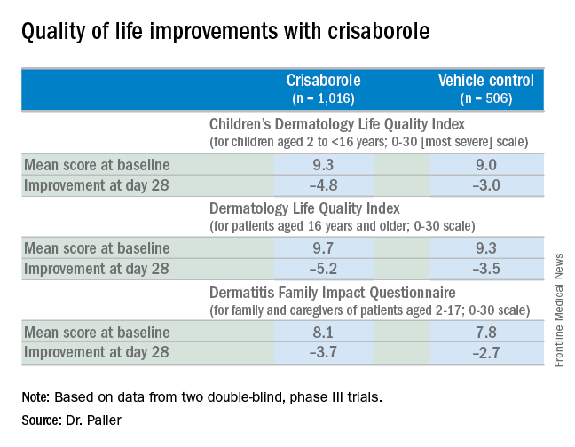
AD in children and adolescents is infamous for the adverse quality of life impact it imposes upon the patients’ parents, family, and caregivers. So the significant improvement seen with crisaborole, compared with its vehicle on the Dermatitis Family Impact (DFI) Questionnaire was particularly gratifying. The DFI questionnaire assesses quality of life in key domains, including family, parent, and caregiver sleep, emotional distress, relationships, family leisure, and ability to do housework or go shopping.
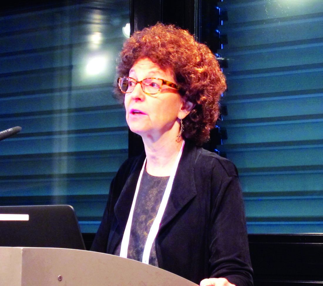
“If approved, crisaborole ... could improve the quality of life for patients with mild to moderate atopic dermatitis and, very importantly, for their families as well,” declared Dr. Paller, professor of dermatology and chair of the department of dermatology and professor of pediatrics at Northwestern University, Chicago.
Crisaborole’s developer, Anacor Pharmaceuticals, has filed an application for approval for treating mild to moderate AD in patients aged 2 years and older, now under review at the Food and Drug Administration.
In a separate presentation at the EADV congress, Lawrence F. Eichenfield, MD, presented the results of a long-term, open-label crisaborole safety study of 48-52 weeks duration. The long-term study involved 517 participants in the two pivotal phase III trials. There were no serious adverse events and no long-term cutaneous adverse events such as the skin atrophy or telangiectasias that can occur with topical steroids. The safety profile was favorable for long-term treatment of patients 2 years of age or older with mild to moderate AD.
Crisaborole 2% topical ointment is a novel, boron-based, nonsteroidal inhibitor of phosphodiesterase 4 (PDE-4). AD is marked by overactivity of PDE-4, which results in decreased levels of cyclic AMP and resultant increased release of inflammatory cytokines.
Dr. Paller noted that, in the previously reported efficacy results of the two pivotal, double-blind, 28-day, phase III trials, crisaborole treatment reduced global disease severity and provided early and sustained improvement in itch severity. She presented the prespecified quality of life results for the two identically designed, parallel pivotal trials, which totaled 1,016 patients on crisaborole and 506 on its vehicle. At baseline, 39% of subjects had mild AD, and 61% had moderate AD. The mean body surface area affected was 18%. Participants’ mean age was 12.3 years, and 14% were aged 8 years old or older.
The structure of the long-term safety study suggests how crisaborole might be used in clinical practice. During the year-long, open-label study, patients were evaluated every 28 days. If their skin was deemed clear or almost clear on the basis of an Investigator’s Static Global Assessment (ISGA) score of 0 or 1, they were taken off crisaborole and could use only emollients for the next 28 days, at which time they would be reevaluated. At that point, if they had an ISGA of 2 or more, they went back on crisaborole twice a day for 28 days until their next evaluation.
Dr. Eichenfield reported that 10.2% of participants in the long-term safety study reported treatment-related adverse events, which were mild to moderate. The most frequently reported of these were mild to moderate flares of AD during a 28-day off-treatment period in 1.3% of patients, application site pain in 2.3%, and application site infection in 1.2%.
Dr. Paller and Dr. Eichenfield reported serving as consultants to Anacor Pharmaceuticals.
The FDA review of crisaborole is expected to be completed by early January 2017, according to Anacor.
VIENNA – Topical crisaborole 2% ointment administered twice a day was consistently associated with clinically meaningful quality of life improvement scores on multiple measures in the two pivotal phase III, randomized, controlled trials of atopic dermatitis (AD) patients aged 2 years old through adulthood, Amy S. Paller, MD, reported at the annual congress of the European Academy of Dermatology and Venereology.
AD in children and adolescents is infamous for the adverse quality of life impact it imposes upon the patients’ parents, family, and caregivers. So the significant improvement seen with crisaborole, compared with its vehicle on the Dermatitis Family Impact (DFI) Questionnaire was particularly gratifying. The DFI questionnaire assesses quality of life in key domains, including family, parent, and caregiver sleep, emotional distress, relationships, family leisure, and ability to do housework or go shopping.
“If approved, crisaborole ... could improve the quality of life for patients with mild to moderate atopic dermatitis and, very importantly, for their families as well,” declared Dr. Paller, professor of dermatology and chair of the department of dermatology and professor of pediatrics at Northwestern University, Chicago.
Crisaborole’s developer, Anacor Pharmaceuticals, has filed an application for approval for treating mild to moderate AD in patients aged 2 years and older, now under review at the Food and Drug Administration.
In a separate presentation at the EADV congress, Lawrence F. Eichenfield, MD, presented the results of a long-term, open-label crisaborole safety study of 48-52 weeks duration. The long-term study involved 517 participants in the two pivotal phase III trials. There were no serious adverse events and no long-term cutaneous adverse events such as the skin atrophy or telangiectasias that can occur with topical steroids. The safety profile was favorable for long-term treatment of patients 2 years of age or older with mild to moderate AD.
Crisaborole 2% topical ointment is a novel, boron-based, nonsteroidal inhibitor of phosphodiesterase 4 (PDE-4). AD is marked by overactivity of PDE-4, which results in decreased levels of cyclic AMP and resultant increased release of inflammatory cytokines.
Dr. Paller noted that, in the previously reported efficacy results of the two pivotal, double-blind, 28-day, phase III trials, crisaborole treatment reduced global disease severity and provided early and sustained improvement in itch severity. She presented the prespecified quality of life results for the two identically designed, parallel pivotal trials, which totaled 1,016 patients on crisaborole and 506 on its vehicle. At baseline, 39% of subjects had mild AD, and 61% had moderate AD. The mean body surface area affected was 18%. Participants’ mean age was 12.3 years, and 14% were aged 8 years old or older.
The structure of the long-term safety study suggests how crisaborole might be used in clinical practice. During the year-long, open-label study, patients were evaluated every 28 days. If their skin was deemed clear or almost clear on the basis of an Investigator’s Static Global Assessment (ISGA) score of 0 or 1, they were taken off crisaborole and could use only emollients for the next 28 days, at which time they would be reevaluated. At that point, if they had an ISGA of 2 or more, they went back on crisaborole twice a day for 28 days until their next evaluation.
Dr. Eichenfield reported that 10.2% of participants in the long-term safety study reported treatment-related adverse events, which were mild to moderate. The most frequently reported of these were mild to moderate flares of AD during a 28-day off-treatment period in 1.3% of patients, application site pain in 2.3%, and application site infection in 1.2%.
Dr. Paller and Dr. Eichenfield reported serving as consultants to Anacor Pharmaceuticals.
The FDA review of crisaborole is expected to be completed by early January 2017, according to Anacor.
EXPERT ANALYSIS FROM THE EADV CONGRESS
Take a Bite of the Big Apple at the VEITHsymposium
New York City is a global destination renowned for its unparalleled variety of things to do and see to match all tastes and interests. In your time between and after the VEITHsymposium sessions, you can enjoy a Broadway show, take in some of the city’s famous museums, go on a shopping spree, or dine at some of the best restaurants in the world. Whether with family or on your own, there is something for everyone.
For a quick look at current entertainment and dining options, pick up a copy of TimeOut New York or visit www.timeout.com/newyork, where you’ll find current listings for what is playing at theaters off and on Broadway as well as live music, special events, shopping, fine dining, and more.
For a more comprehensive listing of Broadway and off-Broadway shows, visit www.Broadway.com.
For the sports fan, you can find a complete online calendar listing of sporting events that are going during the VEITHsymposium (www.nycgo.com/things-to-do/events-in-nyc/sports-calendar). Pick up the New York Knicks basketball team playing the Detroit Pistons on Nov. 16 at Madison Square Garden. College basketball will also be featured at the Garden with the Champions Classic on Nov. 15.
If you’re a classical music enthusiast, you should make time to attend a performance at Lincoln Center (www.lincolncenter.org), which plays host to the New York Philharmonic and the Metropolitan Opera. During the VEITHsymposium, the operas “Aida,” “La Boheme,” and “Manon Lescaut” are all being performed.
And for the forever young crowd, Billy Joel is in concert at Madison Square Garden on Nov. 19.
No matter where your music tastes lie, for the most up-to-date accounting of musical performances in the city, pick up a copy of the free weekly newspaper, the Village Voice, or visit it online (www.villagevoice.com).
For your exercise pleasure and a literal change of pace, there are a number of interesting walking tours available, including ones that amble through the historic lower East Side, the Metropolitan Opera, and Broadway theaters (www.walksofnewyork.com).
The New York food scene is both vast and varied, so to choose your dining from fine to fun, you can visit the venerable New York Times website (www.nytimes.com/reviews/dining) to plan your eating experience by rating, price, neighborhood, and choice of cuisine.
You will find several notable museums and landmarks within a few blocks of the VEITHsymposium. The Museum of Modern Art is the closest to the New York Hilton Midtown, at 11 West 53rd St., between Fifth and Sixth Avenues. MoMA houses more than 150,000 paintings, sculptures, drawings, prints, photographs, architectural models and drawings, and design objects. The Metropolitan Museum of Art is a bit farther away (1000 Fifth Ave. at 82nd St.). The holdings include 2 million works spanning 5,000 years of art history from around the world from Egyptian mummies to Rembrandts and Picassos.
The American Museum of Natural History is on the Upper West Side of Central Park (at 79th St.) and has something for museum goers of all ages: dinosaurs, fossils, stuffed specimens, minerals, gems, and human cultural artifacts.
The memorial at the site of the World Trade Center towers stands witness to a national tragedy and to the spirit of the people of New York. The names of nearly 3,000 men, women, and children killed in the attacks of Sept. 11, 2001, and Feb. 26, 1993, are inscribed on the parapets surrounding the two memorial pools (www.911memorial.org/visit).
In addition the famous Central Park, don’t overlook the less-noted Bryant Park (between 40th and 42nd Streets and Fifth and Sixth Avenues), which offers an opportunity to do some holiday shopping at the annual Holiday Shops fair. This outdoor, European-style market features everything from handcrafted items to gourmet treats.
If you’re a first-time visitor, the Empire State Building, the Statue of Liberty, and Ellis Island remain the must-see symbols of New York. Be prepared for long lines to get to the top of the Empire State Building (www.esbnyc.com). Dress warmly for the ferry ride from Battery Park (www.statuecruises.com) to the Statue of Liberty.
Above all, while you’re at the VEITHsymposium, be sure that you and your guests enjoy some of the many pleasures that New York has to offer. It’s your kind of town.
Prognostic scores helpful in subset of COPD patients
LOS ANGELES – The Sequential Organ Failure Assessment (SOFA) score and the Glasgow Coma Scale (GCS) are simple, accurate tools for risk stratification of hospitalized patients with acute exacerbation of COPD, results from a single-center study showed.
“Acute exacerbations of chronic obstructive pulmonary disease often require hospitalization, may necessitate mechanical ventilation, and can be fatal,” Mohamed Metwally, MD, FCCP, said in an interview in advance of the annual meeting of the American College of Chest Physicians. “There are currently no validated disease-specific scores that measure the severity of acute exacerbation. Prognostic tools are needed to assess acute exacerbations of chronic obstructive pulmonary disease.”
For the 2-year study, Dr. Metwally and his associates prospectively evaluated 250 critically ill ICU AECOPD patients, mean age 65 years, at Assiut University Hospital between December 2012 and December 2014. The primary outcome was in-hospital mortality while the secondary endpoint was need for intubation and mechanical ventilation. The researchers excluded patients who died less than 24 hours after admission, those with underlying COPD who were admitted with another primary diagnosis such as an accident or a stroke, or for elective hospitalizations such as elective surgery or diagnostic procedures.
Dr. Metwally and his associates collected sociodemographic data, vital signs, and other clinical variables, and collected scores from five tools used to measure mortality prediction: the Acute Physiology and Chronic Health Evaluation (APACHE II), the SOFA score, the Early Warning Score (EWS), the GCS, and the Charlson Comorbidity Index (CCI). To assess performance of the scores, they used area under the receiver operating characteristic curve (AUC) analysis and the Hosmer-Lemeshow goodness-of-fit test for logistic regression.
Of the 250 patients, 43 (17%) died during their hospital stay and 54% required mechanical ventilation. All recorded scores were significantly higher in nonsurvivors, compared with survivors, and the risk of clinical deterioration increased with increasing scores. The discriminatory power of each score varied as measured by AUC analysis. The AUC of APACHE II, SOFA, EWS, GCS, and CCI were 0.79, 0.81, 0.76, 0.69, and 0.68, respectively “and all these models had good calibration in mortality prediction,” Dr. Metwally said. The SOFA score was the best in predicting mortality (its predicted mortality was 16%, compared with the actual mortality of 17%), while the APACHE II score overestimated mortality by at least twofold (46% vs. 17%). In addition, the EWS outperformed the GCS in predicting mortality. “This may be due to EWS containing all vital signs plus level of consciousness,” he said in an interview.
The GCS was found to be the most useful in predicting need for mechanical ventilation, with an AUC of 0.81. The AUCs of APACHE II, SOFA, EWS, and CCI were 0.79, 0.80, 0.73, and 0.61, respectively. All of the scores had good calibration in mortality prediction, Dr. Metwally said, with the exception of SOFA.
As for the APACHE II, Dr. Metwally said that instrument “can be used as a tool to predict both mortality and intubation in a specific group of patients, but with low discriminatory power.” He acknowledged certain limitations of the study, including the fact that it was limited to patients with AECOPD. “Future studies should include any critically ill respiratory patients,” he said.
He reported having no financial disclosures.
AT CHEST 2016
Key clinical point:
Major finding: The Sequential Organ Failure Assessment score was the best in predicting mortality of patients with acute exacerbation of COPD (its predicted mortality was 16%, compared with the actual mortality of 17%).
Data source: A prospective evaluation of 250 critically ill ICU patients hospitalized with acute exacerbation of COPD.
Disclosures: Dr. Metwally reported having no financial disclosures.
Balancing speed, safety in device approvals
WASHINGTON – Regulators from two continents are slowly inching toward more common ground when it comes to device approvals, a move that should strike a better balance between the time to approval and safety of the products.
It is no surprise that for devices to be approved in the United States, a much more rigorous evidence standard exists than it does in Europe, but the Food and Drug Administration is looking for ways to make changes to allow for quicker approval times without compromising safety.
Meanwhile, across the Atlantic, European regulators are looking for increased evidence requirements that will not stifle innovation.
However, health information technology may help relieve some inefficiencies going forward.
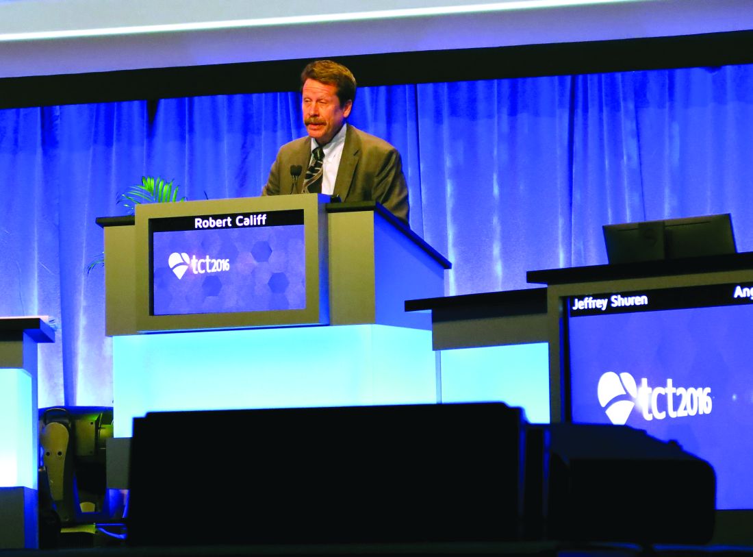
“If we combine quality registries, claims data, the evolving improving electronic health records, and the data warehouses that all of our health systems have, we can conduct prospective, observational, and clinical trials at a dramatically lower cost, answering many more questions per unit of time,” Dr. Califf added.
Other areas that will help improve the efficiency of the U.S. approval process is the work on developing a framework to bring feasibility studies back to America.
“I want to really accelerate that because we are hearing from a lot of people that they want to access the technology early; it’s the right thing to do, and we are hearing from industry that they’d like to work in the United States,” he said.
However, that is going to require one group that is generally more risk adverse to be willing to participate more in the process.
“The main limiting factors at this point in time are the health systems that we are all working in,” Dr. Califf noted. “They are very risk adverse. So while it is nice to argue and advertise that your interventional cardiologists are the best in town on your billboard, protecting those interventional cardiologists to do the high-risk, early studies that are really needed to develop devices is a different matter.”
He called on doctors to go back to their health systems to help better develop those early feasibility pathways to help bring some of that innovation back the United States.
Finally, Dr. Califf addressed the idea of developing evidence that can function not only for device approval, but for payment as well, especially as reimbursement systems are becoming more value driven.
“We are going to move to a system where reimbursement will increasingly move away from fee for service and increasingly gravitate to payment for value,” Dr. Califf said. “In order for this to work, we’ve got to develop the kinds of clinical trials [that include] the calculation of value, so the winners can be promoted on a wider scale and the procedures and technologies that don’t provide incremental value are left behind.”
He continued: “We need your help in figuring out the common source of information so that the FDA can make its decision – which is different from CMS’s decision – but where there’s a continuum of knowledge that allows for products to be approved if they are worthwhile, and then to be paid for and allowed to be marketed.”
Meanwhile, developers are facing a different situation in Europe, where more regulation and more stringent evidence standards could be coming down the pike.
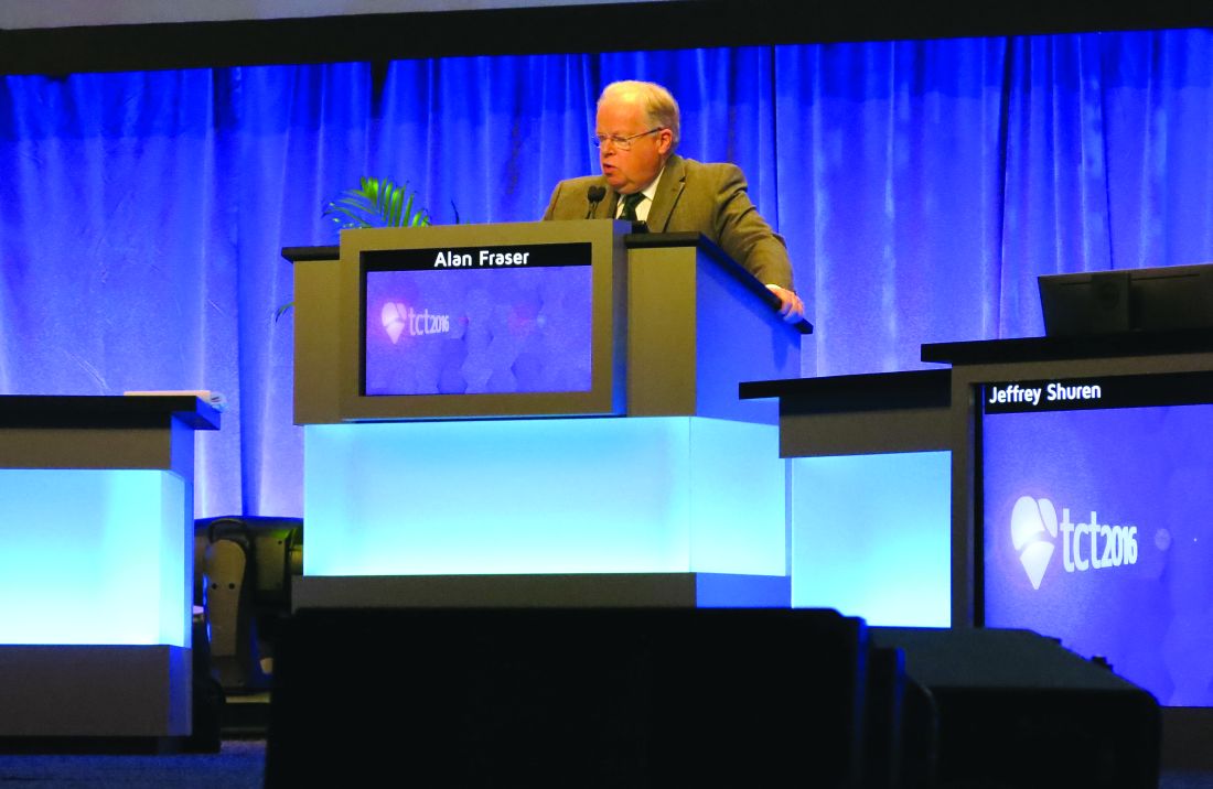
Dr. Fraser noted research that has shown that devices first approved in Europe, compared with their U.S. counterparts, are associated with an increased incidence of recalls or safety alerts.
That being said, he noted that there are several proposed regulations that would increase the evidence requirements related to regulatory approval that manufacturers could face in the coming year, a challenge as it will require some level of harmonization across countries and regulatory systems within each country.
“In Europe, we are on the dawn of a new era regarding medical device assessment, but it is going to pose a very large challenge for our colleagues in the regulatory agencies because of their personnel, funding, and the integration between all these bodies and national systems. There are also tremendous challenges for us as physicians to ensure that we seek the evidence, we assess and contribute to it, and particularly now, that we also routinely take part in new systems for postmarket surveillance,” he said.
WASHINGTON – Regulators from two continents are slowly inching toward more common ground when it comes to device approvals, a move that should strike a better balance between the time to approval and safety of the products.
It is no surprise that for devices to be approved in the United States, a much more rigorous evidence standard exists than it does in Europe, but the Food and Drug Administration is looking for ways to make changes to allow for quicker approval times without compromising safety.
Meanwhile, across the Atlantic, European regulators are looking for increased evidence requirements that will not stifle innovation.
However, health information technology may help relieve some inefficiencies going forward.
“If we combine quality registries, claims data, the evolving improving electronic health records, and the data warehouses that all of our health systems have, we can conduct prospective, observational, and clinical trials at a dramatically lower cost, answering many more questions per unit of time,” Dr. Califf added.
Other areas that will help improve the efficiency of the U.S. approval process is the work on developing a framework to bring feasibility studies back to America.
“I want to really accelerate that because we are hearing from a lot of people that they want to access the technology early; it’s the right thing to do, and we are hearing from industry that they’d like to work in the United States,” he said.
However, that is going to require one group that is generally more risk adverse to be willing to participate more in the process.
“The main limiting factors at this point in time are the health systems that we are all working in,” Dr. Califf noted. “They are very risk adverse. So while it is nice to argue and advertise that your interventional cardiologists are the best in town on your billboard, protecting those interventional cardiologists to do the high-risk, early studies that are really needed to develop devices is a different matter.”
He called on doctors to go back to their health systems to help better develop those early feasibility pathways to help bring some of that innovation back the United States.
Finally, Dr. Califf addressed the idea of developing evidence that can function not only for device approval, but for payment as well, especially as reimbursement systems are becoming more value driven.
“We are going to move to a system where reimbursement will increasingly move away from fee for service and increasingly gravitate to payment for value,” Dr. Califf said. “In order for this to work, we’ve got to develop the kinds of clinical trials [that include] the calculation of value, so the winners can be promoted on a wider scale and the procedures and technologies that don’t provide incremental value are left behind.”
He continued: “We need your help in figuring out the common source of information so that the FDA can make its decision – which is different from CMS’s decision – but where there’s a continuum of knowledge that allows for products to be approved if they are worthwhile, and then to be paid for and allowed to be marketed.”
Meanwhile, developers are facing a different situation in Europe, where more regulation and more stringent evidence standards could be coming down the pike.
Dr. Fraser noted research that has shown that devices first approved in Europe, compared with their U.S. counterparts, are associated with an increased incidence of recalls or safety alerts.
That being said, he noted that there are several proposed regulations that would increase the evidence requirements related to regulatory approval that manufacturers could face in the coming year, a challenge as it will require some level of harmonization across countries and regulatory systems within each country.
“In Europe, we are on the dawn of a new era regarding medical device assessment, but it is going to pose a very large challenge for our colleagues in the regulatory agencies because of their personnel, funding, and the integration between all these bodies and national systems. There are also tremendous challenges for us as physicians to ensure that we seek the evidence, we assess and contribute to it, and particularly now, that we also routinely take part in new systems for postmarket surveillance,” he said.
AT TCT 2016
Welcome to the 2016 VEITHsymposium
Welcome to the 43rd annual Vascular & Endovascular, Issues, Techniques and Horizons Symposium (VEITHsymposium). This year’s program promises to be one of the best, most comprehensive, and most thought-provoking of any of our meetings. This year we celebrate our 43rd anniversary and have introduced several improvements.
Nearly 600 international clinician/educators have gathered to provide attendees with the latest topics and advances that are important to the global vascular community. These data span the breadth of vascular diseases, diagnostic procedures, medical treatments, interventional procedures and open surgical advances for treating vascular disease. As is the hallmark of the VEITHsymposium, the 5-day program will run from dawn to dusk daily and will be fully captured in our online library.

Top vascular experts from around the world will provide updates on the latest clinical trials and offer insight into the real-life application of the most recent data to close the gap between the current state of knowledge and actual clinical practice.
Controversial issues will be approached from multiple perspectives to ensure a balanced, unbiased exposure of topics and to provide audience members with the information they need to make informed choices in their own practices.
This year our meeting continues its increased emphasis on venous disease. Three full days of the meeting are developments in venous disease of all sorts and active endovascular treatments in this rapidly expanding area of opportunity.
Some of the program’s other hot topics will be the continuing controversies surrounding parallel grafts (chimneys, and snorkel and sandwich grafts); multilayer open stents versus fenestrated and branched endografts; new developments in carotid stenting; new developments in the treatment of aortic dissections; a day devoted to the management of arteriovenous malformations (AVMs); new developments in the endovascular treatment of lower-extremity ischemia, particularly below the knee; the latest developments in EVAR and TEVAR including experiences with a plethora of new endovascular grafts and devices that have appeared on the scene in the last year; and improvements in the medical treatment of vascular disease and vascular patients undergoing surgery and other interventions. Important issues to vascular specialists and outpatient vascular treatment will also be highlighted.
This year’s program will include a special session all-day Tuesday, focused in the morning on management options for pulmonary embolism led by Dr. Michael R. Jaff. The afternoon part of the day will focus on new developments in the management of acute and chronic large vein occlusion, and will be led by Dr. Kenneth Ouriel.
This year there will also be sessions devoted to crucial issues for vascular specialists including changing relationships with government and the FDA and how to survive under new reimbursement rules and regulations including Obamacare. Our physician/educators will also offer a glimpse into some new techniques and technologies that have been available overseas, but are just gaining approval in the United States, such as drug-eluting balloons and stents.
Attendees will notice some other exciting changes or additions to this year’s program. We have included a new Job Fair Program on Friday in the Americas Hall 1 on the 3rd floor. In addition, there will be more breaks in the schedule to encourage exploration of state-of-the-art technology, products, and services available in the Exhibit areas and Pavilions. The Exhibit Halls are crowded with displays and booths of particular interest to vascular surgeons and vascular specialists. The Pavilions and Exhibits also offer attendees the chance to meet faculty and to network with other attendees and industry partners. This is one place to learn more about exciting new technologies and developments in our field.
Other new additions to our meeting this year will be an exciting Abbott Pavilion in the Americas Hall as well as an expanded Innovations and Investment Summit which facilitates interaction between innovators, industry and investors. This non-CME Session will be held from 8 AM to 3 PM on Thursday, November 17th in the Gramercy Suites on the 2nd floor, and will be led by Kenneth Ouriel, Jean Bismuth and Chris Cheng.
Also new this year will be an expanded VEITHsymposium mobile app, provided courtesy of Cook Medical. Download the app for your iPhone, iPad or Android phone or tablet! Search the App Store (iPhone/iPad) or Google Play Store (Android) for “VEITHsymposium 2016” and install the app. You will be able to access the complete program, create your personal program, add your notes, view the location of sessions and exhibitors on the floor plan, and much more. After you have installed the app and opened it for the first time, you can continue to use it offline. To receive the latest updates and announcements, you will need to be connected to the internet.
In addition, there will be expanded Associate Faculty programs which will give younger and less well-known vascular specialists the opportunity to present their work at the podium with leading experts as session moderators.
Again this year, an Online Library will be available for a minimal fee of $75 for clinical meeting attendees and will include access to talks, slides, videos, and panels from the meeting. This Library will enable all attendees to see and hear key presentations they may miss because of the concurrent sessions or other reasons. This library will be available 10-14 days after the meeting. Attendees should note in their program talks they wish to hear but could not, and then revisit the missed talks on the Online Library which is indexed exactly to the program. The talks are also indexed in the Library by presenter, topic, or session. This Library is a great resource for study, research or review for any purpose.
On behalf of all the meeting Co-Chairmen and our entire staff, we greatly appreciate you coming to our meeting. We hope it is our best meeting ever and that you find it educational, most useful and exciting so that you return next year.
Welcome to the 43rd annual Vascular & Endovascular, Issues, Techniques and Horizons Symposium (VEITHsymposium). This year’s program promises to be one of the best, most comprehensive, and most thought-provoking of any of our meetings. This year we celebrate our 43rd anniversary and have introduced several improvements.
Nearly 600 international clinician/educators have gathered to provide attendees with the latest topics and advances that are important to the global vascular community. These data span the breadth of vascular diseases, diagnostic procedures, medical treatments, interventional procedures and open surgical advances for treating vascular disease. As is the hallmark of the VEITHsymposium, the 5-day program will run from dawn to dusk daily and will be fully captured in our online library.
Top vascular experts from around the world will provide updates on the latest clinical trials and offer insight into the real-life application of the most recent data to close the gap between the current state of knowledge and actual clinical practice.
Controversial issues will be approached from multiple perspectives to ensure a balanced, unbiased exposure of topics and to provide audience members with the information they need to make informed choices in their own practices.
This year our meeting continues its increased emphasis on venous disease. Three full days of the meeting are developments in venous disease of all sorts and active endovascular treatments in this rapidly expanding area of opportunity.
Some of the program’s other hot topics will be the continuing controversies surrounding parallel grafts (chimneys, and snorkel and sandwich grafts); multilayer open stents versus fenestrated and branched endografts; new developments in carotid stenting; new developments in the treatment of aortic dissections; a day devoted to the management of arteriovenous malformations (AVMs); new developments in the endovascular treatment of lower-extremity ischemia, particularly below the knee; the latest developments in EVAR and TEVAR including experiences with a plethora of new endovascular grafts and devices that have appeared on the scene in the last year; and improvements in the medical treatment of vascular disease and vascular patients undergoing surgery and other interventions. Important issues to vascular specialists and outpatient vascular treatment will also be highlighted.
This year’s program will include a special session all-day Tuesday, focused in the morning on management options for pulmonary embolism led by Dr. Michael R. Jaff. The afternoon part of the day will focus on new developments in the management of acute and chronic large vein occlusion, and will be led by Dr. Kenneth Ouriel.
This year there will also be sessions devoted to crucial issues for vascular specialists including changing relationships with government and the FDA and how to survive under new reimbursement rules and regulations including Obamacare. Our physician/educators will also offer a glimpse into some new techniques and technologies that have been available overseas, but are just gaining approval in the United States, such as drug-eluting balloons and stents.
Attendees will notice some other exciting changes or additions to this year’s program. We have included a new Job Fair Program on Friday in the Americas Hall 1 on the 3rd floor. In addition, there will be more breaks in the schedule to encourage exploration of state-of-the-art technology, products, and services available in the Exhibit areas and Pavilions. The Exhibit Halls are crowded with displays and booths of particular interest to vascular surgeons and vascular specialists. The Pavilions and Exhibits also offer attendees the chance to meet faculty and to network with other attendees and industry partners. This is one place to learn more about exciting new technologies and developments in our field.
Other new additions to our meeting this year will be an exciting Abbott Pavilion in the Americas Hall as well as an expanded Innovations and Investment Summit which facilitates interaction between innovators, industry and investors. This non-CME Session will be held from 8 AM to 3 PM on Thursday, November 17th in the Gramercy Suites on the 2nd floor, and will be led by Kenneth Ouriel, Jean Bismuth and Chris Cheng.
Also new this year will be an expanded VEITHsymposium mobile app, provided courtesy of Cook Medical. Download the app for your iPhone, iPad or Android phone or tablet! Search the App Store (iPhone/iPad) or Google Play Store (Android) for “VEITHsymposium 2016” and install the app. You will be able to access the complete program, create your personal program, add your notes, view the location of sessions and exhibitors on the floor plan, and much more. After you have installed the app and opened it for the first time, you can continue to use it offline. To receive the latest updates and announcements, you will need to be connected to the internet.
In addition, there will be expanded Associate Faculty programs which will give younger and less well-known vascular specialists the opportunity to present their work at the podium with leading experts as session moderators.
Again this year, an Online Library will be available for a minimal fee of $75 for clinical meeting attendees and will include access to talks, slides, videos, and panels from the meeting. This Library will enable all attendees to see and hear key presentations they may miss because of the concurrent sessions or other reasons. This library will be available 10-14 days after the meeting. Attendees should note in their program talks they wish to hear but could not, and then revisit the missed talks on the Online Library which is indexed exactly to the program. The talks are also indexed in the Library by presenter, topic, or session. This Library is a great resource for study, research or review for any purpose.
On behalf of all the meeting Co-Chairmen and our entire staff, we greatly appreciate you coming to our meeting. We hope it is our best meeting ever and that you find it educational, most useful and exciting so that you return next year.
Give guidance, not just Viagra, to men with diabetes and related impotence
Diabetic men with erectile dysfunction might consider a prescription for a drug like Viagra to be a permanent cure for a temporary issue: Take a pill, problem solved. But the truth, a leading urologist says, is entirely different.
“If a diabetic patient has erectile dysfunction, it’s not enough to provide Viagra [sildenafil] or Cialis [tadalafil] and then send him on his merry way,” J. Francois Eid, MD, a New York City urologist, said at the annual meeting of the American Association of Diabetes Educators. “It’s important to let individuals know the drug has not cured the erectile dysfunction. If patients don’t take care of the diabetes, the erectile dysfunction progresses.”
In an interview, Dr. Eid shared several messages for medical professionals who treat men with diabetes and related erectile dysfunction:
• Diabetes has “devastating” effects on the penis, and may even cause it to shrink.
• Long-term uncontrolled diabetes can make ED permanent.
• While ED drugs often fail in men with diabetes, several other options exist; and penis implants may provide significant relief.
How big is the problem? An estimated 50%-75% of men with diabetes experience from some degree of ED, which is thought to be three times more common in diabetic men than other men, according to Dr. Eid.
“Diabetes has a devastating effect on the muscle tissue inside the penis,” he said. “All the tiny little arteries that feed blood to the muscle get occluded. Little by little, the muscle inside the penis shrinks.” Indeed, some diabetics with ED complain that their penises have shrunk, he said.
Diabetic damage doesn’t stop with these small vessels, he said. “You really have two parallel situations: You need blood flow that feeds the muscle of the penis, and you need an artery dedicated to bringing blood rapidly when a man becomes aroused and wants to be sexually active,” he said. “That artery is also affected by diabetes. They’ll say ‘I can get a partial erection, but I can’t maintain it.’ ”
What comes after an ED diagnosis in diabetic patients? Often, Dr. Eid will instantly refer these men to a cardiologist. “If a patient has diabetes and is newly diagnosed, a significant portion of these men are going to develop coronary artery disease in the next 2-3 years,” he said. “One of the things we do is recommend is that they see a cardiologist and perhaps have a stress test or some sort of evaluation.”
Dr. Eid also urges these patients to treat their diabetes in order to avoid developing ED for life. “They need to manage their diabetes and make sure they control it so the ED will not progress and will stabilize, as a result,” he said. “If the diabetes is controlled after the patient is first diagnosed, then the erections will come back. But if the patient has diabetes for many years, and suddenly decides it’s time to control it, they cannot prevent the damage that’s already been done.”
As for treatments, patients with diabetes and related ED should begin with medications like sildenafil and tadalafil, he recommended. But research findings suggest that the drugs will fail in half of men with type 2 diabetes, he said.
Other options include penile self-injections, vacuum devices, and penile implants.
The injections “can work well and are painless, but men detest having to inject themselves before sexual activity,” Dr. Eid said. And he said men rarely have success using vacuum devices, which are available over the counter.
By contrast, penis implants can successfully treat erectile dysfunction in many cases, Dr. Eid said. Men trigger erections by squeezing a pump that is implanted into the scrotum. Fluid then flows from an implanted reservoir into a cylinder implanted in the penis.
Men are often pleased by penis implants because they can have sex spontaneously without having to plan for it ahead of time, as required by medications. “They’re completely transformed,” Dr. Eid said. “They feel that they’re cured.”
Dr. Eid receives or has received research support/grants from American Medical Systems, Coloplast, Lilly ICOS, Bayer, Vivus, Pharmacia-Upjohn, and Pfizer. He is or was a consultant and on the speakers bureau for Coloplast, American Medical Systems, Lilly ICOS, Bayer and Pfizer.
EXPERT ANALYSIS FROM AADE 16
An enlightened approach to weight loss using liraglutide
NEW ORLEANS – Early weight loss on liraglutide – specifically, dropping at least 4% of body weight at 16 weeks – is a strong and clinically useful identifier of patients with a high likelihood of significant weight loss 13 months into treatment, with an accompanying improvement in cardiometabolic risk factors, according to Ken Fujioka, MD.
Conversely, patients who aren’t early responders to subcutaneous liraglutide at 3 mg/day are unlikely to achieve at least 5% weight loss after a full year on the drug, the regulatory benchmark for clinically meaningful weight loss, added Dr. Fujioka, an internist and director of the center for weight management at the Scripps Clinic in La Jolla, Calif.
“Using this early response criterion at week 16 to predict long-term weight loss is to me a very valuable tool. Obesity is an odd disease because it has so many different causes. Finding the right drug is tough, and how long to keep trying with a particular medication is something we haven’t known. So I think the biggest change in obesity medicine is the creation of stopping rules that allow you to say, ‘OK, maybe this isn’t going to work. There’s some other reason you’re gaining weight, so let’s move on to something else,’” Dr. Fujioka said at the meeting presented by the Obesity Society of America and the American Society for Metabolic and Bariatric Surgery.
“When you stop the medication, you improve the risk-benefit ratio by removing all risk. That’s a win-win to me, and I applaud the FDA for getting on the pharmaceutical companies to make sure they put stopping rules in their medication labels,” he added.
He presented a post hoc pooled analysis of two previously published large, double-blind phase III clinical trials of subcutaneous liraglutide at 3 mg/day (Saxenda) or placebo in combination with a diet and exercise intervention for weight loss: the 3,731-patient SCALE Obesity and Prediabetes trial and the 846-patient SCALE Diabetes trial. In both trials liraglutide was started at a dose of 0.6 mg and titrated to 3.0 mg by week 4. The lifestyle intervention entailed a 500-kcal/day deficit diet and a minimum of 150 minutes of physical activity per week.
The purpose of the pooled analysis was to identify the best early predictor of response status at 56 weeks by examining the impact of 3%, 4%, and 5% weight loss after 8, 12, and 16 weeks of treatment as cut points. This post hoc analysis was prespecified at the request of the Food and Drug Administration before the trials were completed.
The bottom line: The best predictor of long-term outcome on liraglutide, a glucagonlike peptide–1 analog, proved to be a weight loss of 4% or greater at 16 weeks. It had an 81% positive predictive value and a 76% negative predictive value for at least a 5% weight loss at 56 weeks. It correctly predicted weight outcomes at 56 weeks in 80.1% of patients, the highest success rate of all the combinations studied. This finding was the impetus for the current product labeling, which contains the stopping rule. Dr. Fujioka shared study data not included in the labeling; namely, the marked contrast in how early responders and early nonresponders fared at 56 weeks.
The mean weight loss at 56 weeks in nondiabetic early responders to liraglutide was 10.8%, compared with only 3% in early nondiabetic nonresponders. Diabetic early responders averaged an 8.5% weight loss at 56 weeks, while early nonresponders had a mean 3.1% weight loss.
In the SCALE Obesity and Prediabetes trial, 50% of early responders to liraglutide ended up with a greater than 10% weight loss at 56 weeks, and 21% had more than 15% weight loss, compared with rates of 6% and 2%, respectively, in early nonresponders.
In the SCALE Diabetes study, 38% of early responders had greater than 10% weight loss long term, a rate nearly fourfold higher than in early nonresponders. Moreover, 10% of early responder diabetic patients had greater than 15% weight loss, versus a mere 2% of early nonresponders, the internist continued.
The ratio of early responders to early nonresponders in the nondiabetic population was 77%:23%. In diabetic patients, it was 63%:37%.
Turning to cardiometabolic endpoints, Dr. Fujioka noted that early responders in the SCALE Obesity and Prediabetes trial went on to show a mean reduction in systolic blood pressure of 5.1 mm Hg at 56 weeks, compared with a 2–mm Hg decrease in early nonresponders. Early responders also averaged a 10.5-cm shrinkage in waist circumference from a baseline of 115 cm, which was more than twice that observed at 56 weeks in early nonresponders. HDL-cholesterol level rose by 3.9% in early responders but remained unchanged over time in early nonresponders.
Diabetic patients who were early responders to liraglutide 3.0 mg/day had a mean 44.2-mg/dL reduction in fasting plasma glucose at 56 weeks from a baseline of 158 mg/dL, compared with a 30.1-mg/dL decrease in early nonresponders.
“The drop in fasting blood glucose is very quick – within a matter of weeks – so if you already have diabetic patients on drugs that are going to bring their blood sugar down, you may have to back titrate those other drugs really quickly. You don’t want to make your patients hypoglycemic,” the physician said.
Mean hemoglobin A1c values in early responder diabetic patients fell by 1.6% from a baseline of 7.9%, a full 0.5% greater reduction than in early nonresponders.
By far the most frequent adverse events in the two SCALE trials were gastrointestinal, with nausea leading the way. Rates were modestly higher in the early responders.
This analysis and the clinical trials on which it was based were sponsored by Novo Nordisk, which markets liraglutide under the brand names Saxenda and Voctoza. The presenter reported receiving research grants from and serving as a consultant to Novo Nordisk and other pharmaceutical companies.
AT OBESITY WEEK 2016
Key clinical point:
Major finding: A 4% or greater weight loss at week 16 on liraglutide 3 mg/day had an 81.4% positive predictive value and a 76% negative predictive value for achievement of at least 5% weight loss at week 56 on the drug.
Data source: This was a prespecified post hoc analysis of two phase III randomized, double-blind clinical trials of liraglutide 3 mg/day for weight loss in more than 4,500 subjects.
Disclosures: This analysis and the clinical trials on which it is based were sponsored by Novo Nordisk, which markets liraglutide under the brand names Saxenda and Voctoza. The presenter reported receiving research grants from and serving as a consultant to Novo Nordisk and other pharmaceutical companies.
