User login
PD-1 inhibitors treat resistant gray zone lymphoma

Three case reports published in The New England Journal of Medicine describe the successful use of PD-1 inhibitors in gray zone lymphoma.
Two patients who had failed treatment with DA-EPOCH-R (dose-adjusted etoposide, doxorubicin, and cyclophosphamide with vincristine, prednisone, and rituximab) ultimately responded to treatment with pembrolizumab.
Another patient—who had previously received 2 chemotherapy regimens, monotherapy with brentuximab vedotin, and radiation—responded to treatment with nivolumab.
Pembrolizumab treatment
An 18-year-old woman with mediastinal gray-zone lymphoma initially had a partial response to DA-EPOCH-R. However, she progressed 6 weeks after salvage radiotherapy.
She went on to receive pembrolizumab, had a complete metabolic response, and proceeded to allogeneic transplant after 235 days of treatment.
A 76-year-old man with mediastinal gray-zone lymphoma also had a partial response to DA-EPOCH-R but later progressed.
He proceeded to pembrolizumab and had a complete metabolic response. He was still in remission on day 381 of treatment.
Nivolumab treatment
The patient who received nivolumab is Bobbie Flexer, an 80-year-old retired mathematics professor.
“For me, trying nivolumab was a binary choice: I could try the drug or I could give up,” Flexer said.
She had initially achieved a complete metabolic response to DA-EPOCH-R, but her disease progressed after 6 cycles of treatment.
“Given Bobbie’s age and her resistance to chemotherapy, it was difficult to simply increase her dose,” said Flexer’s oncologist, Manali Kamdar, MD, of the University of Colorado School of Medicine in Aurora.
“Bobbie’s tumor biopsy expressed a protein called CD30, and so we started her on brentuximab, which targets these CD30 cells. Unfortunately, Bobbie’s disease progressed through multiple cycles of brentuximab. Subsequently, we switched her to another combined chemotherapy—namely, gemcitabine with oxaliplatin [plus rituximab].”
When that regimen and mediastinal radiation both proved unsuccessful, Dr Kamdar started Flexer on nivolumab.
“Within one dose, she was in less pain, and she looked much better,” Dr Kamdar said.
A PET scan after 6 doses showed that Flexer’s disease was in complete remission, and she was still in remission on day 161 of treatment.
She did experience adverse effects, including 2 bouts of pneumonitis, which were successfully treated with prednisone.
Flexer also experienced an uptick in pancreas enzymes that caused high blood glucose levels. She is learning to treat that side effect with insulin. And dietitians helped her manage expected gut-related side effects of nivolumab.
To Dr Kamdar, Flexer’s case was striking enough to warrant submitting a report to NEJM.
Coincidentally, the journal had just received 2 similar case reports from the National Institutes of Health, in which researchers had used pembrolizumab to target gray zone lymphoma in a nearly identical way.
Together, these cases suggest a new strategy for treating gray zone lymphoma. ![]()
Vancomycin research reveals reasons for readmissions and prolonged stays
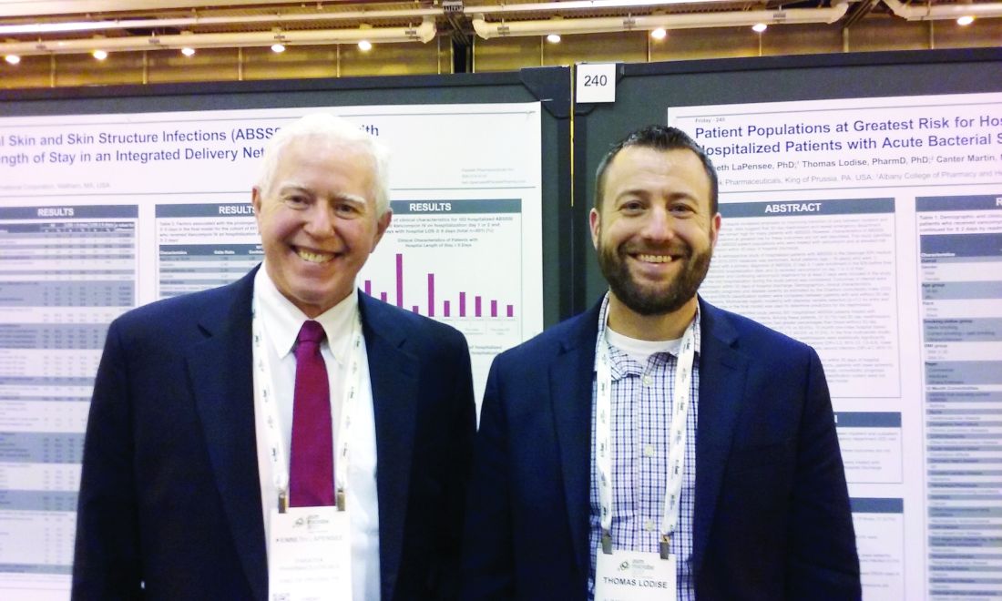
NEW ORLEANS – Approximately 20% of patients treated with vancomycin for an acute bacterial skin and skin structure infection remained in the hospital 8 days or longer, and about 7% experienced a readmission within 30 days, a retrospective study of 507 patients in the Geisinger Health System database showed.
“We found, for those who had a readmission, the major drivers were those who are your ‘health care frequent flyers’ – those who were admitted in the past 6 months,” said Thomas Lodise, PharmD, PhD, professor of pharmacy practice at Albany (N.Y.) College of Pharmacy and Health Sciences. “So, patients with a previous hospitalization are more likely to be treated again for all-cause admission within 30 days of discharge.” In addition, people with a lower-extremity abscess, particularly older patients with diabetes, and those with a traumatic wound were also more likely to return within 30 days.

Identifying the population at highest readmission risk could become more important soon. “Even though hospitals are not penalized for skin infection readmissions, there is some talk of adding that to the Medicare penalty,” Dr. Lodise said.
“It’s under review now by Medicare,” said Kenneth LaPensee, PhD, a consultant for Paratek Pharmaceuticals, King of Prussia, Pa., a firm developing an antibiotic to treat patients with an acute bacterial skin and skin structure infection (ABSSSI).
Dr. Lodise, Dr. LaPensee, and their colleagues studied adults hospitalized for an ABSSSI primary diagnosis based on ICD-9 codes and treated with at least 2 days of vancomycin. Participants were seen within the integrated Geisinger Health System between 2010 and 2015.
A total 6.9% of patients were readmitted within 30 days or had repeat emergency department visits. That group included more people with a body mass index of 36 kg/m2 or greater – 8.4%, compared with 6.2% of those with a BMI of less than 36. Other factors associated with readmission included smoking or a history of smoking (9.8% vs. 2.7% for nonsmokers) and a history of ABSSSI not requiring hospitalization vs. no prior history of ABSSSI, 22.2% vs. 6.6%, respectively.
In addition, those with a prior hospital admission not related to skin and skin structure infection were at higher risk, 8.8%, compared with 6.3% for those with no prior hospital admission. A prior hospital outpatient visit was likewise associated with a higher risk for readmission, 12.0%, compared with 5.4% without such a history.
Greater disease severity did not appear to correlate with a higher risk of 30-day readmission in the study. The researchers compared the groups by both Eron classification and the Charlson Comorbidity Index disease severity measures.
“People with more comorbidities had higher readmission rates, but it wasn’t statistically significant,” Dr. Lodise said. Also, “we saw some signaling – like with older age and some differences with race – [that] nonwhites were more likely to get readmitted. A total 6.7% of readmissions were among people older than 65 years, compared with 7.2% of younger people.” Advanced age was a factor in the bivariate analysis, but not in the logistic regression, Dr. LaPensee said.
“We’re going to repeat this in a larger data set. We’re planning for 10,000 patients,” Dr. Lodise said. “In our next cohort, we’ll be able to delineate more patient covariates.” An unanswered question is whether treatment with an agent other than vancomycin could improve readmission rates.
Using the same Geisinger database, the investigators also examined prolonged length of stay for patients with ABSSSIs treated with vancomycin. Almost one-fifth, 19.9%, met their definition of a prolonged stay of 8 days or longer.
The average length of stay was 7 days. “We were a bit surprised. We anticipated it being closer to 4 or 5 days,” Dr. Lodise said. “Then we wanted to find out who are these patients with these prolonged lengths of stay?
“The interesting thing was, things you think would be predictive, like increased age or high BMI [body mass index], were not,” Dr. Lodise said. “But what we did find is being elderly with diabetes and having a lower-extremity infection or a lower-extremity infection with an abscess – this was the group at greatest risk for a prolonged length of stay.” Those findings make sense, he added, because those patients tend to be slow responders, and because intravenous vancomycin has no oral, step-down formulation.
Unlike the readmission risk factors, Eron disease severity “was … very predictive of a prolonged length of stay,” Dr. Lodise said.
“These are really correlated – those patients with longer length of stay are more likely to get readmitted,” Dr. Lodise said. “In these more-difficult-to-treat patient populations, they really need more attention [to figure out] what is going on with them, why they keep coming back to the hospital, or why are they staying in so long.”
Dr. Lodise is a consultant for Paratek Pharmaceuticals, the study sponsor. Dr. LaPensee is a Paratek employee.
NEW ORLEANS – Approximately 20% of patients treated with vancomycin for an acute bacterial skin and skin structure infection remained in the hospital 8 days or longer, and about 7% experienced a readmission within 30 days, a retrospective study of 507 patients in the Geisinger Health System database showed.
“We found, for those who had a readmission, the major drivers were those who are your ‘health care frequent flyers’ – those who were admitted in the past 6 months,” said Thomas Lodise, PharmD, PhD, professor of pharmacy practice at Albany (N.Y.) College of Pharmacy and Health Sciences. “So, patients with a previous hospitalization are more likely to be treated again for all-cause admission within 30 days of discharge.” In addition, people with a lower-extremity abscess, particularly older patients with diabetes, and those with a traumatic wound were also more likely to return within 30 days.
Identifying the population at highest readmission risk could become more important soon. “Even though hospitals are not penalized for skin infection readmissions, there is some talk of adding that to the Medicare penalty,” Dr. Lodise said.
“It’s under review now by Medicare,” said Kenneth LaPensee, PhD, a consultant for Paratek Pharmaceuticals, King of Prussia, Pa., a firm developing an antibiotic to treat patients with an acute bacterial skin and skin structure infection (ABSSSI).
Dr. Lodise, Dr. LaPensee, and their colleagues studied adults hospitalized for an ABSSSI primary diagnosis based on ICD-9 codes and treated with at least 2 days of vancomycin. Participants were seen within the integrated Geisinger Health System between 2010 and 2015.
A total 6.9% of patients were readmitted within 30 days or had repeat emergency department visits. That group included more people with a body mass index of 36 kg/m2 or greater – 8.4%, compared with 6.2% of those with a BMI of less than 36. Other factors associated with readmission included smoking or a history of smoking (9.8% vs. 2.7% for nonsmokers) and a history of ABSSSI not requiring hospitalization vs. no prior history of ABSSSI, 22.2% vs. 6.6%, respectively.
In addition, those with a prior hospital admission not related to skin and skin structure infection were at higher risk, 8.8%, compared with 6.3% for those with no prior hospital admission. A prior hospital outpatient visit was likewise associated with a higher risk for readmission, 12.0%, compared with 5.4% without such a history.
Greater disease severity did not appear to correlate with a higher risk of 30-day readmission in the study. The researchers compared the groups by both Eron classification and the Charlson Comorbidity Index disease severity measures.
“People with more comorbidities had higher readmission rates, but it wasn’t statistically significant,” Dr. Lodise said. Also, “we saw some signaling – like with older age and some differences with race – [that] nonwhites were more likely to get readmitted. A total 6.7% of readmissions were among people older than 65 years, compared with 7.2% of younger people.” Advanced age was a factor in the bivariate analysis, but not in the logistic regression, Dr. LaPensee said.
“We’re going to repeat this in a larger data set. We’re planning for 10,000 patients,” Dr. Lodise said. “In our next cohort, we’ll be able to delineate more patient covariates.” An unanswered question is whether treatment with an agent other than vancomycin could improve readmission rates.
Using the same Geisinger database, the investigators also examined prolonged length of stay for patients with ABSSSIs treated with vancomycin. Almost one-fifth, 19.9%, met their definition of a prolonged stay of 8 days or longer.
The average length of stay was 7 days. “We were a bit surprised. We anticipated it being closer to 4 or 5 days,” Dr. Lodise said. “Then we wanted to find out who are these patients with these prolonged lengths of stay?
“The interesting thing was, things you think would be predictive, like increased age or high BMI [body mass index], were not,” Dr. Lodise said. “But what we did find is being elderly with diabetes and having a lower-extremity infection or a lower-extremity infection with an abscess – this was the group at greatest risk for a prolonged length of stay.” Those findings make sense, he added, because those patients tend to be slow responders, and because intravenous vancomycin has no oral, step-down formulation.
Unlike the readmission risk factors, Eron disease severity “was … very predictive of a prolonged length of stay,” Dr. Lodise said.
“These are really correlated – those patients with longer length of stay are more likely to get readmitted,” Dr. Lodise said. “In these more-difficult-to-treat patient populations, they really need more attention [to figure out] what is going on with them, why they keep coming back to the hospital, or why are they staying in so long.”
Dr. Lodise is a consultant for Paratek Pharmaceuticals, the study sponsor. Dr. LaPensee is a Paratek employee.
AT ASM MICROBE 2017
Key clinical point: Older patients with diabetes and lower-extremity abscesses are at particularly high risk for readmissions and prolonged length of hospital stay.
Major finding: Approximately 20% of patients treated with vancomycin for an acute bacterial skin and skin structure infection remained in the hospital 8 days or longer.
Data source: A review of 507 Geisinger Health System patients with acute bacterial skin and skin structure infections treated with at least 2 days of vancomycin.
Disclosures: Dr. Lodise is a consultant for Paratek Pharmaceuticals, the study sponsor. Dr. LaPensee is a Paratek employee.
Ixekizumab helps PsA patients who failed a TNFi
MADRID – The anti–interleukin-17 drug ixekizumab, already on the U.S. market for treating psoriasis, showed efficacy and safety for treating psoriatic arthritis in patients who previously failed to respond to or tolerate a tumor necrosis factor inhibitor in a pivotal, phase 3 trial with 363 patients.
Treatment of patients with psoriatic arthritis (PsA) with ixekizumab (Taltz) led to improvements, compared with placebo, in arthritis, physical function, and psoriasis. These patients were unresponsive to or intolerant of a tumor necrosis factor inhibitor (TNFi) at rates similar to previously reported response rates for PsA patients who were TNFi naive, Peter Nash, MD, said at the European Congress of Rheumatology.
A published report with the data presented by Dr. Nash also recently appeared (Lancet. 2017;389[10086]:2317-27).
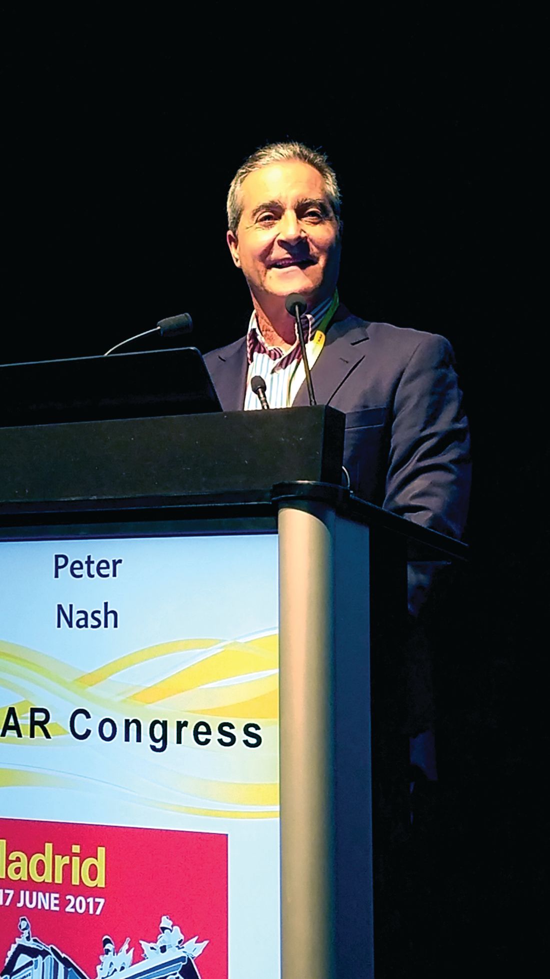
Based in part on the results from this trial, as well as results from a companion phase 3 trial that enrolled PsA patients naive to a TNFi (Ann Rheum Dis. 2017 Jan; 6[1]:79-87), the company that markets ixekizumab, Eli Lilly, filed an application with the Food and Drug Administration in early 2017 to have a new label indication for PsA, said a company spokeswoman.
“At least half of PsA patients don’t get at least a 20% improvement [an ACR20 response] on a TNFi, and so they are looking for something else,” explained Mark C. Genovese, MD, professor of medicine and director of the Rheumatology Clinic at Stanford (Calif.) University and a coinvestigator on the trial reported by Dr. Nash. “There is pent up demand” for an alternative to a TNFi for treating PsA, Dr. Genovese said in an interview.

The finding also sets ixekizumab apart from secukinumab (Cosentyx), another interleukin-17 inhibitor that already has FDA approval for treating PsA but that has not been specifically tested in PsA patients who failed or didn’t tolerate a TNFi, he noted.
The SPIRIT-P2 results also showed superior outcomes for patients treated with an ixekizumab injection once every 2 or 4 weeks, compared with placebo, by several secondary measures, including ACR50 and ACR70 rates and minimal disease activity. The ACR70 rate after 24 weeks on treatment was 23% with a dose of ixekizumab every 4 weeks and none with placebo. Minimal disease activity was reached by about a quarter of patients on either dosage of the active drug and by 3% of patients on placebo.
Despite the apparent role for ixekizumab when TNFi treatment fails, the TNFi drug class remains the clear first-line choice for PsA patients who are starting a biological drug for the first time. Not only do the TNFis have a much longer and more extensive track record but they also generally receive better insurance coverage that minimizes out-of-pocket expenses for patients, Dr. Genovese said.
SPIRIT-P2 was sponsored by Eli Lilly, the company that markets ixekizumab. Dr. Nash has been a speaker for or consultant to and has received research funding from Eli Lily and for several other companies. Dr. Genovese has been a consultant to and has received research funding from Eli Lilly, AbbVie, Astellas, Galapagos, Pfizer, and Vertex.
[email protected]
On Twitter @mitchelzoler
MADRID – The anti–interleukin-17 drug ixekizumab, already on the U.S. market for treating psoriasis, showed efficacy and safety for treating psoriatic arthritis in patients who previously failed to respond to or tolerate a tumor necrosis factor inhibitor in a pivotal, phase 3 trial with 363 patients.
Treatment of patients with psoriatic arthritis (PsA) with ixekizumab (Taltz) led to improvements, compared with placebo, in arthritis, physical function, and psoriasis. These patients were unresponsive to or intolerant of a tumor necrosis factor inhibitor (TNFi) at rates similar to previously reported response rates for PsA patients who were TNFi naive, Peter Nash, MD, said at the European Congress of Rheumatology.
A published report with the data presented by Dr. Nash also recently appeared (Lancet. 2017;389[10086]:2317-27).
Based in part on the results from this trial, as well as results from a companion phase 3 trial that enrolled PsA patients naive to a TNFi (Ann Rheum Dis. 2017 Jan; 6[1]:79-87), the company that markets ixekizumab, Eli Lilly, filed an application with the Food and Drug Administration in early 2017 to have a new label indication for PsA, said a company spokeswoman.
“At least half of PsA patients don’t get at least a 20% improvement [an ACR20 response] on a TNFi, and so they are looking for something else,” explained Mark C. Genovese, MD, professor of medicine and director of the Rheumatology Clinic at Stanford (Calif.) University and a coinvestigator on the trial reported by Dr. Nash. “There is pent up demand” for an alternative to a TNFi for treating PsA, Dr. Genovese said in an interview.
The finding also sets ixekizumab apart from secukinumab (Cosentyx), another interleukin-17 inhibitor that already has FDA approval for treating PsA but that has not been specifically tested in PsA patients who failed or didn’t tolerate a TNFi, he noted.
The SPIRIT-P2 results also showed superior outcomes for patients treated with an ixekizumab injection once every 2 or 4 weeks, compared with placebo, by several secondary measures, including ACR50 and ACR70 rates and minimal disease activity. The ACR70 rate after 24 weeks on treatment was 23% with a dose of ixekizumab every 4 weeks and none with placebo. Minimal disease activity was reached by about a quarter of patients on either dosage of the active drug and by 3% of patients on placebo.
Despite the apparent role for ixekizumab when TNFi treatment fails, the TNFi drug class remains the clear first-line choice for PsA patients who are starting a biological drug for the first time. Not only do the TNFis have a much longer and more extensive track record but they also generally receive better insurance coverage that minimizes out-of-pocket expenses for patients, Dr. Genovese said.
SPIRIT-P2 was sponsored by Eli Lilly, the company that markets ixekizumab. Dr. Nash has been a speaker for or consultant to and has received research funding from Eli Lily and for several other companies. Dr. Genovese has been a consultant to and has received research funding from Eli Lilly, AbbVie, Astellas, Galapagos, Pfizer, and Vertex.
[email protected]
On Twitter @mitchelzoler
AT THE EULAR 2017 CONGRESS
Key clinical point:
Major finding: The ACR20 rate after 24 weeks of treatment was 53% with monthly ixekizumab and 20% on placebo.
Data source: The SPIRIT-P2 trial, a phase 3 multicenter trial with 363 patients.
Disclosures: SPIRIT-P2 was sponsored by Eli Lilly, the company that markets ixekizumab (Taltz). Dr. Nash has been a speaker for or consultant to and has received research funding from Eli Lily and for several other companies. Dr. Genovese has been a consultant to and has received research funding from Eli Lilly, AbbVie, Astellas, Galapagos, Pfizer, and Vertex.
Subsequent squamous cell carcinoma risk higher in HIV patients with low CD4 count
HIV-infected individuals who have experienced a nonmelanoma skin cancer may be at significantly greater risk of subsequent new squamous cell carcinoma if they have a lower CD4 cell count, a new study suggests.
In a study published online July 12 in JAMA Dermatology, researchers reported the results of a retrospective cohort study using medical record data from 455 HIV-infected and 1,952 HIV-uninfected patients who had previously been diagnosed with at least one nonmelanoma skin cancer.
Patients with CD4 cell counts below 200 cells/mcL had a 44% greater risk of a subsequent nonmelanoma skin cancer, compared with uninfected individuals (95% confidence interval, 1.10-1.88), while those with a viral load greater than 10,000 copies/mL had a 31% greater risk (95% CI, 1.00-1.72), after adjusting for age, sex, smoking status, and obesity.
The increase in nonmelanoma skin cancer risk was largely accounted for by an increased risk of squamous cell carcinoma (SCC). Among patients with lower CD4 cell counts and those with higher viral loads, the risk of SCC was more than twofold greater than among uninfected individuals. In contrast, while there was a trend toward a higher risk of basal cell carcinoma in those two groups, it did not reach significance (JAMA Dermatol. 2017 Jul 12. doi: 10.1001/jamadermatol.2017.1716).
Overall, HIV-infected individuals had a significant 15% increase in the risk of subsequent nonmelanoma skin cancer over an average follow-up period of 5 years, compared with uninfected individuals.
“This study addresses key questions regarding subsequent NMSC [nonmelanoma skin cancer] risk among a high-risk subgroup of HIV-infected population who, by virtue of having had a pathologically validated skin cancer, are at increased risk of subsequent NMSCs,” wrote Maryam M. Asgari, MD, of Massachusetts General Hospital, Boston, and her coauthors. “Specifically, it was previously not known precisely which NMSC subtype is increased in high-risk persons with HIV and whether biomarkers of HIV infections, such as degree of immune dysfunction, are associated with subsequent skin cancer risk.”
While the study wasn’t able to control for known skin cancer risk factors such as skin type, the patients were all non-Hispanic white, which the authors said selected for individuals with fair skin and some sun-exposure history.
The findings could have implications for skin cancer screening among individuals with HIV infection, with the authors suggesting more targeted monitoring for SCC among individuals with low CD4 counts or high viral loads.
“Our findings support immune dysfunction as a risk factor for SCCs and dovetail with SCC risk data from iatrogenic immunodeficiency states, such as organ transplantation and autoimmune diseases.”
The study was partly supported by Kaiser Permanente Northern California, and one author was supported by a grant from the National Cancer Institute. Two authors had previously served as investigators on studies funded by the pharmaceutical industry, one author declared research funding from the pharmaceutical industry, and one declared shares in two medical companies.
FROM JAMA DERMATOLOGY
Key clinical point: HIV-infected people who have had a previous nonmelanoma skin cancer are at significantly higher risk of subsequent SCC if they have a lower CD4 count or higher viral load.
Major finding: HIV-infected people with a low CD4 cell count or high viral load have a greater than twofold increased risk of subsequent SCC after a primary nonmelanoma skin cancer than do uninfected people who have had a previous nonmelanoma skin cancer.
Data source: A retrospective cohort study in 455 HIV-infected and 1,945 HIV-uninfected patients.
Disclosures: The study was partly supported by Kaiser Permanente, Northern California, and one author was supported by a grant from the National Cancer Institute. Two authors had previously served as investigators on studies funded by the pharmaceutical industry, one author declared research funding from the pharmaceutical industry, and one declared shares in two medical companies.
Venetoclax-HMA combo promising for AML in patients 65+
MADRID – A combination of the BCL-2 inhibitor venetoclax with a hypomethylating agent may produce high overall response rates among older patients with newly diagnosed acute myeloid leukemia (AML), early data showed.
Among 100 patients age 65 years and older with previously untreated AML, the combination of venetoclax with either decitabine or azacitidine was associated with a 69% overall responses rate, reported Keith W. Pratz, MD, of Johns Hopkins University in Baltimore.

The standard AML induction regimen of cytarabine and an anthracycline with or without cladribine is generally too toxic for patients in their mid-60s or older. For these patients, there are few good therapeutic options, Dr. Pratz said.
Venetoclax has shown good single-agent activity against relapsed/refractory AML and, as reported in a phase 1b study at the 2016 annual meeting of the American Society of Clinical Oncology, induced a combined rate of complete remission (CR) and CR with incomplete marrow recovery (CRi) of 60% when given in escalating doses with a hypomethylating agent.
Dr. Pratz reported on response rates and safety from an expansion cohort of patients 65 years and older treated in that phase 1b study. The patients were treated with either decitabine or azacitidine plus venetoclax at a dose of either 400 mg or 800 mg.
Decitabine was dosed at 20 mg/m2 intravenously on days 1-5 of a 28-day cycle. Azacitidine was dosed at 75 mg/m2 subcutaneously on days 1-7 of every cycle.
The ORRs for each arm were as follows:
- Decitabine plus venetoclax 400 mg: 76% (44% CR, 32% CRi).
- Decitabine plus venetoclax 800 mg: 68% (36% CR, 32% CRi).
- Azacitadine plus venetoclax 400 mg: 72% (28% CR, 44% CRi).
- Azacitadine plus venetoclax 800 mg: 56%: (28% CR, 28% CRi, PR 1% [numbers rounded up]).
The combined CR/CRi rate was 60% among patients with poor-risk cytogenetics and 78% among patients with intermediate-risk disease. In addition, the combination was effective among patients with both primary de novo AML (68%) and secondary AML (related to myelodysplasia or myeloproliferative neoplasms or previous therapy; 73%).
Overall survival after a median of 9 months since the first dose of the study drug was estimated to be 79% at 6 months and 70% at 12 months, with the median not reached.
Treatment-related adverse events were similar between the decitabine- and azacitidine-containing arms at the given dose of venetoclax. Grade 3 or 4 treatment-related adverse events included thrombocytopenia in 56% of patients on the 400-mg dose of venetoclax and in 32% of those on the 800-mg dose. Grade 3/4 febrile neutropenia occurred in 48% and 30%, respectively, and neutropenia in 40% and 32%.
A phase 3 study of venetoclax at the 400-mg dose with azacitidine has been initiated. NCT02993523 is currently enrolling patients, Dr. Pratz said.
The study was supported by Abbvie and Genentech. Dr. Pratz disclosed research funding from Abbvie and other companies.
MADRID – A combination of the BCL-2 inhibitor venetoclax with a hypomethylating agent may produce high overall response rates among older patients with newly diagnosed acute myeloid leukemia (AML), early data showed.
Among 100 patients age 65 years and older with previously untreated AML, the combination of venetoclax with either decitabine or azacitidine was associated with a 69% overall responses rate, reported Keith W. Pratz, MD, of Johns Hopkins University in Baltimore.
The standard AML induction regimen of cytarabine and an anthracycline with or without cladribine is generally too toxic for patients in their mid-60s or older. For these patients, there are few good therapeutic options, Dr. Pratz said.
Venetoclax has shown good single-agent activity against relapsed/refractory AML and, as reported in a phase 1b study at the 2016 annual meeting of the American Society of Clinical Oncology, induced a combined rate of complete remission (CR) and CR with incomplete marrow recovery (CRi) of 60% when given in escalating doses with a hypomethylating agent.
Dr. Pratz reported on response rates and safety from an expansion cohort of patients 65 years and older treated in that phase 1b study. The patients were treated with either decitabine or azacitidine plus venetoclax at a dose of either 400 mg or 800 mg.
Decitabine was dosed at 20 mg/m2 intravenously on days 1-5 of a 28-day cycle. Azacitidine was dosed at 75 mg/m2 subcutaneously on days 1-7 of every cycle.
The ORRs for each arm were as follows:
- Decitabine plus venetoclax 400 mg: 76% (44% CR, 32% CRi).
- Decitabine plus venetoclax 800 mg: 68% (36% CR, 32% CRi).
- Azacitadine plus venetoclax 400 mg: 72% (28% CR, 44% CRi).
- Azacitadine plus venetoclax 800 mg: 56%: (28% CR, 28% CRi, PR 1% [numbers rounded up]).
The combined CR/CRi rate was 60% among patients with poor-risk cytogenetics and 78% among patients with intermediate-risk disease. In addition, the combination was effective among patients with both primary de novo AML (68%) and secondary AML (related to myelodysplasia or myeloproliferative neoplasms or previous therapy; 73%).
Overall survival after a median of 9 months since the first dose of the study drug was estimated to be 79% at 6 months and 70% at 12 months, with the median not reached.
Treatment-related adverse events were similar between the decitabine- and azacitidine-containing arms at the given dose of venetoclax. Grade 3 or 4 treatment-related adverse events included thrombocytopenia in 56% of patients on the 400-mg dose of venetoclax and in 32% of those on the 800-mg dose. Grade 3/4 febrile neutropenia occurred in 48% and 30%, respectively, and neutropenia in 40% and 32%.
A phase 3 study of venetoclax at the 400-mg dose with azacitidine has been initiated. NCT02993523 is currently enrolling patients, Dr. Pratz said.
The study was supported by Abbvie and Genentech. Dr. Pratz disclosed research funding from Abbvie and other companies.
AT EHA 2017
Key clinical point: Induction therapy options are limited for patients 65 years and older with acute myeloid leukemia.
Major finding: Venetoclax and a hypomethylating agent were associated with a 69% overall response rate.
Data source: The expansion portion of a phase 1b trial of venetoclax plus decitabine or azacitidine in 100 patients 65 years and older with de novo AML.
Disclosures: The study was supported by Abbvie and Genentech. Dr. Pratz disclosed research funding from Abbvie and other companies.
Substance use treatment cost $34 billion in 2014
Total spending on the treatment of substance use disorder reached $34 billion in 2014, with outpatient care taking the largest share, according to the National Center for Health Statistics.
That $34 billion represents an increase of 273% from the $9.1 billion spent in 1986 and a considerable shift in the distribution of spending over the last 30 years. In 1986, the largest share of spending – 50%, or $4.6 billion – for substance use disorder went toward inpatient care and only $2.4 billion (27%) was used for outpatient care. In 2014, outpatient treatment of substance use disorder had a 40% share ($13.6 billion) of all spending, and inpatient care was down to 19% ($6.4 billion), the NCHS reported in “Health, United States, 2016.”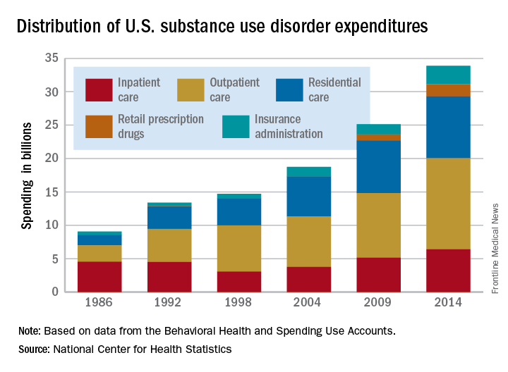
Since methadone is not considered a retail drug by SAMHSA, it is classified under specialty substance use disorder treatment centers, which are included in the estimate for outpatient care, the NCHS noted.
Hugo Aparicio, MD

Proposal would exempt more than half of physicians from MACRA/QPP
, thanks to a Centers for Medicare & Medicaid Services proposal exempting some physicians.
The proposed 2018 update to the Quality Payment Program (QPP), the payment system created as part of the Medicare Access and CHIP Reauthorization Act (MACRA), would increase the low-volume threshold for participation, exempting practices that receive $90,000 or less in Medicare Part B payments or have 200 or fewer Medicare patients would be exempt from participation in either the Merit-based Incentive Payment System (MIPS) or Advanced Alternative Payment Model (APM) tracks of the QPP.
According to the proposed rule, released June 20, the CMS “estimates that approximately 572,000 eligible clinicians would be required to participate in MIPS in the 2018 MIPS performance period. ... After restricting the population of eligible clinician types who are not newly enrolled, the proposed increase in the low-volume threshold is expected to exclude 585,560 clinicians who do not exceed the low-volume threshold.”
Overall, 96.6% of MIPS-eligible physicians will engage in quality reporting in 2020, with 96.1% receiving either a bonus to their Medicare Part B payments or no adjustment, according to CMS estimates. For all eligible clinicians, 76.8% will receive a bonus payment, with all payment bonuses totaling $673.3 million, while those losing money will see their Medicare payments reduced by $173.3 million. The overall aggregate impact will be a 0.9% increase in Part B payments to clinicians.
However, different practice sizes will have different experiences. For example, practices with 1-15 eligible clinicians (114,424 total eligible clinicians in this group) will see in the aggregate a 0.7% increase, while practices with 16-24 eligible clinicians (22,296) will see a 0.4% increase in the aggregate. Practices of 100 or more clinicians (318,841) stand to see the biggest bump in their Medicare payments, with a 1.4% bonus based on the provisions in the proposal.
Ten percent of practices with 1-15 MIPS-eligible clinicians and 10.9% of practices with 16-24 MIPS-eligible clinicians are estimated to receive a decrease in their Medicare payments based on the proposal, while 0.8% of clinicians in practices of 100 or more are expected to see the penalty.
The increased low-volume threshold would help out a lot of physicians who might otherwise struggle to meet the requirements, but some view it as a penalty against those who have made the investment and are ready to fully transition into the new value-based payment program, particularly the larger health care systems.
According to the MACRA legislation language, the MIPS program will be a budget-neutral program – so, the more practices that are exempt from having to participate, the less money will be available for potential bonuses for those who perform well.
“It compresses the potential reward for those who are ready and ready to do well,” Chet Speed, vice president of public policy at AMGA, said in an interview, adding that the projected 1.4% aggregate bonus payments for large practices and health systems “does not really reflect or reward all the work they have done to get to this point.” AMGA is an association representing large practices and health systems.
Comments on the proposed update to the QPP are due to the CMS by Aug. 21, 2017.
New C. difficile drug shows promise
Ridinilazole, a new antibiotic for treatment of Clostridium difficile infection (CDI) proved noninferior to vancomycin, showing promise especially in lower recurrence risk, according to a study funded in part by ridinilazole manufacturer Summit Therapeutics.
In a phase 2, 1:1 randomized, double-blind trial of 69 CDI patients, 24 of 36 (66.7%) ridinilazole patients reported a sustained clinical response, compared with 14 of 33 vancomycin patients (42.4%), indicating statistical noninferiority – and even superiority at the upper confidence level – of ridinilazole (Lancet Infect Dis. 2017 Apr 28. doi: 10.1016/S1473-3099[17]30235-9).
Over the course of 10 days, investigators gave the ridinilazole group 200 mg orally twice per day, as well as two placebo doses. Those in the vancomycin group took 125 mg orally four times per day.
Investigators assessed both groups on days 4-6, 10-11, 12-14, and routine weekly follow-ups until 30 days after treatment.
Most of the patients were white females, with an average age of 58 years for the ridinilazole group, and 56 years for the vancomycin group.
Ridinilazole correlated with more sustained clinical responses across almost all subgroups as well, including with treatment differences (respectively) of 42.7%, 15.9%, 19.9%, and 8.9% for those over 75 years, with a more severe diagnosis, more than one previous CDI episode, and those taking other antibiotics before study participation, according to the investigators. Both groups saw similar rates of adverse events related to treatment.
The outcome of this trial could be significant in reducing recurrence risk in CDI patients, which occurs in up to 30% of patients after first treatment, and can increase up to 65% after multiple reinfections, according to Richard Vickers, PhD, chief scientific officer on antimicrobials at Summit Therapeutics PLC, and his fellow investigators.
CDI patients also are subject to significantly higher inpatient mortality, spend longer periods in intensive care, and have higher rates of all-cause readmission over 3 months than do matched controls, the investigators noted.
Unlike the three common CDI drugs on the market, metronidazole, vancomycin, and fidaxomicin, ridinilazole is restricted to the gastrointestinal tract and, according to the investigators, has shown encouraging results in previous studies.
“In vitro studies have shown its high inhibitory activity against C. difficile and minimal activity against both Gram-positive and Gram-negative aerobic and anaerobic intestinal microorganisms,” they wrote. “In a phase 1 study, ridinilazole was safe and well tolerated in healthy human volunteers, with little systemic absorption and little effect on normal gut microbiota.”
They asserted it was this lack of effect that caused the drug to show success in phase 2, noting ridinilazole’s superiority was “likely to be due to the highly selective activity of ridinilazole against CDI and the absence of collateral damage to the microbiota during therapy.”
The study was limited by its sample, which was younger and had a milder form of CDI than is usually represented. The study also formed its power calculations based on the original sample size of 100, and not the adjusted, intended-to-treat population of 69 which became its primary analysis.
Finally, the investigators advised further trials be conducted with a follow-up schedule longer than 30 days.
Dr. Vickers, Dr. Bina Tejura, and Dr. David Roblin reported working for Summit Therapeutics, the drug manufacturer, and hold share options with the company. Other authors reported holding close relationships with other, similar drug manufacturing companies.
[email protected]
On Twitter @eaztweets
With a 30-day mortality margin of 9%-38% and recurrence risk baseline of 15%-25%, Clostridium difficile infection continues to be a significant global problem. Yet, for decades there had only been a minimal number of drugs available to treat this disease, namely metronidazole and vancomycin.
As new resistant strains of this disease phased out metronidazole, even in mild cases, fidaxomicin emerged as an adequate replacement, although it was soon clear that because of its high price, fidaxomicin would remain as an initial treatment, disadvantaging those with multiple episodes.
This lack of reliable, effective antimicrobials puts into sharp relief the need for new drug development, and ridinilazole may be a step in the right direction.
While the study conducted by Richard Vickers and colleagues was limited by a slightly younger sample with a milder form of the disease than a true representative sample, the superior recurrence reduction related to ridinilazole is an advantage.
It is important that we build upon this study, and push further to expand the array of tools we have to fight C. difficile to optimize treatment for patients at all stages of this disease.
Simon D. Goldenberg, MBBS, MSc, FRCPath, MD, DipHIC, is a consultant microbiologist at the Centre for Clinical Infection and Diagnostics Research, King’s College London. He has received grants and personal fees from Astellas, BD, Luminex, Abbott, Orion Diagnostics, Qiagen, MSD, and DNA electronics.
Ridinilazole, a new antibiotic for treatment of Clostridium difficile infection (CDI) proved noninferior to vancomycin, showing promise especially in lower recurrence risk, according to a study funded in part by ridinilazole manufacturer Summit Therapeutics.
In a phase 2, 1:1 randomized, double-blind trial of 69 CDI patients, 24 of 36 (66.7%) ridinilazole patients reported a sustained clinical response, compared with 14 of 33 vancomycin patients (42.4%), indicating statistical noninferiority – and even superiority at the upper confidence level – of ridinilazole (Lancet Infect Dis. 2017 Apr 28. doi: 10.1016/S1473-3099[17]30235-9).
Over the course of 10 days, investigators gave the ridinilazole group 200 mg orally twice per day, as well as two placebo doses. Those in the vancomycin group took 125 mg orally four times per day.
Investigators assessed both groups on days 4-6, 10-11, 12-14, and routine weekly follow-ups until 30 days after treatment.
Most of the patients were white females, with an average age of 58 years for the ridinilazole group, and 56 years for the vancomycin group.
Ridinilazole correlated with more sustained clinical responses across almost all subgroups as well, including with treatment differences (respectively) of 42.7%, 15.9%, 19.9%, and 8.9% for those over 75 years, with a more severe diagnosis, more than one previous CDI episode, and those taking other antibiotics before study participation, according to the investigators. Both groups saw similar rates of adverse events related to treatment.
The outcome of this trial could be significant in reducing recurrence risk in CDI patients, which occurs in up to 30% of patients after first treatment, and can increase up to 65% after multiple reinfections, according to Richard Vickers, PhD, chief scientific officer on antimicrobials at Summit Therapeutics PLC, and his fellow investigators.
CDI patients also are subject to significantly higher inpatient mortality, spend longer periods in intensive care, and have higher rates of all-cause readmission over 3 months than do matched controls, the investigators noted.
Unlike the three common CDI drugs on the market, metronidazole, vancomycin, and fidaxomicin, ridinilazole is restricted to the gastrointestinal tract and, according to the investigators, has shown encouraging results in previous studies.
“In vitro studies have shown its high inhibitory activity against C. difficile and minimal activity against both Gram-positive and Gram-negative aerobic and anaerobic intestinal microorganisms,” they wrote. “In a phase 1 study, ridinilazole was safe and well tolerated in healthy human volunteers, with little systemic absorption and little effect on normal gut microbiota.”
They asserted it was this lack of effect that caused the drug to show success in phase 2, noting ridinilazole’s superiority was “likely to be due to the highly selective activity of ridinilazole against CDI and the absence of collateral damage to the microbiota during therapy.”
The study was limited by its sample, which was younger and had a milder form of CDI than is usually represented. The study also formed its power calculations based on the original sample size of 100, and not the adjusted, intended-to-treat population of 69 which became its primary analysis.
Finally, the investigators advised further trials be conducted with a follow-up schedule longer than 30 days.
Dr. Vickers, Dr. Bina Tejura, and Dr. David Roblin reported working for Summit Therapeutics, the drug manufacturer, and hold share options with the company. Other authors reported holding close relationships with other, similar drug manufacturing companies.
[email protected]
On Twitter @eaztweets
FROM LANCET INFECTIOUS DISEASES
Key clinical point:
Major finding: A total of 24 of 36 (66.7%) of the ridinilazole patients showed sustained clinical response, compared with 14 of 33 (42.4%) of vancomycin patients, proving ridinilazole noninferior (treatment difference: 21.1%, CI, 90% [3.1-39.1] P = .0004).
Data source: A phase 2, double-blind, randomized noninferiority study of 69 patients gathered from 21 North American sites between June 16, 2014, and Aug. 31, 2015.
Disclosures: Dr. Richard Vickers, Dr. Bina Tejura, and Dr. David Roblin reported working for Summit Therapeutics, the drug manufacturer, and hold share options with the company. Other authors reported holding close relationships with other, similar drug manufacturing companies.