User login
A state of mind
Are you happy with your current situation? Do you enjoy your job and look forward to getting home at the end of the day? Or, do you find your work unrewarding? Do you consider your home simply a place to wait impatiently until you can hop on a plane for your next getaway vacation?

Maybe you should consider relocating to Montana. According to the headline in an article by Richard Franki in Pediatric News (“Montana named ‘best state to practice medicine’ in 2019,” Mar. 28, 2019) the Treasure State is currently the best state to practice medicine. Big Sky Country earned this distinction by outdistancing 49 states and Washington, D.C., in a ranking by WalletHub. The personal finance website used 18 metrics ranging from average annual wage adjusted for cost of living to malpractice award payment per capita. One category of metrics grouped data related to “competition and opportunity” and the other “medical environment.”
I suspect that you are as skeptical as I am of surveys that claim to rank complex entities across broad geographic landscapes. I hope you are neither depressed or elated when your alma mater moves three positions on U.S. News and World Report’s ranking of colleges and universities. However, there are a few pearls hidden in this WalletHub attempt at choosing the most physician-friendly states.
New York was again ranked the worst state to practice medicine, a distinction it had “earned” in 2017 with a highest cost of malpractice insurance. This consistency suggests that there is a litigious atmosphere, at least in some parts of New York, that could make forging a trusting doctor-patient relationship difficult. Heading off to work each morning under the dark cloud of malpractice must take a lot of the fun out of practicing medicine.
The other interesting association buried in the ranking is that Montana is at the top of the list because it also was the state with the highest percentage of “medical residents retained.” This concurrence suggests that living and working in Big Sky Country provided a balance that young physicians found not just tolerable but so enjoyable they wanted to stay. I have been unable to find a complete listing of the raw data, but I suspect that Maine also could boast a high percentage of medical residents who choose to remain at the end of their training. It has been and continues to be a wonderful place to live and raise a family.

While there may be days when you feel as though the practice of medicine has consumed your every waking moment, the truth is that there is more to life than being a physician. Of course, one must be able to earn enough to support oneself and family, but this survey that purports to rank the best place to practice is too heavily weighted to the financial side of the equation and ignores the more difficult to quantify lifestyle qualities.
You may have found a position that pays well enough but requires a time-gobbling and stress-inducing commute to a place you feel comfortable living. Or, you may like your work, but find the community where you have settled lacks the suite of recreational and/or cultural opportunities you enjoy. Not everyone gets it right the first time. Sometimes it is a matter of making compromises and then continuing to reassess whether these compromises have been the best ones.
Regardless of its ranking on any survey, every state has multiple communities in which a physician can have a satisfying career and a lifestyle he or she enjoys. However, achieving this balanced mix may require the physician to invest something of him or herself into making that community one that feels like home.
Dr. Wilkoff practiced primary care pediatrics in Brunswick, Maine for nearly 40 years. He has authored several books on behavioral pediatrics, including “How to Say No to Your Toddler.” Email him at [email protected].
Are you happy with your current situation? Do you enjoy your job and look forward to getting home at the end of the day? Or, do you find your work unrewarding? Do you consider your home simply a place to wait impatiently until you can hop on a plane for your next getaway vacation?
Maybe you should consider relocating to Montana. According to the headline in an article by Richard Franki in Pediatric News (“Montana named ‘best state to practice medicine’ in 2019,” Mar. 28, 2019) the Treasure State is currently the best state to practice medicine. Big Sky Country earned this distinction by outdistancing 49 states and Washington, D.C., in a ranking by WalletHub. The personal finance website used 18 metrics ranging from average annual wage adjusted for cost of living to malpractice award payment per capita. One category of metrics grouped data related to “competition and opportunity” and the other “medical environment.”
I suspect that you are as skeptical as I am of surveys that claim to rank complex entities across broad geographic landscapes. I hope you are neither depressed or elated when your alma mater moves three positions on U.S. News and World Report’s ranking of colleges and universities. However, there are a few pearls hidden in this WalletHub attempt at choosing the most physician-friendly states.
New York was again ranked the worst state to practice medicine, a distinction it had “earned” in 2017 with a highest cost of malpractice insurance. This consistency suggests that there is a litigious atmosphere, at least in some parts of New York, that could make forging a trusting doctor-patient relationship difficult. Heading off to work each morning under the dark cloud of malpractice must take a lot of the fun out of practicing medicine.
The other interesting association buried in the ranking is that Montana is at the top of the list because it also was the state with the highest percentage of “medical residents retained.” This concurrence suggests that living and working in Big Sky Country provided a balance that young physicians found not just tolerable but so enjoyable they wanted to stay. I have been unable to find a complete listing of the raw data, but I suspect that Maine also could boast a high percentage of medical residents who choose to remain at the end of their training. It has been and continues to be a wonderful place to live and raise a family.
While there may be days when you feel as though the practice of medicine has consumed your every waking moment, the truth is that there is more to life than being a physician. Of course, one must be able to earn enough to support oneself and family, but this survey that purports to rank the best place to practice is too heavily weighted to the financial side of the equation and ignores the more difficult to quantify lifestyle qualities.
You may have found a position that pays well enough but requires a time-gobbling and stress-inducing commute to a place you feel comfortable living. Or, you may like your work, but find the community where you have settled lacks the suite of recreational and/or cultural opportunities you enjoy. Not everyone gets it right the first time. Sometimes it is a matter of making compromises and then continuing to reassess whether these compromises have been the best ones.
Regardless of its ranking on any survey, every state has multiple communities in which a physician can have a satisfying career and a lifestyle he or she enjoys. However, achieving this balanced mix may require the physician to invest something of him or herself into making that community one that feels like home.
Dr. Wilkoff practiced primary care pediatrics in Brunswick, Maine for nearly 40 years. He has authored several books on behavioral pediatrics, including “How to Say No to Your Toddler.” Email him at [email protected].
Predictive analytics with large data sets are being pursued to individualize IBD therapy
SAN FRANCISCO – The potential power of machine learning to improve therapeutic choices in inflammatory bowel disease (IBD) is at the threshold of clinical applications, according to data presented at the 2019 AGA Tech Summit, sponsored by the AGA Center for GI Innovation and Technology.
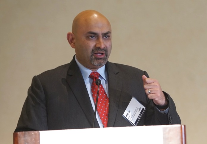
This type of work is relevant to many fields of medicine, but studies conducted in IBD have provided particularly compelling evidence that predictive analytics will improve outcomes and lead to more cost-effective delivery of care, according to Akbar K. Waljee, MD, MSc, an associate professor in the division of gastroenterology, University of Michigan, Ann Arbor, and a staff physician and researcher at the VA Ann Arbor Healthcare System.
Having now published several papers on the role of precision analytics and big data to improve treatment choices in IBD as well as other diseases, Dr. Waljee said, “We collect large amounts of clinical data every day in the delivery of health care but we are now only just beginning to leverage [these] data to guide treatment.”
Based on the experience in IBD, these analyses are relevant for selecting who to treat and not to treat with a given drug.
In one study of how this technology can be applied, data from 1,080 IBD patients taking thiopurines were used to develop a machine learning algorithm that analyzed multiple readily available variables, such as a complete blood count with differential and a chemistry panel, to predict if the patient was in remission. The area under the receiver operating characteristics curve (AuROC) was 0.79 or much higher than previous prediction using drug metabolites, according to Dr. Waljee.
The mean yearly rate of clinical events (new steroid prescriptions, hospitalizations, and abdominal surgeries) was then compared between those who did and those who did not have an algorithm-predicted remission. The lower mean rate in those predicted to be in remission (1.08 vs. 3.95 events) provided support for the conclusions that the algorithm is clinically viable.
“The heterogeneity of response to therapies for IBD is well established. If machine learning predicts effective choices, there will be an opportunity to accelerate the time to disease control as well as save costs by avoiding therapies not likely to be effective,” Dr. Waljee explained.
In another example, the focus was on predicting response to vedolizumab, a monoclonal antibody targeted at a gut-specific mediator of inflammation. In this case, machine learning was applied to predicting corticosteroid-free remission at 1 year in patients with Crohn’s disease patients evaluated 6 weeks after initiating therapy. The machine-learning algorithm was based on an analysis of numerous variables, including laboratory data, sex, and race. Based on the model drawn from the analysis of 472 patients, 35.8% of the patients predicted to be in corticosteroid-free biologic remission at 1 year achieved this endpoint, whereas only 6.7% of the patients predicted to fail achieved the endpoint.
“This suggests that we can use an algorithm relatively early in the course of this biologic to predict who is going to respond,” reported Dr. Waljee. Again, patients with a low likelihood of response at 6 weeks can be started on an alternative therapy, potentially accelerating the time to disease control and avoiding the costs of ineffective and expensive treatments.
IBD is a particularly attractive focus of precision analytics with big data. IBD has a relatively unpredictable relapsing-remitting course and a variable heterogeneous response to available therapies. Algorithms predictive of response circumvent the inherent delays from evaluating disease control over an extended period.
“With ever increasing concern about costs of care and access to care, these treatment algorithms promise to use resources more efficiently,” Dr. Waljee said.
SAN FRANCISCO – The potential power of machine learning to improve therapeutic choices in inflammatory bowel disease (IBD) is at the threshold of clinical applications, according to data presented at the 2019 AGA Tech Summit, sponsored by the AGA Center for GI Innovation and Technology.
This type of work is relevant to many fields of medicine, but studies conducted in IBD have provided particularly compelling evidence that predictive analytics will improve outcomes and lead to more cost-effective delivery of care, according to Akbar K. Waljee, MD, MSc, an associate professor in the division of gastroenterology, University of Michigan, Ann Arbor, and a staff physician and researcher at the VA Ann Arbor Healthcare System.
Having now published several papers on the role of precision analytics and big data to improve treatment choices in IBD as well as other diseases, Dr. Waljee said, “We collect large amounts of clinical data every day in the delivery of health care but we are now only just beginning to leverage [these] data to guide treatment.”
Based on the experience in IBD, these analyses are relevant for selecting who to treat and not to treat with a given drug.
In one study of how this technology can be applied, data from 1,080 IBD patients taking thiopurines were used to develop a machine learning algorithm that analyzed multiple readily available variables, such as a complete blood count with differential and a chemistry panel, to predict if the patient was in remission. The area under the receiver operating characteristics curve (AuROC) was 0.79 or much higher than previous prediction using drug metabolites, according to Dr. Waljee.
The mean yearly rate of clinical events (new steroid prescriptions, hospitalizations, and abdominal surgeries) was then compared between those who did and those who did not have an algorithm-predicted remission. The lower mean rate in those predicted to be in remission (1.08 vs. 3.95 events) provided support for the conclusions that the algorithm is clinically viable.
“The heterogeneity of response to therapies for IBD is well established. If machine learning predicts effective choices, there will be an opportunity to accelerate the time to disease control as well as save costs by avoiding therapies not likely to be effective,” Dr. Waljee explained.
In another example, the focus was on predicting response to vedolizumab, a monoclonal antibody targeted at a gut-specific mediator of inflammation. In this case, machine learning was applied to predicting corticosteroid-free remission at 1 year in patients with Crohn’s disease patients evaluated 6 weeks after initiating therapy. The machine-learning algorithm was based on an analysis of numerous variables, including laboratory data, sex, and race. Based on the model drawn from the analysis of 472 patients, 35.8% of the patients predicted to be in corticosteroid-free biologic remission at 1 year achieved this endpoint, whereas only 6.7% of the patients predicted to fail achieved the endpoint.
“This suggests that we can use an algorithm relatively early in the course of this biologic to predict who is going to respond,” reported Dr. Waljee. Again, patients with a low likelihood of response at 6 weeks can be started on an alternative therapy, potentially accelerating the time to disease control and avoiding the costs of ineffective and expensive treatments.
IBD is a particularly attractive focus of precision analytics with big data. IBD has a relatively unpredictable relapsing-remitting course and a variable heterogeneous response to available therapies. Algorithms predictive of response circumvent the inherent delays from evaluating disease control over an extended period.
“With ever increasing concern about costs of care and access to care, these treatment algorithms promise to use resources more efficiently,” Dr. Waljee said.
EXPERT ANALYSIS FROM 2019 AGA TECH SUMMIT
More evidence shows value of CBT for schizophrenia
ORLANDO – Cognitive-behavioral therapy (CBT) should be more widely offered to patients with psychosis, and a growing body of evidence is demonstrating why, an expert said at the annual congress of the Schizophrenia International Research Society.

Tony Morrison, ClinPsyD, said that, while antipsychotic medications are appropriate for many patients with disorders such as schizophrenia, they are not offered cognitive therapy nearly often enough.
“What I am against is the sole reliance on antipsychotics,” said Dr. Morrison, professor of clinical psychology at the University of Manchester (England). “Putting all of our eggs in that basket is not particularly sensible.” That’s because so many patients who are started on antipsychotic medications discontinue them, either because they are ineffective or because they conclude that the risk of side effects – weight gain, cardiovascular problems, and sexual dysfunction among them – are not worth it.
In a trial published last year, 75 patients with psychosis who were not taking antipsychotic medication were randomized to receive up to 26 sessions of CBT over 6 months, antipsychotic medication, or both. After a year, no difference was found in Positive and Negative Symptom Scale (PANSS) scores between the three groups, researchers found, although the combination group saw an improvement in PANSS scores at 24 weeks before later falling in line with the other groups (Lancet Psychiatry. 2018 May;5[5]:411-23).
Patients in the medical treatment arm mostly received aripiprazole, olanzapine, or quetiapine. Patients received an average of 14 sessions of CBT, with 80% attending at least 6 sessions. Researchers did not see any significant level adverse events in the CBT group.
The findings show that this is fertile ground to explore further, Dr. Morrison said. “It is safe to conduct trials in which people with psychosis are not taking antipsychotics,” he said.
A larger, more definitive trial is needed in this area.
Treatment of patients for whom antipsychotic medication has already been shown not to work is a bigger challenge, but a recent study Dr. Morrison led shows that CBT could work in these patients as well (Health Technol Assess. 2019 Feb;23[7]:1-144).
In that study, 487 patients with schizophrenia who were resistant to treatment with clozapine were randomized to CBT – up to 30 sessions over 9 months – or treatment as usual, without CBT. Researchers saw a slight benefit in PANSS scores for CBT at 9 months (P = .049). But this benefit was not maintained after patients stopped their cognitive therapy, and no difference in PANSS scores was seen at 21 months, the study’s primary endpoint.
Nonetheless, the findings were encouraging, Dr. Morrison said, showing that CBT “compares favorably with augmenting treatment with a second antipsychotic.” Researchers, however, were not able to determine which patients were likely to benefit most from CBT, despite “lots of subanalyses” in an effort to do so.
Patients resistant to initial medical therapy will need their illness tackled from a variety of fronts – subsequent antipsychotic therapy, psychological therapy, social-activity based therapy, and peer support.
“I think we probably need lots of things in combination – a much more rich package of care,” Dr. Morrison said.
Dr. Morrison reported no disclosures.
ORLANDO – Cognitive-behavioral therapy (CBT) should be more widely offered to patients with psychosis, and a growing body of evidence is demonstrating why, an expert said at the annual congress of the Schizophrenia International Research Society.
Tony Morrison, ClinPsyD, said that, while antipsychotic medications are appropriate for many patients with disorders such as schizophrenia, they are not offered cognitive therapy nearly often enough.
“What I am against is the sole reliance on antipsychotics,” said Dr. Morrison, professor of clinical psychology at the University of Manchester (England). “Putting all of our eggs in that basket is not particularly sensible.” That’s because so many patients who are started on antipsychotic medications discontinue them, either because they are ineffective or because they conclude that the risk of side effects – weight gain, cardiovascular problems, and sexual dysfunction among them – are not worth it.
In a trial published last year, 75 patients with psychosis who were not taking antipsychotic medication were randomized to receive up to 26 sessions of CBT over 6 months, antipsychotic medication, or both. After a year, no difference was found in Positive and Negative Symptom Scale (PANSS) scores between the three groups, researchers found, although the combination group saw an improvement in PANSS scores at 24 weeks before later falling in line with the other groups (Lancet Psychiatry. 2018 May;5[5]:411-23).
Patients in the medical treatment arm mostly received aripiprazole, olanzapine, or quetiapine. Patients received an average of 14 sessions of CBT, with 80% attending at least 6 sessions. Researchers did not see any significant level adverse events in the CBT group.
The findings show that this is fertile ground to explore further, Dr. Morrison said. “It is safe to conduct trials in which people with psychosis are not taking antipsychotics,” he said.
A larger, more definitive trial is needed in this area.
Treatment of patients for whom antipsychotic medication has already been shown not to work is a bigger challenge, but a recent study Dr. Morrison led shows that CBT could work in these patients as well (Health Technol Assess. 2019 Feb;23[7]:1-144).
In that study, 487 patients with schizophrenia who were resistant to treatment with clozapine were randomized to CBT – up to 30 sessions over 9 months – or treatment as usual, without CBT. Researchers saw a slight benefit in PANSS scores for CBT at 9 months (P = .049). But this benefit was not maintained after patients stopped their cognitive therapy, and no difference in PANSS scores was seen at 21 months, the study’s primary endpoint.
Nonetheless, the findings were encouraging, Dr. Morrison said, showing that CBT “compares favorably with augmenting treatment with a second antipsychotic.” Researchers, however, were not able to determine which patients were likely to benefit most from CBT, despite “lots of subanalyses” in an effort to do so.
Patients resistant to initial medical therapy will need their illness tackled from a variety of fronts – subsequent antipsychotic therapy, psychological therapy, social-activity based therapy, and peer support.
“I think we probably need lots of things in combination – a much more rich package of care,” Dr. Morrison said.
Dr. Morrison reported no disclosures.
REPORTING FROM SIRS 2019
The promise of endoscopy simulation–based training and gaps as targets for innovation
SAN FRANCISCO – The growing sophistication of simulation technology has the potential to improve training and assessment of skills in gastrointestinal endoscopy, but there are gaps between the promise and evidence, according to an overview of this form of training at the 2019 AGA Tech Summit, sponsored by the AGA Center for GI Innovation and Technology.
For endoscopy, the term simulator encompasses a broad expanse of tools that can range from a simple physical model with holes used to practice endoscope navigation skills to a complex virtual world that challenges technical skills as well as decision-making processes. In 2012, the American Society of Gastrointestinal Endoscopy (ASGE) issued a statement encouraging endoscopy simulation-based training in the context of other strategies to gain skills, but Catharine Walsh, MD, MEd, PhD, of the University of Toronto, warned of the current limits as well as the advantages of simulation training.
“There are many companies making simulators of varying price and complexity. For early skill acquisition, more expensive devices may not necessarily be better,” Dr. Walsh said. She highlighted that “one’s choice of simulator should be based on the educational goals as opposed to technology, as the effectiveness of simulation depends highly on a close match between the training goals and the simulation tool.” A longstanding issue in the field of simulation relates to cost and access. Simulation will not have widespread impact unless it is accessible. “There is a need for future development of inexpensive, portable simulators targeting specific skills to help facilitate uptake of simulation across endoscopy units and training programs,” said Dr. Walsh. Other strategies to increase uptake include specific learning modules designed to complement simulators.
Although simulators are increasingly being used during training to help endoscopists develop basic endoscopic skills, Dr. Walsh focused on the gap in development of simulator devices targeting practicing endoscopists and research examining their use for training new skills within practice, preventing skills decay, and remediating performance deficits. She explained that “currently, there is a lack of evidence that simulation adoption by practicing endoscopists leads to better patient outcomes. This remains a priority area for simulation education, research, and development.” It also remains to be seen how cost-effective simulators are compared with other reaching modalities, she said.
“The potential is certainly there, but it is essential to develop simulators targeting training for low-volume, higher stakes therapeutic techniques, emerging procedures, and techniques and advanced endoscopic procedures, and perform well-controlled studies to demonstrate their effectiveness in practice,” Dr. Walsh said. “Embedding assessments within emerging simulation technology is key as it permits identification of skills requiring further practice and can form the basis of virtual coaching employing endoscopic simulation to improve skills and outcomes.”
Simulators offer the very important advantage of giving the physician the chance to acquire skills before participating in a clinical case and allowing errors to occur in a risk-free environment, suggesting that this type of training will only grow. For example, Dr. Walsh described emerging simulation-based team training that allows endoscopy teams to practice both technical skills as well as nontechnical skills, such as communication and decision making, which may be particularly important in the event of a crisis. Gamification is also being pursued as a potential adjunct strategy to help improve engagement and skill acquisition.
Current simulators are limited in their ability to train and assess cognitive and nontechnical skills. Development of simulation-based cognitive training tools for key areas such as lesion recognition, classification, and management decision-making skills is also a promising area to pursue. Such education could be delivered via portable electronic devices and incorporate assessment and feedback to facilitate skills acquisition.
“There remains a substantial gap between the promise of many types of simulation training and objective evidence that these are helping endoscopists gain skills,” Dr. Walsh said. This in no way diminishes the enormous promise of new simulation technology to be an effective and safe approach for clinicians to learn and maintain performance of endoscopic skills, but Dr. Walsh focused on the need for the development of new simulation technologies and controlled studies that will render these approaches evidence based.
REPORTING FROM 2019 AGA TECH SUMMIT
Virtual reality emerges as a therapeutic tool in gastroenterology
SAN FRANCISCO – The body of evidence to support virtual reality (VR) as a therapeutic modality will increasingly involve the GI tract, according to evidence summarized at the 2019 AGA Tech Summit, sponsored by the AGA Center for GI Innovation and Technology. Evolving from its early use in acute or chronic pain, where its function was to simply divert attention from symptoms, VR computer-generated environments are now being applied to alter patient perceptions and behavior that may involve changes in brain function, according to Brennan Spiegel, MD, AGAF, director of Health Services Research for Cedars-Sinai Medical Center, Los Angeles.
“The field of gastroenterology is a particularly promising area for treatment based on VR because of the well-established brain-gut interaction,” Dr. Spiegel explained. He said this tool has now been shown repeatedly to change how patients experience their symptoms in a variety of clinical contexts.
The field is not entirely new. Already by 2017, 11 randomized controlled trials of VR for therapeutic purposes were identified in a systematic review (Innov Clin Neurosci 2017;14:14-21). These trials, dating back to 2010, have explored this technology in depression, cognitive and motor rehabilitation, and eating disorders. Most showed significant benefit. In eating disorders, for example, response at one year was 44% in those receiving VR as an adjunct to cognitive behavioral therapy versus 10% in the controls.
“VR may not just alter perception. In studies being conducted with functional MRI imaging, changes in brain function similar to those observed in patients taking opioids have been observed,” said Dr. Spiegel, outlining objective evidence that VR has physiological effects.
VR already has an established role as a training tool for physicians in GI and other areas of medicine, but Dr. Spiegel focused on the evidence of its applications in treatment. Earlier this year, an expert panel in which he participated published a methodology for VR clinical trials to help move the field forward by defining how to establish evidence of benefit (JMIR Mental Health 2019;6:e11973). With a growing body of data suggesting VR has measurable clinical benefits, the field is poised to grow quickly.
In gastroenterology specifically, Dr. Spiegel envisions applications in functional diseases, such as irritable bowel syndrome (IBS), in which there is already strong evidence of a mind-gut component to symptom flares. He said, “VR can help patients to engage with their body differently, changing how they react to symptoms and leading to better coping mechanisms.”
In one example, Dr. Spiegel displayed a video depicting a woman with severe pain due to liver ascites testifying to substantial pain relief after a VR experience that included images that took her far from the hospital room in which she was sitting at the time. He reported that gastrointestinal pain relief is so consistent with VR that failure to respond prompts him to reevaluate patients for missed organic pathology.
Implementation of VR as a therapeutic tool is not without obstacles. For example, patients susceptible to motion sickness can react poorly to the three-dimensional environment created by VR, according to Dr. Spiegel. Many patients have expressed reluctance to try VR for any one of a number of reasons, including skepticism. However, there are many potential advantages. In the management of pain, for example, VR circumvents a long list of adverse events related to opioids or other analgesics.
This technology is only being used in a few centers, but there is enough evidence of clinical benefit that Dr. Siegel expects it to be more broadly adopted as indications expand. With more controlled trials being performed to measure and establish benefits, he envisions an evidence-based VR pharmacy that will allow clinicians to prescribe specific VR software suitable not only for the target condition but matched to patient preferences for VR environments.
“We have good evidence that VR is a powerful tool to manage mood disorders and pain perception. Although there is so far a fairly limited about of research specific to GI conditions, this is coming,” Dr. Spiegel said.
EXPERT ANALYSIS FROM 2019 AGA TECH SUMMIT
AI boosts gastroendoscopy, supports clinicians
SAN FRANCISCO – Artificial intelligence (AI) can improve video recording and physician support during colonoscopy procedures, and the data being collected could eventually pave the way to AI systems powerful enough to detect polyps on their own.
AI has been touted as a means to reduce costs and improve patient outcomes, but there’s another benefit that is sometimes overlooked – physician satisfaction. Examining colonoscopy after colonoscopy can get a little overwhelming. “There’s only so much the human eye can see, and when you’re doing multiple colonoscopies a day, there’s a possibility that you’re just seeing too much. [AI] could help lessen the burden in making those diagnoses,” said Kurt Heine in an interview.
Mr. Heine is vice president of the endoscopy division at Olympus America. He joined other speakers at a panel on artificial intelligence at the 2019 AGA Tech Summit sponsored by the AGA Center for GI Innovation and Technology.
Physicians are under an increasing burden with the aging population and recommendations from the American Cancer Society that colonoscopies should begin at age 45. “We need a more efficient way to perform colonoscopies. Ideally AI would aid in the adenoma detection rate and perhaps someday in diagnosis. We’re trying to build a support tool to assist in that procedure,” said Heine.
That point was echoed by Matt Schwartz, CEO of Virgo, which specializes in video recording of colonoscopies. In an effort to ease physician burden, it is automating some simple tasks, like starting and stopping video recording during a procedure. That’s a time-consuming process, and “it’s the sort of task that AI is really good at, so it’s a natural fit to get our first foray of AI on the market,” said Mr. Schwartz in an interview. Virgo currently provides a small, Apple TV–sized box that connects directly to an existing endoscopy system that independently records procedures.
The system uploads captured video into the cloud, and automatically creates highlights for easy viewing. That feature is available now. In the future, Virgo hopes to offer advanced analysis of withdrawal time, cecal intubation rate, and other features. “We want to provide video analytics that can help doctors and physician groups make informed decisions on how to improve quality,” said Mr. Schwartz.
Developing AI poses distinct challenges, and can be costly and time-consuming, said Jason Tucker-Schwartz, PhD, director of marketing for NinePoint Medical. During the session, he outlined the development of the AI component of the company’s NvisionVLE Imaging System, which is the only Food and Drug Administration–approved AI product for upper GI imaging. It reveals layers inside the esophageal tissue wall with high resolution, where 90% of cancers begin.
“It’s a new type of data that gastroenterologists are not used to seeing,” said Dr. Tucker-Schwartz. But that has presented a challenge to physicians attempting to identify and process all that novel information.
That called for AI, but because NinePoint is a small company with limited resources, it needed to control development costs. To streamline matters, they developed a system that focuses on three features that are most useful in making esophageal diagnoses. “The AI algorithms find those features as a function of depth so we can flatten them and use them to create a roadmap that physicians can use to guide their [interpretation],” said Dr. Tucker-Schwartz.
The resulting system has garnered lots of positive feedback, according to Dr. Tucker-Schwartz. The experience highlights the need to incorporate physician input into product development. “You need to involve them in all the steps along the path to end with a product that meets not only their goals but your business goals as well,” he said.
The electric dream
The long-term goal for AI in colonoscopy is automated polyp detection, a so-called optical biopsy, but that vision lies well in the future, said Mr. Schwartz. The primary issue is that only still images are available as training sets, and these don’t capture the diversity of patients, endoscopy systems, and operators that will be required to create a robust, generalizable polyp detection system. Existing efforts have shown promise on training sets, but struggle in real-world tests. “AI is good at tricking you into thinking it’s a working system when it’s only looking at retrospective data,” said Mr. Schwartz.
Olympus signed an agreement last year with ai4gi, a commercial initiative applying deep learning to gastrointestinal cancer, to combine its AI systems with Olympus’ colonoscopy line, but Mr. Heine agreed that optical biopsies won’t appear any time soon: “We’re not ready right now to launch anything that’s making a diagnosis claim. It’s not about optical biopsies at this point. It’s about supporting the physician,” he said.
Along with improving video capture and quality-control efforts, Mr. Schwartz believes that Virgo’s systems can help solve the problem of limited training data. By capturing and storing video data from a wide range of procedures, it is generating a resource that could boost the field and may one day make optical biopsies a reality. “It becomes the training set to build the AI video systems of the future,” he said.
Mr. Heine is an employee of Olympus. Dr. Tucker-Schwartz is an employee of NinePoint Medical. Mr. Schwartz is an employee of Virgo.
REPORTING FROM THE 2019 AGA TECH SUMMIT
Interventional endoscopic ultrasonography is hitting its stride

SAN FRANCISCO – Interventional endoscopic ultrasonography (EUS) recently has brought transluminal stent placement to problems like walled off pancreatic necroses, Joo Ha Hwang, MD, PhD, professor of medicine at Stanford (Calif.) University, said in an interview at the AGA Tech Summit, sponsored by the AGA Center for GI Innovation and Technology. He sees EUS taking over surgical territory beyond the usual biopsies in the gastrointestinal and biliary tracts and soon moving into tumor treatment in these areas. The field is “rapidly expanding,” he said, and the value of these minimally invasive procedures means that the field will soon need to train more interventional endoscopists.
SAN FRANCISCO – Interventional endoscopic ultrasonography (EUS) recently has brought transluminal stent placement to problems like walled off pancreatic necroses, Joo Ha Hwang, MD, PhD, professor of medicine at Stanford (Calif.) University, said in an interview at the AGA Tech Summit, sponsored by the AGA Center for GI Innovation and Technology. He sees EUS taking over surgical territory beyond the usual biopsies in the gastrointestinal and biliary tracts and soon moving into tumor treatment in these areas. The field is “rapidly expanding,” he said, and the value of these minimally invasive procedures means that the field will soon need to train more interventional endoscopists.
SAN FRANCISCO – Interventional endoscopic ultrasonography (EUS) recently has brought transluminal stent placement to problems like walled off pancreatic necroses, Joo Ha Hwang, MD, PhD, professor of medicine at Stanford (Calif.) University, said in an interview at the AGA Tech Summit, sponsored by the AGA Center for GI Innovation and Technology. He sees EUS taking over surgical territory beyond the usual biopsies in the gastrointestinal and biliary tracts and soon moving into tumor treatment in these areas. The field is “rapidly expanding,” he said, and the value of these minimally invasive procedures means that the field will soon need to train more interventional endoscopists.
REPORTING FROM 2019 AGA TECH SUMMIT

Dolutegravir passes late pregnancy test
SEATTLE – In HIV-positive pregnant women who had not previously been on antiretroviral therapy (ART), initiation of treatment with dolutegravir during the third trimester led to lower viral loads at delivery and reduced HIV transmission to offspring compared to treatment with efavirenz. The result comes from a study conducted in South Africa and Uganda. Both drugs were combined with two nucleoside reverse transcriptase inhibitors (NRTIs).

ART-naive women who start ART during the third trimester often do not achieve sufficient reduction of viral load, and the researchers believed that integrase inhibitors like dolutegravir may lead to greater success because they can lead to viral clearance faster than other classes of drugs, which aren’t powerful enough to get viral loads to undetectable levels by the time of delivery. In fact, women who start therapy in the third trimester are at a 600% higher risk of transmitting the virus to offspring, and infant mortality is doubled in the first year of life, according to Saye Khoo, MD, professor of molecular and clinical pharmacology at the University of Liverpool (England).
“Clearly the viral load and undetectable viral load at the time of delivery is the best proxy for the lowest risk of mother to child transmission,” Dr. Khoo said at the Conference on Retroviruses & Opportunistic Infections. “This study clearly showed that there were big differences between dolutegravir and efavirenz. Viral load was undetectable in 74% in the dolutegravir group compared to 43% in efavirenz, which was highly significant.”
In the open-label DolPHIN-2 study, 268 women were randomized after week 28 of their pregnancy (median, 31 weeks) to receive dolutegravir or efavirenz plus two NRTIs. The dolutegravir group was more likely to achieve viral load less than 50 copies/mL at delivery (74% vs. 43%; adjusted risk ratio, 1.66; 95% confidence interval, 1.32-2.09). The trend occurred across subanalyses that included baseline viral load, baseline CD4 count, and gestation at initiation. 93% of the women in the dolutegravir group had a viral load less than 1,000 copies/mL, compared with 83% in the efavirenz group (RR, 1.11; 95% CI, 1.00-1.23).
There were no differences between the two groups with respect to frequency of severe adverse events, organ class of severe adverse events, gestational age at delivery, births earlier than 34 weeks, or births earlier than 37 weeks.
Four stillbirths occurred in the dolutegravir arm, and none in the efavirenz group. “Each of these was examined in great detail, as you might imagine. There were clear explanations for the cause of death, and it was related to maternal infection in two cases and obstetric complications in the other two,” said Dr. Khoo. There were no observations of neural tube defects – a concern that has been raised in the use of dolutegravir at the time of conception.
“I think we did see clear differences in virologic response in the arms, and so that would argue that dolutegravir certainly could be considered in these high-risk scenarios,” said Dr. Khoo.
The study was funded by the University of Liverpool. Dr. Khoo reported consulting for and receiving research funding from ViiV.
SOURCE: Khoo S et al. CROI 2019, Abstract 40 LB.
SEATTLE – In HIV-positive pregnant women who had not previously been on antiretroviral therapy (ART), initiation of treatment with dolutegravir during the third trimester led to lower viral loads at delivery and reduced HIV transmission to offspring compared to treatment with efavirenz. The result comes from a study conducted in South Africa and Uganda. Both drugs were combined with two nucleoside reverse transcriptase inhibitors (NRTIs).
ART-naive women who start ART during the third trimester often do not achieve sufficient reduction of viral load, and the researchers believed that integrase inhibitors like dolutegravir may lead to greater success because they can lead to viral clearance faster than other classes of drugs, which aren’t powerful enough to get viral loads to undetectable levels by the time of delivery. In fact, women who start therapy in the third trimester are at a 600% higher risk of transmitting the virus to offspring, and infant mortality is doubled in the first year of life, according to Saye Khoo, MD, professor of molecular and clinical pharmacology at the University of Liverpool (England).
“Clearly the viral load and undetectable viral load at the time of delivery is the best proxy for the lowest risk of mother to child transmission,” Dr. Khoo said at the Conference on Retroviruses & Opportunistic Infections. “This study clearly showed that there were big differences between dolutegravir and efavirenz. Viral load was undetectable in 74% in the dolutegravir group compared to 43% in efavirenz, which was highly significant.”
In the open-label DolPHIN-2 study, 268 women were randomized after week 28 of their pregnancy (median, 31 weeks) to receive dolutegravir or efavirenz plus two NRTIs. The dolutegravir group was more likely to achieve viral load less than 50 copies/mL at delivery (74% vs. 43%; adjusted risk ratio, 1.66; 95% confidence interval, 1.32-2.09). The trend occurred across subanalyses that included baseline viral load, baseline CD4 count, and gestation at initiation. 93% of the women in the dolutegravir group had a viral load less than 1,000 copies/mL, compared with 83% in the efavirenz group (RR, 1.11; 95% CI, 1.00-1.23).
There were no differences between the two groups with respect to frequency of severe adverse events, organ class of severe adverse events, gestational age at delivery, births earlier than 34 weeks, or births earlier than 37 weeks.
Four stillbirths occurred in the dolutegravir arm, and none in the efavirenz group. “Each of these was examined in great detail, as you might imagine. There were clear explanations for the cause of death, and it was related to maternal infection in two cases and obstetric complications in the other two,” said Dr. Khoo. There were no observations of neural tube defects – a concern that has been raised in the use of dolutegravir at the time of conception.
“I think we did see clear differences in virologic response in the arms, and so that would argue that dolutegravir certainly could be considered in these high-risk scenarios,” said Dr. Khoo.
The study was funded by the University of Liverpool. Dr. Khoo reported consulting for and receiving research funding from ViiV.
SOURCE: Khoo S et al. CROI 2019, Abstract 40 LB.
REPORTING FROM CROI 2019
Should we defend the unrestrained availability of patented psychotropics?
Since the biological revolution in psychiatry, with the introduction of chlorpromazine in the 1950s,1 psychiatrists have been introduced to the economic questions inherent in the tension between funding psychotropic medications for the treatment of mental illness versus funding psychosocial interventions. Of course, our natural inclination is to advocate for all available treatments for our patients, but the economic realities of medical care – especially government-subsidized or regulated medical care – force us to weigh the relative advantages of these treatments and to promote our patients’ interests with a wise allocation of limited resources.

It has become common practice for the American Psychiatric Association to advocate for additional funds for both research into mental illness as well as treatment. The promotion of mental health parity and the demonization of prior authorizations are examples of our natural priorities in the debates over funding for medical care. A bias has played out in the national conversation about medical care in general regarding the right to said care, but economists understand that medical care is a limited resource and, as such, treating it as a “right,” per se, does not make sense: One has to make hard decisions about its allocation or simply leave it to the free market to make said decisions.
Recently, the government has proposed to eliminate certain psychotropic medications from their protected status within Medicare Part D. Those medications include all drugs labeled as antidepressants, antipsychotics, and anticonvulsants. As expected, the APA’s medical director has written a formal statement opposing the proposal. His statement includes warnings about suicides and overwhelmed emergency departments. He compared the mental health situation in the United States to a crisis. He described the availability of expensive and new psychotropics to be “lifesaving.”2
The goals of the APA and its leaders are honorable. We are inspired by the dedication that some psychiatrists have to advocate for us all as well as for our patients. However, we are concerned that unfounded claims are being made. We are even more troubled when those claims promote the interests of a fallible pharmaceutical industry, an industry that has opened up our field to extensive critical scrutiny over the past few years. We wonder whether a brief examination of the scientific evidence warrants the statements made by the APA.
After reviewing clinical textbooks and search engines, we were not able to find reliable and convincing evidence that newer psychotropics reduce emergency department stays or that lengths of stay in the hospital correlate with the use of newer agents. We have actually not even heard of that claim made before in any serious forum. Reviews of predictors of length of stay in psychiatric hospitals have typically included demographic factors, diagnostic factors, logistical factors such as time of day, and social factors, such as insurance status and homelessness.3,4 We found no review mentioning the use of patented drugs as a predictor of shorter stays.
At a larger level, The Food and Drug Administration approves psychiatric medications based on superiority to placebo and not superiority to existing – and usually much cheaper – medications. Our subscription to Epocrates informs us that a 1-month supply of once-a-day brand-name Abilify, Invega, or Latuda is more than $1,000.5 Alternatively, a 1-month supply of generic olanzapine, risperidone, or quetiapine is available for $4 at Walmart.6 As famously described in the CATIE trial7 of patients with schizophrenia, newer antipsychotics are not particularly better than older ones. In addition, a more recent meta-analysis8 did not find significant differences among antipsychotics’ efficacy.

A similar analysis can be made of antidepressants without addressing debates surrounding the effectiveness of antidepressants as a class and the value of psychological interventions over chemical ones. Reviews of the literature do not suggest that newer antidepressants are more effective than older ones. A recent meta-analysis of antidepressant efficacy did not find significant differences among antidepressants and, when looking at trends, amitriptyline, a much older antidepressant, was most effective.9
The most surprising part of the APA medical director’s statement was the claim of reduced suicidality. While lithium and clozapine have some evidence for reducing the risk of suicide, the evidence that antidepressants reduce suicide is equivocal. Quite the contrary, some evidence exists that antidepressants may increase the risk of suicide,10 and we are not aware of evidence suggesting that any newer agents can reduce suicide at any higher rate. One psychiatrist has even made a career out of testifying that antidepressants increase impulsivity and suicide.11
We are not politicians, and we trust the APA to have good intentions with a desire to help patients suffering from mental illness. We understand the need to advocate for any measure that provides additional resources for the treatment of mental illness. We have no doubt that a publicly funded and appropriately regulated mental health system is a wise goal from both an ethical as well as a societal perspective. The APA has an imperative to advocate for our patients with the goal to improve our society.
However, we are concerned when our field makes unfounded claims. Advocating that insurance companies and the government provide most psychotropics without prior authorization and without discrimination does not appear to be based on scientific evidence and has serious economic implications that are not being weighed in a transparent manner. Whatever funding levels the APA recommends for the treatment of mental illness, said treatments will remain a limited resource, and then it becomes a question not just of ethics but of economics. What combination of resources produce the most benefit for the most people in question? Would the increased cost of a newer psychotropic be better spent on a system with more elaborate psychosocial interventions? In making this argument, does one risk repeating the historical blunder made when, in the 1960s, long-term psychiatric hospitals were closed with the intention of replacing their costs with outpatient treatments that then never materialized?
A review of the literature does not support the claim that newer psychotropic agents are more effective from either a clinical or an economic perspective. Cost-saving measures are ethical and possibly beneficial if they permit a more justifiable allocation of resources.
Dr. Lehman is an associate professor of psychiatry at the University of California, San Diego. He is codirector of all acute and intensive psychiatric treatment at the Veterans Affairs Medical Center in San Diego, where he practices clinical psychiatry. He is also the course director for the UCSD third-year medical student psychiatry clerkship. Dr. Badre is a forensic psychiatrist in San Diego and an expert in correctional mental health. He holds teaching positions at the University of California, San Diego, and the University of San Diego. Among his writings is Chapter 7 in the new book “Critical Psychiatry: Controversies and Clinical Implications” (Cham, Switzerland: Springer, 2019).
References
1. Ann Med Psychol (Paris). 1952 Jun;110(2 1):112-7.
2. https://psychnews.psychiatryonline.org/doi/10.1176/appi.pn.2019.3b26.
3. Am J Emerg Med. 2016 Feb;34(2):133-9.
4. Eur Psychiatry. 2018 Feb;48:6-12.
5. https://online.epocrates.com/drugs. Retrieved March 3, 2019.
6. https://www.walmart.com/cp/$4-prescriptions/1078664. Retrieved March 27, 2019.
7. N Engl J Med. 2005 Sep 22;353(12):1209-23.
8. Am J Psychiatry. 2017. 174(10):927-42.
9. Lancet. 2018 Apr 7. 391(10128):P1357-66.
10. BMJ. 2009.339;b2880.
11. https://breggin.com/.
Since the biological revolution in psychiatry, with the introduction of chlorpromazine in the 1950s,1 psychiatrists have been introduced to the economic questions inherent in the tension between funding psychotropic medications for the treatment of mental illness versus funding psychosocial interventions. Of course, our natural inclination is to advocate for all available treatments for our patients, but the economic realities of medical care – especially government-subsidized or regulated medical care – force us to weigh the relative advantages of these treatments and to promote our patients’ interests with a wise allocation of limited resources.
It has become common practice for the American Psychiatric Association to advocate for additional funds for both research into mental illness as well as treatment. The promotion of mental health parity and the demonization of prior authorizations are examples of our natural priorities in the debates over funding for medical care. A bias has played out in the national conversation about medical care in general regarding the right to said care, but economists understand that medical care is a limited resource and, as such, treating it as a “right,” per se, does not make sense: One has to make hard decisions about its allocation or simply leave it to the free market to make said decisions.
Recently, the government has proposed to eliminate certain psychotropic medications from their protected status within Medicare Part D. Those medications include all drugs labeled as antidepressants, antipsychotics, and anticonvulsants. As expected, the APA’s medical director has written a formal statement opposing the proposal. His statement includes warnings about suicides and overwhelmed emergency departments. He compared the mental health situation in the United States to a crisis. He described the availability of expensive and new psychotropics to be “lifesaving.”2
The goals of the APA and its leaders are honorable. We are inspired by the dedication that some psychiatrists have to advocate for us all as well as for our patients. However, we are concerned that unfounded claims are being made. We are even more troubled when those claims promote the interests of a fallible pharmaceutical industry, an industry that has opened up our field to extensive critical scrutiny over the past few years. We wonder whether a brief examination of the scientific evidence warrants the statements made by the APA.
After reviewing clinical textbooks and search engines, we were not able to find reliable and convincing evidence that newer psychotropics reduce emergency department stays or that lengths of stay in the hospital correlate with the use of newer agents. We have actually not even heard of that claim made before in any serious forum. Reviews of predictors of length of stay in psychiatric hospitals have typically included demographic factors, diagnostic factors, logistical factors such as time of day, and social factors, such as insurance status and homelessness.3,4 We found no review mentioning the use of patented drugs as a predictor of shorter stays.
At a larger level, The Food and Drug Administration approves psychiatric medications based on superiority to placebo and not superiority to existing – and usually much cheaper – medications. Our subscription to Epocrates informs us that a 1-month supply of once-a-day brand-name Abilify, Invega, or Latuda is more than $1,000.5 Alternatively, a 1-month supply of generic olanzapine, risperidone, or quetiapine is available for $4 at Walmart.6 As famously described in the CATIE trial7 of patients with schizophrenia, newer antipsychotics are not particularly better than older ones. In addition, a more recent meta-analysis8 did not find significant differences among antipsychotics’ efficacy.
A similar analysis can be made of antidepressants without addressing debates surrounding the effectiveness of antidepressants as a class and the value of psychological interventions over chemical ones. Reviews of the literature do not suggest that newer antidepressants are more effective than older ones. A recent meta-analysis of antidepressant efficacy did not find significant differences among antidepressants and, when looking at trends, amitriptyline, a much older antidepressant, was most effective.9
The most surprising part of the APA medical director’s statement was the claim of reduced suicidality. While lithium and clozapine have some evidence for reducing the risk of suicide, the evidence that antidepressants reduce suicide is equivocal. Quite the contrary, some evidence exists that antidepressants may increase the risk of suicide,10 and we are not aware of evidence suggesting that any newer agents can reduce suicide at any higher rate. One psychiatrist has even made a career out of testifying that antidepressants increase impulsivity and suicide.11
We are not politicians, and we trust the APA to have good intentions with a desire to help patients suffering from mental illness. We understand the need to advocate for any measure that provides additional resources for the treatment of mental illness. We have no doubt that a publicly funded and appropriately regulated mental health system is a wise goal from both an ethical as well as a societal perspective. The APA has an imperative to advocate for our patients with the goal to improve our society.
However, we are concerned when our field makes unfounded claims. Advocating that insurance companies and the government provide most psychotropics without prior authorization and without discrimination does not appear to be based on scientific evidence and has serious economic implications that are not being weighed in a transparent manner. Whatever funding levels the APA recommends for the treatment of mental illness, said treatments will remain a limited resource, and then it becomes a question not just of ethics but of economics. What combination of resources produce the most benefit for the most people in question? Would the increased cost of a newer psychotropic be better spent on a system with more elaborate psychosocial interventions? In making this argument, does one risk repeating the historical blunder made when, in the 1960s, long-term psychiatric hospitals were closed with the intention of replacing their costs with outpatient treatments that then never materialized?
A review of the literature does not support the claim that newer psychotropic agents are more effective from either a clinical or an economic perspective. Cost-saving measures are ethical and possibly beneficial if they permit a more justifiable allocation of resources.
Dr. Lehman is an associate professor of psychiatry at the University of California, San Diego. He is codirector of all acute and intensive psychiatric treatment at the Veterans Affairs Medical Center in San Diego, where he practices clinical psychiatry. He is also the course director for the UCSD third-year medical student psychiatry clerkship. Dr. Badre is a forensic psychiatrist in San Diego and an expert in correctional mental health. He holds teaching positions at the University of California, San Diego, and the University of San Diego. Among his writings is Chapter 7 in the new book “Critical Psychiatry: Controversies and Clinical Implications” (Cham, Switzerland: Springer, 2019).
References
1. Ann Med Psychol (Paris). 1952 Jun;110(2 1):112-7.
2. https://psychnews.psychiatryonline.org/doi/10.1176/appi.pn.2019.3b26.
3. Am J Emerg Med. 2016 Feb;34(2):133-9.
4. Eur Psychiatry. 2018 Feb;48:6-12.
5. https://online.epocrates.com/drugs. Retrieved March 3, 2019.
6. https://www.walmart.com/cp/$4-prescriptions/1078664. Retrieved March 27, 2019.
7. N Engl J Med. 2005 Sep 22;353(12):1209-23.
8. Am J Psychiatry. 2017. 174(10):927-42.
9. Lancet. 2018 Apr 7. 391(10128):P1357-66.
10. BMJ. 2009.339;b2880.
11. https://breggin.com/.
Anastrozole-fulvestrant in breast cancer and daily aspirin to prevent HCC
Welcome to the first edition of “How I will treat my next patient,” a regular column analyzing the practical clinical relevance of the latest literature. In this first column, I will take a look at two interesting studies – a combination hormonal treatment for metastatic ER-positive breast cancer and aspirin therapy for prevention of hepatocellular cancer.
Anastrozole plus fulvestrant

In a large SWOG trial for postmenopausal patients with stage IV, hormonally responsive breast cancer whose metastatic disease could be treated with frontline hormonal therapy, long-term survival analysis showed that the combination of anastrozole plus fulvestrant was superior to anastrozole alone, with essentially no increase in toxicity.
The study – “Overall Survival With Fulvestrant Plus Anastrozole in Metastatic Breast Cancer” – was published in the New England Journal of Medicine (2019;380:1226-34).
The overall survival difference was not only statistically and clinically significant, but impressively so among patients who had not received prior adjuvant hormonal therapy. That is despite the fact that 45% of patients who were assigned to initial treatment with anastrozole received single agent fulvestrant at first relapse.
What this means in practice
Because of the generally negative results of combined hormonal therapy in comparison with sequential use of the same agents and the potency of CDK4/6 inhibitors in combination with hormonal agents in the frontline setting, many oncologists have forgotten the initial publication of this regimen in 2012. In that study, the combination demonstrated improved progression-free survival over anastrozole alone, particularly in the subset of patients who presented with stage IV breast cancer as their initial presentation.
Although the benefits for CDK4/6 inhibitors as an addition to hormonal therapy are truly impressive, the practical aspects of utilizing the CDK4/6 inhibitors may be prohibitive for a small subset of our most vulnerable, medically underserved patients. Specifically, these are patients who are unable to return for frequent blood counts in the initial few months of therapy, patients with limited financial resources who cannot afford the out-of-pocket costs of an expensive oral medication and required laboratory testing, those with difficulty adhering to oral medication regimens, or those with constitutional neutropenia.
Not coincidentally, many of these patients are exactly the ones who present with stage IV disease as their initial manifestation of breast cancer. For such patients, the combination of anastrozole plus fulvestrant is an attractive alternative and may offer competitive survival benefits. This is not “yesterday’s therapy” in the era of CDK4/6 inhibitors, but rather represents a valuable option for treatment in at-risk populations.
When I see my next patient who presents with stage IV breast cancer, I will consider combined hormonal therapy among the various available treatment options.
Aspirin to prevent HCC
Investigators at Taichung (Taiwan) Veterans General Hospital recently analyzed 16 years of data from a cohort of more than 10,000 patients with chronic hepatitis B virus (HBV) infection and found statistically significantly fewer cases of hepatocellular cancer (HCC) in patients who took antiviral antinucleoside analogue therapy, statins, and aspirin.
The study – “Association of Daily Aspirin Therapy With Risk of Hepatocellular Carcinoma in Patients With Chronic Hepatitis B” – was published in JAMA Internal Medicine (doi: 10.1001/jamainternmed.2018.8342).
Although there were more impressive reductions in relative risk of HCC among statin and antinucleoside analogue users, the authors highlighted that HCC developed in 5.20% of the approximately 2,100 chronic aspirin users and in 7.87% of nonusers – a 29% relative reduction in risk in this cohort study. Toxicity, including upper GI bleeding, was low.
What this means in practice
This is a hypothesis-generating analysis at best. Although the authors highlight possible mechanisms by which aspirin use could lead to reduction in HCC among patients with chronic inflammatory conditions affecting the liver, the study produces more questions than it answers (dose, chronicity of use, duration of protection, biomarkers for benefit).
Owing to the simplicity and low cost of the intervention, it may be worth studying prospectively in chronic HBV-infected patients and other populations at risk for HCC, but the intervention should not be adopted at this point based on an international cohort study.
The practicality of conducting such a large, complicated, prospective study of a widely available medication that has widely publicized additional health benefits is an open question.
When I see my next patient with a high risk for HCC, I won’t prescribe aspirin for chemoprevention.
Dr. Lyss has been a community-based medical oncologist and clinical researcher for more than 35 years, practicing in St. Louis. His clinical and research interests are in the prevention, diagnosis, and treatment of breast and lung cancers, and in expanding access to clinical trials to medically underserved populations.
Welcome to the first edition of “How I will treat my next patient,” a regular column analyzing the practical clinical relevance of the latest literature. In this first column, I will take a look at two interesting studies – a combination hormonal treatment for metastatic ER-positive breast cancer and aspirin therapy for prevention of hepatocellular cancer.
Anastrozole plus fulvestrant
In a large SWOG trial for postmenopausal patients with stage IV, hormonally responsive breast cancer whose metastatic disease could be treated with frontline hormonal therapy, long-term survival analysis showed that the combination of anastrozole plus fulvestrant was superior to anastrozole alone, with essentially no increase in toxicity.
The study – “Overall Survival With Fulvestrant Plus Anastrozole in Metastatic Breast Cancer” – was published in the New England Journal of Medicine (2019;380:1226-34).
The overall survival difference was not only statistically and clinically significant, but impressively so among patients who had not received prior adjuvant hormonal therapy. That is despite the fact that 45% of patients who were assigned to initial treatment with anastrozole received single agent fulvestrant at first relapse.
What this means in practice
Because of the generally negative results of combined hormonal therapy in comparison with sequential use of the same agents and the potency of CDK4/6 inhibitors in combination with hormonal agents in the frontline setting, many oncologists have forgotten the initial publication of this regimen in 2012. In that study, the combination demonstrated improved progression-free survival over anastrozole alone, particularly in the subset of patients who presented with stage IV breast cancer as their initial presentation.
Although the benefits for CDK4/6 inhibitors as an addition to hormonal therapy are truly impressive, the practical aspects of utilizing the CDK4/6 inhibitors may be prohibitive for a small subset of our most vulnerable, medically underserved patients. Specifically, these are patients who are unable to return for frequent blood counts in the initial few months of therapy, patients with limited financial resources who cannot afford the out-of-pocket costs of an expensive oral medication and required laboratory testing, those with difficulty adhering to oral medication regimens, or those with constitutional neutropenia.
Not coincidentally, many of these patients are exactly the ones who present with stage IV disease as their initial manifestation of breast cancer. For such patients, the combination of anastrozole plus fulvestrant is an attractive alternative and may offer competitive survival benefits. This is not “yesterday’s therapy” in the era of CDK4/6 inhibitors, but rather represents a valuable option for treatment in at-risk populations.
When I see my next patient who presents with stage IV breast cancer, I will consider combined hormonal therapy among the various available treatment options.
Aspirin to prevent HCC
Investigators at Taichung (Taiwan) Veterans General Hospital recently analyzed 16 years of data from a cohort of more than 10,000 patients with chronic hepatitis B virus (HBV) infection and found statistically significantly fewer cases of hepatocellular cancer (HCC) in patients who took antiviral antinucleoside analogue therapy, statins, and aspirin.
The study – “Association of Daily Aspirin Therapy With Risk of Hepatocellular Carcinoma in Patients With Chronic Hepatitis B” – was published in JAMA Internal Medicine (doi: 10.1001/jamainternmed.2018.8342).
Although there were more impressive reductions in relative risk of HCC among statin and antinucleoside analogue users, the authors highlighted that HCC developed in 5.20% of the approximately 2,100 chronic aspirin users and in 7.87% of nonusers – a 29% relative reduction in risk in this cohort study. Toxicity, including upper GI bleeding, was low.
What this means in practice
This is a hypothesis-generating analysis at best. Although the authors highlight possible mechanisms by which aspirin use could lead to reduction in HCC among patients with chronic inflammatory conditions affecting the liver, the study produces more questions than it answers (dose, chronicity of use, duration of protection, biomarkers for benefit).
Owing to the simplicity and low cost of the intervention, it may be worth studying prospectively in chronic HBV-infected patients and other populations at risk for HCC, but the intervention should not be adopted at this point based on an international cohort study.
The practicality of conducting such a large, complicated, prospective study of a widely available medication that has widely publicized additional health benefits is an open question.
When I see my next patient with a high risk for HCC, I won’t prescribe aspirin for chemoprevention.
Dr. Lyss has been a community-based medical oncologist and clinical researcher for more than 35 years, practicing in St. Louis. His clinical and research interests are in the prevention, diagnosis, and treatment of breast and lung cancers, and in expanding access to clinical trials to medically underserved populations.