User login
Calcium supplement use linked to cancer death
PHILADELPHIA – a nutrition specialist noted at the annual meeting of the American College of Physicians.
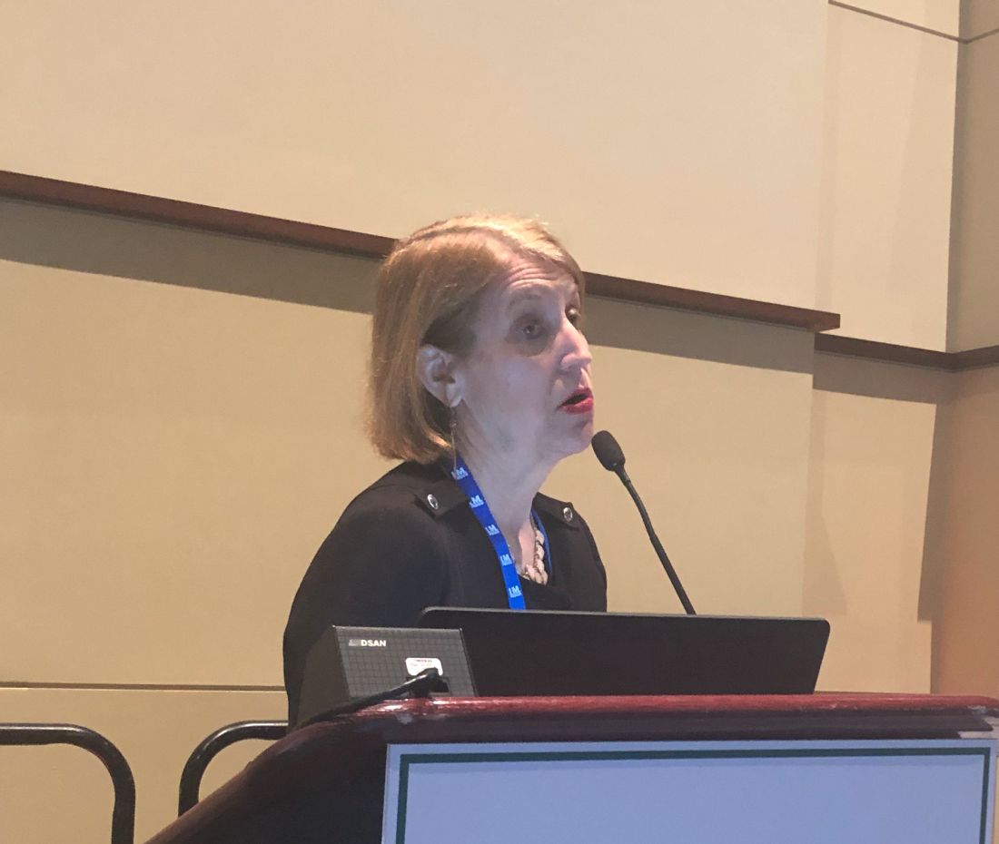
The report, published (Ann Intern Med. 2019 Apr 9. doi: 10.7326/M18-2478) just 2 days before the start of the Internal Medicine meeting, found no mortality benefits associated with any reported dietary supplement use among nearly 31,000 adults in the National Health and Nutrition Examination Survey.
On the contrary, they found that excess calcium consumption was associated with increased risk for cancer-related deaths. Calcium supplements were specifically implicated in the excess of mortality, according to the investigators, with a rate ratio of 1.53 (95% confidence interval, 1.04-2.25) for intakes of 1,000 mg/day versus no intake.
“It’s better to get all of your vitamins from your food, over supplements,” said Marijane Hynes, MD, director of weight management at George Washington University, Washington, in a meet-the-professor session at the conference.
The amount of calcium patients are getting from food can be estimated with one rule of thumb: Multiply the number of dairy servings per day by 300 mg, Dr. Hynes said, who added that a serving is 8 ounces of milk or 1 ounce of hard cheese. Dark green vegetables, breads, cereals, and some nuts can provide 100-200 mg of calcium per day.
Calcium carbonate can be taken with food to enhance calcium absorption, according to Dr. Hynes, while calcium citrate can be taken without food, and is preferred for patients taking acid reflux medications.
Because calcium absorption is reduced at higher doses, patients who need more than 600 mg/day should be taking divided doses, she said.
Bone health goes beyond the dairy aisle, Dr. Hynes added. High vitamin K intake was linked to reduced hip fracture risk among the Framingham Heart Study participants. To get the recommended amount of vitamin K in the diet, patients can consume one or more servings of broccoli, kale, collard greens, or dark green lettuce.
Dr. Hynes reported she that had no relationships with entities producing, marketing, reselling, or distributing health care goods or services consumed by, or used on, patients.
These are observational data. This is not saying we put someone on calcium, and they ended up with cancer, and when you look at this whole thing it’s amazing to me that nobody is discussing the benefits that were found in patients taking magnesium, vitamin K2, and other vitamins. The other thing I would like to point out is that, for at least a decade, it has been really well established that we shouldn’t be using more than 1,000 milligrams of calcium a day, especially from a supplements source. In this study, supplemental calcium intake of 1,000 mg/d or higher was associated with increased risk of cancer death, so what’s the big deal?
The big thing with calcium is calcium comes in 7 different forms. When you eat a variety of fruits and vegetables the source of calcium you get is mixed. The problem with supplements is you are using one or maybe two forms of calcium, but if your body doesn’t recognize that form of calcium then you aren’t getting calcium and it may not be beneficial to you.
What we need to do here, in my opinion, is we need to look at the whole picture. We know that dieting alone or exercising alone does not improve outcomes. It’s the combination of diet, exercise, hormone balance, nutrients from supplements, and emotional balance that makes you healthy. Similarly, you can’t say if you just take this one nutrient you are going to improve your quality of life.
With calcium and vitamin D, you have to take vitamin K2, because vitamin K2 activates osteocalcin, a protein that rebuilds the matrix of the bone. Without vitamin K2, you can’t deposit calcium in the bones. K2 also prevents the deposition of calcium in the blood vessels.
Magnesium is another tremendously important mineral, and magnesium deficiency is the most common mineral deficiency in the United States.
Probably one of the most common causes of magnesium deficiency is the use of acid blockers. I would be very curious to know how many people were taking proton pump inhibitors or acid blockers in general. I bet you most of them were.
Derrick DeSilva Jr., MD, is an internist, practicing in Edison, N.J. He made these comments in an interview. He reported serving as a consultant for Common Sense Supplements, a company that produces dietary supplements.
PHILADELPHIA – a nutrition specialist noted at the annual meeting of the American College of Physicians.
The report, published (Ann Intern Med. 2019 Apr 9. doi: 10.7326/M18-2478) just 2 days before the start of the Internal Medicine meeting, found no mortality benefits associated with any reported dietary supplement use among nearly 31,000 adults in the National Health and Nutrition Examination Survey.
On the contrary, they found that excess calcium consumption was associated with increased risk for cancer-related deaths. Calcium supplements were specifically implicated in the excess of mortality, according to the investigators, with a rate ratio of 1.53 (95% confidence interval, 1.04-2.25) for intakes of 1,000 mg/day versus no intake.
“It’s better to get all of your vitamins from your food, over supplements,” said Marijane Hynes, MD, director of weight management at George Washington University, Washington, in a meet-the-professor session at the conference.
The amount of calcium patients are getting from food can be estimated with one rule of thumb: Multiply the number of dairy servings per day by 300 mg, Dr. Hynes said, who added that a serving is 8 ounces of milk or 1 ounce of hard cheese. Dark green vegetables, breads, cereals, and some nuts can provide 100-200 mg of calcium per day.
Calcium carbonate can be taken with food to enhance calcium absorption, according to Dr. Hynes, while calcium citrate can be taken without food, and is preferred for patients taking acid reflux medications.
Because calcium absorption is reduced at higher doses, patients who need more than 600 mg/day should be taking divided doses, she said.
Bone health goes beyond the dairy aisle, Dr. Hynes added. High vitamin K intake was linked to reduced hip fracture risk among the Framingham Heart Study participants. To get the recommended amount of vitamin K in the diet, patients can consume one or more servings of broccoli, kale, collard greens, or dark green lettuce.
Dr. Hynes reported she that had no relationships with entities producing, marketing, reselling, or distributing health care goods or services consumed by, or used on, patients.
REPORTING FROM INTERNAL MEDICINE 2019
In defense of hospital administrators
Improving relationships between leaders and clinicians
In the March 2019 issue of The Hospitalist, I wrote about some key findings from a 2018 survey of U.S. physicians by The Physicians Foundation. It’s no surprise to anyone working in health care today that the survey found alarming levels of professional dissatisfaction, burnout, and pessimism about the future of medicine among respondent physicians. Sadly, it appears that much of that pessimism is directed toward hospitals and their leaders: 46% of survey respondents viewed the relationships between physicians and hospitals as somewhat or mostly negative and adversarial.

Several physicians posted comments online, and they deeply saddened me. My heart hurt for those doctors who wrote, “I loved medicine. It was good for my soul, but medicine left me. Doctors gave up most of their power and large corporations without an ethical foundation and no god, but money took over.” Or “They are waiting so all the senior physicians will retire. Nurses will become leaders who will follow administration’s lead and control physicians. Money and cost cutting is the major driver. Physicians are not valuable anymore because they have different opinions which cost a lot. There is a lot of window dressing, but they actually don’t care. They just want to run a business.” I also read “I was tossed out like dirty laundry water at age 59.” And “On a personal basis, I will try to reason with management exactly once before I bail.” Sigh.
These commenters are well-meaning physicians who had bad experiences with hospital leaders they saw as uncaring and unresponsive to their concerns as clinicians. Their experiences left them demoralized and embittered. I’m truly sorry for that.
I’m a recovering hospital administrator myself. My business partner John Nelson, MD, MHM, likes to tell people that he has successfully deprogrammed me from the way most administrators think about doctors, but he’s mostly joking (at least I think he is). I can tell you that most of the hospital leaders I have met – both when I was still an administrator and now in my consulting work – are well-intentioned people who care deeply about patients and their fellow health care professionals and are trying hard to do the right thing. Many of them could have earned more and had better career opportunities doing similar work in a field other than health care, but they chose health care out of a sincere desire to do good and help people.
A big part of the problem is that doctors and administrators come to health care from very different starting points, and so have very different perspectives. They generally function in separate silos, each paying attention to their own comfortable little part of that monster we call a health care delivery system. Often, neither administrators nor doctors have made enough effort to cross over and understand the issues and perspectives of people in other silos. As a result, it’s easy for assumptions and biases to creep in and poison our interactions.
When we interpret the behavior of others, we humans tend to overemphasize dispositional factors, such as personality or motives, and to discount situational factors, such as external stressors. Psychologists call this the fundamental attribution error or correspondence bias, and the result is usually heightened conflict as a result of presumed negative intentions on the part of others (“All she cares about is making a profit”) and discounting circumstantial factors that might be influencing others’ behavior (“She is facing reduced market share and a funding shortfall, and she’s fearful for the future of the institution”).
Add in another phenomenon known as the actor-observer bias, in which we tend to attribute others’ behavior to their dispositions but attribute our own behavior to the circumstances (“That administrator lost his temper because he’s a demanding jerk, but I only lost my temper because he pushed me over the edge”).
Is it possible that hospital leaders and doctors are reading each other inaccurately and that they’re making assumptions about each other’s intentions that get in the way of having constructive dialogue? How can we get to a place of greater trust? I don’t know the whole answer, of course, but I have a few ideas to offer.
Read the full post at hospitalleader.org.
Improving relationships between leaders and clinicians
Improving relationships between leaders and clinicians
In the March 2019 issue of The Hospitalist, I wrote about some key findings from a 2018 survey of U.S. physicians by The Physicians Foundation. It’s no surprise to anyone working in health care today that the survey found alarming levels of professional dissatisfaction, burnout, and pessimism about the future of medicine among respondent physicians. Sadly, it appears that much of that pessimism is directed toward hospitals and their leaders: 46% of survey respondents viewed the relationships between physicians and hospitals as somewhat or mostly negative and adversarial.
Several physicians posted comments online, and they deeply saddened me. My heart hurt for those doctors who wrote, “I loved medicine. It was good for my soul, but medicine left me. Doctors gave up most of their power and large corporations without an ethical foundation and no god, but money took over.” Or “They are waiting so all the senior physicians will retire. Nurses will become leaders who will follow administration’s lead and control physicians. Money and cost cutting is the major driver. Physicians are not valuable anymore because they have different opinions which cost a lot. There is a lot of window dressing, but they actually don’t care. They just want to run a business.” I also read “I was tossed out like dirty laundry water at age 59.” And “On a personal basis, I will try to reason with management exactly once before I bail.” Sigh.
These commenters are well-meaning physicians who had bad experiences with hospital leaders they saw as uncaring and unresponsive to their concerns as clinicians. Their experiences left them demoralized and embittered. I’m truly sorry for that.
I’m a recovering hospital administrator myself. My business partner John Nelson, MD, MHM, likes to tell people that he has successfully deprogrammed me from the way most administrators think about doctors, but he’s mostly joking (at least I think he is). I can tell you that most of the hospital leaders I have met – both when I was still an administrator and now in my consulting work – are well-intentioned people who care deeply about patients and their fellow health care professionals and are trying hard to do the right thing. Many of them could have earned more and had better career opportunities doing similar work in a field other than health care, but they chose health care out of a sincere desire to do good and help people.
A big part of the problem is that doctors and administrators come to health care from very different starting points, and so have very different perspectives. They generally function in separate silos, each paying attention to their own comfortable little part of that monster we call a health care delivery system. Often, neither administrators nor doctors have made enough effort to cross over and understand the issues and perspectives of people in other silos. As a result, it’s easy for assumptions and biases to creep in and poison our interactions.
When we interpret the behavior of others, we humans tend to overemphasize dispositional factors, such as personality or motives, and to discount situational factors, such as external stressors. Psychologists call this the fundamental attribution error or correspondence bias, and the result is usually heightened conflict as a result of presumed negative intentions on the part of others (“All she cares about is making a profit”) and discounting circumstantial factors that might be influencing others’ behavior (“She is facing reduced market share and a funding shortfall, and she’s fearful for the future of the institution”).
Add in another phenomenon known as the actor-observer bias, in which we tend to attribute others’ behavior to their dispositions but attribute our own behavior to the circumstances (“That administrator lost his temper because he’s a demanding jerk, but I only lost my temper because he pushed me over the edge”).
Is it possible that hospital leaders and doctors are reading each other inaccurately and that they’re making assumptions about each other’s intentions that get in the way of having constructive dialogue? How can we get to a place of greater trust? I don’t know the whole answer, of course, but I have a few ideas to offer.
Read the full post at hospitalleader.org.
Embracing an executive leadership role
Dr. Bryce Gartland says hospitalists thrive as leaders
Bryce Gartland, MD, was working as a full-time hospitalist at Emory University Hospital in Atlanta when hospital administrators first started asking him to take on administrative roles, such as clinical site director or medical director of care coordination.
Today, Dr. Gartland is hospital group president and cochief of clinical operations for Emory Healthcare, with responsibility for overall performance and achievement across all 11 Emory hospitals. In that role, he keeps his eyes open for similar talent and leadership potential in younger physicians.
Following internal medicine residency at Cedars-Sinai Medical Center in Los Angeles, Dr. Gartland moved into a traditional private practice setting in Beverly Hills. “Two years later, my wife and I decided to move back to my home town of Atlanta. This was 2005 and hospital medicine was a nascent movement in health care. I was intrigued, and Emory had a strong hospitalist program based in a major academic medical setting, which has since grown from approximately 20 physicians to over 120 across seven hospitals,” he said.
Senior leaders at Emory recognized something in Dr. Gartland and more administrative offers were forthcoming.
“After a year of practicing at Emory, the system’s chief financial officer knocked on my door to ask if I would be interested in becoming medical director for care coordination. This role afforded me tremendous opportunities to get involved in clinical/administrative activities at Emory – utilization review, hospice and palliative care, transitions of care, interface with managed care organizations. The role was very rewarding. In some ways, I became a kind of chief translator at the hospital for anything clinical that also had financial implications,” he recalled.
“Then we went through a reorganization and I was offered the opportunity to step into the chief operating officer position at Emory University Hospital. Shortly thereafter, there was leadership turnover within the division of hospital medicine and I was asked by the CEO of Emory Healthcare and chair of the department of medicine to serve as section head for hospital medicine.” Dr. Gartland wore both of those hats for about 2 years, later becoming the CEO of Emory University Hospital and two other facilities within the system. He was appointed to his current position as hospital group president and cochief of clinical operations for Emory Healthcare in 2018.
Consumed with administrative responsibilities, he largely had to step away from patient care, although with mixed emotions.
“Over the years, I worked hard to maintain a strong clinical role, but the reality is that if you are not delivering patient care routinely, it’s difficult to practice at the highest level of current medical practice,” he said. Nonetheless, Dr. Gartland tries to keep a hand in patient care by routinely rounding with hospitalist teams and attending care conferences.
Fixing the larger health care system
“I am a huge supporter of more physicians becoming actively engaged in administrative positions in health care. They are key to helping us best fix the larger health care system,” Dr. Gartland said. “However, we’ve all seen clinicians drafted into administrative positions who were not great administrators. One needs to be bilingual in both medicine and business. While some skills, such as strong communication, may cross over, it’s important to recognize that clinical strength and success do not necessarily equate to administrative achievement.”
Dr. Gartland also believes in the importance of mentorship in developing future leaders and in seeking and engaging mentors from other disciplines outside of one’s own specialty. “I’ve been fortunate to have a number of mentors who saw something in me and supported investment in my personal and professional development. I am now fortunate to be in the position to give back by mentoring a number of younger hospitalists who are interested in growing their nonclinical roles.”
“One bit of advice from a mentor that really resonated with me was: Don’t let the urgent get in the way of the important,” Dr. Gartland said. “Life is busy and full of urgent day-to-day fires. It’s important to take the time to pause and consider where you are going and what you are doing to enhance your career development. Are you getting the right kinds of feedback?” He explained that a coach or mentor who can provide constructive feedback is important and is something he has relied upon throughout his own professional development.
Different paths to learning business
Dr. Gartland did not pursue formal business training before the administrative opportunities started to multiply for him at Emory, although in college he had a strong interest in both business and medicine and at one time contemplated going into either.
“Over the years, my mentors have given me a lot of advice, one of which was that a medical degree can be a passport to a lot of different career paths, with real opportunities for merging business and medicine,” he said.
He has since intentionally pursued business training opportunities wherever they came up, such as courses offered by the American College of Physician Executives (now the American Association for Physician Leadership). “At one point, I considered going back to college in an MBA program, but that’s when John Fox – then Emory Healthcare’s CEO – called and said he wanted to send me to the Harvard Business School’s Managing Health Care Delivery executive education program, with an Emory team comprising the chief nurse executive, chief of human resources, and CEO for one of our hospitals.” Harvard’s roughly 9-month program involves 3 weeks on campus with assignments between the on-campus visits.
“In my current role as hospital group president, I have direct responsibility for our hospitals’ and system’s clinically essential services such as radiology, laboratory, pharmacy, and perioperative medicine. I also still serve as CEO for Emory University Hospital while we recruit my replacement,” Dr. Gartland said. “Overall, my work time breaks down roughly into thirds. One-third is spent on strategy and strategic initiatives – such as organizational and program design. Our system recently acquired a large community health system whose strategic and operational integration I am actively leading.”
Another third of his time is focused on operations, and the final third is focused on talent management and development. “People are truly the most valuable asset any organization has, particularly in health care,” he noted. “Being intentional about organizational design, coaching, and supporting the development and deployment of talent at all levels of the organization helps everyone achieve their full potential. It is one of the most important roles a leader can play.”
Dr. Gartland said that Emory is committed to Lean-based management systems, using both horizontal and vertical strategies for process improvement and waste reduction, with implementation beginning in urology, transplant, and heart and vascular services. Experts say Lean success starts at the very top, and Emory and Dr. Gartland are all in.
“These types of changes are measured in 5- to 7-year increments or more, not in months. We believe this is key to creating the best workplace to support the highest quality, experience, and value in health care delivery. It creates and supports the right culture within an organization, and we have made the commitment to following that path,” he said.
Recognizing leadership potential
What does Dr. Gartland look for in physicians with leadership potential?
“Are you someone who collaborates well?” he asked. “Someone who raises your hand at meetings or gets engaged with the issues? Do you volunteer to take on assignments? Are you someone with a balanced perspective, system minded in thinking and inquisitive, with a positive approach to problem solving?”
A lot of physicians might come to a meeting with the hospital or their boss and complain about all the things that aren’t working, he said, but “it’s rarer for them to come in and say: ‘I see these problems, and here’s where I think we can make improvements. How can I help?’ ” Dr. Gartland looks for evidence of emotional intelligence and the ability to effect change management across disciplines. Another skill with ever-greater importance is comfort with data and data-driven decision making.
“When our national health care system is experiencing so much change and upheaval, much of which is captured in newspaper headlines, it can sound scary,” he said. “I encourage people to see that complex, dynamic times like these, filled with so much change, are also a tremendous opportunity. Run towards and embrace the opportunity for change. Hospitalists, by nature, bring with them a tremendous background and experience set that is invaluable to help lead positive change in these dynamic times.”
The SHM has offerings for hospitalists wanting to advance in leadership positions, Dr. Gartland said, including its annual Leadership Academy. The next one is scheduled to be held in Nashville, Tenn., Nov. 4-7, 2019.
“The Leadership Academy is a great initial step for physicians, especially those early in their careers. Also, try to gain exposure to a variety of perspectives outside of hospital medicine,” he said. “I’d love to see further advances in leadership for our specialty – growing the number of hospitalists who serve as hospital CEOs or CMOs and in other leadership roles. We have more to learn collectively about leadership as a specialty, and I’d love to see us grow that capacity by offering further learning opportunities and bringing together hospitalists who have an interest in advancing leadership.”
Dr. Bryce Gartland says hospitalists thrive as leaders
Dr. Bryce Gartland says hospitalists thrive as leaders
Bryce Gartland, MD, was working as a full-time hospitalist at Emory University Hospital in Atlanta when hospital administrators first started asking him to take on administrative roles, such as clinical site director or medical director of care coordination.
Today, Dr. Gartland is hospital group president and cochief of clinical operations for Emory Healthcare, with responsibility for overall performance and achievement across all 11 Emory hospitals. In that role, he keeps his eyes open for similar talent and leadership potential in younger physicians.
Following internal medicine residency at Cedars-Sinai Medical Center in Los Angeles, Dr. Gartland moved into a traditional private practice setting in Beverly Hills. “Two years later, my wife and I decided to move back to my home town of Atlanta. This was 2005 and hospital medicine was a nascent movement in health care. I was intrigued, and Emory had a strong hospitalist program based in a major academic medical setting, which has since grown from approximately 20 physicians to over 120 across seven hospitals,” he said.
Senior leaders at Emory recognized something in Dr. Gartland and more administrative offers were forthcoming.
“After a year of practicing at Emory, the system’s chief financial officer knocked on my door to ask if I would be interested in becoming medical director for care coordination. This role afforded me tremendous opportunities to get involved in clinical/administrative activities at Emory – utilization review, hospice and palliative care, transitions of care, interface with managed care organizations. The role was very rewarding. In some ways, I became a kind of chief translator at the hospital for anything clinical that also had financial implications,” he recalled.
“Then we went through a reorganization and I was offered the opportunity to step into the chief operating officer position at Emory University Hospital. Shortly thereafter, there was leadership turnover within the division of hospital medicine and I was asked by the CEO of Emory Healthcare and chair of the department of medicine to serve as section head for hospital medicine.” Dr. Gartland wore both of those hats for about 2 years, later becoming the CEO of Emory University Hospital and two other facilities within the system. He was appointed to his current position as hospital group president and cochief of clinical operations for Emory Healthcare in 2018.
Consumed with administrative responsibilities, he largely had to step away from patient care, although with mixed emotions.
“Over the years, I worked hard to maintain a strong clinical role, but the reality is that if you are not delivering patient care routinely, it’s difficult to practice at the highest level of current medical practice,” he said. Nonetheless, Dr. Gartland tries to keep a hand in patient care by routinely rounding with hospitalist teams and attending care conferences.
Fixing the larger health care system
“I am a huge supporter of more physicians becoming actively engaged in administrative positions in health care. They are key to helping us best fix the larger health care system,” Dr. Gartland said. “However, we’ve all seen clinicians drafted into administrative positions who were not great administrators. One needs to be bilingual in both medicine and business. While some skills, such as strong communication, may cross over, it’s important to recognize that clinical strength and success do not necessarily equate to administrative achievement.”
Dr. Gartland also believes in the importance of mentorship in developing future leaders and in seeking and engaging mentors from other disciplines outside of one’s own specialty. “I’ve been fortunate to have a number of mentors who saw something in me and supported investment in my personal and professional development. I am now fortunate to be in the position to give back by mentoring a number of younger hospitalists who are interested in growing their nonclinical roles.”
“One bit of advice from a mentor that really resonated with me was: Don’t let the urgent get in the way of the important,” Dr. Gartland said. “Life is busy and full of urgent day-to-day fires. It’s important to take the time to pause and consider where you are going and what you are doing to enhance your career development. Are you getting the right kinds of feedback?” He explained that a coach or mentor who can provide constructive feedback is important and is something he has relied upon throughout his own professional development.
Different paths to learning business
Dr. Gartland did not pursue formal business training before the administrative opportunities started to multiply for him at Emory, although in college he had a strong interest in both business and medicine and at one time contemplated going into either.
“Over the years, my mentors have given me a lot of advice, one of which was that a medical degree can be a passport to a lot of different career paths, with real opportunities for merging business and medicine,” he said.
He has since intentionally pursued business training opportunities wherever they came up, such as courses offered by the American College of Physician Executives (now the American Association for Physician Leadership). “At one point, I considered going back to college in an MBA program, but that’s when John Fox – then Emory Healthcare’s CEO – called and said he wanted to send me to the Harvard Business School’s Managing Health Care Delivery executive education program, with an Emory team comprising the chief nurse executive, chief of human resources, and CEO for one of our hospitals.” Harvard’s roughly 9-month program involves 3 weeks on campus with assignments between the on-campus visits.
“In my current role as hospital group president, I have direct responsibility for our hospitals’ and system’s clinically essential services such as radiology, laboratory, pharmacy, and perioperative medicine. I also still serve as CEO for Emory University Hospital while we recruit my replacement,” Dr. Gartland said. “Overall, my work time breaks down roughly into thirds. One-third is spent on strategy and strategic initiatives – such as organizational and program design. Our system recently acquired a large community health system whose strategic and operational integration I am actively leading.”
Another third of his time is focused on operations, and the final third is focused on talent management and development. “People are truly the most valuable asset any organization has, particularly in health care,” he noted. “Being intentional about organizational design, coaching, and supporting the development and deployment of talent at all levels of the organization helps everyone achieve their full potential. It is one of the most important roles a leader can play.”
Dr. Gartland said that Emory is committed to Lean-based management systems, using both horizontal and vertical strategies for process improvement and waste reduction, with implementation beginning in urology, transplant, and heart and vascular services. Experts say Lean success starts at the very top, and Emory and Dr. Gartland are all in.
“These types of changes are measured in 5- to 7-year increments or more, not in months. We believe this is key to creating the best workplace to support the highest quality, experience, and value in health care delivery. It creates and supports the right culture within an organization, and we have made the commitment to following that path,” he said.
Recognizing leadership potential
What does Dr. Gartland look for in physicians with leadership potential?
“Are you someone who collaborates well?” he asked. “Someone who raises your hand at meetings or gets engaged with the issues? Do you volunteer to take on assignments? Are you someone with a balanced perspective, system minded in thinking and inquisitive, with a positive approach to problem solving?”
A lot of physicians might come to a meeting with the hospital or their boss and complain about all the things that aren’t working, he said, but “it’s rarer for them to come in and say: ‘I see these problems, and here’s where I think we can make improvements. How can I help?’ ” Dr. Gartland looks for evidence of emotional intelligence and the ability to effect change management across disciplines. Another skill with ever-greater importance is comfort with data and data-driven decision making.
“When our national health care system is experiencing so much change and upheaval, much of which is captured in newspaper headlines, it can sound scary,” he said. “I encourage people to see that complex, dynamic times like these, filled with so much change, are also a tremendous opportunity. Run towards and embrace the opportunity for change. Hospitalists, by nature, bring with them a tremendous background and experience set that is invaluable to help lead positive change in these dynamic times.”
The SHM has offerings for hospitalists wanting to advance in leadership positions, Dr. Gartland said, including its annual Leadership Academy. The next one is scheduled to be held in Nashville, Tenn., Nov. 4-7, 2019.
“The Leadership Academy is a great initial step for physicians, especially those early in their careers. Also, try to gain exposure to a variety of perspectives outside of hospital medicine,” he said. “I’d love to see further advances in leadership for our specialty – growing the number of hospitalists who serve as hospital CEOs or CMOs and in other leadership roles. We have more to learn collectively about leadership as a specialty, and I’d love to see us grow that capacity by offering further learning opportunities and bringing together hospitalists who have an interest in advancing leadership.”
Postvaccination febrile seizures are no more severe than other febrile seizures
according to a study in Pediatrics.

Lucy Deng, MBBS, of the University of Sydney and her colleagues investigated 1,022 index febrile seizures in children aged 6 years or less, of which 6% (n = 67) were VP-FSs and 94% (n = 955) were NVP-FSs. Both univariate and multivariate analyses showed no increased risk of severe seizure associated with VP-FSs, compared with NVP-FS. Most of the febrile seizures of either type were brief (15 minutes or less) and had a length of stay of 1 day or less; there also were no differences in 24-hour recurrence. The most common symptom was respiratory, and the rates were similar in each group (62.7% with VP-FS vs. 62.8% with NVP-FS). In keeping with a known 100% increased risk associated with measles vaccination, 84% of VP-FSs were associated with measles-containing vaccines. The majority of the remaining VP-FSs occurred after combination vaccines.
One limitation is that, because these cases were documented in sentinel tertiary pediatric hospitals, the case ascertainment may not be representative. Also, the small proportion of VP-FSs and limited cohort size means the study may not have been powered to detect true differences in prolonged seizures between the groups, Dr. Deng and her colleagues wrote.
“This study confirms that VP-FSs are clinically not any different from NVP-FSs and should be managed the same way,” the researchers concluded.
The authors reported no relevant financial disclosures, although Dr. Deng is supported by the University of Sydney Training Program scholarship, and two other study authors are supported by Australian National Health and Medical Research Council Career Development Fellowships. The study was funded by a grant from the Australian Government Department of Health and the National Health and Medical Research Council.
SOURCE: Deng L et al. Pediatrics. 2019 Apr 19. doi: 10.1542/peds.2018-2120.
according to a study in Pediatrics.
Lucy Deng, MBBS, of the University of Sydney and her colleagues investigated 1,022 index febrile seizures in children aged 6 years or less, of which 6% (n = 67) were VP-FSs and 94% (n = 955) were NVP-FSs. Both univariate and multivariate analyses showed no increased risk of severe seizure associated with VP-FSs, compared with NVP-FS. Most of the febrile seizures of either type were brief (15 minutes or less) and had a length of stay of 1 day or less; there also were no differences in 24-hour recurrence. The most common symptom was respiratory, and the rates were similar in each group (62.7% with VP-FS vs. 62.8% with NVP-FS). In keeping with a known 100% increased risk associated with measles vaccination, 84% of VP-FSs were associated with measles-containing vaccines. The majority of the remaining VP-FSs occurred after combination vaccines.
One limitation is that, because these cases were documented in sentinel tertiary pediatric hospitals, the case ascertainment may not be representative. Also, the small proportion of VP-FSs and limited cohort size means the study may not have been powered to detect true differences in prolonged seizures between the groups, Dr. Deng and her colleagues wrote.
“This study confirms that VP-FSs are clinically not any different from NVP-FSs and should be managed the same way,” the researchers concluded.
The authors reported no relevant financial disclosures, although Dr. Deng is supported by the University of Sydney Training Program scholarship, and two other study authors are supported by Australian National Health and Medical Research Council Career Development Fellowships. The study was funded by a grant from the Australian Government Department of Health and the National Health and Medical Research Council.
SOURCE: Deng L et al. Pediatrics. 2019 Apr 19. doi: 10.1542/peds.2018-2120.
FROM PEDIATRICS
Advanced degree programs to consider when changing careers
I have been in private practice as a gastroenterologist for 18 years. Many of us in gastroenterology and related fields have wondered how to navigate toward the next step in our careers. There are resources available to further our knowledge, add new skills, and fine tune personal talents to help position us for that next step.

Questions to ask at this stage are: What do I really want to do? Where do I see myself in 5-10 years? How do I go about achieving my target?
We come from different backgrounds including, broadly, academic clinical, academic research, basic science, clinical practice, and education. The next stage of these career paths can vary, and that should be kept in mind while choosing courses/programs. I reached out to two well-known gastroenterologists who have successfully changed their career paths after starting with different backgrounds.
Ronald Vender, MD, professor of medicine, associate dean of clinical affairs, chief medical officer, Yale University, New Haven, Conn.
Dr. Vender began in private practice gastroenterology after fellowship. His own trajectory has been one of “evolution” and has grown to the above titles through “incremental opportunity.” While reflecting on his career, Dr. Vender felt three main attributes were responsible: involvement in medical/GI societies, involvement in non-GI organizations, and engagement of needs for improvement at the hospital of practice. Opportunities became available by speaking up, raising issues, and demanding improvements. Dr. Vender’s involvement in both the private practice sector and hospital administration made his transition to hospital administration possible. This change was based on a “change in [him] and change in what [he] wanted to do.” His advice for all is to learn to say “yes” often in your early career and recognize when to say “no” later in your career.
John Allen, MD, MBA, clinical professor of medicine, University of Michigan, Ann Arbor
Dr. Allen started his career in the Veterans Affairs (VA) system, and during this time, he was exposed to research activities and learned research skills. His initial interest was in health care delivery, but this eventually changed to private practice gastroenterology. His exposure to information and the opportunity to learn about variations in practice and outcomes allowed him to maintain his interest in quality, which ultimately led to publications on colonoscopy quality. In his 40s he decided to obtain an executive master of business administration (EMBA), which he feels one should embark upon “when you have a problem to solve.” He has effectively moved from the VA system to private practice and now to academic medicine. Dr. Allen identified attending leadership conferences, engaging executive coaches, and participation in key committees as further opportunities to help you change careers. His prior work experience, education, and exposure enables him in his current position to help oversee a large department of medicine with 160 care sites, with quality and financials as key factors.
As we can see, there is no correct answer or set path for those of us wanting to change career directions. What was clear while speaking with both Dr. Vender and Dr. Allen was the importance of enthusiasm in solving issues, a willingness to commit to new projects, and an interest in exploring new areas.
Below is a brief overview of some degree programs that may help promote a change in your career path.
Masters in health care
This degree is aimed at those looking to advance their career in the field of health care in various locations, such as hospitals, clinics, and nonprofit organizations.1 Length of prior health care experience will vary based upon program. Programs are administered on a full-time and part-time basis, as well as online and study abroad. Numerous specialties are offered such as medicine, nutrition, psychiatry, nursing, veterinary medicine, physiotherapy, biomedical engineering, medical laboratory studies, radiology, alternative medicine, and health care management, administration, or leadership.
Health care MBA
Master of business administration (MBA) programs in health care administration management are offered by several universities. Given their aim of imparting essential information on a broad range of topics relevant to the health care industry, they are usually quite rigorous. It is recommended that you pursue an MBA only after a few years of working in your chosen field of practice. Many institutions require GMAT scores with the application.2-4
Executive MBA
EMBA programs are similar to health care MBAs in that they also include rigorous course work.5 EMBA programs are developed to meet the educational needs of managers and executives or physicians hoping to advance or change their career. Typically, students can earn an MBA in 2 years or less while working full-time. GMAT scores are required by most institutions offering EMBA.
Certification leadership programs
A benefit of leadership programs is that they help to develop a clear vision by creating a mission statement, goals, and action plans. Some notable programs include:
- American Gastroenterological Association leadership programs: Future Leaders Program, Women’s Leadership Conference, and Forward Program (new for 2019). These programs offer robust sessions which are well thought out and intended to advance the careers of traditionally underrepresented groups within the field of gastroenterology.6
- American Medical Association’s Women’s Leadership Certificate: This program was developed to promote knowledge and strong leadership, both within oneself and among others. Attendees are awarded with the “AMA Women’s Leadership Certificate” following the 2-day event.7
- Executive leadership training and development programs: As with many universities offer programs like these; for example, Harvard Medical School, Boston, offers a popular – and usually sold out – event for women titled “Career Advancement and Leadership Skills for Women in Health Care.”8
- American College of Healthcare Executives: Various conferences and educational meetings in addition to the annual ACHE Congress on Healthcare Leadership.9
- American Association of Physician Leadership: Physician executive accreditation along with career services, continued education, and publications.10
After reviewing the experiences of two well-known gastroenterologists and several of the available programs, the question to ask yourself is, “What’s next?” Most will likely have this question already in mind, so here are a few potential career directions/positions to consider:
Academic medicine: department chief, program director, director of endoscopy, chief medical officer
Private practice: managing director, director of endoscopy, finance director
Private sector: pharmaceutical industry, scientific advisor, medical director, medical insurance industry, malpractice insurance industry, medical informatics, public policy, private equity, entrepreneurial
Conclusion
In summary, there is no single answer nor a single program that fits everyone’s needs. Health care delivery and management/administration are complicated and will only continue to evolve. Consideration must be given to the fact that any change in one’s career direction needs time and commitment.
Here are some take-home points:
- You needs to be introspective about personal strengths and weaknesses and areas to focus on.
- Asking questions raised in the second paragraph will help you narrow options and choose the correct program.
- Enrolling in, and completing, your chosen program is crucial.
- Experience and exposure to issues are invaluable in building your skill set. As our featured leaders advised: “Put yourself out there.”
- Build your resume by listing any activity outside of clinical work that has contributed to enhancing your skills.
Good luck!
References
1. HealthcareAdministrationEDU.org. Master’s in Health Administration. https://www.healthcareadministrationedu.org.
2. Healthcare Management Degree Guide. https://www.healthcare-management-degree.net.
3. The Best Schools: The 15 Best Online MBA in Healthcare Management Degree Programs. https://thebestschools.org/rankings/best-online-mba-healthcare-management/.
4. US News. Best Executive MBA Programs. 2019. https://www.usnews.com/best-graduate-schools/top-business-schools/executive-rankings.
5. The Best Schools: The Best Executive MBA Programs Online & On-Campus. https://thebestschools.org/rankings/best-executive-mba-programs/.
6. AGA. https://www.gastro.org/.
7. AMA. https://www.ama-assn.org/about/leadership-development-institute.
8. Harvard Medical School. Career Advancement and Leadership Skills for Women in Healthcare. https://womensleadership.hmscme.com/.
9. American College of Healthcare Executives. https://www.ache.org/.
10. American Association for Physician Leadership. https://www.physicianleaders.org.
Dr. Alaparthi is in private practice in Hamden, Conn.; assistant clinical professor, Yale University, New Haven, Conn.; and assistant clinical professor, Quinnipiac University, Hamden. She is also an ex-officio member of the AGA Women’s Committee.
I have been in private practice as a gastroenterologist for 18 years. Many of us in gastroenterology and related fields have wondered how to navigate toward the next step in our careers. There are resources available to further our knowledge, add new skills, and fine tune personal talents to help position us for that next step.
Questions to ask at this stage are: What do I really want to do? Where do I see myself in 5-10 years? How do I go about achieving my target?
We come from different backgrounds including, broadly, academic clinical, academic research, basic science, clinical practice, and education. The next stage of these career paths can vary, and that should be kept in mind while choosing courses/programs. I reached out to two well-known gastroenterologists who have successfully changed their career paths after starting with different backgrounds.
Ronald Vender, MD, professor of medicine, associate dean of clinical affairs, chief medical officer, Yale University, New Haven, Conn.
Dr. Vender began in private practice gastroenterology after fellowship. His own trajectory has been one of “evolution” and has grown to the above titles through “incremental opportunity.” While reflecting on his career, Dr. Vender felt three main attributes were responsible: involvement in medical/GI societies, involvement in non-GI organizations, and engagement of needs for improvement at the hospital of practice. Opportunities became available by speaking up, raising issues, and demanding improvements. Dr. Vender’s involvement in both the private practice sector and hospital administration made his transition to hospital administration possible. This change was based on a “change in [him] and change in what [he] wanted to do.” His advice for all is to learn to say “yes” often in your early career and recognize when to say “no” later in your career.
John Allen, MD, MBA, clinical professor of medicine, University of Michigan, Ann Arbor
Dr. Allen started his career in the Veterans Affairs (VA) system, and during this time, he was exposed to research activities and learned research skills. His initial interest was in health care delivery, but this eventually changed to private practice gastroenterology. His exposure to information and the opportunity to learn about variations in practice and outcomes allowed him to maintain his interest in quality, which ultimately led to publications on colonoscopy quality. In his 40s he decided to obtain an executive master of business administration (EMBA), which he feels one should embark upon “when you have a problem to solve.” He has effectively moved from the VA system to private practice and now to academic medicine. Dr. Allen identified attending leadership conferences, engaging executive coaches, and participation in key committees as further opportunities to help you change careers. His prior work experience, education, and exposure enables him in his current position to help oversee a large department of medicine with 160 care sites, with quality and financials as key factors.
As we can see, there is no correct answer or set path for those of us wanting to change career directions. What was clear while speaking with both Dr. Vender and Dr. Allen was the importance of enthusiasm in solving issues, a willingness to commit to new projects, and an interest in exploring new areas.
Below is a brief overview of some degree programs that may help promote a change in your career path.
Masters in health care
This degree is aimed at those looking to advance their career in the field of health care in various locations, such as hospitals, clinics, and nonprofit organizations.1 Length of prior health care experience will vary based upon program. Programs are administered on a full-time and part-time basis, as well as online and study abroad. Numerous specialties are offered such as medicine, nutrition, psychiatry, nursing, veterinary medicine, physiotherapy, biomedical engineering, medical laboratory studies, radiology, alternative medicine, and health care management, administration, or leadership.
Health care MBA
Master of business administration (MBA) programs in health care administration management are offered by several universities. Given their aim of imparting essential information on a broad range of topics relevant to the health care industry, they are usually quite rigorous. It is recommended that you pursue an MBA only after a few years of working in your chosen field of practice. Many institutions require GMAT scores with the application.2-4
Executive MBA
EMBA programs are similar to health care MBAs in that they also include rigorous course work.5 EMBA programs are developed to meet the educational needs of managers and executives or physicians hoping to advance or change their career. Typically, students can earn an MBA in 2 years or less while working full-time. GMAT scores are required by most institutions offering EMBA.
Certification leadership programs
A benefit of leadership programs is that they help to develop a clear vision by creating a mission statement, goals, and action plans. Some notable programs include:
- American Gastroenterological Association leadership programs: Future Leaders Program, Women’s Leadership Conference, and Forward Program (new for 2019). These programs offer robust sessions which are well thought out and intended to advance the careers of traditionally underrepresented groups within the field of gastroenterology.6
- American Medical Association’s Women’s Leadership Certificate: This program was developed to promote knowledge and strong leadership, both within oneself and among others. Attendees are awarded with the “AMA Women’s Leadership Certificate” following the 2-day event.7
- Executive leadership training and development programs: As with many universities offer programs like these; for example, Harvard Medical School, Boston, offers a popular – and usually sold out – event for women titled “Career Advancement and Leadership Skills for Women in Health Care.”8
- American College of Healthcare Executives: Various conferences and educational meetings in addition to the annual ACHE Congress on Healthcare Leadership.9
- American Association of Physician Leadership: Physician executive accreditation along with career services, continued education, and publications.10
After reviewing the experiences of two well-known gastroenterologists and several of the available programs, the question to ask yourself is, “What’s next?” Most will likely have this question already in mind, so here are a few potential career directions/positions to consider:
Academic medicine: department chief, program director, director of endoscopy, chief medical officer
Private practice: managing director, director of endoscopy, finance director
Private sector: pharmaceutical industry, scientific advisor, medical director, medical insurance industry, malpractice insurance industry, medical informatics, public policy, private equity, entrepreneurial
Conclusion
In summary, there is no single answer nor a single program that fits everyone’s needs. Health care delivery and management/administration are complicated and will only continue to evolve. Consideration must be given to the fact that any change in one’s career direction needs time and commitment.
Here are some take-home points:
- You needs to be introspective about personal strengths and weaknesses and areas to focus on.
- Asking questions raised in the second paragraph will help you narrow options and choose the correct program.
- Enrolling in, and completing, your chosen program is crucial.
- Experience and exposure to issues are invaluable in building your skill set. As our featured leaders advised: “Put yourself out there.”
- Build your resume by listing any activity outside of clinical work that has contributed to enhancing your skills.
Good luck!
References
1. HealthcareAdministrationEDU.org. Master’s in Health Administration. https://www.healthcareadministrationedu.org.
2. Healthcare Management Degree Guide. https://www.healthcare-management-degree.net.
3. The Best Schools: The 15 Best Online MBA in Healthcare Management Degree Programs. https://thebestschools.org/rankings/best-online-mba-healthcare-management/.
4. US News. Best Executive MBA Programs. 2019. https://www.usnews.com/best-graduate-schools/top-business-schools/executive-rankings.
5. The Best Schools: The Best Executive MBA Programs Online & On-Campus. https://thebestschools.org/rankings/best-executive-mba-programs/.
6. AGA. https://www.gastro.org/.
7. AMA. https://www.ama-assn.org/about/leadership-development-institute.
8. Harvard Medical School. Career Advancement and Leadership Skills for Women in Healthcare. https://womensleadership.hmscme.com/.
9. American College of Healthcare Executives. https://www.ache.org/.
10. American Association for Physician Leadership. https://www.physicianleaders.org.
Dr. Alaparthi is in private practice in Hamden, Conn.; assistant clinical professor, Yale University, New Haven, Conn.; and assistant clinical professor, Quinnipiac University, Hamden. She is also an ex-officio member of the AGA Women’s Committee.
Keys to becoming an effective educator in gastroenterology
Introduction
For many young faculty, the transition from trainee to educator can be daunting. We may accrue valuable experiences as a senior resident or fellow on the floors, but nothing fully prepares you for the challenge of integrating education into your daily life as a new attending. This challenge is all the greater in a procedural field such as gastroenterology, in which educators need to turn their tangible skills into verbal instructions for a trainee.

The aim of this article is to ease that transition, whether it be on the wards, in the clinic, or in the endoscopy suite. Below are a few key tips on becoming an effective educator for the new gastroenterology attending.
In the clinic and on the wards
Don’t try to do too much: It is impossible to effectively teach every component of a single case. If you attempt to teach on multiple topics at once, the major points of the case may be missed. Choose a salient point from the specific case in front of you and explain how it changed your management. For example, “How did the ulcer stigmata change your management in the case of Mrs. B?” The clinical learning pearl in this case might be the bleeding risk of clean-based ulcers rather than the timing of endoscopy or PPI dosing. By focusing on one takeaway point per case, you can maximize the yield for the learner.
Make them commit: While reviewing a case with a trainee, you want to learn not just what they are thinking but also why they are thinking the way they are. By encouraging trainees to explain why they believe the diagnosis to be a particular disease or why a particular test should be the next step in a work-up, they are forced to explain their decision making. This allows you to truly understand their critical reasoning and ultimately correct any faulty logic along the way. In addition, trainees need practice in making clinical decisions. It is all too easy for them to let the attending drive clinical plans while on a busy service. Having them commit to a diagnosis or a plan will keep them engaged and is a key part of effective teaching frameworks such as the One-Minute Preceptor or SNAPPS.1
Correct mistakes: Trainee mistakes are a tremendous learning opportunity. A preceptor ought not gloss over these but rather address them directly. Clearly stating that something is wrong and then explaining why it is wrong and what the correct decision should be allows you to demonstrate clinical reasoning for your trainee. On a busy clinical service, it is easy to just say something is wrong, but the trainee will gain little from that experience.
In the endoscopy suite
Understand the learner’s objective: Depending on the trainee’s experience, the learning objective for a procedure may be different. A beginning endoscopist may hope to “reach the cecum,” while a more seasoned endoscopist may hope to effectively snare a flat polyp. The available procedural cognitive load for each trainee is different, and a beginning trainee may not be able to effectively integrate advanced techniques no matter how well you communicate with them.2 Establishment of the learner’s specific learning objectives for a procedure allows them to identify where they are and provides an opportunity for you to provide specific feedback and assistance to that individual.
Use specific language: Utilizing a common language between yourself and the trainee is very important. Phrases such as “Go right” or “Put your snare at the bottom” may not be specific enough for your learner. More exact language, such as “Little knob upward” or “Move your working channel to the 6 o’clock position,” will help the trainee comprehend your instruction and hopefully achieve the endoscopic objective at hand.3
Create an effective learning environment: Removing distractions from the endoscopy suite such as “multiple separate conversations” or “loud music” may be beneficial for trainees by minimizing extraneous load. Active engagement by the attending during a procedure has also been shown to be helpful in creating an effective learning environment.4 Examples of this include giving positive motivation or clear advice at a difficult junction of the case or just being engaged and watching the entire case rather than answering emails.
For all locations
Give feedback: Feedback should be given to the trainees on a regular basis in a comfortable, private setting away from the distractions of clinical responsibility. Feedback sandwiches – in which constructive comments are put between positive feedback – are no longer advised because trainees have been shown to not retain the topics they need to improve on but retain only the positive feedback from the end. Instead, utilize a format of soliciting self-reflection from the trainee, providing direct feedback on strengths and targets for improvement, and then concluding with an action plan for improvement.5
Get feedback: Do not be afraid of asking your trainees what you can do better. Don’t wait for the formal evaluations to be reviewed with your chairperson. Ask your trainees what you are doing well and what you can improve on. This feedback is a wealth of knowledge just waiting to be tapped.
Use your resources: There are many local, regional, and national resources available to educators. Senior faculty and fellowship directors at your institution can likely assist you. The office of graduate medical education in your institution likely has educational resources that are available for all faculty. Many institutions have some form of an institute for medical education that offers mentorship, online resources, and medical education journal clubs. The journal Gastroenterology includes a “Mentoring, Education, and Training” section in each issue that has many tips for educators. Lastly, there are national resources such as the AGA Academy of Educators that offer plenary sessions on medical education at Digestive Disease Week® and a collaborative network of faculty interested in medical education within gastroenterology.
References
1. Pascoe J et al. J Hosp Med. 2015 Feb;10(2):125-30.
2. Sewell JL et al. Acad Med. 2017 Nov;92(11):1622-31.
3. Dilly CK, Sewell JL. Gastroenterology 2017 Sept;153(3):632-6.
4. Pourmand K et al. J Surg Edu. 2018;75(5):1195-9.
5. Ramani S, Krackov SK. Med Teach. 2012;34(10):787-91.
Dr. Whitson is associate fellowship director, gastroenterology, assistant professor of medicine, The Donald and Barbara Zucker School of Medicine at Hofstra-Northwell, New York. Twitter: @MJWhitsonMD
Introduction
For many young faculty, the transition from trainee to educator can be daunting. We may accrue valuable experiences as a senior resident or fellow on the floors, but nothing fully prepares you for the challenge of integrating education into your daily life as a new attending. This challenge is all the greater in a procedural field such as gastroenterology, in which educators need to turn their tangible skills into verbal instructions for a trainee.
The aim of this article is to ease that transition, whether it be on the wards, in the clinic, or in the endoscopy suite. Below are a few key tips on becoming an effective educator for the new gastroenterology attending.
In the clinic and on the wards
Don’t try to do too much: It is impossible to effectively teach every component of a single case. If you attempt to teach on multiple topics at once, the major points of the case may be missed. Choose a salient point from the specific case in front of you and explain how it changed your management. For example, “How did the ulcer stigmata change your management in the case of Mrs. B?” The clinical learning pearl in this case might be the bleeding risk of clean-based ulcers rather than the timing of endoscopy or PPI dosing. By focusing on one takeaway point per case, you can maximize the yield for the learner.
Make them commit: While reviewing a case with a trainee, you want to learn not just what they are thinking but also why they are thinking the way they are. By encouraging trainees to explain why they believe the diagnosis to be a particular disease or why a particular test should be the next step in a work-up, they are forced to explain their decision making. This allows you to truly understand their critical reasoning and ultimately correct any faulty logic along the way. In addition, trainees need practice in making clinical decisions. It is all too easy for them to let the attending drive clinical plans while on a busy service. Having them commit to a diagnosis or a plan will keep them engaged and is a key part of effective teaching frameworks such as the One-Minute Preceptor or SNAPPS.1
Correct mistakes: Trainee mistakes are a tremendous learning opportunity. A preceptor ought not gloss over these but rather address them directly. Clearly stating that something is wrong and then explaining why it is wrong and what the correct decision should be allows you to demonstrate clinical reasoning for your trainee. On a busy clinical service, it is easy to just say something is wrong, but the trainee will gain little from that experience.
In the endoscopy suite
Understand the learner’s objective: Depending on the trainee’s experience, the learning objective for a procedure may be different. A beginning endoscopist may hope to “reach the cecum,” while a more seasoned endoscopist may hope to effectively snare a flat polyp. The available procedural cognitive load for each trainee is different, and a beginning trainee may not be able to effectively integrate advanced techniques no matter how well you communicate with them.2 Establishment of the learner’s specific learning objectives for a procedure allows them to identify where they are and provides an opportunity for you to provide specific feedback and assistance to that individual.
Use specific language: Utilizing a common language between yourself and the trainee is very important. Phrases such as “Go right” or “Put your snare at the bottom” may not be specific enough for your learner. More exact language, such as “Little knob upward” or “Move your working channel to the 6 o’clock position,” will help the trainee comprehend your instruction and hopefully achieve the endoscopic objective at hand.3
Create an effective learning environment: Removing distractions from the endoscopy suite such as “multiple separate conversations” or “loud music” may be beneficial for trainees by minimizing extraneous load. Active engagement by the attending during a procedure has also been shown to be helpful in creating an effective learning environment.4 Examples of this include giving positive motivation or clear advice at a difficult junction of the case or just being engaged and watching the entire case rather than answering emails.
For all locations
Give feedback: Feedback should be given to the trainees on a regular basis in a comfortable, private setting away from the distractions of clinical responsibility. Feedback sandwiches – in which constructive comments are put between positive feedback – are no longer advised because trainees have been shown to not retain the topics they need to improve on but retain only the positive feedback from the end. Instead, utilize a format of soliciting self-reflection from the trainee, providing direct feedback on strengths and targets for improvement, and then concluding with an action plan for improvement.5
Get feedback: Do not be afraid of asking your trainees what you can do better. Don’t wait for the formal evaluations to be reviewed with your chairperson. Ask your trainees what you are doing well and what you can improve on. This feedback is a wealth of knowledge just waiting to be tapped.
Use your resources: There are many local, regional, and national resources available to educators. Senior faculty and fellowship directors at your institution can likely assist you. The office of graduate medical education in your institution likely has educational resources that are available for all faculty. Many institutions have some form of an institute for medical education that offers mentorship, online resources, and medical education journal clubs. The journal Gastroenterology includes a “Mentoring, Education, and Training” section in each issue that has many tips for educators. Lastly, there are national resources such as the AGA Academy of Educators that offer plenary sessions on medical education at Digestive Disease Week® and a collaborative network of faculty interested in medical education within gastroenterology.
References
1. Pascoe J et al. J Hosp Med. 2015 Feb;10(2):125-30.
2. Sewell JL et al. Acad Med. 2017 Nov;92(11):1622-31.
3. Dilly CK, Sewell JL. Gastroenterology 2017 Sept;153(3):632-6.
4. Pourmand K et al. J Surg Edu. 2018;75(5):1195-9.
5. Ramani S, Krackov SK. Med Teach. 2012;34(10):787-91.
Dr. Whitson is associate fellowship director, gastroenterology, assistant professor of medicine, The Donald and Barbara Zucker School of Medicine at Hofstra-Northwell, New York. Twitter: @MJWhitsonMD
New sleep apnea guidelines offer evidence-based recommendations
New guidelines on treating obstructive sleep apnea with positive airway pressure include recommendations for using positive airway pressure (PAP) versus no therapy, using either continuous PAP (CPAP) or automatic PAP (APAP) for ongoing treatment, and providing educational interventions to patients starting PAP. The complete guidelines, issued by the American Academy of Sleep Medicine, were published in the Journal of Clinical Sleep Medicine.
The guidelines were driven by improvements in PAP adherence and device technology, wrote lead author Susheel P. Patil, MD, of Johns Hopkins University, Baltimore, and his colleagues.
The guidelines begin with a pair of Good Practice Statements to ensure effective and appropriate management of obstructive sleep apnea (OSA) in adults. First, “Treatment of OSA with PAP therapy should be based on a diagnosis of OSA established using objective sleep apnea testing.” Second, “Adequate follow-up, including troubleshooting and monitoring of objective efficacy and usage data to ensure adequate treatment and adherence, should occur following PAP therapy initiation and during treatment of OSA.”
The nine recommendations, approved by the AASM board of directors, include four strong recommendations that clinicians should follow under most circumstances, and five conditional recommendations that are suggested but lack strong clinical support for their appropriateness for all patients in all circumstances.
The first of the strong recommendations, for using PAP versus no therapy to treat adults with OSA and excessive sleepiness, was based on a high level of evidence from a meta-analysis of 38 randomized, controlled trials and the conclusion that the benefits of PAP outweighed the harms.
The second strong recommendation for using either CPAP or APAP for ongoing treatment was based on data from 26 trials that showed no clinically significant difference between the two. The third strong recommendation that PAP therapy be initiated using either APAP at home or in-laboratory PAP titration in adults with OSA and no significant comorbidities was supported by a meta-analysis of 10 trials that showed no clinically significant difference between at-home and laboratory initiation, and that each option has its benefits. The authors noted that “the majority of well-informed adult patients with OSA and without significant comorbidities would prefer initiation of PAP using the most rapid, convenient, and cost-effective strategy.” This comment supports the fourth strong recommendation for providing educational interventions to patients starting PAP.
The conditional recommendations include using PAP versus no therapy for adults with OSA and impaired quality of life related to poor sleep, such as insomnia, snoring, morning headaches, and daytime fatigue. Other conditional recommendations include using PAP versus no therapy for adults with OSA and comorbid hypertension, choosing CPAP or APAP over bilateral PAP for routine treatment of OSA in adults, providing behavioral interventions or troubleshooting during patients’ initial use of PAP, and using telemonitoring-guided interventions to monitor patients during their initial use of PAP.
“The ultimate judgment regarding any specific care must be made by the treating clinician and the patient, taking into consideration the individual circumstances of the patient, available treatment options, and resources,” the authors noted.
“When implementing the recommendations, providers should consider additional strategies that will maximize the individual patient’s comfort and adherence such as nasal/intranasal over oronasal mask interface and heated humidification,” they added.
The guidelines were developed by a task force commissioned by the AASM that included board-certified sleep specialists and experts in PAP use, and will be reviewed and updated as new information surfaces, the authors wrote.
Dr. Patil reported no financial conflicts; several coauthors reported conflicts that were managed by their not voting on guidelines related to those conflicts.
SOURCE: Patil SP et al. J Clin Sleep Med. 2018 Feb 15;15(2):335-43.
Octavian C. Ioachimescu, MD, FCCP, comments: The last guidelines and practice parameters for the use of positive airway pressure (PAP) as therapy for adult patients with obstructive sleep apnea, were published in 2006 and 2008, respectively. Since then, new technological advances, an ever-growing body of literature, and shifting practice patterns led to an acute need for a thorough reassessment, a comprehensive update of the previous recommendations, and the potential of issuing new ones for emerging areas. As such, the American Academy of Sleep Medicine commissioned a task force of content experts to review the existing evidence, to issue new guidelines and to publish an associated systematic review and a meta-analysis of the literature on this topic.
A welcome recommendation is the endorsement by the task force of the use of telemedicine capabilities in monitoring patients’ adherence to PAP therapy. Another interesting aspect is that, while our literature is represented by a mix of both randomized and nonrandomized controlled trials, occasionally there seems to be an interesting dichotomy in the results: Randomized trials tend to point in one direction, while nonrandomized studies pooled in the meta-analysis seem to point to the contrary or to give the impression of more definitive effects. While this is clearly not the place to make an extensive analysis of the strengths and the potential pitfalls of randomized versus nonrandomized studies, this clearly raises some issues. One is that our randomized studies are typically small, underpowered, and hence with nonconvincing risk or hazard reduction assessments. Second, the dichotomy in the results may be driven by publication bias, expense, and difficulty in performing adequately-powered, long-term trials that essentially may be studying small effects.
Guidelines are not intended to be used in an Occam’s razor approach, but in a fashion that would allow individualization of therapy while critically appraising the existing evidence for various interventions in specific conditions and maintaining a very stringent and critical view on generalizability, expected results, and adequate management of reasonable expectations. In addition, the areas that are unclear, with conflicting evidence or in which the guidelines allow “too much” latitude to the treating clinician, may be seen as either an invitation to remain “creative,” or one for abstaining from action in the name of equipoise. I would advise that both extremes are to be avoided.
New guidelines on treating obstructive sleep apnea with positive airway pressure include recommendations for using positive airway pressure (PAP) versus no therapy, using either continuous PAP (CPAP) or automatic PAP (APAP) for ongoing treatment, and providing educational interventions to patients starting PAP. The complete guidelines, issued by the American Academy of Sleep Medicine, were published in the Journal of Clinical Sleep Medicine.
The guidelines were driven by improvements in PAP adherence and device technology, wrote lead author Susheel P. Patil, MD, of Johns Hopkins University, Baltimore, and his colleagues.
The guidelines begin with a pair of Good Practice Statements to ensure effective and appropriate management of obstructive sleep apnea (OSA) in adults. First, “Treatment of OSA with PAP therapy should be based on a diagnosis of OSA established using objective sleep apnea testing.” Second, “Adequate follow-up, including troubleshooting and monitoring of objective efficacy and usage data to ensure adequate treatment and adherence, should occur following PAP therapy initiation and during treatment of OSA.”
The nine recommendations, approved by the AASM board of directors, include four strong recommendations that clinicians should follow under most circumstances, and five conditional recommendations that are suggested but lack strong clinical support for their appropriateness for all patients in all circumstances.
The first of the strong recommendations, for using PAP versus no therapy to treat adults with OSA and excessive sleepiness, was based on a high level of evidence from a meta-analysis of 38 randomized, controlled trials and the conclusion that the benefits of PAP outweighed the harms.
The second strong recommendation for using either CPAP or APAP for ongoing treatment was based on data from 26 trials that showed no clinically significant difference between the two. The third strong recommendation that PAP therapy be initiated using either APAP at home or in-laboratory PAP titration in adults with OSA and no significant comorbidities was supported by a meta-analysis of 10 trials that showed no clinically significant difference between at-home and laboratory initiation, and that each option has its benefits. The authors noted that “the majority of well-informed adult patients with OSA and without significant comorbidities would prefer initiation of PAP using the most rapid, convenient, and cost-effective strategy.” This comment supports the fourth strong recommendation for providing educational interventions to patients starting PAP.
The conditional recommendations include using PAP versus no therapy for adults with OSA and impaired quality of life related to poor sleep, such as insomnia, snoring, morning headaches, and daytime fatigue. Other conditional recommendations include using PAP versus no therapy for adults with OSA and comorbid hypertension, choosing CPAP or APAP over bilateral PAP for routine treatment of OSA in adults, providing behavioral interventions or troubleshooting during patients’ initial use of PAP, and using telemonitoring-guided interventions to monitor patients during their initial use of PAP.
“The ultimate judgment regarding any specific care must be made by the treating clinician and the patient, taking into consideration the individual circumstances of the patient, available treatment options, and resources,” the authors noted.
“When implementing the recommendations, providers should consider additional strategies that will maximize the individual patient’s comfort and adherence such as nasal/intranasal over oronasal mask interface and heated humidification,” they added.
The guidelines were developed by a task force commissioned by the AASM that included board-certified sleep specialists and experts in PAP use, and will be reviewed and updated as new information surfaces, the authors wrote.
Dr. Patil reported no financial conflicts; several coauthors reported conflicts that were managed by their not voting on guidelines related to those conflicts.
SOURCE: Patil SP et al. J Clin Sleep Med. 2018 Feb 15;15(2):335-43.
FROM THE JOURNAL OF CLINICAL SLEEP MEDICINE
May 2019 – ICYMI
Gastroenterology
How to write an effective business plan in medicine. Jazayeri A; Park KT. 2019 April;156(5):1243-7.
doi.org/10.1053/j.gastro.2019.03.003 https://www.gastrojournal.org/article/S0016-5085(19)32513-2/fulltext
How to deliver safer and effective patient care: Tips for team leaders and educators. Shah BJ. 2019 March;156(4):852-5.
doi.org/10.1053/j.gastro.2019.02.017 https://www.gastrojournal.org/article/S0016-5085(19)30390-7/fulltext
AGA Clinical Practice Update on diagnosis and monitoring of celiac disease – changing utility of serology and histologic measures: expert review. Husby S; Murray JA; Kattzka DA. 2019 March;156(4):885-9.
doi.org/10.1053/j.gastro.2018.12.010 https://www.gastrojournal.org/article/S0016-5085(18)35408-8/fulltext
How to get involved in global health. Proctor DD. 2019 Feb;156(3):542-4.
doi.org/10.1053/j.gastro.2019.01.012 https://www.gastrojournal.org/article/S0016-5085(19)30033-2/fulltext
AGA Clinical Practice Guidelines on the management of mild-to-moderate ulcerative colitis. Ko CW; Singh S; Feuerstein JD; Falck-Yytter C; Falck-Ytter Y; Cross RK; on behalf of the American Gastroenterological Association Institute Clinical Guidelines Committee. 2019 Feb;156(3);748-64.
doi.org/10.1053/j.gastro.2018.12.009 https://www.gastrojournal.org/article/S0016-5085(18)35407-6/fulltext
Clin Gastro Hepatol
Translating best practices to meaningful quality measures: From measure conceptualization to implementation. Adams MA; Allen JI; Saini SD. 2019 April;17(5):805-8.
doi.org/10.1016/j.cgh.2018.10.027 https://www.cghjournal.org/article/S1542-3565(18)31149-2/fulltext
Switching between biologics and biosimilars in inflammatory bowel diseases. Raffals LE; Nguyen GC; Rubin DT. 2019 April;17(5):818-23.
doi.org/10.1016/j.cgh.2018.08.064 https://www.cghjournal.org/article/S1542-3565(18)30943-1/fulltext
Preventive medicine in inflammatory bowel disease. Weaver KN; Long MD. 2019 April;17(5):824-8.
doi.org/10.1016/j.cgh.2018.11.054 https://www.cghjournal.org/article/S1542-3565(18)31331-4/fulltext
Innovating in your practice: Overcoming barriers to create new opportunities. Muthusamy VR; Komanduri S. 2019 March;17(4):580-3.
doi.org/10.1016/j.cgh.2018.09.016 https://www.cghjournal.org/article/S1542-3565(18)30978-9/fulltext
Incorporating advanced practice providers into gastroenterology practice. Nandwani MDR; Clarke JO. 2019 Feb;17(3):365-9.
doi.org/10.1016/j.cgh.2018.09.015 https://www.cghjournal.org/article/S1542-3565(18)30977-7/fulltext
AGA Clinical Practice Update on functional gastrointestinal symptoms in patients with inflammatory bowel disease: expert review. Colombel J-F; Shin A; Gibson PR. 2019 Feb;17(3):380-90.
doi.org/10.1016/j.cgh.2018.08.001 https://www.cghjournal.org/article/S1542-3565(18)30810-3/fulltext
AGA News
Meet a rising star in fecal incontinence research
The AGA Research Foundation offers its flagship grant, the AGA Research Scholar Award, to the most promising early career investigators. Kyle Staller, MD, MPH, assistant professor of medicine at Harvard Medical School in Boston, is no exception. We’re thrilled to highlight Dr. Staller – a 2016 AGA Research Scholar Award winner – as our AGA Research Foundation researcher of the month.
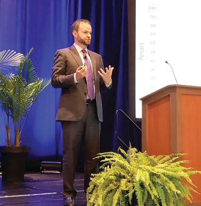
The Staller lab’s AGA-funded project is specifically focused on the risk factors for fecal incontinence, which have not been well studied. One in 10 women over age 80 suffers from this debilitating condition. Dr. Staller looked at the lifestyles and dietary factors of female study participants in research databases to determine whether they were predisposed to developing fecal incontinence beyond the usual risk factors such as childbirth, which can cause injury to the pelvic floor, and diabetes. Dr. Staller believes that understanding and modifying risk factors could decrease the chance of women developing this condition, or could even prevent it.
With his AGA Research Foundation grant, Dr. Staller found that consumption of dietary fiber in higher quantities, and an increase of moderate exercise up to a point, lowered the risk of developing fecal incontinence. “This tells us that not only is fiber healthy but also preventative to fecal incontinence,” he said.
Dr. Staller says that he became interested in this area of study after patients, who were getting excited about their impending retirement or enjoying their retirement years, developed this life-altering condition. His compassion for his patients inspired him to study the factors leading to fecal incontinence, which will likely become more prevalent as the U.S. population ages.
Dr. Staller is using the baseline data from his AGA Research Foundation grant to support his application for a 5-year NIH grant designed to help young investigators learn new research skills to further their careers.
“This is the next step in my career,” he said. “If I didn’t have the AGA Research Foundation grant, I don’t know if the opportunity would be there for me to go on to the next level. The AGA grant gives you the opportunity to get that baseline data so you can become a competitive applicant for longer-term grants.”
Another benefit of Dr. Staller’s AGA Research Foundation grant: It got him involved with AGA. In March 2019, Dr. Staller joined the new class of AGA Future Leaders, AGA’s competitive leadership development program designed to prepare early career GIs for future leadership roles in AGA, at their home institutions, and within the field of digestive diseases. The program kicked off at AGA’s inaugural leadership development conference.
“It is a true honor to participate in the AGA Future Leaders Program. During the AGA Leadership Development Conference, we learned concrete tips about effective leadership strategies across the spectrum of GI practice from research to clinical practice. Among our mentors were prominent researchers, clinical innovators, and division and department heads from across the U.S. – there was no shortage of inspiration. Perhaps most importantly, I was able to form what I hope to be career-long connections with both my fellow future leaders program participants and our mentors,” he said.
Dr. Staller’s qualifications as a clinician and researcher of bowel issues are put to good use as a father of two boys, ages 4 and 6, who are at the peak of the potty humor stage.
“They’re interested in the GI tract as well,” Dr. Staller said with a laugh. “My mom likes to say I never got out of the potty phase and made it a career. It’s important to feel comfortable talking about these uncomfortable topics. That’s what people want from their physician. If you can talk about this and the physician doesn’t bat an eyelash, that’s a good setup to have a good therapeutic relationship.”
‘Put your own oxygen mask on first’
Takeaways from the leadership conference stress the importance of self-care, emotional intelligence and remaining optimistic.

“Leadership 101: Put your own oxygen mask on first @DarwinConwell #AGAleads #AGAForward @AmerGastroAssn”– Dr Michelle T. Long (@DrMTLong)
The inaugural Leadership Development Conference combined participants from three AGA programs for a weekend of networking, mentorship and mapping out goals and initiatives.
Attendees included the 2020 class of AGA Future Leaders and mentors, Women’s Leadership Conference participants, and mentors and scholars of the new AGA FORWARD Program, an National Institutes of Health–funded initiative that supports underrepresented minority physicians and scientists.
“Got to meet one of my tweeps heroes today! She’s even more awesome in real life!! #AGALeads #WomenInMedicine #WomenInGI @drfolamay @AmerGastroAssn” – Dr Aline Charabaty (@DCharabaty)
“Dr. Boland (Lynch syndrome) discussing career success in an ever changing scientific environment #AGALeads #AGAForward” – Eric J. Vargas M.D. (@EricJVargasMD)
“7 AGA Presidents, moderated by Dr. Anandasabapathy on Pathways to Leadership and Overcoming Challenges of the Era Presidential Panel @AmerGastroAssn Leadership conference program @SeragHashem @BCMDeptMedicine @KanwalFasiha @Aketwaroo @richashukla84” – Ruben Hernaez (@ruben_hernaez)
The event coincided with International Women’s Day, giving Women’s Leadership Conference attendees the chance to celebrate their journeys and grow into leadership roles with other #WomenInGI.
“#AGALeads #womenleadershipconference #womeninGI #InternationWomensDay with some amazing ladies in GI!! @AmerGastroAssn @AlisonGoldinMD @ibddocmaria @joanwchen” – ReezwanaCMD (@reezwanc)
“#AGAleads #WomeninGI women negotiating in a group are perceived favorably-Ellen Zimmerman, MD”
– Fazia Mir-Shaffi, MD (@Faiziya) March 9, 2019
“What I learned at @AmerGastroAssn #womeninGI Leadership course (after waiting a bit to see what stuck w me)
1. If you say yes to a request, you’re saying yes to doing it well.
2. Knowing your limitations will serve you better than being great at everything” – Laura Targownik (@UofM_GI_Head)
Aline Charabaty Pishvaian, MD, shared some takeaways in the AGA Community forum (community.gastro.org) about challenges women in GI face – a breakout discussion from the Women’s Leadership Conference.
View more insight and takeaways from participants on Twitter using #AGALeads.
Dr. Vaibhav Wadhwa advocates for step therapy reform in Florida
Vaibhav Wadhwa, MD, met with Ms. Laurie Flink, deputy district director for Rep. Debbie Wasserman Schultz (FL-23), to discuss AGA’s legislative priorities.
Dr. Wadhwa thanked Ms. Flink for Rep. Wasserman Schultz’s support of the Removing Barriers to Colorectal Screening Act and NIH funding. Dr. Wadhwa also mentioned that Rep. Wasserman Schultz is not a cosponsor of the Restoring the Patient’s Voice Act and explained in detail about why this is an important resolution that needs to be passed.
Dr. Wadhwa gave examples of patients from his own practice and discussed the challenges they face. Ms. Flink was very interested in hearing about patients with chronic conditions such as inflammatory bowel disease (IBD) not being able to get the appropriate regimen because of the barriers created by step therapy. Ms. Flink was very appreciative of the visit and stated that these in-person visits along with personal stories about these issues go a long way in helping congressional offices understand the implications that these bills have.
Ms. Flink assured Dr. Wadhwa that she will raise these points with Rep. Wasserman Schultz and will discuss cosponsoring the Restoring the Patient’s Voice Act once it is reintroduced.
Dr. Wadhwa is a fellow at the Cleveland Clinic Florida in Weston, and is the AGA Congressional Advocates Program state leader for Florida. He is interested in therapeutic endoscopy and advocating for appropriate reimbursement for endoscopic procedures.
How to get involved in advocacy
Interested in advocacy but not sure how or whether you have time in your busy schedule? AGA has an array of options for how you can be active in advocacy. Some take as little as 5 minutes.
Letter writing. AGA uses GovPredict, an online advocacy platform that allows members to contact their representatives in Congress with just a few clicks. AGA develops messages on significant pieces of legislation, key efforts in Congress, or on issues being advanced by federal agencies that have a great impact on gastroenterology. AGA’s ongoing letter writing campaigns can always be found at gastro.org, but be sure to keep an eye out for advocacy emails, AGA eDigest, and social media, so you do not miss your opportunity to take action on timely issues. AGA encourages its members to share letter writing campaigns with their colleagues, as well as posting them on social media.
Meetings with members of Congress. In-person meetings are an excellent opportunity to share with your representatives in Congress, or their staff, how the issues that impact gastroenterology affect you, your patients, and your practice. AGA has a plethora of resources to help you set up such meetings, including up-to-date issue briefs, tips and tricks for productive meetings, and webinars on how to host an on-site visit. AGA staff is always more than happy to help you arrange a meeting either in Washington, D.C., or in your home state. If you are interested in arranging such a meeting, please contact AGA Public Policy Coordinator Jonathan Sollish, at [email protected] or 240-482-3228.
AGA PAC. AGA PAC is a voluntary, nonpartisan political organization affiliated with and supported by AGA. It is the only political action committee supported by a national gastroenterology society, and its mission is to give gastroenterologists a greater presence on Capitol Hill and a more effective voice in policy discussions. AGA PAC supports candidates who support our policy priorities, such as fair reimbursement, cutting regulatory red tape, supporting patient protections, and access to specialty care, and sustained federal funding of digestive disease research. If you are interested in learning more, contact AGA Government and Political Affairs Manager Navneet Buttar, at [email protected] or 240-482-3221.
Congressional Advocates Program. This grassroots program is aimed at establishing a stronger foundation for our current and future advocacy initiatives by creating state teams to work on advocacy on the local, state, and national levels. Participation can include a wide variety of activities, ranging from creating educational posts on social media to meeting with members of Congress. Members of the Congressional Advocates Program are mentored and receive advocacy training by AGA leadership and staff. Participating members receive an AGA Congressional Advocate Program Certificate, a Digestive Disease Week® (DDW) badge ribbon, policy badge on the AGA Community, and recognition on AGA’s website. Applications for the next cycle will be released in 2019.
Introducing the AGA Future Leaders class of 2020
AGA has announced the 18 early career physicians and scientists selected to participate in its Future Leaders Program, which was created in 2015 to provide a pathway for leadership development within AGA for early career physicians and scientists who have the potential to make a significant impact on the specialty. These 18 participants will embark on an 18-month-long program designed to develop the skills necessary to become future leaders in the AGA, at their home institutions, and within the field of digestive diseases.
“The 2020 class of AGA Future Leaders represents the next generation of leaders in our field,” said Darrell S. Pardi, MD, MSc, AGAF, co–program chair for the AGA Future Leaders Program. “Along with my cochair, Sheryl Pfeil, MD, AGAF, and the esteemed mentors and faculty participating in this program, we look forward to cultivating these rising stars who stand out for their current achievements, commitment to advancing the field, and potential for future success.”
Class of 2020 Future Leaders
- Christen Klochan Dilly, MD, MEHP, Indiana University School of Medicine and Roudebush VA Medical Center
- Daniel Freedberg, MD, MS, Columbia University
- Wendy A. Henderson, PhD, National Institutes of Health
- Ruben Hernaez, MD, MPH, PhD, Baylor College of Medicine and Michael E. DeBakey VA Medical Center
- Animesh Jain, MD, University of North Carolina at Chapel Hill
- Avinash Ketwaroo, MD, Baylor College of Medicine and Michael E. DeBakey VA Medical Center
- Bharati Kochar, MD, MSCR, University of North Carolina at Chapel Hill
- David Leiman, MD, MSHP, Duke University Medical Center
- James Lin, MD, City of Hope National Medical Center in Duarte
- Michelle Long, MD, Boston Medical Center
- Aimee Lucas, MD, MS, Icahn School of Medicine at Mount Sinai
- Miguel Malespin, MD, Tampa General Hospital
- Simon C. Mathews, MD, Johns Hopkins Medicine
- Karthik Ravi, MD, Mayo Clinic (Rochester, Minnesota)
- Florian Rieder, MD, Cleveland Clinic Foundation
- Kyle Staller, MD, MPH, Harvard Medical School
- Christina Twyman-Saint Victor, MD, University of Pennsylvania Perelman School of Medicine
- Ryan Ungaro, MD, MS, Icahn School of Medicine at Mount Sinai
View Future Leader Bios
The AGA Future Leaders Program will kick off with the AGA Leadership Development Conference March 8-10, 2019, at the Hilton Rockville Executive conference center in Rockville, Maryland, and will continue through Digestive Disease Week® (DDW) 2020 in Chicago, Illinois. Throughout the course of the program, participants will work closely with AGA mentors on projects linked to AGA’s Strategic Plan.
Learn more about the AGA Future Leaders Program.
Sessions at DDW® 2019 designed for fellows and early career GIs
AGA has an agenda of special sessions at Digestive Disease Week® (DDW) 2019 to meet the unique needs of physicians who are new to the field. Participants will learn about all aspects of starting a career in clinical practice or research, have the opportunity to network with mentors and peers, and review board material.
With the exception of the AGA Postgraduate Course, all of the sessions are free, but you must register for DDW to attend. Visit AGA University for a full list and additional details.
- AGA Postgraduate Course Saturday, May 18, and Sunday, May 19
- Introduction to GI Practice: A Trainee Boot Camp, Monday, May 20, 10-11:30 a.m.
- AGA Board Review CourseMonday, May 20, 1:30-5:30 p.m.
- Advancing Clinical Practice: GI Fellow-Directed Quality Improvement ProjectsMonday, May 20, 2-3:30 p.m.
- GI in the Digital Age, Monday, May 20, 4-5:30 p.m.
DDW Trainee and Early Career Lounge
Included with the cost of DDW registration, trainee and early career GI attendees have access to this lounge in the Sails Pavilion. It’s a great way to meet and network with peers from around the world over a cup of coffee and will feature new programming in 2019. Meet with experts to have your questions answered about practical issues of career choice, contracting, or how to write a manuscript.
Meet a rising star in fecal incontinence research
The AGA Research Foundation offers its flagship grant, the AGA Research Scholar Award, to the most promising early career investigators. Kyle Staller, MD, MPH, assistant professor of medicine at Harvard Medical School in Boston, is no exception. We’re thrilled to highlight Dr. Staller – a 2016 AGA Research Scholar Award winner – as our AGA Research Foundation researcher of the month.
The Staller lab’s AGA-funded project is specifically focused on the risk factors for fecal incontinence, which have not been well studied. One in 10 women over age 80 suffers from this debilitating condition. Dr. Staller looked at the lifestyles and dietary factors of female study participants in research databases to determine whether they were predisposed to developing fecal incontinence beyond the usual risk factors such as childbirth, which can cause injury to the pelvic floor, and diabetes. Dr. Staller believes that understanding and modifying risk factors could decrease the chance of women developing this condition, or could even prevent it.
With his AGA Research Foundation grant, Dr. Staller found that consumption of dietary fiber in higher quantities, and an increase of moderate exercise up to a point, lowered the risk of developing fecal incontinence. “This tells us that not only is fiber healthy but also preventative to fecal incontinence,” he said.
Dr. Staller says that he became interested in this area of study after patients, who were getting excited about their impending retirement or enjoying their retirement years, developed this life-altering condition. His compassion for his patients inspired him to study the factors leading to fecal incontinence, which will likely become more prevalent as the U.S. population ages.
Dr. Staller is using the baseline data from his AGA Research Foundation grant to support his application for a 5-year NIH grant designed to help young investigators learn new research skills to further their careers.
“This is the next step in my career,” he said. “If I didn’t have the AGA Research Foundation grant, I don’t know if the opportunity would be there for me to go on to the next level. The AGA grant gives you the opportunity to get that baseline data so you can become a competitive applicant for longer-term grants.”
Another benefit of Dr. Staller’s AGA Research Foundation grant: It got him involved with AGA. In March 2019, Dr. Staller joined the new class of AGA Future Leaders, AGA’s competitive leadership development program designed to prepare early career GIs for future leadership roles in AGA, at their home institutions, and within the field of digestive diseases. The program kicked off at AGA’s inaugural leadership development conference.
“It is a true honor to participate in the AGA Future Leaders Program. During the AGA Leadership Development Conference, we learned concrete tips about effective leadership strategies across the spectrum of GI practice from research to clinical practice. Among our mentors were prominent researchers, clinical innovators, and division and department heads from across the U.S. – there was no shortage of inspiration. Perhaps most importantly, I was able to form what I hope to be career-long connections with both my fellow future leaders program participants and our mentors,” he said.
Dr. Staller’s qualifications as a clinician and researcher of bowel issues are put to good use as a father of two boys, ages 4 and 6, who are at the peak of the potty humor stage.
“They’re interested in the GI tract as well,” Dr. Staller said with a laugh. “My mom likes to say I never got out of the potty phase and made it a career. It’s important to feel comfortable talking about these uncomfortable topics. That’s what people want from their physician. If you can talk about this and the physician doesn’t bat an eyelash, that’s a good setup to have a good therapeutic relationship.”
‘Put your own oxygen mask on first’
Takeaways from the leadership conference stress the importance of self-care, emotional intelligence and remaining optimistic.
“Leadership 101: Put your own oxygen mask on first @DarwinConwell #AGAleads #AGAForward @AmerGastroAssn”– Dr Michelle T. Long (@DrMTLong)
The inaugural Leadership Development Conference combined participants from three AGA programs for a weekend of networking, mentorship and mapping out goals and initiatives.
Attendees included the 2020 class of AGA Future Leaders and mentors, Women’s Leadership Conference participants, and mentors and scholars of the new AGA FORWARD Program, an National Institutes of Health–funded initiative that supports underrepresented minority physicians and scientists.
“Got to meet one of my tweeps heroes today! She’s even more awesome in real life!! #AGALeads #WomenInMedicine #WomenInGI @drfolamay @AmerGastroAssn” – Dr Aline Charabaty (@DCharabaty)
“Dr. Boland (Lynch syndrome) discussing career success in an ever changing scientific environment #AGALeads #AGAForward” – Eric J. Vargas M.D. (@EricJVargasMD)
“7 AGA Presidents, moderated by Dr. Anandasabapathy on Pathways to Leadership and Overcoming Challenges of the Era Presidential Panel @AmerGastroAssn Leadership conference program @SeragHashem @BCMDeptMedicine @KanwalFasiha @Aketwaroo @richashukla84” – Ruben Hernaez (@ruben_hernaez)
The event coincided with International Women’s Day, giving Women’s Leadership Conference attendees the chance to celebrate their journeys and grow into leadership roles with other #WomenInGI.
“#AGALeads #womenleadershipconference #womeninGI #InternationWomensDay with some amazing ladies in GI!! @AmerGastroAssn @AlisonGoldinMD @ibddocmaria @joanwchen” – ReezwanaCMD (@reezwanc)
“#AGAleads #WomeninGI women negotiating in a group are perceived favorably-Ellen Zimmerman, MD”
– Fazia Mir-Shaffi, MD (@Faiziya) March 9, 2019
“What I learned at @AmerGastroAssn #womeninGI Leadership course (after waiting a bit to see what stuck w me)
1. If you say yes to a request, you’re saying yes to doing it well.
2. Knowing your limitations will serve you better than being great at everything” – Laura Targownik (@UofM_GI_Head)
Aline Charabaty Pishvaian, MD, shared some takeaways in the AGA Community forum (community.gastro.org) about challenges women in GI face – a breakout discussion from the Women’s Leadership Conference.
View more insight and takeaways from participants on Twitter using #AGALeads.
Dr. Vaibhav Wadhwa advocates for step therapy reform in Florida
Vaibhav Wadhwa, MD, met with Ms. Laurie Flink, deputy district director for Rep. Debbie Wasserman Schultz (FL-23), to discuss AGA’s legislative priorities.
Dr. Wadhwa thanked Ms. Flink for Rep. Wasserman Schultz’s support of the Removing Barriers to Colorectal Screening Act and NIH funding. Dr. Wadhwa also mentioned that Rep. Wasserman Schultz is not a cosponsor of the Restoring the Patient’s Voice Act and explained in detail about why this is an important resolution that needs to be passed.
Dr. Wadhwa gave examples of patients from his own practice and discussed the challenges they face. Ms. Flink was very interested in hearing about patients with chronic conditions such as inflammatory bowel disease (IBD) not being able to get the appropriate regimen because of the barriers created by step therapy. Ms. Flink was very appreciative of the visit and stated that these in-person visits along with personal stories about these issues go a long way in helping congressional offices understand the implications that these bills have.
Ms. Flink assured Dr. Wadhwa that she will raise these points with Rep. Wasserman Schultz and will discuss cosponsoring the Restoring the Patient’s Voice Act once it is reintroduced.
Dr. Wadhwa is a fellow at the Cleveland Clinic Florida in Weston, and is the AGA Congressional Advocates Program state leader for Florida. He is interested in therapeutic endoscopy and advocating for appropriate reimbursement for endoscopic procedures.
How to get involved in advocacy
Interested in advocacy but not sure how or whether you have time in your busy schedule? AGA has an array of options for how you can be active in advocacy. Some take as little as 5 minutes.
Letter writing. AGA uses GovPredict, an online advocacy platform that allows members to contact their representatives in Congress with just a few clicks. AGA develops messages on significant pieces of legislation, key efforts in Congress, or on issues being advanced by federal agencies that have a great impact on gastroenterology. AGA’s ongoing letter writing campaigns can always be found at gastro.org, but be sure to keep an eye out for advocacy emails, AGA eDigest, and social media, so you do not miss your opportunity to take action on timely issues. AGA encourages its members to share letter writing campaigns with their colleagues, as well as posting them on social media.
Meetings with members of Congress. In-person meetings are an excellent opportunity to share with your representatives in Congress, or their staff, how the issues that impact gastroenterology affect you, your patients, and your practice. AGA has a plethora of resources to help you set up such meetings, including up-to-date issue briefs, tips and tricks for productive meetings, and webinars on how to host an on-site visit. AGA staff is always more than happy to help you arrange a meeting either in Washington, D.C., or in your home state. If you are interested in arranging such a meeting, please contact AGA Public Policy Coordinator Jonathan Sollish, at [email protected] or 240-482-3228.
AGA PAC. AGA PAC is a voluntary, nonpartisan political organization affiliated with and supported by AGA. It is the only political action committee supported by a national gastroenterology society, and its mission is to give gastroenterologists a greater presence on Capitol Hill and a more effective voice in policy discussions. AGA PAC supports candidates who support our policy priorities, such as fair reimbursement, cutting regulatory red tape, supporting patient protections, and access to specialty care, and sustained federal funding of digestive disease research. If you are interested in learning more, contact AGA Government and Political Affairs Manager Navneet Buttar, at [email protected] or 240-482-3221.
Congressional Advocates Program. This grassroots program is aimed at establishing a stronger foundation for our current and future advocacy initiatives by creating state teams to work on advocacy on the local, state, and national levels. Participation can include a wide variety of activities, ranging from creating educational posts on social media to meeting with members of Congress. Members of the Congressional Advocates Program are mentored and receive advocacy training by AGA leadership and staff. Participating members receive an AGA Congressional Advocate Program Certificate, a Digestive Disease Week® (DDW) badge ribbon, policy badge on the AGA Community, and recognition on AGA’s website. Applications for the next cycle will be released in 2019.
Introducing the AGA Future Leaders class of 2020
AGA has announced the 18 early career physicians and scientists selected to participate in its Future Leaders Program, which was created in 2015 to provide a pathway for leadership development within AGA for early career physicians and scientists who have the potential to make a significant impact on the specialty. These 18 participants will embark on an 18-month-long program designed to develop the skills necessary to become future leaders in the AGA, at their home institutions, and within the field of digestive diseases.
“The 2020 class of AGA Future Leaders represents the next generation of leaders in our field,” said Darrell S. Pardi, MD, MSc, AGAF, co–program chair for the AGA Future Leaders Program. “Along with my cochair, Sheryl Pfeil, MD, AGAF, and the esteemed mentors and faculty participating in this program, we look forward to cultivating these rising stars who stand out for their current achievements, commitment to advancing the field, and potential for future success.”
Class of 2020 Future Leaders
- Christen Klochan Dilly, MD, MEHP, Indiana University School of Medicine and Roudebush VA Medical Center
- Daniel Freedberg, MD, MS, Columbia University
- Wendy A. Henderson, PhD, National Institutes of Health
- Ruben Hernaez, MD, MPH, PhD, Baylor College of Medicine and Michael E. DeBakey VA Medical Center
- Animesh Jain, MD, University of North Carolina at Chapel Hill
- Avinash Ketwaroo, MD, Baylor College of Medicine and Michael E. DeBakey VA Medical Center
- Bharati Kochar, MD, MSCR, University of North Carolina at Chapel Hill
- David Leiman, MD, MSHP, Duke University Medical Center
- James Lin, MD, City of Hope National Medical Center in Duarte
- Michelle Long, MD, Boston Medical Center
- Aimee Lucas, MD, MS, Icahn School of Medicine at Mount Sinai
- Miguel Malespin, MD, Tampa General Hospital
- Simon C. Mathews, MD, Johns Hopkins Medicine
- Karthik Ravi, MD, Mayo Clinic (Rochester, Minnesota)
- Florian Rieder, MD, Cleveland Clinic Foundation
- Kyle Staller, MD, MPH, Harvard Medical School
- Christina Twyman-Saint Victor, MD, University of Pennsylvania Perelman School of Medicine
- Ryan Ungaro, MD, MS, Icahn School of Medicine at Mount Sinai
View Future Leader Bios
The AGA Future Leaders Program will kick off with the AGA Leadership Development Conference March 8-10, 2019, at the Hilton Rockville Executive conference center in Rockville, Maryland, and will continue through Digestive Disease Week® (DDW) 2020 in Chicago, Illinois. Throughout the course of the program, participants will work closely with AGA mentors on projects linked to AGA’s Strategic Plan.
Learn more about the AGA Future Leaders Program.
Sessions at DDW® 2019 designed for fellows and early career GIs
AGA has an agenda of special sessions at Digestive Disease Week® (DDW) 2019 to meet the unique needs of physicians who are new to the field. Participants will learn about all aspects of starting a career in clinical practice or research, have the opportunity to network with mentors and peers, and review board material.
With the exception of the AGA Postgraduate Course, all of the sessions are free, but you must register for DDW to attend. Visit AGA University for a full list and additional details.
- AGA Postgraduate Course Saturday, May 18, and Sunday, May 19
- Introduction to GI Practice: A Trainee Boot Camp, Monday, May 20, 10-11:30 a.m.
- AGA Board Review CourseMonday, May 20, 1:30-5:30 p.m.
- Advancing Clinical Practice: GI Fellow-Directed Quality Improvement ProjectsMonday, May 20, 2-3:30 p.m.
- GI in the Digital Age, Monday, May 20, 4-5:30 p.m.
DDW Trainee and Early Career Lounge
Included with the cost of DDW registration, trainee and early career GI attendees have access to this lounge in the Sails Pavilion. It’s a great way to meet and network with peers from around the world over a cup of coffee and will feature new programming in 2019. Meet with experts to have your questions answered about practical issues of career choice, contracting, or how to write a manuscript.