User login
Empty words, Captain Bacteria, and the perils of vampire facials
And now, 37 words from our sponsor
If you’re looking for gluten-free news of the health and medical sciences that’s low in sugar, we here at LOTME Farms promise to use no artificial colors or flavors to tell you about a study of the health claims on cereal boxes.

Researchers identified 37 such claims that appeared on the boxes of 460 different breakfast cereals and grouped them into four categories: adding positives (high fiber, probiotics), not adding negatives (GMO free, no high-fructose corn syrup), removing negatives (low cholesterol, no trans fat), and not removing positives (made with whole grains, fresh).
What they found is that words matter: None of the 37 claims explicitly said that the product inside would make people healthier or help them lose weight, but that was how respondents interpreted them. There is, of course, a reason none of the products claimed to improve health. “The correlation between the type of ‘healthy’ claim made and the actual nutritional quality of the breakfast cereal was almost zero,” investigator Pierre Chandon said.
This is, perhaps, not such a surprise. But we here at the pure, all-natural LOTME deal with facts, which are low in calories and contain no artificial growth hormones, and we would never insult (NEW LOTME LIGHT! NO ARTIFICIAL SWEETENERS!) our wholesome, low-fat readers by resorting (TRY FRESH LOTME ORGANIC!) to hyperbole or doublespeak. Not a chance (MMM … HOMEMADE LOTME).
Now, who’s up for a bowl of Froot Loops?
Captain Bacteria: Civil War
In the never-ending struggle of bacteria versus the world, Clostridium difficile has become a particularly stubborn foe. It is far more likely to be resistant to antibiotics, and the antibiotics that can do the job are either incredibly expensive or destroy the patient’s entire microbiota. However, we may have a new ally in the fight against C. diff: other bacteria.

Specifically, we’re talking about fecal transplants. According to an article published in the Journal of the American Osteopathic Association, the wide variety of bacteria that get transferred into the body along with the poop can block C. diff’s ability to germinate and produce the toxins that affect the human body. The treatment is especially beneficial for patients with C. diff whose microbiotas have been compromised by some other treatment, like chemotherapy, antibiotics, or proton pump inhibitors.
We here at LOTME would like to take a moment to salute the brave bacteria in our guts, fighting the good fight against those who would do us harm, and to the fecal transplants that let our own bacteria join the battle. Poop, you never let us down!
Dracula side effects
The Kim Kardashian effect is having dire ramifications. Back in 2013, the social media influencer posted a photo of herself getting a “vampire facial” – a dermatologic procedure in which a person’s own blood is injected into their face as a way to freshen and rejuvenate their skin. At least they aren’t drinking it.

Vampire facials have skyrocketed in popularity since, despite the ick factor. Unfortunately, it seems to be as dangerous as an encounter with a real vampire: Recently, two people in New Mexico were diagnosed with HIV after getting vampire facials.
The New Mexico Department of Health noted that both cases have been traced to the same spa, which was shut down in 2018 after at least three government agencies noted its lack of attention to hygiene and cleanliness. Warning to all: Just because a Kardashian does it doesn’t mean you should, too.
Shave that beard, Fido
The adage “a dog’s mouth is cleaner than a person’s” is getting an upgrade and this time it’s backed by science. A clinic in Zurich looked at the pathogenic microorganisms that reside in men’s beards and dogs’ fur and guess what – the beards were dirtier.
Despite beards’ manly cache among Millennials and hipsters, the results many have some reaching for the razor. The clinicians took a look at the bacterial load of colony-forming units of human-pathogenic microorganisms, and compared samples of beards and dog’s necks (since dogs don’t have beards, but how cute would that be?). They found high microbial counts in all beard samples, but only 23 of 30 dogs’ hair samples. Half of the human subjects carried so much bacteria that they were in danger of illness, the researchers noted.
Does this mean men are dirty? Or are dogs clean? The clinicians didn’t survey the men on their habits so it could be likely that they spend lots of time rolling around in the grass or drinking water from the toilet. More research is definitely needed here.

And now, 37 words from our sponsor
If you’re looking for gluten-free news of the health and medical sciences that’s low in sugar, we here at LOTME Farms promise to use no artificial colors or flavors to tell you about a study of the health claims on cereal boxes.
Researchers identified 37 such claims that appeared on the boxes of 460 different breakfast cereals and grouped them into four categories: adding positives (high fiber, probiotics), not adding negatives (GMO free, no high-fructose corn syrup), removing negatives (low cholesterol, no trans fat), and not removing positives (made with whole grains, fresh).
What they found is that words matter: None of the 37 claims explicitly said that the product inside would make people healthier or help them lose weight, but that was how respondents interpreted them. There is, of course, a reason none of the products claimed to improve health. “The correlation between the type of ‘healthy’ claim made and the actual nutritional quality of the breakfast cereal was almost zero,” investigator Pierre Chandon said.
This is, perhaps, not such a surprise. But we here at the pure, all-natural LOTME deal with facts, which are low in calories and contain no artificial growth hormones, and we would never insult (NEW LOTME LIGHT! NO ARTIFICIAL SWEETENERS!) our wholesome, low-fat readers by resorting (TRY FRESH LOTME ORGANIC!) to hyperbole or doublespeak. Not a chance (MMM … HOMEMADE LOTME).
Now, who’s up for a bowl of Froot Loops?
Captain Bacteria: Civil War
In the never-ending struggle of bacteria versus the world, Clostridium difficile has become a particularly stubborn foe. It is far more likely to be resistant to antibiotics, and the antibiotics that can do the job are either incredibly expensive or destroy the patient’s entire microbiota. However, we may have a new ally in the fight against C. diff: other bacteria.
Specifically, we’re talking about fecal transplants. According to an article published in the Journal of the American Osteopathic Association, the wide variety of bacteria that get transferred into the body along with the poop can block C. diff’s ability to germinate and produce the toxins that affect the human body. The treatment is especially beneficial for patients with C. diff whose microbiotas have been compromised by some other treatment, like chemotherapy, antibiotics, or proton pump inhibitors.
We here at LOTME would like to take a moment to salute the brave bacteria in our guts, fighting the good fight against those who would do us harm, and to the fecal transplants that let our own bacteria join the battle. Poop, you never let us down!
Dracula side effects
The Kim Kardashian effect is having dire ramifications. Back in 2013, the social media influencer posted a photo of herself getting a “vampire facial” – a dermatologic procedure in which a person’s own blood is injected into their face as a way to freshen and rejuvenate their skin. At least they aren’t drinking it.
Vampire facials have skyrocketed in popularity since, despite the ick factor. Unfortunately, it seems to be as dangerous as an encounter with a real vampire: Recently, two people in New Mexico were diagnosed with HIV after getting vampire facials.
The New Mexico Department of Health noted that both cases have been traced to the same spa, which was shut down in 2018 after at least three government agencies noted its lack of attention to hygiene and cleanliness. Warning to all: Just because a Kardashian does it doesn’t mean you should, too.
Shave that beard, Fido
The adage “a dog’s mouth is cleaner than a person’s” is getting an upgrade and this time it’s backed by science. A clinic in Zurich looked at the pathogenic microorganisms that reside in men’s beards and dogs’ fur and guess what – the beards were dirtier.
Despite beards’ manly cache among Millennials and hipsters, the results many have some reaching for the razor. The clinicians took a look at the bacterial load of colony-forming units of human-pathogenic microorganisms, and compared samples of beards and dog’s necks (since dogs don’t have beards, but how cute would that be?). They found high microbial counts in all beard samples, but only 23 of 30 dogs’ hair samples. Half of the human subjects carried so much bacteria that they were in danger of illness, the researchers noted.
Does this mean men are dirty? Or are dogs clean? The clinicians didn’t survey the men on their habits so it could be likely that they spend lots of time rolling around in the grass or drinking water from the toilet. More research is definitely needed here.
Are guidelines relevant?
The recent publication of the changes and current levels of evidence for the guidelines from the American College of Cardiology, American Heart Association, and the European Society of Cardiology once again indicates that the level of high-quality scientific data supporting the decision making process is limited.

Of the thousands of recommendations among the guidelines provided, level A data – which is supported by at least two randomized, controlled trials (RCT), arguably the gold standard for the proof of benefit of a treatment program – was available in only 8.5% of ACC/AHA recommendations and in 14.2% of ESC recommendations. The recent review covering guidelines published from 2008 to 2018 has changed very little since the previous review carried out on data collected between 1999 and 2014 (JAMA. 2019 Mar 19;321[11]:1069-80). Admittedly, by the nature of their design, RCTs represent a very special subset of any disease process but they are the best we can do. The remainder of the guidelines is supported by nonrandomized data and consensus opinion of “experts.” The absence of randomized data in the rest of the guidelines creates an area of uncertainty between clinical practice and outcomes.
Guidelines have been with us for almost 30 years and have provided a near “cookbook recipe” for the treatment of almost the totality of cardiovascular care, both in the United States and Europe. There is little evidence that guideline dissemination results in a beneficial effect on cardiovascular care, instead reflecting the contemporary published research and informed opinion of our generation.
Despite the limitation of hard data based on RCTs, cardiovascular mortality has leveled off in the last few years. Although there are a number of areas that could be investigated with more focused and practical RCTs, there has been little enthusiasm by either the National Heart, Lung, and Blood Institute or the pharmaceutical or device industries to investigate existing cardiovascular drugs and/or devices to better elucidate nuanced therapy for subsets of heart disease that remain in the uncertain data areas. In fact, the current report shows that there has been little change in guidelines in the last 10 years. Most effort has been focused on new drug development and application.
Much of the advance in cardiovascular care and decrease in mortality is a result of the development of new drugs for the treatment of hypertension, management of hypercholesterolemia, and MI, together with coronary angiography and its interventional permutations of the previous 50 years. Those advances are now part of our knowledge base, our therapeutic DNA, and in the absence of some new scientific breakthrough, there is little to expect from future guideline development. Medical students and house officers are being taught as fact what we found as exciting new therapy just a few short years ago. Guidelines are now part of textbook knowledge and until new clinical outcome are reported, guidelines will probably serve a useful reference material but will not lead to much change in clinical care.
Even so, treating the patient in the clinic and in the hospital still requires the doctor to practice the “art” of medicine and to synthesize therapy based on the paucity of scientific data and quasi knowledge acquired over a generation.
The appreciation that much is unknown gives the doctor the humility required to be sensitive to patient needs and their symptoms and the aspiration for new, science-based data. This is still part of the excitement of being a doctor.
Dr. Goldstein, medical editor of Cardiology News, is professor of medicine at Wayne State University and division head emeritus of cardiovascular medicine at Henry Ford Hospital, both in Detroit. He is on data safety monitoring committees for the National Institutes of Health and several pharmaceutical companies.
The recent publication of the changes and current levels of evidence for the guidelines from the American College of Cardiology, American Heart Association, and the European Society of Cardiology once again indicates that the level of high-quality scientific data supporting the decision making process is limited.
Of the thousands of recommendations among the guidelines provided, level A data – which is supported by at least two randomized, controlled trials (RCT), arguably the gold standard for the proof of benefit of a treatment program – was available in only 8.5% of ACC/AHA recommendations and in 14.2% of ESC recommendations. The recent review covering guidelines published from 2008 to 2018 has changed very little since the previous review carried out on data collected between 1999 and 2014 (JAMA. 2019 Mar 19;321[11]:1069-80). Admittedly, by the nature of their design, RCTs represent a very special subset of any disease process but they are the best we can do. The remainder of the guidelines is supported by nonrandomized data and consensus opinion of “experts.” The absence of randomized data in the rest of the guidelines creates an area of uncertainty between clinical practice and outcomes.
Guidelines have been with us for almost 30 years and have provided a near “cookbook recipe” for the treatment of almost the totality of cardiovascular care, both in the United States and Europe. There is little evidence that guideline dissemination results in a beneficial effect on cardiovascular care, instead reflecting the contemporary published research and informed opinion of our generation.
Despite the limitation of hard data based on RCTs, cardiovascular mortality has leveled off in the last few years. Although there are a number of areas that could be investigated with more focused and practical RCTs, there has been little enthusiasm by either the National Heart, Lung, and Blood Institute or the pharmaceutical or device industries to investigate existing cardiovascular drugs and/or devices to better elucidate nuanced therapy for subsets of heart disease that remain in the uncertain data areas. In fact, the current report shows that there has been little change in guidelines in the last 10 years. Most effort has been focused on new drug development and application.
Much of the advance in cardiovascular care and decrease in mortality is a result of the development of new drugs for the treatment of hypertension, management of hypercholesterolemia, and MI, together with coronary angiography and its interventional permutations of the previous 50 years. Those advances are now part of our knowledge base, our therapeutic DNA, and in the absence of some new scientific breakthrough, there is little to expect from future guideline development. Medical students and house officers are being taught as fact what we found as exciting new therapy just a few short years ago. Guidelines are now part of textbook knowledge and until new clinical outcome are reported, guidelines will probably serve a useful reference material but will not lead to much change in clinical care.
Even so, treating the patient in the clinic and in the hospital still requires the doctor to practice the “art” of medicine and to synthesize therapy based on the paucity of scientific data and quasi knowledge acquired over a generation.
The appreciation that much is unknown gives the doctor the humility required to be sensitive to patient needs and their symptoms and the aspiration for new, science-based data. This is still part of the excitement of being a doctor.
Dr. Goldstein, medical editor of Cardiology News, is professor of medicine at Wayne State University and division head emeritus of cardiovascular medicine at Henry Ford Hospital, both in Detroit. He is on data safety monitoring committees for the National Institutes of Health and several pharmaceutical companies.
Increase in pediatric thyroid cancers: overdiagnosis or something more?
Thyroid cancer rates are on the rise in U.S. pediatric patients, a large epidemiologic study shows, and researchers say the trend can’t be explained away purely as overdiagnosis.
However, the extent of a real increase in clinically significant cancers and what might be causing that increase remains unclear, according to authors of the study and two related editorials appearing in the journal Cancer.
Cases of pediatric differentiated thyroid cancer (DTC) cases increased by 4.43% per year in the study, which was based on data from 39 U.S. cancer registries. Increases were seen in both smaller early-stage and larger late-stage tumors, leading study authors to assert that the trend was “unlikely to be explained solely by increased medical surveillance or improved detection.”
“Environmental and individual factors may also have affected rising trends,” said Meredith S. Shiels, PhD, of the National Cancer Institute and coauthors in a report on the study.
A true increase in pediatric thyroid cancer incidence is a possibility, authors of a related editorial said; however, they also expressed concern that inferences drawn from this U.S. cancer epidemiology data may be “artifactual.”
“We believe that first it is most important to closely examine the reasons for the increase that could be attributable to overdiagnosis, given this appears to be a likely explanation,” said authors of one editorial, including Amy Y. Chen, MD, MPH, of Emory University, Atlanta, and Louise Davies, MD, MS, of the Dartmouth Institute for Health Policy and Clinical Practice in Lebanon, N.H.
Overdiagnosis does occur, but the “real component” of the rise in thyroid cancer incidence cannot be ignored, according to a second editorial by David Goldenberg, MD, of Penn State University, Hershey.
“Although many authors are quick to explain the rise in thyroid cancer as an artifact of the overdiagnosis of clinically insignificant thyroid cancers, multiple groups all over the world have shown that this is not sufficient to explain the rise in thyroid cancer,” Dr. Goldenberg said in his editorial.
New data in pediatric patients
Pediatric DTCs are rare, representing just 2%-4% of pediatric malignancies, and they’re particularly rare in relation to adult cases, comprising about 2.3% of thyroid cancer diagnoses overall, according to Dr. Shiels and coauthors of the current study.
“The rarity of pediatric DTC, the lack of information on histologic features, or both have prevented prior studies from analyzing trends by tumor size or cancer stage at diagnosis,” they said.
To address this knowledge gap, Dr. Shiels and colleagues analyzed a total of 7,296 primary DTCs in children aged 0-19 years in data obtained from the North American Association of Central Cancer Registries. Ninety-one percent of these pediatric patients had papillary thyroid cancer, 83% were female, and 76% were between the ages of 15 to 19 years.
The rate of pediatric DTCs increased from 4.77 per million in 1998 to 8.82 per million in 2013, representing an increase of 4.43% per year, they found.
Both localized and more aggressive tumors increased in incidence over that time period, they also found. The annual increase was 4.06% for local stage at diagnosis, 5.68% for regional, and 8.55% for distant disease.
Similarly, increases were seen in small and large tumors alike. The annual percentage increase was 9.46% for tumors smaller than 1 cm, and 4.69% for tumors greater than 2 cm, according to the report.
Looking at age, investigators found that the increases in incidence were significant only for 10- to 19-year-olds, while significant increases were consistently observed for both sexes and for all races and ethnicities.
Overdiagnosis vs. environmental factors and lifestyle changes
If further investigations point to detection of subclinical disease as the cause of the increases in pediatric DTC incidence then initiatives may be needed to curtail use of CT scans, ultrasound, and needle biopsies, as has been done in adults, Dr. Chen and Dr. Davies said in their editorial.
“When the American Thyroid Association modified their guidelines for needle biopsy of nodules to discourage sampling of small lesions, a corresponding decrease in incidence rates was observed, suggesting that, indeed, overdiagnosis was the culprit,” they said.
Although it’s not clear what environmental factors or lifestyle changes are driving an increase, obesity has been consistently linked to increases in thyroid cancer, Dr. Goldenberg said. Conversely, smoking has been linked to reduced thyroid cancer risk, which means reduced prevalence of smoking in the community could potentially contribute to increased thyroid cancer incidence.
“It is our role as physicians to protect our patients from complacency and undertreatment,” he concluded in his editorial. “Explaining away thyroid cancers as being subclinical or clinically insignificant is reminiscent of days past when we told our patients: ‘Don’t worry, it’s good cancer.’ ”
The research by Dr. Shiels and colleagues was supported by the Intramural Research Program of the National Cancer Institute. Dr. Shiels and coauthors made no conflict of interest disclosures related to their report.
SOURCE: Shiels MO et al. Cancer. 2019 Apr 23. doi: 10.1002/cncr.32125.
FROM CANCER
Type 2 diabetes bumps up short-term risk for bone fracture
LOS ANGELES – results from a large community-based study have shown.
“Osteoporotic fractures are a significant public health burden, causing high morbidity, mortality, and associated health care costs,” Elizabeth J. Samelson, PhD, said in an interview in advance of the annual scientific and clinical congress of the American Association of Clinical Endocrinologists. “Risk of fractures are higher in patients with type 2 diabetes. Further, outcomes are worse in type 2 diabetes patients, with greater frequency of complications following a fracture.”
Given the projected increase in type 2 diabetes in the U.S. population, Dr. Samelson, an associate scientist at the Marcus Institute for Aging Research at Hebrew SeniorLife and Harvard Medical School, Boston, and colleagues set out to evaluate the short- and long-term risks of bone fractures associated with the disease. They drew from 2,105 women and 1,130 men who participated in the Framingham Original and Offspring Cohorts and whose baseline osteoporosis visit was around 1990. Type 2 diabetes was defined as having a fasting plasma glucose of greater than 125 mg/dL or being on treatment for the disease. Incident fractures excluded finger, toe, skull, face, and pathologic fractures, and the researchers used repeated measures analyses to estimate hazard ratios for the association between type 2 diabetes, type 2 diabetes medication use, and type 2 diabetes duration and incident fracture, adjusted for age, sex, height, and weight.
The mean age of the study participants was 67 years, and the mean follow-up was 9 years. The prevalence of type 2 diabetes in women and men was 7% and 13%, respectively, and 63% and 51% of those were on medication for the disease. The mean duration of diabetes was 8 years.
Dr. Samelson and colleagues found that the cumulative incidence of fracture was 37% in women with type 2 diabetes and 30% in those without the disease. Meanwhile, the cumulative incidence of fracture was 11% in men with type 2 diabetes and 16% in those without the disease. The researchers also found that type 2 diabetes was associated with 1-year fracture risk in women (hazard ratio, 2.23), but not in men.
In the entire study population, longer duration of type 2 diabetes increased the 2-year fracture risk (HR, 1.28), as did the use of any type 2 diabetes medication (HR, 1.70). The researchers observed no statistically significant differences between type 2 diabetes and long-term incidence of fracture.
“Previous studies have contributed to understanding the higher incidence of fractures and worse outcomes in type 2 diabetes, [but] the current study demonstrated that patients [with type 2 diabetes] have 50% to 100% higher short-term [1- to 2-year] risk of fracture independent of clinical risk factors, whereas long-term [10-year] risk of fracture was similar in [patients with] type 2 diabetes and those who do not have [the disease],” Dr. Samelson said. “The current study has some inherent limitations of observational studies, including a lack of definitive determination of causality and that the results are not generalizable to patients with similar demographics. The study, however, is robust in the availability of detailed clinical information, which allows for control of multiple confounding variables.”
Dr. Samelson reported having no financial disclosures. Coauthors Setareh Williams, PhD, and Rich Weiss, MD, are employees and shareholders of Radius Health Inc.
REPORTING FROM AACE 2019
Induction trough levels predicted ustekinumab response in Crohn’s disease
For patients with Crohn’s disease, therapeutic drug monitoring helped identify early primary nonresponders to induction with ustekinumab, according to researchers. The report is in Clinical Gastroenterology and Hepatology.
At week 8, median trough levels of ustekinumab were 6.0 mcg per mL (interquartile range, 3.1-8.0) among patients who achieved a primary response to induction at week 16, versus 1.3 mcg/mL (IQR, 0.9-5.6 ) among primary nonresponders (P = .03). An 8-week ustekinumab trough level cutoff of 2.0 mcg/mL distinguished week 16 responders from nonresponders with an area under the receiver operating curve (AUROC) of 0.75, wrote Ninon Soufflet of University Claude Bernard Lyon 1 in France, and associates. The researchers recommended “dedicated studies” to assess whether escalating the dose of ustekinumab can benefit patients with lower trough levels at week 8.
Few studies have explored biomarkers for response to ustekinumab induction therapy. Hence, the researchers assessed the relative utility of ustekinumab trough levels, C-reactive protein (CRP) levels, and fecal calprotectin levels for predicting early primary nonresponse. All 51 study participants had active luminal Crohn’s disease and received body weight–based intravenous infusions of ustekinumab at baseline, followed by subcutaneous injections of 90 mg. Primary nonresponders did not achieve steroid-free clinical and biochemical remission at week 16, defined as a Harvey-Bradshaw Index (HBI) of 4 points or less, a CRP level under 5 mg/L, and a fecal calprotectin level under 250 mcg/g. Week 16 was chosen to account for any delayed responders, the researchers noted.
A total of 32 patients (63%) achieved remission to ustekinumab induction therapy by week 16. An 8-week trough level of 2.0 mcg/mL was found to be optimal and distinguished primary nonresponders from responders with a sensitivity of 87%, a specificity of 66%, a positive predictive value of 82%, and a negative predictive value of 75%. In prior studies, optimal thresholds exceeded 3.3 mcg/mL for achieving remission and 4.5 mcg/mL at week 26 for achieving endoscopic response, the researchers noted. They said that this discrepancy might reflect different time points for evaluation, assays for measuring ustekinumab, patient populations, and a lack of endoscopic data in their study. “The relatively small sample size and the short period of follow-up evaluation [were] substantial limitations” they acknowledged.
In this study, levels of CRP did not change significantly between weeks 0 and 16 among either responders or nonresponders. In contrast, fecal calprotectin levels dropped rapidly and significantly over time only in responders. Median fecal calprotectin levels were 1,612 mcg/g of stools at week 0 versus 374 mcg/g at week 4 and 339 mcg/g at week 8. The finding “confirms the value of this biomarker, as previously shown in inflammatory bowel disease with anti–tumor necrosis factor,” the researchers wrote.
The investigators did not acknowledge external funding sources. Dr. Soufflet reported having no conflicts of interest. The senior author and three coinvestigators disclosed ties to MSD, AbbVie, Tillots, and several other pharmaceutical companies.
SOURCE: Soufflet N et al. Clin Gastroenterol Hepatol. 2019 Mar 6. doi: 10.1016/j.cgh.2019.02.042. https://www.cghjournal.org/article/S1542-3565(19)30248-4/abstract
FROM CLINICAL GASTROENTEROLOGY AND HEPATOLOGY
Ribavirin boosts HCV genotype 3 eradication in compensated cirrhotic patients
VIENNA – In patients with compensated cirrhosis infected with genotype 3 hepatitis C virus, adding ribavirin to a usual antiviral regimen of sofosbuvir and velpatasvir significantly boosted the rate of sustained virologic response in a review of more than 14,000 English residents entered in a national registry starting in 2017.
With ribavirin added to a sofosbuvir plus velpatasvir regimen for 12 weeks of treatment, the three-drug combination produced a 98% rate of sustained virologic response after 12 weeks (SVR12) in 196 treated patients, Kate Drysdale, MBBCh, said at the meeting sponsored by the European Association for the Study of the Liver. In contrast, 218 compensated cirrhosis patients who received a 12-week regimen of sofosbuvir plus velpatasvir (Epclusa) but without ribavirin had an SVR12 rate of just under 92%, a statistically significant difference, compared with the rate among patients who also received ribavirin, said Dr. Drysdale, a gastroenterologist at Bart’s Health and Queen Mary University of London. The SVR12 rate among 167 compensated cirrhotic patients treated for 12 weeks with the combination of glecaprevir plus pibrentasvir (Mavyret) was 96%, and not statistically different from the patients who received three drugs including ribavirin. The sofosbuvir, velpatasvir, ribavirin combination also outperformed the combination of sofosbuvir plus daclatasvir (Daklinza) and ribavirin, which produced an SVR12 of 92% in 868 patients. The SVR12 rate is the percentage of patients with undetectable hepatitis C virus (HCV) 12 or more weeks after the end of treatment.
Dr. Drysdale cautioned that the data have not yet been put through a multivariate analysis, but the results so far provide “a strong indication that ribavirin may not be as insignificant” as many have recently presumed. “Ribavirin has been set aside because it was thought not to add to the SVR12, but if patients get only one go at treatment, we must be sure their first treatment is the best one,” Dr. Drysdale said in an interview. If ribavirin can be shown to make a significant contribution to treatment efficacy “then we should think more widely about using it when patients tolerate it.”
The analysis included too few patients with either current decompensated cirrhosis or a history of decompensated cirrhosis to make any statistically meaningful comparisons of the treatment subgroups among these patients. And among patients with genotype 3 HCV infection and without cirrhosis, none of the treatments used in practice showed any statistically significant differences in the SVR12 rates they produced. Among patients without cirrhosis the most commonly used regimens by far were an 8-week course of glecaprevir plus pibrentasvir in 731 patients or a 12-week course of sofosbuvir plus velpatasvir in 1,184 patients. Both regimens had SVR12 rates in noncirrhotic patients of 97%, regardless of whether patients had no, mild, or moderate liver fibrosis.
The study used data collected in an English national registry of HCV-infected patients treated with direct-acting antiviral drugs starting in 2017. Dr. Drysdale and her associates narrowed down the total database of more than 37,000 English adults who received some HCV therapy during the period to 14,603 who received a complete, valid regimen and had follow-up SVR12 information available. The overall SVR12 rate among all these patients was 95.59%, and among the patients infected by genotype 3 virus the SVR12 rate was 95.03%. Dr. Drysdale’s analysis focused primarily on the roughly one-third of patients in the study group infected with genotype 3 HCV, the genotype that historically has presented unique treatment challenges (Drugs. 2017 Feb;77[2]:131-44).
Another finding Dr. Drysdale reported was that as liver disease severity worsened from no fibrosis to mild or moderate fibrosis, and then to compensated cirrhosis or decompensation, the SVR12 rate steadily diminished. Among genotype 3 patients, the SVR12 rate fell from about 97% among patients without any fibrosis to about 87% among those with decompensated cirrhosis. Although this observation had been made before, this finding in such a large number of treated patients adds significant new evidence to support this pattern. It also adds further support to the idea of screening for HCV infection among higher-risk, asymptomatic people to optimize their prospects for virus eradication with treatment.
“If patients get much better treatment outcomes before they become cirrhotic then we should try to find these HCV-infected people before they develop symptoms,” Dr. Drysdale said.
Dr. Drysdale reported no disclosures.
SOURCE: Drysdale K et al. J Hepatol. 2019 April;70(1):e131.
The results from Dr. Drysdale’s analysis confirm what had previously been proposed by other investigators that, in a subgroup of patients with cirrhosis and infected with hepatitis C virus (HCV) genotype 3, adding ribavirin to a regimen of direct-acting antiviral drugs can increase efficacy. But the new study included no data to address the prevalence of HCV genetic variants with resistance mutations that necessitate adding ribavirin. We have known that, in patients with cirrhosis and infected with resistant genotype 3 HCV, adding ribavirin is necessary. In many locations resistance testing is not possible; in those circumstances, adding ribavirin to the treatment should be routinely done.

Thomas Berg, MD, is professor and head of hepatology at University Hospital in Leipzig, Germany. He has received personal fees and research support from several companies. He made these comments in an interview.
The results from Dr. Drysdale’s analysis confirm what had previously been proposed by other investigators that, in a subgroup of patients with cirrhosis and infected with hepatitis C virus (HCV) genotype 3, adding ribavirin to a regimen of direct-acting antiviral drugs can increase efficacy. But the new study included no data to address the prevalence of HCV genetic variants with resistance mutations that necessitate adding ribavirin. We have known that, in patients with cirrhosis and infected with resistant genotype 3 HCV, adding ribavirin is necessary. In many locations resistance testing is not possible; in those circumstances, adding ribavirin to the treatment should be routinely done.
Thomas Berg, MD, is professor and head of hepatology at University Hospital in Leipzig, Germany. He has received personal fees and research support from several companies. He made these comments in an interview.
VIENNA – In patients with compensated cirrhosis infected with genotype 3 hepatitis C virus, adding ribavirin to a usual antiviral regimen of sofosbuvir and velpatasvir significantly boosted the rate of sustained virologic response in a review of more than 14,000 English residents entered in a national registry starting in 2017.
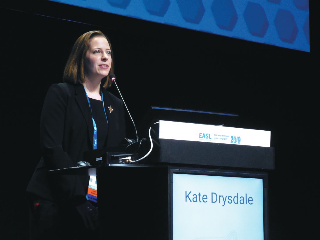
With ribavirin added to a sofosbuvir plus velpatasvir regimen for 12 weeks of treatment, the three-drug combination produced a 98% rate of sustained virologic response after 12 weeks (SVR12) in 196 treated patients, Kate Drysdale, MBBCh, said at the meeting sponsored by the European Association for the Study of the Liver. In contrast, 218 compensated cirrhosis patients who received a 12-week regimen of sofosbuvir plus velpatasvir (Epclusa) but without ribavirin had an SVR12 rate of just under 92%, a statistically significant difference, compared with the rate among patients who also received ribavirin, said Dr. Drysdale, a gastroenterologist at Bart’s Health and Queen Mary University of London. The SVR12 rate among 167 compensated cirrhotic patients treated for 12 weeks with the combination of glecaprevir plus pibrentasvir (Mavyret) was 96%, and not statistically different from the patients who received three drugs including ribavirin. The sofosbuvir, velpatasvir, ribavirin combination also outperformed the combination of sofosbuvir plus daclatasvir (Daklinza) and ribavirin, which produced an SVR12 of 92% in 868 patients. The SVR12 rate is the percentage of patients with undetectable hepatitis C virus (HCV) 12 or more weeks after the end of treatment.
Dr. Drysdale cautioned that the data have not yet been put through a multivariate analysis, but the results so far provide “a strong indication that ribavirin may not be as insignificant” as many have recently presumed. “Ribavirin has been set aside because it was thought not to add to the SVR12, but if patients get only one go at treatment, we must be sure their first treatment is the best one,” Dr. Drysdale said in an interview. If ribavirin can be shown to make a significant contribution to treatment efficacy “then we should think more widely about using it when patients tolerate it.”
The analysis included too few patients with either current decompensated cirrhosis or a history of decompensated cirrhosis to make any statistically meaningful comparisons of the treatment subgroups among these patients. And among patients with genotype 3 HCV infection and without cirrhosis, none of the treatments used in practice showed any statistically significant differences in the SVR12 rates they produced. Among patients without cirrhosis the most commonly used regimens by far were an 8-week course of glecaprevir plus pibrentasvir in 731 patients or a 12-week course of sofosbuvir plus velpatasvir in 1,184 patients. Both regimens had SVR12 rates in noncirrhotic patients of 97%, regardless of whether patients had no, mild, or moderate liver fibrosis.
The study used data collected in an English national registry of HCV-infected patients treated with direct-acting antiviral drugs starting in 2017. Dr. Drysdale and her associates narrowed down the total database of more than 37,000 English adults who received some HCV therapy during the period to 14,603 who received a complete, valid regimen and had follow-up SVR12 information available. The overall SVR12 rate among all these patients was 95.59%, and among the patients infected by genotype 3 virus the SVR12 rate was 95.03%. Dr. Drysdale’s analysis focused primarily on the roughly one-third of patients in the study group infected with genotype 3 HCV, the genotype that historically has presented unique treatment challenges (Drugs. 2017 Feb;77[2]:131-44).
Another finding Dr. Drysdale reported was that as liver disease severity worsened from no fibrosis to mild or moderate fibrosis, and then to compensated cirrhosis or decompensation, the SVR12 rate steadily diminished. Among genotype 3 patients, the SVR12 rate fell from about 97% among patients without any fibrosis to about 87% among those with decompensated cirrhosis. Although this observation had been made before, this finding in such a large number of treated patients adds significant new evidence to support this pattern. It also adds further support to the idea of screening for HCV infection among higher-risk, asymptomatic people to optimize their prospects for virus eradication with treatment.
“If patients get much better treatment outcomes before they become cirrhotic then we should try to find these HCV-infected people before they develop symptoms,” Dr. Drysdale said.
Dr. Drysdale reported no disclosures.
SOURCE: Drysdale K et al. J Hepatol. 2019 April;70(1):e131.
VIENNA – In patients with compensated cirrhosis infected with genotype 3 hepatitis C virus, adding ribavirin to a usual antiviral regimen of sofosbuvir and velpatasvir significantly boosted the rate of sustained virologic response in a review of more than 14,000 English residents entered in a national registry starting in 2017.
With ribavirin added to a sofosbuvir plus velpatasvir regimen for 12 weeks of treatment, the three-drug combination produced a 98% rate of sustained virologic response after 12 weeks (SVR12) in 196 treated patients, Kate Drysdale, MBBCh, said at the meeting sponsored by the European Association for the Study of the Liver. In contrast, 218 compensated cirrhosis patients who received a 12-week regimen of sofosbuvir plus velpatasvir (Epclusa) but without ribavirin had an SVR12 rate of just under 92%, a statistically significant difference, compared with the rate among patients who also received ribavirin, said Dr. Drysdale, a gastroenterologist at Bart’s Health and Queen Mary University of London. The SVR12 rate among 167 compensated cirrhotic patients treated for 12 weeks with the combination of glecaprevir plus pibrentasvir (Mavyret) was 96%, and not statistically different from the patients who received three drugs including ribavirin. The sofosbuvir, velpatasvir, ribavirin combination also outperformed the combination of sofosbuvir plus daclatasvir (Daklinza) and ribavirin, which produced an SVR12 of 92% in 868 patients. The SVR12 rate is the percentage of patients with undetectable hepatitis C virus (HCV) 12 or more weeks after the end of treatment.
Dr. Drysdale cautioned that the data have not yet been put through a multivariate analysis, but the results so far provide “a strong indication that ribavirin may not be as insignificant” as many have recently presumed. “Ribavirin has been set aside because it was thought not to add to the SVR12, but if patients get only one go at treatment, we must be sure their first treatment is the best one,” Dr. Drysdale said in an interview. If ribavirin can be shown to make a significant contribution to treatment efficacy “then we should think more widely about using it when patients tolerate it.”
The analysis included too few patients with either current decompensated cirrhosis or a history of decompensated cirrhosis to make any statistically meaningful comparisons of the treatment subgroups among these patients. And among patients with genotype 3 HCV infection and without cirrhosis, none of the treatments used in practice showed any statistically significant differences in the SVR12 rates they produced. Among patients without cirrhosis the most commonly used regimens by far were an 8-week course of glecaprevir plus pibrentasvir in 731 patients or a 12-week course of sofosbuvir plus velpatasvir in 1,184 patients. Both regimens had SVR12 rates in noncirrhotic patients of 97%, regardless of whether patients had no, mild, or moderate liver fibrosis.
The study used data collected in an English national registry of HCV-infected patients treated with direct-acting antiviral drugs starting in 2017. Dr. Drysdale and her associates narrowed down the total database of more than 37,000 English adults who received some HCV therapy during the period to 14,603 who received a complete, valid regimen and had follow-up SVR12 information available. The overall SVR12 rate among all these patients was 95.59%, and among the patients infected by genotype 3 virus the SVR12 rate was 95.03%. Dr. Drysdale’s analysis focused primarily on the roughly one-third of patients in the study group infected with genotype 3 HCV, the genotype that historically has presented unique treatment challenges (Drugs. 2017 Feb;77[2]:131-44).
Another finding Dr. Drysdale reported was that as liver disease severity worsened from no fibrosis to mild or moderate fibrosis, and then to compensated cirrhosis or decompensation, the SVR12 rate steadily diminished. Among genotype 3 patients, the SVR12 rate fell from about 97% among patients without any fibrosis to about 87% among those with decompensated cirrhosis. Although this observation had been made before, this finding in such a large number of treated patients adds significant new evidence to support this pattern. It also adds further support to the idea of screening for HCV infection among higher-risk, asymptomatic people to optimize their prospects for virus eradication with treatment.
“If patients get much better treatment outcomes before they become cirrhotic then we should try to find these HCV-infected people before they develop symptoms,” Dr. Drysdale said.
Dr. Drysdale reported no disclosures.
SOURCE: Drysdale K et al. J Hepatol. 2019 April;70(1):e131.
REPORTING FROM ILC 2019
2019 CAQ Exam Preparation: Migraine & Headache Overview
The National Headache Foundation is offering a Certificate in Added Qualification (CAQ) exam in Headache Medicine from September 16 through October 1, 2019. In preparation for the CAQ, this supplement to Neurology Reviews walks readers through pertinent topics in migraine and headache that will be covered in the exam.

Benefits of completing the CAQ exam include:
- Validation of a level in expertise in headache medicine
- Possibility of more patient referrals
- Enhanced credibility and satisfaction of providing your patients with the best possible care
- Recognition of skills when dealing with managed care and government agencies
Click here to read the supplement and learn more about CAQ exam enrollment.
The National Headache Foundation is offering a Certificate in Added Qualification (CAQ) exam in Headache Medicine from September 16 through October 1, 2019. In preparation for the CAQ, this supplement to Neurology Reviews walks readers through pertinent topics in migraine and headache that will be covered in the exam.
Benefits of completing the CAQ exam include:
- Validation of a level in expertise in headache medicine
- Possibility of more patient referrals
- Enhanced credibility and satisfaction of providing your patients with the best possible care
- Recognition of skills when dealing with managed care and government agencies
Click here to read the supplement and learn more about CAQ exam enrollment.

Bipolar disorder during pregnancy: Lessons learned
Careful management of bipolar disorder during pregnancy is critical because for so many patients with this illness, the road to emotional well-being has been a long one, requiring a combination of careful pharmacologic and nonpharmacologic strategies.

Half of referrals to our Center for Women’s Mental Health – where we evaluate and treat women before, during, and after pregnancy – are for women who have histories of bipolar disorder. My colleagues and I are asked at continuing medical education programs what we “always do” and “never do” with respect to the treatment of these patients.
What about discontinuation of mood stabilizers during pregnancy and risk of relapse?
We never abruptly stop mood stabilizers if a patient has an unplanned pregnancy – a common scenario, with 50% of pregnancies across the country being unplanned across sociodemographic lines – save for sodium valproate, which is a clearly a documented teratogen; it increases risk for organ malformation and behavioral difficulties in exposed offspring. In our center, we typically view the use of sodium valproate in reproductive age women as contraindicated.
One may then question the circumstances under which lithium might be used during pregnancy, because many clinicians are faced with patients who have been exquisite responders to lithium. Such a patient may present with a history of mania, but there are obvious concerns given the historical literature, and even some more recent reports, that describe an increased risk of teratogenicity with fetal exposure to lithium.
not only to decrease the risk of relapse following discontinuation of mood stabilizers, but because recurrence of illness during pregnancy for these patients is a very strong predictor of risk for postpartum depression. Women with bipolar disorder already are at a fivefold increased risk for postpartum depression, so discussion of sustaining euthymia during pregnancy for bipolar women is particularly timely given the focus nationally on treatment and prevention of postpartum depression.
In patients with history of mania, what about stopping treatment with lithium and other effective treatments during pregnancy?
Historically, we sometimes divided patients with bipolar disorder into those with “more severe recurrent disease” compared with those with more distant, circumscribed disease. In patients with more remote histories of mood dysregulation, we tended to discontinue treatment with mood stabilizers such as lithium or even newer second-generation atypical antipsychotics to see if patients could at least get through earlier stages of pregnancy before going back on anti-manic treatment.
Our experience now over several decades has revealed that this can be a risky clinical move. What we see is that even in patients with histories of mania years in the past (i.e., a circumscribed episode of mania during college in a woman now 35 years old with intervening sustained well-being), discontinuation of treatment that got patients well can lead to recurrence. Hence, we should not confuse an exquisite response to treatment with long periods of well-being as suggesting that the patient has a less severe form of bipolar disorder and hence the capacity to sustain that well-being when treatment is removed.
What about increasing/decreasing lithium dose during pregnancy and around time of delivery?
Select patients may be sensitive to changes in plasma levels of lithium, but the literature suggests that the clinical utility of arbitrarily sustaining plasma levels at the upper limit of the accepted range may be of only modest advantage, if any. With this as a backdrop and even while knowing that increased plasma volume of pregnancy is associated with a fall in plasma level of most medications, we do not arbitrarily increase the dose of lithium across pregnancy merely to sustain a level in the absence of a change in clinical symptoms. Indeed, to my knowledge, currently available data supporting a clear correlation of decline in plasma levels and frank change in symptoms during pregnancy are very sparse, if existent.
Earlier work had suggested that lithium dosage should be reduced proximate to delivery, a period characterized by rapid shifts in plasma volume during the acute peripartum period. Because physicians in our center do not alter lithium dose across pregnancy, we never reduce the dose of lithium proximate to delivery because of a theoretical concern for increased risk of either neonatal toxicity or maternal lithium toxicity, which is essentially nonexistent in terms of systematic reports in the literature.
Obvious concerns about lithium during pregnancy have focused on increased risk of teratogenesis, with the earliest reports supporting an increased risk of Epstein’s anomaly (0.05%-0.1%). More recent reports suggest an increased risk of cardiovascular malformations, which according to some investigators may be dose dependent.
For those patients who are exquisitely responsive to lithium, we typically leave them on the medicine and avail ourselves of current fetal echocardiographic evaluation at 16 weeks to 18 weeks to document the integrity of the fetal cardiac anatomy. Although the risk for cardiac malformations associated with lithium exposure during the first trimester is still exceedingly small, it is still extremely reassuring to patients to know that they are safely on the other side of a teratogenic window.
What about lamotrigine levels across pregnancy?
The last decade has seen a dramatic decrease in the administration of lithium to women with bipolar disorder, and growing use of both lamotrigine and second-generation atypical antipsychotics (frequently in combination) as an alternative. The changes in plasma level of lamotrigine across pregnancy are being increasingly well documented based on rigorous studies (Obstet Gynecol Clin North Am. 2018 Sep;45[3]:403-17).
These are welcome data, but the correlation between plasma concentration of lamotrigine and clinical response is a poor one. To date, there are sparse data to suggest that maintaining plasma levels of lithium or lamotrigine at a certain level during pregnancy changes clinical outcome. Following lamotrigine plasma levels during pregnancy seems more like an academic exercise than a procedure associated with particular clinical value.
As in the case of lithium, we never change lamotrigine doses proximate to pregnancy because of the absence of reports of neonatal toxicity associated with using lamotrigine during the peripartum period. The rationale for removing or minimizing the use of an effective medicine proximate to delivery, a period of risk for bipolar women, is lacking.

In 2019, we clearly are seeing a growing use of atypical antipsychotics for the treatment of bipolar disorder during pregnancy frequently coadministered with medicines such as lamotrigine as opposed to lithium. The accumulated data to date on second-generation atypical antipsychotics are not definitive, but increasingly are reassuring in terms of absence of a clear signal for teratogenicity; hence, our comfort in using this class of medicines is only growing, which is important given the prevalence of use of these agents in reproductive-age women.
If there is a single critical guiding principle for the clinician when it comes to managing bipolar women during pregnancy and the postpartum period, it is sustaining euthymia. With the recent focus of the U.S. Preventive Services Task Force on prevention of postpartum depression, nothing is more helpful perhaps than keeping women with bipolar disorder well, both proximate to pregnancy and during an actual pregnancy. Keeping those patients well maximizes the likelihood that they will proceed across the peripartum and into the postpartum period with a level of emotional well-being that optimizes and maximizes positive long-term outcomes for both patients and families.
Dr. Cohen is the director of the Ammon-Pinizzotto Center for Women’s Mental Health at Massachusetts General Hospital in Boston, which provides information resources and conducts clinical care and research in reproductive mental health. He is also the Edmund and Carroll Carpenter professor of psychiatry at Harvard Medical School. He has been a consultant to manufacturers of psychiatric medications. Email Dr. Cohen at [email protected].
Careful management of bipolar disorder during pregnancy is critical because for so many patients with this illness, the road to emotional well-being has been a long one, requiring a combination of careful pharmacologic and nonpharmacologic strategies.
Half of referrals to our Center for Women’s Mental Health – where we evaluate and treat women before, during, and after pregnancy – are for women who have histories of bipolar disorder. My colleagues and I are asked at continuing medical education programs what we “always do” and “never do” with respect to the treatment of these patients.
What about discontinuation of mood stabilizers during pregnancy and risk of relapse?
We never abruptly stop mood stabilizers if a patient has an unplanned pregnancy – a common scenario, with 50% of pregnancies across the country being unplanned across sociodemographic lines – save for sodium valproate, which is a clearly a documented teratogen; it increases risk for organ malformation and behavioral difficulties in exposed offspring. In our center, we typically view the use of sodium valproate in reproductive age women as contraindicated.
One may then question the circumstances under which lithium might be used during pregnancy, because many clinicians are faced with patients who have been exquisite responders to lithium. Such a patient may present with a history of mania, but there are obvious concerns given the historical literature, and even some more recent reports, that describe an increased risk of teratogenicity with fetal exposure to lithium.
not only to decrease the risk of relapse following discontinuation of mood stabilizers, but because recurrence of illness during pregnancy for these patients is a very strong predictor of risk for postpartum depression. Women with bipolar disorder already are at a fivefold increased risk for postpartum depression, so discussion of sustaining euthymia during pregnancy for bipolar women is particularly timely given the focus nationally on treatment and prevention of postpartum depression.
In patients with history of mania, what about stopping treatment with lithium and other effective treatments during pregnancy?
Historically, we sometimes divided patients with bipolar disorder into those with “more severe recurrent disease” compared with those with more distant, circumscribed disease. In patients with more remote histories of mood dysregulation, we tended to discontinue treatment with mood stabilizers such as lithium or even newer second-generation atypical antipsychotics to see if patients could at least get through earlier stages of pregnancy before going back on anti-manic treatment.
Our experience now over several decades has revealed that this can be a risky clinical move. What we see is that even in patients with histories of mania years in the past (i.e., a circumscribed episode of mania during college in a woman now 35 years old with intervening sustained well-being), discontinuation of treatment that got patients well can lead to recurrence. Hence, we should not confuse an exquisite response to treatment with long periods of well-being as suggesting that the patient has a less severe form of bipolar disorder and hence the capacity to sustain that well-being when treatment is removed.
What about increasing/decreasing lithium dose during pregnancy and around time of delivery?
Select patients may be sensitive to changes in plasma levels of lithium, but the literature suggests that the clinical utility of arbitrarily sustaining plasma levels at the upper limit of the accepted range may be of only modest advantage, if any. With this as a backdrop and even while knowing that increased plasma volume of pregnancy is associated with a fall in plasma level of most medications, we do not arbitrarily increase the dose of lithium across pregnancy merely to sustain a level in the absence of a change in clinical symptoms. Indeed, to my knowledge, currently available data supporting a clear correlation of decline in plasma levels and frank change in symptoms during pregnancy are very sparse, if existent.
Earlier work had suggested that lithium dosage should be reduced proximate to delivery, a period characterized by rapid shifts in plasma volume during the acute peripartum period. Because physicians in our center do not alter lithium dose across pregnancy, we never reduce the dose of lithium proximate to delivery because of a theoretical concern for increased risk of either neonatal toxicity or maternal lithium toxicity, which is essentially nonexistent in terms of systematic reports in the literature.
Obvious concerns about lithium during pregnancy have focused on increased risk of teratogenesis, with the earliest reports supporting an increased risk of Epstein’s anomaly (0.05%-0.1%). More recent reports suggest an increased risk of cardiovascular malformations, which according to some investigators may be dose dependent.
For those patients who are exquisitely responsive to lithium, we typically leave them on the medicine and avail ourselves of current fetal echocardiographic evaluation at 16 weeks to 18 weeks to document the integrity of the fetal cardiac anatomy. Although the risk for cardiac malformations associated with lithium exposure during the first trimester is still exceedingly small, it is still extremely reassuring to patients to know that they are safely on the other side of a teratogenic window.
What about lamotrigine levels across pregnancy?
The last decade has seen a dramatic decrease in the administration of lithium to women with bipolar disorder, and growing use of both lamotrigine and second-generation atypical antipsychotics (frequently in combination) as an alternative. The changes in plasma level of lamotrigine across pregnancy are being increasingly well documented based on rigorous studies (Obstet Gynecol Clin North Am. 2018 Sep;45[3]:403-17).
These are welcome data, but the correlation between plasma concentration of lamotrigine and clinical response is a poor one. To date, there are sparse data to suggest that maintaining plasma levels of lithium or lamotrigine at a certain level during pregnancy changes clinical outcome. Following lamotrigine plasma levels during pregnancy seems more like an academic exercise than a procedure associated with particular clinical value.
As in the case of lithium, we never change lamotrigine doses proximate to pregnancy because of the absence of reports of neonatal toxicity associated with using lamotrigine during the peripartum period. The rationale for removing or minimizing the use of an effective medicine proximate to delivery, a period of risk for bipolar women, is lacking.
In 2019, we clearly are seeing a growing use of atypical antipsychotics for the treatment of bipolar disorder during pregnancy frequently coadministered with medicines such as lamotrigine as opposed to lithium. The accumulated data to date on second-generation atypical antipsychotics are not definitive, but increasingly are reassuring in terms of absence of a clear signal for teratogenicity; hence, our comfort in using this class of medicines is only growing, which is important given the prevalence of use of these agents in reproductive-age women.
If there is a single critical guiding principle for the clinician when it comes to managing bipolar women during pregnancy and the postpartum period, it is sustaining euthymia. With the recent focus of the U.S. Preventive Services Task Force on prevention of postpartum depression, nothing is more helpful perhaps than keeping women with bipolar disorder well, both proximate to pregnancy and during an actual pregnancy. Keeping those patients well maximizes the likelihood that they will proceed across the peripartum and into the postpartum period with a level of emotional well-being that optimizes and maximizes positive long-term outcomes for both patients and families.
Dr. Cohen is the director of the Ammon-Pinizzotto Center for Women’s Mental Health at Massachusetts General Hospital in Boston, which provides information resources and conducts clinical care and research in reproductive mental health. He is also the Edmund and Carroll Carpenter professor of psychiatry at Harvard Medical School. He has been a consultant to manufacturers of psychiatric medications. Email Dr. Cohen at [email protected].
Opicapone increased on-time without dyskinesia in patients with Parkinson’s disease
PHILADELPHIA -
The 2-hour improvement was considered clinically meaningful, although the average patient in the studies had about 6 hours of off-time, said investigator Peter LeWitt, MD, of Henry Ford Hospital in West Bloomfield, Mich., and the department of neurology at Wayne State University, Detroit. Dr. LeWitt and colleagues will present the data at the annual meeting of the American Academy of Neurology.
“While this is a substantial improvement, it is 2 hours improvement over a total of 6 hours of off-time, which is not perfect,” Dr. LeWitt said in an interview. “So how could we do better is the challenge for all of us who are doing research.”
Opicapone is under development in the United States; it is currently approved in the European Union as adjunctive therapy to preparations of levodopa/DOPA decarboxylase inhibitors for patients with Parkinson’s disease and end-of-dose motor fluctuations.
The ability of opicapone to prolong the clinical actions of levodopa has been evaluated in BIPARK-1 and BIPARK-2. These two international phase 3 studies evaluated the third-generation COMT inhibitor against placebo and, in the case of BIPARK-1, against the COMT inhibitor entacapone as an active control. Each study was 14-15 weeks in duration and included a 1-year open-label phase.
In BIPARK-1, on-time without troublesome dyskinesia was significantly increased for opicapone 50 mg versus placebo, with an absolute increase of 1.9 versus 0.9 hours, respectively, from baseline to week 14 or 15 (P = .002), investigators said. Similarly, BIPARK-2 data showed an increase in this endpoint, at 1.7 versus 0.9 hours for opicapone and placebo, respectively (P = .025).
The 50-mg dose of opicapone was received by 115 patients in BIPARK-1 and 147 patients in BIPARK-2, while placebo was received by 120 and 135 patients in those two studies, respectively.
In the long-term extension studies, the mean change in on-time without dyskinesia from baseline to the end of the open-label endpoint was 2.0 hours for all 494 opicapone-treated patients in BIPARK-1 and 1.8 hours for all 339 opicapone-treated patients in BIPARK-2.
Dyskinesia was reported as a treatment-emergent adverse effect for 17.4% of opicapone-treated patients and 6.2% of placebo-treated patients, according to results of a pooled safety analysis of BIPARK-1 and BIPARK-2. However, only 1.9% of opicapone-treated patients and 0.4% of placebo-treated patients had treatment-emergent dyskinesia leading to discontinuation, and the dyskinesia was considered serious in 0.3% of the opicapone group and 0.0% of the placebo group, investigators added.
Neurocrine Biosciences has announced plans to file a New Drug Application for opicapone for Parkinson’s disease in the United States. That filing is expected to take place in the second quarter of 2019, according to an April 29 press release.
Dr. LeWitt disclosed that he has served as an advisor to Neurocrine Biosciences. He also provided disclosures related to Acadia, Acorda, Adamas, BioElectron Technology, Biotie, Britannia, Intec, Jazz Pharmaceuticals, Lundbeck, the Michael J. Fox Foundation for Parkinson’s Research, Merz, NeuroDerm, the Parkinson Study Group, Pfizer, Prexton, Sage, Scion, Sunovion, SynAgile, and US WorldMeds.
SOURCE: LeWitt P et al. AAN 2019, Abstract S4.003.
FROM AAN 2019
No benefit to infants from e-books over board books
BALTIMORE – Despite the greater portability and seeming convenience of electronic tablets or smartphones, compared with giving them physical board books, according to preliminary findings.

James Guevara, MD, MPH, professor of pediatrics at the Children’s Hospital of Philadelphia, presented his findings at the annual meeting of Pediatric Academic Societies in a session focused on the changing nature of children’s digital media use as digital natives.
The session chair, Danielle C. Erkoboni, MD, of the University of Pennsylvania, Philadelphia, opened the session with a review of research to date regarding children’s frequency of digital use and relative benefits of independent and shared reading, both with traditional books and e-books.
Nearly all U.S. children (98%) live in homes with mobile devices, and about a third of U.S. children (35%) use those devices, according to a 2017 Common Sense Media Report. The same survey found that children aged under 2 years get an average 42 minutes a day of overall screen time, but children in low-income households get twice as much screen time as those in middle- and upper-income homes.
Research into e-books exists for preschoolers, showing that animated pictures and sounds directly matching an e-book’s story text can potentially promote language memory, but that too many interactive features or other bells and whistles can overwhelm children and contribute to poor vocabulary development and comprehension.
Researchers also have found that parent reading of e-books has greater benefits for children than children listening to an e-book’s audio narration. However, little to no research has looked into e-reading for infants and toddlers, a gap especially relevant for physicians who participate in the Reach Out and Read program.
Dr Guevara’s study enrolled 100 Medicaid-eligible children aged 5-7 months from three participating practices in a single geographic area. Only English- or Spanish-speaking parents who owned a smartphone or tablet participated.
At the children’s 6-month well visit, the clinicians gave parents information about the importance of early parent-child reading. Then parents received Dr. Seuss’s “Hop on Pop” as either an e-book download (n = 45) or a physical board book (n = 54). The parents similarly received “Barnyard Dance” at the 9-month visit and “Goodnight Moon” at the 12-month visit.
No significant differences between the two groups existed in terms of race/ethnicity, parent age, household income, education level, marital status or the total number of adults or children in the household. Maternal depression rates (based on the Edinburgh Postnatal Depression Scale), Adverse Childhood Experience scale scores, health literacy scores (Short Assessment of Health Literacy) and scores on the StimQ home cognitive environment assessment also were similar at baseline between the two groups.
The children were assessed with the StimQ Infant measure at 7-8 months and 10-11 months, and then with the StimQ Toddler and Bayley Scale of Infant and Toddler Development (BSID-III) at 13-15 months. At the final check-up, 37 parents remained in the e-book group and 43 parents remained in the board book group.
A similar proportion of parents in both groups reported reading to their children, and parents in both groups said they read an average of 5 days a week to their child.
However, parents who received the e-books had read an average 18 books at the final follow-up, compared with an average of 31 books among parents who received the board books (P = .039).
No significant differences in children’s StimQ scores or any of the BSID-III scores (cognitive, language, motor) existed between those who received e-books versus those who received board books.
The study results showed the feasibility of promoting literacy by providing e-books to parents of older infants, but doing so did not appear to confer any advantage over providing families with traditional, physical board books. Further, language development among children in both groups remained below average, albeit not statistically different from one another.
“Pediatric clinicians should exercise caution in recommending e-books to parents of young children.” Dr Guevara said. “Additional strategies beyond clinic-based literacy promotion are needed to enhance language development among poor children.”
The research was funded by the Vanguard Strong Start for Kids Program. Dr. Guevara reported no disclosures.
BALTIMORE – Despite the greater portability and seeming convenience of electronic tablets or smartphones, compared with giving them physical board books, according to preliminary findings.
James Guevara, MD, MPH, professor of pediatrics at the Children’s Hospital of Philadelphia, presented his findings at the annual meeting of Pediatric Academic Societies in a session focused on the changing nature of children’s digital media use as digital natives.
The session chair, Danielle C. Erkoboni, MD, of the University of Pennsylvania, Philadelphia, opened the session with a review of research to date regarding children’s frequency of digital use and relative benefits of independent and shared reading, both with traditional books and e-books.
Nearly all U.S. children (98%) live in homes with mobile devices, and about a third of U.S. children (35%) use those devices, according to a 2017 Common Sense Media Report. The same survey found that children aged under 2 years get an average 42 minutes a day of overall screen time, but children in low-income households get twice as much screen time as those in middle- and upper-income homes.
Research into e-books exists for preschoolers, showing that animated pictures and sounds directly matching an e-book’s story text can potentially promote language memory, but that too many interactive features or other bells and whistles can overwhelm children and contribute to poor vocabulary development and comprehension.
Researchers also have found that parent reading of e-books has greater benefits for children than children listening to an e-book’s audio narration. However, little to no research has looked into e-reading for infants and toddlers, a gap especially relevant for physicians who participate in the Reach Out and Read program.
Dr Guevara’s study enrolled 100 Medicaid-eligible children aged 5-7 months from three participating practices in a single geographic area. Only English- or Spanish-speaking parents who owned a smartphone or tablet participated.
At the children’s 6-month well visit, the clinicians gave parents information about the importance of early parent-child reading. Then parents received Dr. Seuss’s “Hop on Pop” as either an e-book download (n = 45) or a physical board book (n = 54). The parents similarly received “Barnyard Dance” at the 9-month visit and “Goodnight Moon” at the 12-month visit.
No significant differences between the two groups existed in terms of race/ethnicity, parent age, household income, education level, marital status or the total number of adults or children in the household. Maternal depression rates (based on the Edinburgh Postnatal Depression Scale), Adverse Childhood Experience scale scores, health literacy scores (Short Assessment of Health Literacy) and scores on the StimQ home cognitive environment assessment also were similar at baseline between the two groups.
The children were assessed with the StimQ Infant measure at 7-8 months and 10-11 months, and then with the StimQ Toddler and Bayley Scale of Infant and Toddler Development (BSID-III) at 13-15 months. At the final check-up, 37 parents remained in the e-book group and 43 parents remained in the board book group.
A similar proportion of parents in both groups reported reading to their children, and parents in both groups said they read an average of 5 days a week to their child.
However, parents who received the e-books had read an average 18 books at the final follow-up, compared with an average of 31 books among parents who received the board books (P = .039).
No significant differences in children’s StimQ scores or any of the BSID-III scores (cognitive, language, motor) existed between those who received e-books versus those who received board books.
The study results showed the feasibility of promoting literacy by providing e-books to parents of older infants, but doing so did not appear to confer any advantage over providing families with traditional, physical board books. Further, language development among children in both groups remained below average, albeit not statistically different from one another.
“Pediatric clinicians should exercise caution in recommending e-books to parents of young children.” Dr Guevara said. “Additional strategies beyond clinic-based literacy promotion are needed to enhance language development among poor children.”
The research was funded by the Vanguard Strong Start for Kids Program. Dr. Guevara reported no disclosures.
REPORTING FROM PAS 2019