User login
Commentary: New Research on BC Chemotherapies, March 2024

The phase 3 KEYNOTE-355 trial established the role of chemotherapy in combination with pembrolizumab in the first-line setting for programmed death-ligand 1 (PD-L1)–positive advanced triple-negative breast cancer (TNBC). Patients unselected for PD-L1 status in this trial who received platinum- or taxane-based chemotherapy with placebo had a median progression-free survival of 5.6 months.[3] Strategies to improve upon efficacy and tolerability are desired in this space, and various trials have evaluated "switch maintenance" that involves receipt of an intensive induction regimen followed by a switch to an alternative/more tolerable regimen after response is achieved.[4] The phase II DORA trial randomized 45 patients with advanced TNBC and ongoing stable disease or complete or partial response from first- or second-line platinum-based chemotherapy to a maintenance regimen of olaparib (300 mg orally twice daily) with or without durvalumab (1500 mg on day 1 and every 4 weeks) (Tan et al). At a median follow-up of 9.8 months, median progression-free survival was 4.0 months (95% CI 2.6-6.1) with olaparib and 6.1 months (95% CI 3.7-10.1) with the combination; both were significantly longer than the historical control of continued platinum-based therapy (P = .0023 and P < .0001, respectively). Durable disease control appeared more pronounced in patients with complete or partial response to prior platinum therapy, and no new safety signals were observed. Future efforts to study this approach include the phase 2/3 KEYLYNK-009 trial, which is evaluating olaparib plus pembrolizumab maintenance therapy after first-line chemotherapy plus pembrolizumab for TNBC.[5]
TNBC is a heterogenous subtype, characterized by aggressive biology, and it benefits from chemotherapy and immunotherapy treatment approaches. Presently, the management of early-stage TNBC often involves neoadjuvant systemic therapy; however, a proportion of patients receive treatment in the postoperative setting, highlighting the relevance of time to initiation of adjuvant therapy as well.[6] Various prior studies have showed that delayed administration of adjuvant chemotherapy for EBC can lead to adverse survival outcomes. Furthermore, this effect is subtype-dependent, with more aggressive tumors (luminal B, triple-negative, human epidermal growth factor receptor 2 [HER2]-positive) exhibiting inferior outcomes with delayed chemotherapy.[7] A retrospective cohort study that included 245 patients with early TNBC who received adjuvant chemotherapy after surgery evaluated the impact of time to initiation of adjuvant therapy in this population (Hatzipanagiotou et al). Superior survival outcomes were observed for the group receiving systemic therapy 22-28 days after surgery (median overall survival 10.2 years) compared with those receiving adjuvant chemotherapy at later time points (29-35 days, 36-42 days, and >6 weeks after surgery; median overall survival 8.3 years, 7.8 years, and 6.9 years, respectively). Patients receiving chemotherapy 22-28 days after surgery had significantly better survival than those receiving chemotherapy 29-35 days (P = .043) and >6 weeks (P = 0.033) postoperatively. This study emphasizes the importance of timely administration of adjuvant chemotherapy for early TNBC, and efforts aimed to identify potential challenges and propose solutions to optimize outcomes in this space are valuable.
Additional References
- Gnant M, Frantal S, Pfeiler G, et al, for the Austrian Breast & Colorectal Cancer Study Group. Long-term outcomes of adjuvant denosumab in breast cancer. NEJM Evid. 2022;1:EVIDoa2200162. doi: 10.1056/EVIDoa2200162 Source
- Fassio A, Idolazzi L, Rossini M, et al. The obesity paradox and osteoporosis. Eating and Weight Disorders - Studies on Anorexia, Bulimia and Obesity. 2018;23:293-30 doi: 10.1007/s40519-018-0505-2 Source
- Cortes J, Cescon DW, Rugo HS, et al, for the KEYNOTE-355 Investigators. Pembrolizumab plus chemotherapy versus placebo plus chemotherapy for previously untreated locally recurrent inoperable or metastatic triple-negative breast cancer (KEYNOTE-355): A randomised, placebo-controlled, double-blind, phase 3 clinical trial. Lancet. 2020;396:1817-1828. doi: 10.1016/S0140-6736(20)32531-9 Source
- Bachelot T, Filleron T, Bieche I, et al. Durvalumab compared to maintenance chemotherapy in metastatic breast cancer: The randomized phase II SAFIR02-BREAST IMMUNO trial. Nat Med. 2021;27:250-255. doi: 10.1038/s41591-020-01189-2 Source
- Saji S, Cussac AL, Andre F, et al. 68TiP KEYLYNK-009: a phase II/III, open-label, randomized study of pembrolizumab (pembro) + olaparib (ola) vs pembro + chemotherapy after induction with first-line (1L) pembro + chemo in patients (pts) with locally recurrent inoperable or metastatic TNBC (abstract). Ann Oncol. 2020;31(Suppl 6):S1268. doi: 10.1016/j.annonc.2020.10.088 Source
- Ortmann O, Blohmer JU, Sibert NT, et al for 55 breast cancer centers certified by the German Cancer Society. Current clinical practice and outcome of neoadjuvant chemotherapy for early breast cancer: Analysis of individual data from 94,638 patients treated in 55 breast cancer centers. J Cancer Res Clin Oncol. 2023;149:1195-1209. doi: 10.1007/s00432-022-03938-x Source
- Yu KD, Fan L, Qiu LX, et al. Influence of delayed initiation of adjuvant chemotherapy on breast cancer survival is subtype-dependent. Oncotarget. 2017;8:46549-46556. doi: 10.18632/oncotarget.10551 Source
Gout Increases the Risk for a Wide Range of Cardiovascular Diseases
People with gout are 58% more likely to develop cardiovascular disease (CVD), according to a new analysis. This increased risk was observed across 12 different cardiovascular conditions, including heart failure, arrhythmias, and valve diseases.
“These findings suggest that the organ damage associated with gout is likely to be much broader than originally thought,” Nathalie Conrad, PhD, senior author of the research and cardiovascular epidemiologist at KU Leuven, Leuven, Belgium, said in an email. This could be useful for future research on underlying biological mechanisms driving CVD risk in gout, she added.
While previous research has tied gout to increased cardiovascular risk, these studies “largely focused on coronary heart disease, stroke, and thromboembolic outcomes,” she explained, and have been smaller in size.
This new study included more than 862,000 individuals, which permitted researchers to investigate rarer CVD outcomes such as myocarditis and pericarditis.
For the study, researchers used electronic health records from the UK Clinical Practice Research Datalink, a primary care database that contains anonymized health data for about 22 million individuals. Using these data, they identified more than 152,600 individuals with gout. Patients included in the analysis were diagnosed between 2000 and 2017, younger than 80 years at diagnosis, and free of CVD for at least 12 months after their gout diagnosis.
Patients with gout were compared with nearly 710,000 controls, matched on demographic factors such as age, sex, and geographic region.
Researchers then investigated the incidence of 12 CVDs, including atherosclerotic diseases, degenerative and thromboembolic diseases, and arrythmias, between the two groups from January 1, 2000, to June 30, 2019.
The findings were published in the March 2024 issue of The Lancet Rheumatology. Overall, patients with gout were 58% more likely to develop any CVD than their matched comparators without gout. There was a higher disease incidence among patients with gout for each of the 12 conditions. This association was more pronounced in women (hazard ratio [HR], 1.88) than in men (HR, 1.49), and gout amplified the risk for CVD in younger individuals to a greater extent.
Individuals younger than 45 years with gout were more than twice as likely to develop CVD compared with similarly aged individuals without gout. For comparison, individuals aged 45-54 years with gout were 84% more likely to develop CVD, and individuals aged 55-64 years were 57% more likely to develop CVD than matched controls.
Conduction system disease had the highest incident risk (HR, 1.88), followed by heart failure and valve disease (HR, 1.85 for both).
Individuals with gout had higher rates of comorbidities than the controls, including hypertension, obesity, and dyslipidemia. Overall, CVD risk was slightly attenuated after adjustment for traditional CVD risk factors such as smoking, blood pressure, and body mass index but still significant: Patients with gout had a 31% higher risk for CVD than comparators.
This shows “that known CVD risk factors only explain part of the CVD risks seen in patients with gout,” Dr. Conrad said. Other factors such as inflammation and other disease activity factors could be at play, she explained, which would need to be explored in future research.
The study “shows the whole landscape” of CVD and gout, Michael H. Pillinger, MD, rheumatologist and professor of medicine, biochemistry, and molecular pharmacology at NYU Grossman School of Medicine in New York City, said in an interview. He was not involved with the research.
“Every possible cardiovascular disease that they could think of was something that gout patients had more of than the non-gout patients,” he added. “I think this is going to be a paper that gets cited a lot, at minimum when describing the background of risk when we look at gout patients.”
The study had some limitations, including that researchers were unable to account for how medications such as nonsteroidal anti-inflammatory drugs, corticosteroids, colchicine, or allopurinol may have affected the association between gout and CVD.
“This is because analyses of nonrandomized treatment can be confounded by indication, wherein it is difficult to differentiate the effects of the treatment from underlying disease severity,” the authors wrote.
There was also a large amount of missing data on blood pressure, body mass index, smoking status, and other health information relevant to cardiovascular risk, so sensitivity analyses adjusting for these factors “should be interpreted with caution,” they added.
Dr. Pillinger also noted that the rates of comorbidities in the gout study population were lower than what have been found in US study populations. For example, about 40% of patients with gout in the analysis had hypertension, while other studies have suggested higher rates of 60%-70%, he said. However, it’s not clear if these differences could have affected outcomes. He added that these limitations do not “in any way weaken [the authors’] conclusion.”
The findings call for better strategies to reduce CVD risk in patients with gout, Dr. Conrad noted.
“Further improvements could come from better recognition and intervention on CVD risk factors (eg, through lifestyle changes or drug therapies where they are indicated), as well as proactive screening for heart disease in patients with gout, which could allow early diagnosis and interventions to delay more severe outcomes,” she added.
This study was funded by Research Foundation Flanders. Dr. Conrad was funded by a personal fellowship from the Research Foundation Flanders and a European Society of Cardiology research grant. She received royalties from Oxford University Innovation. Four of Dr. Conrad’s eight coauthors also reported financial relationships with pharmaceutical companies. Dr. Pillinger served as a consultant to Amgen, Federation Bio, Fortress Biotech, and Scilex, and he holds an investigator-initiated grant from Hikma.
A version of this article appeared on Medscape.com.
Is Stretching Now Underrated? Accumulating Research Says Yes
For many, stretching is the fitness equivalent of awkward small talk. It’s the opening act, the thing you tolerate because you know it will be over soon.
Others have challenged the practice, suggesting that stretching isn’t necessary at all. Some research has found that a preworkout stretch may even be disadvantageous, weakening muscles and hindering performance.
To put it plainly, no one seems terribly enthusiastic about touching their toes.
That’s why a 2020 study on exercise and mortality was such a head-scratcher. The study found that stretching was uniquely associated with a lower risk for all-cause mortality among American adults. That’s after controlling for participation in other types of exercise.
The finding seemed like a fluke, until a 2023 study found essentially the same thing.
That was slightly better than the risk reduction associated with high volumes of aerobic exercise and resistance training.
How can that be ? It turns out, stretching is linked to several health benefits that you might not expect.
The Surprising Benefits of Stretching
When we talk about stretching, we usually mean static stretching — getting into and holding a position that challenges a muscle, with the goal of improving range of motion around a joint.
It doesn’t need to be a big challenge. “Research shows you can get increases in flexibility by stretching to the initial point of discomfort,” said David Behm, PhD, an exercise scientist at Memorial University of Newfoundland in Canada who’s published dozens of studies on stretching over the past quarter-century.
That brings us to the first benefit.
Stretching Benefit #1: More Strength
At first glance, flexibility training and strength training have little in common. You lengthen muscles in the former and contract them in the latter.
But in both cases, Dr. Behm said, you’re applying tension to muscles and connective tissues. Tension activates proteins called integrins, which send and receive signals across cellular membranes. Those signals are the start of a cascade that leads to protein synthesis. That’s how muscles get bigger and stronger when you lift weights.
That mechanism could explain the small gains in muscle strength and size associated with static stretching, Dr. Behm said.
But can you really stretch your way to muscle growth? Theoretically, yes. But strength training is far more time-efficient, Dr. Behm says. Studies showing increases in muscle mass have typically stretched a single muscle (usually the calves, using a specialized device) for > 30 min/session, 6 d/wk for 6 weeks. And that’s for just one leg.
Still, stretching may be more accessible for some patients — research suggested that older and more sedentary people are most likely to benefit from stretching-induced gains in strength.
Stretching Benefit #2: Reduced Arterial Stiffness
“Most people don’t think about the cardiovascular benefits of stretching,” Dr. Behm said. There are some big ones.
If your body doesn’t move well, it’s not unreasonable to assume your blood doesn’t flow well. That is indeed the case: Poor flexibility is associated with arterial stiffness.
Stretching is associated not only with improved arterial function but also with reductions in resting heart rate and blood pressure and increased vasodilation.
Mobility improvements may have an indirect benefit on cardiovascular health as well.
“Studies show runners are more economical when they’re more flexible,” Dr. Behm said. If your movement is more efficient, you’ll probably do more of it. Doing more, in turn, would lead to improved fitness.
Stretching Benefit #3: Improved Performance
Research is equivocal on whether stretching improves athletic performance, said Joe Yoon, a sports massage therapist in Orlando, Florida, and author of Better Stretching.
“But I’ve always taken the approach that if you can improve your range of motion and get into positions” required for your sport, you’ll probably perform better, with less risk for injury, Mr. Yoon said.
It’s worth noting that some research over the past 30 years has linked pre-exercise static stretching with a loss of strength, power, and/or speed.
But consider this: In a 2016 review, Dr. Behm and his coauthors showed that performance reductions were most likely to occur in two situations:
When participants did extremely long stretches (duration, ≥ 60 sec per muscle).
When researchers tested the participants’ strength, power, or speed immediately after they stretched.
Avoiding those problems is easy, Dr. Behm said: Stretch each muscle for < 60 sec, and combine static stretches with more active warm-up exercises.
“Stretching can impair your performance but only if you do it wrong,” he said.
Stretching Benefit #4: Fewer Injuries
When you stretch, the point where you feel tension is where the muscle is most vulnerable. “That’s where injuries usually happen,” Dr. Behm said.
More flexibility in those areas allows your muscles to safely generate force at longer lengths. For an athlete, that means fewer injuries when they’re doing explosive movements or changing direction.
For nonathletes, flexibility reduces injuries by improving balance. Better balance reduces the risk of falling and helps mitigate the damage if you do take a tumble.
Help Your Patients Get the Benefits of Stretching
Stretching, like training for endurance or strength, can be as complex as you want to make it. But Mr. Yoon advocates a simpler approach.
“You see this flashy stuff online,” he said. “But if you see those trainers in real life or you book a session with them, they go right back to the basics.”
Ideally, Mr. Yoon said, a flexibility routine will work the entire body. But if that’s too big a stretch for your patient, he recommends starting with one or two stretches for the most problematic area.
For example, for a stiff back, try doing the puppy pose at least once a day, although twice is better. Hold the position for 30 seconds to 2 minutes, said Mr. Yoon. Even if you combine it with a dynamic movement like the cat-cow, the two exercises would take just a few minutes a day.
“There’s this misconception that you have to do a lot of it to be successful,” Mr. Yoon said.
Consistency is far more important than volume. Mr. Yoon recommends “a little bit every day — the minimum viable dose.”
As a bonus, stretching an area like your upper back will probably improve your shoulder mobility, Mr. Yoon said. Same with your lower body: Stretches for your hips, over time, should also benefit your knees and lower back.
And thanks to a phenomenon called nonlocal flexibility transfer, lower-body stretches should improve upper-body flexibility, at least temporarily. Shoulder stretches can also have an immediate effect on hip mobility.
“It’s all connected,” Mr. Yoon said, which brings us back to where we started.
If stretching can indeed reduce mortality risk, it’s probably because of interconnected pathways, rather than any single mechanism.
Most obviously, stretching improves flexibility, which makes movement easier, improves balance, and reduces the risk for falls and other types of injuries. It can also lead to small improvements in strength. Less obviously, stretching improves several aspects of cardiovascular function, including circulation.
“There seems to be a global effect in everything we do,” Dr. Behm said. “Whether you’re stretching or weight training, the message is sent throughout your body."
A version of this article appeared on Medscape.com.
Commentary: Medication Timing and Other Dupilumab Concerns, March 2024
When skin diseases affect the palm or sole, they can have a disproportionately large negative effect on patients' lives. Hand and foot dermatitis can be disabling. Simpson and colleagues find that dupilumab is an effective treatment for AD of the hands and feet. Having safe and effective treatment for hand and foot dermatitis will be life-changing for many of our patients.
Patients often do very well with biologic treatment. When they do, they often wonder, Do I need to continue taking the medication? Lasheras-Pérez and colleagues found that the great majority of patients doing well taking dupilumab for AD could stretch out their dosing interval. I suspect a lot of our patients are doing this already. I used to worry that stretching out the dosing interval might lead to antidrug antibodies and loss of activity. Such loss of activity doesn't appear common. Because we also have multiple alternative treatments for severe AD, I think it may be quite reasonable for patients to try spreading out their doses after their disease has been well controlled for a good long time.
Superficial skin infections aren't rare in children, particularly children with AD. Paller and colleagues' study is informative about the safety of dupilumab in children. The drug, which blocks a pathway of the immune system, was associated with fewer infections. This is good news. The reduction in infections could be through restoring "immune balance" (whatever that means) or by improving skin barrier function. Perhaps the low rate of infection explains why dupilumab is not considered immunosuppressive.
I love studies of drug survival because I think that knowing the percentage of patients who stay with drug treatment is a good measure of overall safety and efficacy. Pezzolo and colleagues found — perhaps not surprisingly given the extraordinary efficacy of upadacitinib for AD — that almost no one discontinued the drug over 1.5 years due to lack of efficacy. There were patients who discontinued due to adverse events (and additional patients lost to follow-up who perhaps also discontinued the drug), but 80% of patients were still in the study at the end of 1.5 years. Three patients who weren't vaccinated for shingles developed shingles; encouraging patients to get the shingles vaccine may be a prudent measure when starting patients taking upadacitinib.
Scents May Improve Memory in Major Depression
TOPLINE:
Scents that trigger specific, vivid autobiographical memories (AMs) could improve deficits in memory recall in patients with major depressive disorder (MDD), new research suggests.
METHODOLOGY:
- Participants included 32 individuals aged 18-55 years (mean age, 30 years; 26 females) with a diagnosis of MDD recruited from the community.
- Those with psychosis, bipolar I or II, neurological disorders, or drug or alcohol abuse were excluded.
- Participants were presented with a series of 12 words and 12 odors, such as cough syrup, tobacco ash, and Vicks VapoRub, and asked to recall a specific memory in response to each cue.
- AMs were rated in terms of vividness, frequency, and whether they were associated with positive or negative emotions.
TAKEAWAY:
- Although participants only guessed correct stimulus odors 30% of the time, they recalled more specific memories from odor cues than from word cues (68% vs 52%; P < .001).
- Odor-cued recall was more arousing and vivid (P < .001) than recall responses generated by word cues.
- Compared with the population mean for responses to word cues in healthy controls, study participants recalled fewer specific memories in response to words (P < .001), but the percentage of specific memories recalled in response to odor cues did not differ from the healthy control population mean.
- Investigators hoped to further their research by investigating the mechanisms underlying odor-cued AMs, particularly to test if the amygdala and hippocampus are activated during recall.
IN PRACTICE:
“This study suggests the potential for increasing autobiographical memory specificity in individuals with MDD, with the future goal of reducing depression symptoms for this population and informing a better understanding of the neural mechanisms influencing odor-based AM recall,” the authors wrote. “We hope this initial study spurs larger studies in more diverse samples that include healthy control participants to further investigate and explain these associations.”
SOURCE:
Kymberly D. Young, PhD, of the University of Pittsburgh, Pennsylvania, led the study, which was published online on February 13, 2024, in JAMA Network Open.
LIMITATIONS:
Study limitations included the lack of a healthy control group and the small sample size.
DISCLOSURES:
The study was funded internally by the University of Pittsburgh School of Medicine, Pennsylvania. No disclosures were reported.
A version of this article appeared on Medscape.com.
Judge Won’t Overturn Invalidated USMLE Scores
(USMLE).
In a February 23 order, Judge Christopher R. Cooper, of the US District Court for the District of Columbia, denied Latika Giri’s emergency motion to block the National Board of Medical Examiners (NBME) from invalidating the scores, ruling the public interest plainly weighs against granting the request.
“First and foremost, is the overriding interest in public safety,” Cooper wrote in his 32-page order. “This is a case about the credentials of doctors applying to medical residency programs…Granting the preliminary injunction would create an unacceptable risk that individuals who lack the requisite knowledge and skills they purport to possess because they achieved their exam scores fraudulently will be administering medical care to unsuspecting patients across the nation.”
Attorneys for Giri did not return messages seeking comment about the order.
The NBME also did not return messages seeking comment. The board previously said it does not comment on pending litigation.
The decision is the latest development in a widespread cheating scandal. Giri, an international medical graduate (IMG) from Kathmandu, sued NBME earlier this month claiming the board discriminated against Nepali medical graduates when it invalidated hundreds of exam scores linked to the country.
Giri also accused NBME of violating its own procedures when it voided the scores before giving examinees a chance to argue and appeal. She asked the district court to block NBME from invalidating her exam scores while the lawsuit continues and restore her original results.
In court documents, NBME argued that it did not invalidate the scores because the examinees were Nepali but because staff concluded that there was “a good faith basis for questioning the validity of the scores.”
The invalidations were based on concerns that the results reflected prior access to secure exam content rather than knowledge and understanding of the medical principles and skills the exams are intended to assess, according to the NBME’s legal response.
“The USMLE program took reasonable and appropriate actions to prevent the significant harm and disruption that would result from allowing potentially unqualified individuals to participate in the 2024 residency Match,” the NBME stated in court documents. “If granted, the requested injunction would cause enormous harm not only to NBME… but also to state licensing authorities, which rely upon USMLE results to help ensure that physicians have the minimum competencies needed to provide safe and effective health care.”
In his order, Cooper wrote that Giri has not proven the board’s actions were discriminatory against Nepali doctors.
“Nothing in the present record suggests that NBME went looking for a problem in Nepal out of ethnicity-or national-origin based [sic] suspicion,” Cooper wrote. “[It] followed the trail of evidence, including tips about organized cheating taking place in medical schools and at a testing center located in Nepal, and on an online forum for which a ‘nexus to Nepal’ was a ticket to admission.”
NBME: Nepal Outperformed All Other Countries on USMLE
Court documents shed more light on NBME’s investigation into the suspected cheating and on the anomalous patterns the board allegedly discovered from Nepal medical graduates.
In response to anonymous tips, the USMLE program in early 2023 asked the NBME Psychometrics and Data Analysis (PADA) unit to analyze examinee performance data for test centers in Jordan, Nepal, and Pakistan, according to court records. Within the initial data analysis, the data involving the single test center in Nepal was “the most extreme,” the unit found.
Out of more than 400 test centers across the world, including those in the United States, the test center in Nepal produced the highest test scores in the world for Step 1 in 2021 and 2022 and the highest test scores in the world for Step 2 CK in 2022, according to court documents. For the 2022 Step 1 exam for example, the average score of examinees testing in the Nepal test center was 240. No other test center in the world had an average examinee score above 227, according to the NBME’s legal response.
The median item response time for examinees who tested at the Nepal test center in 2022 was also among the fastest of all international test centers for Step 1 and Step 2 CK, investigators found.
In addition, the volume of examinees taking the USMLE Step 1 and Step 2 CK at the Nepal test center in Nepal had sharply increased. Step 1 volume more than doubled in the Nepal test center from 281 examinees in 2019 to 662 examinees in 2022, according to court documents.
The rapid increase continued in 2023, when examinee volume was nearly three-and-a-half times higher than the 2019 volume. The data were consistent with anonymous tips received by the USMLE program office, suggesting there may be wide-scale collection and sharing of live USMLE exam content within Nepal.
Investigation Finds Similar Correct and Incorrect Answers
Agreement similarity among the exams analyzed also raised red flags. Investigators ran an “agreement analysis” for all examinees who tested at centers in Jordan, Nepal, and Pakistan as well as two centers in India, according to court documents.
For the 2022 Step 1 exam and the 2021 and 2022 Step 2 CK exam, the analysis showed a substantially higher percentage of examinees with a statistically significant level of agreement matches in the examine group that tested at centers in Jordan, Nepal, Pakistan, and India compared with the baseline group, according to legal records.
The vast majority of examinees with a statistically significant number of matching incorrect answers tested at the Nepal test center, data showed.
Further analysis found that examinee volumes increased considerably at the Nepal test center in the months prior to the USMLE program releasing new test items, “suggesting that candidates who had prior access to disclosed exam questions wanted to test before new questions came into the item pool.”
Investigators also identified posts on social medial and in online chat rooms suggesting groups were collecting and sharing large amounts of secure exam material in private groups. Some posts advised examinees to use the full examination time when taking the USMLE “to avoid raising suspicion about having had prior access to secure exam materials,” according to court documents.
From its investigation and analysis, the USMLE program identified 832 examinees who had passing level exam results whose validity the USMLE program had a significant and good faith basis for questioning, according to court records.
Of the total, 618 examinees had one Step score flagged as being of questioned validity, 202 examinees had two Step exam scores flagged, and 12 examinees had scores flagged on all three Step exams.
NBME Defends Departure From Traditional Procedures
In court documents, NBME disputed claims that it violated its own procedures by invalidating the exam scores. Giri’s report contends that examinees suspected of cheating are typically first advised of the matter, given an opportunity to share relevant information, and provided the right to appeal — during which time, their scores are treated as valid.
But the NBME said the USMLE program is authorized to take any actions it deems appropriate in response to concerns regarding score validity if the USMLE Committee for Individualized Review or the USMLE Composite Committee concludes that alternative or supplemental procedures are warranted in response to a given set of facts or circumstances.
“Following the month-long investigation and analysis…the USMLE program concluded that alternative procedures were warranted to address the score invalidity concerns identified in the interest of providing a process that is timely, efficient, effective, and fair, and given the large number of examinees involved in the investigation,” the board stated in its legal response.
In his order, Cooper wrote the current scenario, which implicates that more than 800 test-takers, is “clearly a situation calling for a procedure geared toward efficiency.” No evidence shows the board would not have taken similarly swift action if confronted with evidence of cheating on a comparable scale elsewhere, he wrote.
The judge also denied Giri’s motion to certify the lawsuit as a class action. The motion was denied without prejudice, meaning the plaintiff has the option to renew the motion should the case proceed.
A version of this article appeared on Medscape.com.
MOC Woes? This System Might Be the Solution
, and what he hopes will prove less stressful approach to maintaining his credentials: The Longitudinal Knowledge Assessment (LKA).
Dr. Ali, assistant professor at the Icahn School of Medicine at Mount Sinai in New York City, is far from alone. Since the American Board of Internal Medicine (ABIM) launched the new method in 2022, approximately 80% of internists have chosen the LKA to maintain their board certification over the 10-year Maintenance of Certification (MOC) exam coupled with continuing education requirements.
“You have to keep learning. I think the LKA is good in that regard, as long as the questions are relevantly updated,” said Dr. Ali, who was first board-certified in 2018 and obtained his geriatrics certification in 2020.
Many other internists contend the MOC is too time-consuming and expensive and have taken action.
Some specialists, including a group of oncologists, argue the exam contains too much information that has become irrelevant to clinical practice. Members of the American College of Cardiology have even left ABIM over the certification process, as this news organization previously reported. After receiving criticism, the ABIM introduced longitudinal assessment as a less onerous means to maintain certification — although the group denies it succumbed to negative feedback.
One and Done, or More Flexibility?
Both the traditional 10-year exam and the LKA have their advantages and disadvantages, according to Helen Chen, MD, the chair of the Geriatric Medicine Board Exam–Writing Committee at ABIM.
The LKA is arguably easier to access and available for most internal medicine disciplines. It requires no preparation for studying, and internists can complete exam questions on their phone, computer, or tablet.
Participants receive 30 questions per quarter for 5 years. Feedback is immediate and includes links to references for further learning. Once the process is completed and a physician meets the performance standard, the next 5-year cycle begins.
Still, some physicians still prefer the traditional 10-year, long-form exam. Studying for the test can be intense and take months. Physicians also must travel to an exam center on a designated date. However, once the test is over, the certification test does not roll around for another decade.
“It’s really about choice. Some doctors want to sit down and do it all at once and get it over with; others prefer to do a few questions at a time and never feel rushed,” said Dr. Chen, who is triple-boarded in geriatrics, internal medicine, and hospice and palliative medicine.
In 2022, Dr. Chen opted to begin the LKA cycle; a cross-country move and new job would not have allowed her enough time to prepare for the long-form exam, she said.
The new exam challenged her knowledge in smaller bites, provided immediate feedback, and allowed her to satisfy her curiosity through additional reading, she said, even if some questions were not relevant to her clinical practice.
The LKA is not yet as specialized, and ABIM is working to refine questions to be more relevant for some subspecialties.
Questions for both the LKA and long-form exam are developed from physician input, according to Dr. Chen. They are regularly assessed for relevance, accuracy, and changes to practice guidelines.
She acknowledged that questions can sometimes become outdated in a relatively short time, particularly for those taking the 10-year exam. But feedback from physicians helps committees analyze the relevancy of questions and how intensely an area should be tested. Committee members will even throw out questions if the literature changes significantly.
An Unnecessary Exercise
As criticism has mounted over the MOC, physicians have questioned whether recertification is necessary.
According to a survey of 1700 members of the American Society of Clinical Oncology (ASCO), most (64%) backed initial ABIM certification, but three quarters said the recertification process did not benefit their knowledge of clinical practice. More than 80% reported that Continuing Medical Education (CME) credits should suffice for ongoing learning, without having to be supplemented by the MOC exam. ASCO is considering alternative pathways to the current process based on their member feedback and plans to release a proposal to members in the first half of 2024.
Meanwhile, some cardiologists have called the MOC process “an onerous and unnecessary addition to continuing medical education requirements they already must meet at the state and hospital levels.”
The ABIM responded in part in a recent JAMA Viewpoint written by several members of the ABIM board of directors. They said board-certified physicians save the health system about $5 billion annually, compared with those who are not.
“Patients who are cared for by physicians who demonstrate more medical knowledge through certification and MOC have a better prognosis for a host of important outcomes including lower mortality from cardiovascular disease, fewer emergency department visits, and fewer unplanned hospitalizations,” the group wrote.
Certification provides a significant benefit, according to Dr. Ali. Some of his patients do ask about his credentials. He said he also finds keeping up with the latest information essential. Ongoing learning shows patients he is committed to providing the best care, he said. “It benefits me, and I’ve benefited my patients. When they come in with questions, I can speak knowledgeably,” he said.
Maintaining board certification is also not unique to internal medicine physicians or subspecialists. Other physician specialties mandate more frequent exams, include both oral and written portions, or administer exams totally online. The American Academy of Family Physicians (AAFP) has a longitudinal option, similar to the LKA, as an alternative to their 1-day exam.
Margo Savoy, MD, MPH, senior vice president of education, inclusiveness, and physician well-being at AAFP, said physicians should make the best choice for them.
“The AAFP welcomes the opportunity for family physicians to have options for how to demonstrate their competence and strongly encourages a balanced approach that avoids undue administrative burdens and fosters a culture of physician well-being and high-quality care,” Dr. Savoy said.
The ABIM has also been criticized for the fee structure for MOC, which some physicians consider excessive: $220 per year for the first certification and $120 for each additional certification. Physicians choosing to take the 10-year exam are charged an additional $700 testing center fee. Those charges do not include the cost of attending CME-related activities. One analysis estimated the cost of maintaining certification could reach into the tens of thousands of dollars, primarily from the time physicians must spend preparing for the long-form exam.
Dr. Chen pushed back on the contention that the ABIM is making a huge profit off of the 10-year exam. She called MOC fees reasonable when amortized over a 10-year cycle and noted the costs for longitudinal assessment are included in those charges.
Meanwhile, she encouraged physicians who were on the fence about maintaining board certification at all to consider both the benefit to their practice and to their patients, especially since the LKA has already demonstrated such popularity.
“There’s nothing like continuous learning to keep you humble,” Dr. Chen said. “You just don’t know everything.”
A version of this article appeared on Medscape.com.
DMARDs Restore GI Microbiota Balance in RA But Ability to Predict Response Falls Short
Patients with rheumatoid arthritis (RA) whose symptoms improved after they started taking nonbiologic disease-modifying antirheumatic drugs also demonstrated restored balance in their oral and gut flora, which could potentially serve as a marker of how they’ll respond to DMARDs, an observational study in the United Kingdom found.
Reporting in the journal Rheumatology, researchers led by Nathan Danckert, PhD, a genetic epidemiology researcher at King’s College London, and Maxim Freidin, PhD, of the Queen Mary University of London, London, England, evaluated stool and saliva samples of 144 people recently diagnosed with RA before and after they started DMARD therapy.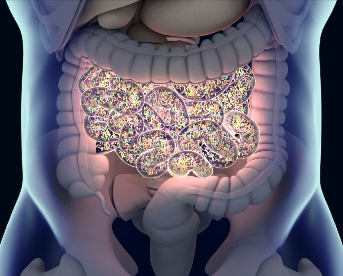
“We identified a partial restoration of the microbiome to a more eubiotic state in RA patients at 6 weeks and 12 weeks of DMARD treatment in participants [who] responded well to DMARD therapy,” they wrote. “This was further supported by long-term (> 1 year) treated DMARD RA participants with similar community shifts.” Microbiomes, they said, are “a promising diagnostic tool” for directing DMARD therapy.
Study Goal Not Met
The goal of the study was to determine whether the microbiome of patients before they began treatment with DMARDs could predict their response to therapy. The patients were enrolled in the IMRABIOME study. Eligible patients had inflammatory arthritis symptoms for a year or less and met the clinical criteria for RA. Most patients were taking methotrexate (134 at baseline, 77 at 12 weeks), but study participants were also taking sulfasalazine (16 at baseline, 14 at 12 weeks) or hydroxychloroquine (58 at baseline, 45 at 12 weeks) either in combination or as a stand-alone treatment.
The study found a total of 26 different stool microbes that decreased in patients who had a minimal clinically important improvement (MCII) after starting DMARD therapy. At 6 weeks, the most significant declines were in Prevotella species. At 12 weeks, the greatest declines were in Streptococcus.
The researchers also developed models that used gut and oral metagenomes to predict MCII in patients starting DMARD therapy. They used a previously published microbiome dataset as a validation cohort for the model, but they acknowledged their models “were not as strong” as three previously published models. “Our findings support the hypothesis of DMARD restoration of a eubiotic gut microbiome when patient and treatment align,” the authors wrote.
They noted they had anticipated finding baseline microbiome samples that would help predict treatment responses. While baseline evaluation didn’t differentiate between responders and nonresponders, they wrote that a longitudinal analysis demonstrated changing microbiota and a positive response to therapy, with declining levels of Prevotella and Streptococcus species most pronounced at 6 and 12 weeks, respectively.
“Microbiomes provide a promising diagnostic tool for guiding therapeutic decisions in the future,” the study authors wrote.
Commentary
In commenting on the study, Gregg J. Silverman, MD, professor of medicine and pathology at the New York University School of Medicine, New York City, said it “was carefully performed, technically it was actually quite impressive, and the scale of the study actually was quite suitable.”

However, the study fell short of achieving its primary goal of using the microbiome to predict treatment response, he said. “Basically, they could not find there was anything they could correlate with clinical response rates, although they did find that the presence or absence of certain bacteria at 6 weeks or 12 weeks into treatment correlated with a clinical response,” he said.
The multiplicity of DMARDs used by the study population was “one of the complicating factors” of the study, Dr. Silverman said. “It would’ve been a much more easily interpreted study if it used just a single agent like methotrexate,” he said. “I think that’s problematic, but I do think this contributes to getting us a little further down the road of understanding how the microbiome can influence the pathogenesis of rheumatoid arthritis response to treatment.”
One of the questions surrounding the microbiome changes is whether they occurred because of the effect of the therapy itself or because the disease activity subsides, Dr. Silverman said. “So, you’re not sure if it’s cause or effect. There’s evidence to suggest that either could be true.”
This study adds to a 2022 study that found a similar effect with methotrexate, Dr. Silverman said. “They considered a lot of variables, and they considered a lot of potential confounding effects,” he said. “So, their data were well-considered, and they will actually hold up over time and contribute to the next range of studies that will be performed, no doubt, in this area.”
It would be better if those future studies focused on just one DMARD drug and studied the recovered bacteria in animal models to gain a better understanding of how they correlate to pathogenesis, Dr. Silverman added.
The study received funding from Versus Arthritis. Dr. Danckert, Dr. Freidin, and coauthors, as well as Dr. Silverman, reported no relevant financial relationships.
A version of this article appeared on Medscape.com.
Patients with rheumatoid arthritis (RA) whose symptoms improved after they started taking nonbiologic disease-modifying antirheumatic drugs also demonstrated restored balance in their oral and gut flora, which could potentially serve as a marker of how they’ll respond to DMARDs, an observational study in the United Kingdom found.
Reporting in the journal Rheumatology, researchers led by Nathan Danckert, PhD, a genetic epidemiology researcher at King’s College London, and Maxim Freidin, PhD, of the Queen Mary University of London, London, England, evaluated stool and saliva samples of 144 people recently diagnosed with RA before and after they started DMARD therapy.
“We identified a partial restoration of the microbiome to a more eubiotic state in RA patients at 6 weeks and 12 weeks of DMARD treatment in participants [who] responded well to DMARD therapy,” they wrote. “This was further supported by long-term (> 1 year) treated DMARD RA participants with similar community shifts.” Microbiomes, they said, are “a promising diagnostic tool” for directing DMARD therapy.
Study Goal Not Met
The goal of the study was to determine whether the microbiome of patients before they began treatment with DMARDs could predict their response to therapy. The patients were enrolled in the IMRABIOME study. Eligible patients had inflammatory arthritis symptoms for a year or less and met the clinical criteria for RA. Most patients were taking methotrexate (134 at baseline, 77 at 12 weeks), but study participants were also taking sulfasalazine (16 at baseline, 14 at 12 weeks) or hydroxychloroquine (58 at baseline, 45 at 12 weeks) either in combination or as a stand-alone treatment.
The study found a total of 26 different stool microbes that decreased in patients who had a minimal clinically important improvement (MCII) after starting DMARD therapy. At 6 weeks, the most significant declines were in Prevotella species. At 12 weeks, the greatest declines were in Streptococcus.
The researchers also developed models that used gut and oral metagenomes to predict MCII in patients starting DMARD therapy. They used a previously published microbiome dataset as a validation cohort for the model, but they acknowledged their models “were not as strong” as three previously published models. “Our findings support the hypothesis of DMARD restoration of a eubiotic gut microbiome when patient and treatment align,” the authors wrote.
They noted they had anticipated finding baseline microbiome samples that would help predict treatment responses. While baseline evaluation didn’t differentiate between responders and nonresponders, they wrote that a longitudinal analysis demonstrated changing microbiota and a positive response to therapy, with declining levels of Prevotella and Streptococcus species most pronounced at 6 and 12 weeks, respectively.
“Microbiomes provide a promising diagnostic tool for guiding therapeutic decisions in the future,” the study authors wrote.
Commentary
In commenting on the study, Gregg J. Silverman, MD, professor of medicine and pathology at the New York University School of Medicine, New York City, said it “was carefully performed, technically it was actually quite impressive, and the scale of the study actually was quite suitable.”
However, the study fell short of achieving its primary goal of using the microbiome to predict treatment response, he said. “Basically, they could not find there was anything they could correlate with clinical response rates, although they did find that the presence or absence of certain bacteria at 6 weeks or 12 weeks into treatment correlated with a clinical response,” he said.
The multiplicity of DMARDs used by the study population was “one of the complicating factors” of the study, Dr. Silverman said. “It would’ve been a much more easily interpreted study if it used just a single agent like methotrexate,” he said. “I think that’s problematic, but I do think this contributes to getting us a little further down the road of understanding how the microbiome can influence the pathogenesis of rheumatoid arthritis response to treatment.”
One of the questions surrounding the microbiome changes is whether they occurred because of the effect of the therapy itself or because the disease activity subsides, Dr. Silverman said. “So, you’re not sure if it’s cause or effect. There’s evidence to suggest that either could be true.”
This study adds to a 2022 study that found a similar effect with methotrexate, Dr. Silverman said. “They considered a lot of variables, and they considered a lot of potential confounding effects,” he said. “So, their data were well-considered, and they will actually hold up over time and contribute to the next range of studies that will be performed, no doubt, in this area.”
It would be better if those future studies focused on just one DMARD drug and studied the recovered bacteria in animal models to gain a better understanding of how they correlate to pathogenesis, Dr. Silverman added.
The study received funding from Versus Arthritis. Dr. Danckert, Dr. Freidin, and coauthors, as well as Dr. Silverman, reported no relevant financial relationships.
A version of this article appeared on Medscape.com.
FROM RHEUMATOLOGY
FDA Approves 10th Humira Biosimilar, With Interchangeability
The US Food and Drug Administration has approved the first interchangeable, high-concentration, citrate-free adalimumab biosimilar, adalimumab-ryvk (Simlandi).
This is the 10th adalimumab biosimilar approved by the regulatory agency and the first biosimilar approval in the US market for the Icelandic pharmaceutical company Alvotech in partnership with Teva Pharmaceuticals.
“An interchangeable citrate-free, high-concentration biosimilar adalimumab has the potential to change the market dynamics in a rapidly evolving environment for biosimilars in the U.S.,” said Robert Wessman, chairman and CEO of Alvotech, in a company press release on February 23.
Adalimumab-ryvk was approved in the European Union in 2021 and in Australia and Canada in 2022.
Adalimumab-ryvk is indicated for adults with rheumatoid arthritis, psoriatic arthritis, ankylosing spondylitis, ulcerative colitis, Crohn’s disease, plaque psoriasis, hidradenitis suppurativa, and noninfectious intermediate and posterior uveitis and panuveitis. In pediatric patients, it is indicated for polyarticular juvenile idiopathic arthritis in children 2 years of age and older and Crohn’s disease in children 6 years of age and older.
Adalimumab-ryvk is the third Humira biosimilar overall granted interchangeability status, which allows pharmacists (depending on state law) to substitute the biosimilar for the reference product without involving the prescribing clinician. Adalimumab-adbm (Cyltezo), manufactured by Boehringer Ingelheim, and adalimumab-afzb (Abrilada), manufactured by Pfizer, were previously granted interchangeability status; however, they both are interchangeable with the low-concentration formulation of Humira, which make up only an estimated 15% of Humira prescriptions, according to a report by Goodroot.
Adalimumab-ryvk will be launched “imminently” in the United States, according to the press release, but no specific dates were provided. It is also not yet known how the biosimilar will be priced compared with Humira. Other adalimumab biosimilars have launched with discounts from 5% to 85% of Humira’s list price.
A version of this article appeared on Medscape.com.
Is Metformin a ‘Drug for All Diseases’?
In 2021 alone, clinicians wrote more than 91 million orders for the medication — up from 40 million 2004.
But is metformin just getting started? Emerging evidence suggests the drug may be effective for a much broader range of conditions beyond managing high blood glucose, including various cancers, obesity, liver disease, cardiovascular, neurodegenerative, and renal diseases. As the evidence for diverse uses accumulates, many trials have launched, with researchers looking to expand metformin’s indications and validate or explore new directions.
Metformin’s long history as a pharmaceutical includes an herbal ancestry, recognition in 1918 for its ability to lower blood glucose, being cast aside because of toxicity fears in the 1930s, rediscovery and synthesis in Europe in the 1940s, the first reported use for diabetes in 1957, and approval in the United States in 1994.
The drug has maintained its place as the preferred first-line treatment for type 2 diabetes since 2011, when it was first included in the World Health Organization’s essential medicines list.
“The focus hitherto has been primarily on its insulin sensitization effects,” Akshay Jain, MD, a clinical and research endocrinologist at TLC Diabetes and Endocrinology, in Surrey, British Columbia, Canada, told this news organization.
“The recent surge of renewed interest is in part related to its postulated effects on multiple other receptors,” he said. “In my mind, the metformin data on coronary artery disease reduction and cancer-protective effects have come farther along than other disease states.”
Cardiovascular Outcomes
Gregory G. Schwartz, MD, PhD, chief of the cardiology section at Rocky Mountain Regional VA Medical Center and professor of medicine at the University of Colorado School of Medicine in Aurora, is leading the VA-IMPACT trial. Despite metformin’s long history and widespread use, he said his study is the first placebo-controlled cardiovascular outcomes trial of the drug.
Launched in 2023, the study tests the hypothesis that metformin reduces the risk for death or nonfatal ischemic cardiovascular events in patients with prediabetes and established coronary, cerebrovascular, or peripheral artery disease, Dr. Schwartz said. The trial is being conducted at roughly 40 VA medical centers, with a planned enrollment of 7410 patients. The estimated completion date is March 2029.
“The principal mechanism of action of metformin is through activation of AMP [adenosine monophosphate]–activated protein kinase, a central pathway in metabolic regulation, cell protection, and survival,” Dr. Schwartz explained. “Experimental data have demonstrated attenuated development of atherosclerosis, reduced myocardial infarct size, improved endothelial function, and antiarrhythmic actions — none of those dependent on the presence of diabetes.”
Dr. Schwartz and his colleagues decided to test their hypothesis in people with prediabetes, rather than diabetes, to create a “true placebo-controlled comparison,” he said.
“If patients with type 2 diabetes had been chosen, there would be potential for confounding because a placebo group would require more treatment with other active antihyperglycemic medications to achieve the same degree of glycemic control as a metformin group,” Dr. Schwartz said.
“If proven efficacious in the VA-IMPACT trial, metformin could provide an inexpensive, generally safe, and well-tolerated approach to reduce cardiovascular morbidity and mortality in a large segment of the population,” Dr. Schwartz added. “Perhaps the old dog can learn some new tricks.”
Other recruiting trials looking at cardiovascular-related outcomes include Met-PEF, LIMIT, and Metformin as an Adjunctive Therapy to Catheter Ablation in Atrial Fibrillation.
Reducing Cancer Risks
Sai Yendamuri, MD, chair of the Department of Thoracic Surgery and director of the Thoracic Surgery Laboratory at Roswell Park Comprehensive Cancer Center in Buffalo, New York, is leading a phase 2 trial exploring whether metformin can prevent lung cancer in people with overweight or obesity who are at a high risk for the malignancy.
The study, which has accrued about 60% of its estimated enrollment, also will assess whether metformin can reprogram participants’ immune systems, with a view toward reducing the activity of regulatory T cells that are linked to development of tumors.
“In our preclinical and retrospective clinical data, we found that metformin had anticancer effects but only if the patients were overweight,” Dr. Yendamuri said. “In mice, we find that obesity increases regulatory T-cell function, which suppresses the immune system of the lungs. This effect is reversed by metformin.” The team is conducting the current study to examine if this happens in patients, as well. Results are expected next year.
Research is underway in other tumor types, including oral and endometrial, and brain cancers.
Preventing Alzheimer’s Disease
Cognitive function — or at least delaying its erosion — represents another front for metformin. José A. Luchsinger, MD, MPH, vice-chair for clinical and epidemiological research and director of the section on geriatrics, gerontology, and aging at Columbia University Irving Medical Center in New York City, is heading a phase 2/3 randomized controlled trial assessing the ability of the drug to prevent Alzheimer›s disease.
The study investigators hope to enroll 326 men and women aged 55-90 years with early and late mild cognitive impairment, overweight or obesity, and no diabetes.
“The hypothesis is that improving insulin and glucose levels can lead to lowering the risk of Alzheimer’s disease,” Dr. Luchsinger said. Recruitment should be complete by the end of 2024 and results are expected in late 2026.
Similar studies are underway in Europe and Asia.
Other areas of investigation, while tantalizing, are mostly in early stages, although bolstered by preclinical and mechanistic studies. The authors of a recent review on the potential mechanisms of action of metformin and existing evidence of the drug›s effectiveness — or lack thereof — in treating diseases other than diabetes, wrote: “Collectively, these data raise the question: Is metformin a drug for all diseases? It remains unclear as to whether all of these putative beneficial effects are secondary to its actions as an antihyperglycemic and insulin-sensitizing drug, or result from other cellular actions, including inhibition of mTOR (mammalian target for rapamycin), or direct antiviral actions.”
Off-Label Uses
Metformin currently is approved by the US Food and Drug Administration only for the treatment of type 2 diabetes, although it is also the only antidiabetic medication for prediabetes currently recommended by the American Diabetes Association.
Some studies currently are looking at its use in a variety of off-label indications, including obesity, gestational diabetes, weight gain from antipsychotics, and polycystic ovary syndrome.
For the most part, metformin is considered a safe drug, but it is not risk-free, Dr. Jain cautioned.
“Although it would certainly be helpful to see if this inexpensive medication that’s universally available can help in disease states, one shouldn’t overlook the potential risk of adverse effects, such as gastrointestinal, potential vitamin B12 deficiency, blunting of skeletal muscle development and the rare risk of lactic acidosis in those with kidney impairment,” he said.
“Similarly, with recent reports of the carcinogenic potential of certain formulations of long-acting metformin that contained NDMA [N-nitrosodimethylamine], it would be imperative that these kinks are removed before we incorporate metformin as the gift that keeps giving.”
Dr. Jain reported financial relationships with Abbott, Amgen, Boehringer Ingelheim, Dexcom, Eli Lilly, Janssen, Medtronic, Merck, and Novo Nordisk. Dr. Yendamuri disclosed serving on the scientific advisory board member of Karkinos Healthcare and research funding from Lumeda for the metformin study. Dr. Luchsinger reported receiving donated metformin and matching placebo from EMD Serono, a subsidiary of Merck, for the MAP study. Dr. Schwartz received research support from the US Department of Veterans Affairs as National Chair of the VA-IMPACT trial.
A version of this article appeared on Medscape.com.