User login
Obesity and overweight declined among lower-income kids
The combined rate of , according to a study in JAMA.
Liping Pan, MD, MPH, of the Centers for Disease Control and Prevention and colleagues used data from the WIC Participant and Program Characteristics survey from 2010, 2012, 2014, and 2016 for 12,403,629 children aged 2-4 years from 50 states, Washington, D.C., and 5 territories. In addition to a –3.2% change (95% confidence interval, –3.3% to –3.2%) in adjusted prevalence difference for the combined rate of obesity and overweight seen between 2010 and 2016, the researchers found the crude prevalence decreased from 32.5% to 29.1%. A decrease was also seen for obesity alone (crude prevalence, 15.9% to 13.9%; adjusted prevalence difference, –1.9%; 95% CI, –1.9% to –1.8%).
One of the limitations of the study is that the characteristics of enrolled children might differ from those of children not enrolled in this WIC program; however, the researchers noted that they accounted for many demographic characteristics in the trend analyses.
“Reasons for the declines in obesity among young children in WIC remain undetermined but may include WIC food package revisions and local, state, and national initiatives,” they wrote.
SOURCE: Pan L et al. JAMA. 2019 Jun 18;321(23):2364-6.
FROM JAMA
Suicide rates rise in U.S. adolescents and young adults
Suicides in teens and young adults reached 6,241 in 2017, the highest since 2000, according to data from a review of the Centers for Disease Control and Prevention Underlying Cause of Death database.

The suicide rate overall was 12 per 100,000 in 2017 for 15-19 year olds.
Although suicide rates have increased across all age groups in the United States since 2000, “adolescents are of particular concern, with increases in social media use, anxiety, depression, and self-inflicted injuries,” wrote Oren Miron of Harvard Medical School, Boston, and colleagues.
In a research letter published in JAMA, the researchers analyzed trends in teen and young adult suicides from 2000 to 2017. The combined suicide rate for males and females aged 15-19 years in 2000 was 8 per 100,000 with no significant changes until 2007, followed by an annual percentage change (APC) of 3% from 2007 to 2014 and 10% from 2014 to 2017.
When the data were broken out by gender, Of note, these young men showed a decreasing trend in APC of –2% from 2000 to 2007 before increasing.
Among females aged 15-19 years, no increase was noted until 2010, then researchers identified an APC of 8% from 2010 to 2017.
For ages 20-24 years, the combined suicide rate for males and females was 13 per 100,000 in 2000, which rose to 17 per 100,000 in 2017. The APC in the older group was 1% from 2000 to 2013 and 6% from 2013 to 2017. Increasing trends were observed for both males and females over the study period.
The study was limited by the potential inaccuracy in cause of death listed on death certificates, such as mistaking a suicide for an accidental overdose, and the increased suicide rate could reflect more accurate reporting, the researchers noted.
Nonetheless, the results support the need for more studies of contributing factors to teen and young adult suicides to help develop prevention strategies and analysis of factors that may have contributed to declines in suicide rates in the past, they said.
Coauthor Dr. Yu was supported by the Harvard Data Science Fellowship. The researchers had no relevant financial disclosures.
SOURCE: Miron O et al. JAMA. 2019 Jun 28;321:2362-4.
Suicides in teens and young adults reached 6,241 in 2017, the highest since 2000, according to data from a review of the Centers for Disease Control and Prevention Underlying Cause of Death database.
The suicide rate overall was 12 per 100,000 in 2017 for 15-19 year olds.
Although suicide rates have increased across all age groups in the United States since 2000, “adolescents are of particular concern, with increases in social media use, anxiety, depression, and self-inflicted injuries,” wrote Oren Miron of Harvard Medical School, Boston, and colleagues.
In a research letter published in JAMA, the researchers analyzed trends in teen and young adult suicides from 2000 to 2017. The combined suicide rate for males and females aged 15-19 years in 2000 was 8 per 100,000 with no significant changes until 2007, followed by an annual percentage change (APC) of 3% from 2007 to 2014 and 10% from 2014 to 2017.
When the data were broken out by gender, Of note, these young men showed a decreasing trend in APC of –2% from 2000 to 2007 before increasing.
Among females aged 15-19 years, no increase was noted until 2010, then researchers identified an APC of 8% from 2010 to 2017.
For ages 20-24 years, the combined suicide rate for males and females was 13 per 100,000 in 2000, which rose to 17 per 100,000 in 2017. The APC in the older group was 1% from 2000 to 2013 and 6% from 2013 to 2017. Increasing trends were observed for both males and females over the study period.
The study was limited by the potential inaccuracy in cause of death listed on death certificates, such as mistaking a suicide for an accidental overdose, and the increased suicide rate could reflect more accurate reporting, the researchers noted.
Nonetheless, the results support the need for more studies of contributing factors to teen and young adult suicides to help develop prevention strategies and analysis of factors that may have contributed to declines in suicide rates in the past, they said.
Coauthor Dr. Yu was supported by the Harvard Data Science Fellowship. The researchers had no relevant financial disclosures.
SOURCE: Miron O et al. JAMA. 2019 Jun 28;321:2362-4.
FROM JAMA
Key clinical point: Suicide rates in U.S. adolescents and young adults have increased since 2000.
Major finding: The combined suicide rate for males and females aged 15-19 years underwent an annual percentage change of 3% from 2007 to 2014 and 10% from 2014 to 2017.
Study details: The data come from the CDC Underlying Cause of Death database.
Disclosures: Coauthor Dr. Yu was supported by the Harvard Data Science Fellowship. The researchers had no relevant financial disclosures.
Source: Miron O et al. JAMA. 2019 Jun 28;321:2362-4.
Cyclic Vomiting Syndrome and Primary Headache
Cyclic Vomiting Syndrome and Primary Headache
Cyclic vomiting syndrome and benign paroxysmal torticollis are associated with a very high risk of developing headache, mostly migraine, later in life. This according to a longitudinal study that assessed the rate of headache in patients presenting with cyclic vomiting syndrome and benign paroxysmal torticollis during infancy and defined the main clinical features of the disorder. Researchers administered a questionnaire to the parents of pediatric patients with previous diagnosis of cyclic vomiting syndrome and/or benign paroxysmal torticollis. They found:
- 82 patients with cyclic vomiting syndrome and 33 with benign paroxysmal torticollis were included in the final analysis.
- 79% of patients with cyclic vomiting syndrome presented with headache during the follow-up with a mean age at onset of 6 years.
- 67% of patients with benign paroxysmal torticollis suffered from headache during the follow-up with a mean age at onset of 5 years.
Moavero R, et al. Cyclic vomiting syndrome and benign paroxysmal torticollis are associated with a high risk of developing primary headache: A longitudinal study. [Published online ahead of print April 13, 2019]. Cephalalgia. doi: 10.1177/0333102419844542.
Migraine in Patients with Calcified Neurocysticercosis
The clinical characteristics of migraine attacks are sufficiently different in patients with and without neurocysticercosis, a new study found. Researchers investigated the characteristics of migraine attacks in patients with calcified neurocysticercosis (NCC) on brain imaging. Of 350 migraine patients, 166 had undergone brain imaging. Seventy-two patients with migraines had calcified NCC. The migraine attacks of the patients with calcification (MiC) were compared with those of 94 patients without calcification (MiNC). Among the findings:
- Side-locked headaches were seen in 48.6% of the MiC patients.
- Aura preceding the migraine attack was more common in the MiC group vs the MiNC group.
- The MiC group had fewer headache episodes per month with fewer common associated features and required fewer drugs for secondary prophylaxis.
Pradhan S, et al. Clinical characteristics of migraine in patients with calcified neurocysticercosis. [Published online ahead of print April 6, 2019]. Trans R Soc Trop Med Hyg. doi: 10.1093/trstmh/trz018.
Modest cognitive changes deemed inherent in ‘normal’ aging
Interventions leading to improved gray matter volume tied to reducing dementia risk
CRYSTAL CITY, VA. – As technology advances and the population becomes older, clinicians should understand how modest age-related declines in cognition affect older adults’ ability to learn new technological skills, Philip D. Harvey, PhD, said at Focus on Neuropsychiatry presented by Current Psychiatry and the American Academy of Clinical Psychiatrists.

According to the U.S. Census Bureau, the number of adults in the United States above age 65 is slated to increase over the next several decades, and by 2030, one in five adults in the United States will be at retirement age. By 2050, “a significant number of people” in the United States are expected to be age 90, Dr. Harvey said at the meeting, presented by Global Academy for Medical Education.
“What we need to do is to understand what are the normal things that happen to people as they become 90 years of age,” said Dr. Harvey, of the department of psychiatry and behavioral sciences at the University of Miami.
Within the technology industry, significant advancements were made over the last 40 years with the advent of the personal computer in the 1980s, mobile phones in the 1990s, and wireless Internet, smartphones, and wireless devices in the 2000s. Many interactions that used to be person-to-person are now performed online, and it is feasible for a 90-year-old living today to never have encountered this technology during their careers. “Utilizing technology is a central requirement for independent living today,” Dr. Harvey said.
Most people passively adapt to these new changes in technology. However, Dr. Harvey noted that adults in their 80s and 90s who are retired can have difficulty using or learning about new technology as they age. “Human-technology interaction involves information processing, and places demands on memory and other cognitive abilities,” he said. “Age is associated with declines specifically in the kind of abilities that are required to master new technology.”
Learning about and using technology requires different elements of cognition that include different types of memory, such as working, episodic, declarative, procedural, semantic, long-term factual, and emotional. A decline in any of those kinds of memory could result in failures in forgetting, learning or recalling material, and learning new motor skills, among other problems. Crystallized intelligence is more likely to be retained over time, but fluid cognition in the form of processing speed, working and episodic memory, and the ability to solve abstract problems tend to decline over time as people age, Dr. Harvey said.
Base cognitive abilities do play a role in how crystallized and fluid cognition decline over time. For example, while vocabulary might increase as one ages, a person’s working memory, processing speed, and episodic memory decline over time. Evidence also suggests that speed training and exercise appear to improve cognition. (J Am Geriatr Soc. 13 Jan 2014. doi: 10.1111/jgs.12607).
Cyrus Raji, MD, PhD, and colleagues also explored the relationship between caloric expenditure and gray matter volume in the Cardiovascular Health Study, and found that exercise of various types improved gray matter volume and reduced the risk of dementia in people aged 65 or older. Furthermore, Dr. Raji and colleagues found, caloric expenditures, rather than intensity of exercise, may alone predict increases in gray matter volume (J Alzheimers Dis. 2016. doi: 10.3233/JAD-160057).
“If you want to improve your memory, grow your hippocampus,” Dr. Harvey said at the meeting.
Dr. Harvey reported serving as a consultant for Alkermes, Boehringer-Ingelheim, Lundbeck, Otsuka Digital Health, Sanofi, Sunovion Pharmaceuticals, Takeda, and Teva. He also reported receiving a grant from Takeda, and is the founder and CSO of i-Function.
Global Academy and this news organization are owned by the same parent company.
Interventions leading to improved gray matter volume tied to reducing dementia risk
Interventions leading to improved gray matter volume tied to reducing dementia risk
CRYSTAL CITY, VA. – As technology advances and the population becomes older, clinicians should understand how modest age-related declines in cognition affect older adults’ ability to learn new technological skills, Philip D. Harvey, PhD, said at Focus on Neuropsychiatry presented by Current Psychiatry and the American Academy of Clinical Psychiatrists.
According to the U.S. Census Bureau, the number of adults in the United States above age 65 is slated to increase over the next several decades, and by 2030, one in five adults in the United States will be at retirement age. By 2050, “a significant number of people” in the United States are expected to be age 90, Dr. Harvey said at the meeting, presented by Global Academy for Medical Education.
“What we need to do is to understand what are the normal things that happen to people as they become 90 years of age,” said Dr. Harvey, of the department of psychiatry and behavioral sciences at the University of Miami.
Within the technology industry, significant advancements were made over the last 40 years with the advent of the personal computer in the 1980s, mobile phones in the 1990s, and wireless Internet, smartphones, and wireless devices in the 2000s. Many interactions that used to be person-to-person are now performed online, and it is feasible for a 90-year-old living today to never have encountered this technology during their careers. “Utilizing technology is a central requirement for independent living today,” Dr. Harvey said.
Most people passively adapt to these new changes in technology. However, Dr. Harvey noted that adults in their 80s and 90s who are retired can have difficulty using or learning about new technology as they age. “Human-technology interaction involves information processing, and places demands on memory and other cognitive abilities,” he said. “Age is associated with declines specifically in the kind of abilities that are required to master new technology.”
Learning about and using technology requires different elements of cognition that include different types of memory, such as working, episodic, declarative, procedural, semantic, long-term factual, and emotional. A decline in any of those kinds of memory could result in failures in forgetting, learning or recalling material, and learning new motor skills, among other problems. Crystallized intelligence is more likely to be retained over time, but fluid cognition in the form of processing speed, working and episodic memory, and the ability to solve abstract problems tend to decline over time as people age, Dr. Harvey said.
Base cognitive abilities do play a role in how crystallized and fluid cognition decline over time. For example, while vocabulary might increase as one ages, a person’s working memory, processing speed, and episodic memory decline over time. Evidence also suggests that speed training and exercise appear to improve cognition. (J Am Geriatr Soc. 13 Jan 2014. doi: 10.1111/jgs.12607).
Cyrus Raji, MD, PhD, and colleagues also explored the relationship between caloric expenditure and gray matter volume in the Cardiovascular Health Study, and found that exercise of various types improved gray matter volume and reduced the risk of dementia in people aged 65 or older. Furthermore, Dr. Raji and colleagues found, caloric expenditures, rather than intensity of exercise, may alone predict increases in gray matter volume (J Alzheimers Dis. 2016. doi: 10.3233/JAD-160057).
“If you want to improve your memory, grow your hippocampus,” Dr. Harvey said at the meeting.
Dr. Harvey reported serving as a consultant for Alkermes, Boehringer-Ingelheim, Lundbeck, Otsuka Digital Health, Sanofi, Sunovion Pharmaceuticals, Takeda, and Teva. He also reported receiving a grant from Takeda, and is the founder and CSO of i-Function.
Global Academy and this news organization are owned by the same parent company.
REPORTING FROM FOCUS ON NEUROPSYCHIATRY 2019
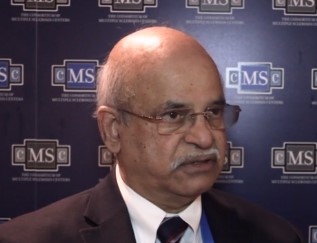
Patient registry sheds light on the economic impact of MS
SEATTLE –
“MS seems to prevent people with MS from realizing their full potential at work or home,” said study coauthor Kottil Rammohan, MD, who summarized the study results in a video interview. Dr. Rammohan is professor of clinical neurology, director of the MS center of excellence, and chief of the multiple sclerosis division at the University of Miami. The study findings were presented at the annual meeting of the Consortium of Multiple Sclerosis Centers.
The North American Registry for Care and Research in Multiple Sclerosis (NARCRMS) prospectively collects information about the health care economics of patients with MS and its effects on daily life. In 2017, NARCRMS established the health care economics outcomes research (HEOR) advisory group. NARCRMS developed a Health-Related Productivity Questionnaire and Health Resource Utilization Questionnaire. The questionnaires were incorporated into the existing case report forms that are completed by patients at enrollment, annual, and exacerbation visits.
This analysis was based on 480 patients who had completed HEOR case report forms. Among those, 77% are employed either full or part time; however, of those 15% were underemployed, meaning they wanted to work more than their current work levels. About 13% are on disability.
“What we found was there was a significant impact at home as well,” said Dr. Rammohan. Patients reported that MS kept them from completing household chores. “MS is a disease that seems to impact not only the work environment, but also the home environment.”
When polled to determine the main reason why these MS patients are not able to function, “what we found was that it was not because of gait or immobility, it was difficulty related to fatigue,” Dr. Rammohan said. The second most common impairment was related to cognition.
“These are what we call the silent or the transparent symptoms of MS.”
Dr. Rammohan disclosed consulting fees from EMD Serono, Biogen, Sanofi-Aventis, Genzyme, Novartis, Teva Neurosciences, Acorda, and Roche/Genentech.
EXPERT ANALYSIS FROM CMSC 2019
Leading By Example: How Medical Journals Can Improve Representation in Academic Medicine
Women and racial and ethnic minorities remain underrepresented in senior faculty roles and academic leadership positions.1 Participation in peer review and publication in medical journals are important components of academic advancement that are emphasized in the promotion process. These efforts offer recognition of expertise and increase visibility in the scientific community, which may enhance opportunities for networking and collaboration, and provide other opportunities for career advancement. In addition, abundant evidence shows that organizations benefit from diverse teams, with better quality decisions and increased productivity resulting from diverse ideas and perspectives.2
Numerous studies have highlighted the prevalence and persistence of disparities in peer review and authorship.3,4 Much of this work has focused on gender though gaps in these measures likely exist for racial and ethnic minorities. Yet, there are few examples of journals implementing strategies to address disparities and track results of such efforts.5 While institutional barriers to advancement must be addressed, we believe that medical journals have an obligation to address unequal opportunities.
At the Journal of Hospital Medicine, we are committed to leading by example and developing approaches to create equity in all facets of journal leadership and authorship.6 The first step towards progress is to assess the current representation of women and racial and ethnic minorities in our journal community, including first and senior authors, invited expert contributors, reviewers, and editorial team members. Like most journals, we have not collected demographic information from authors or reviewers. But now, as part of the journal’s commitment to this cause, we request that everyone in the journal community (author, reviewer, editor) update their journal account (accessible at https://mc.manuscriptcentral.com/jhm) with demographic data, including gender, race, and ethnicity.
Inclusion of these data is voluntary. While each individual will be able to access and edit their personal demographic data, the individual data will remain private and unviewable to others. As such, it will not be available for nor will it be used in the manuscript review or decision process but rather for assessing our own inclusiveness. We will review these data in aggregate to broadly inform outreach efforts to promote diversity and inclusion in our author, invited expert contributor, reviewer, and journal leadership pools. We will report on the progress of these efforts in upcoming years.
We are committed to equity in providing opportunities for academic advancement across the journal community. Diversity and inclusion are important in raising the quality of the work that we publish. Different perspectives strengthen our journal and will help us continue to advance the field of Hospital Medicine.
Disclosures
The authors have nothing to disclose.
1. American Association of Medical Colleges. U.S. Medical School Faculty, 2018. https://www.aamc.org/data/facultyroster/reports/494946/usmsf18.html. Accessed May 6, 2019.
2. Turban S, Wu D, Zhang L. “When Gender Diversity Makes Firms More Productive” Harvard Business Review Feb 2019. https://hbr.org/2019/02/research-when-gender-diversity-makes-firms-more-productive. Accessed May 6, 2019.
3. Silver JK, Poorman JA, Reilly JM, Spector ND, Goldstein R, Zafonte RD. Assessment of women physicians among authors of perspective-type articles published in high-impact pediatric journals. JAMA Netw Open. 2018;1(3):e180802. doi: 10.1001/jamanetworkopen.2018.0802. PubMed
4. Jagsi R, Guancial EA, Worobey CC, Henault LE, Chang Y, Starr R, Tarbell NJ, Hylek EM. The “gender gap” in authorship of academic medical literature- a 35-year perspective. N Engl J Med. 2006;355(3):281-287. doi: 10.1056/NEJMsa053910. PubMed
5. Nature’s under-representation of women. Nature. 2018;558:344. doi: 10.1038/d41586-018-05465-7. PubMed
6. Shah SS. The Journal of Hospital Medicine in 2019 and beyond. J Hosp Med. 2019;14(1):7. doi: 10.12788/jhm.3143. PubMed
Women and racial and ethnic minorities remain underrepresented in senior faculty roles and academic leadership positions.1 Participation in peer review and publication in medical journals are important components of academic advancement that are emphasized in the promotion process. These efforts offer recognition of expertise and increase visibility in the scientific community, which may enhance opportunities for networking and collaboration, and provide other opportunities for career advancement. In addition, abundant evidence shows that organizations benefit from diverse teams, with better quality decisions and increased productivity resulting from diverse ideas and perspectives.2
Numerous studies have highlighted the prevalence and persistence of disparities in peer review and authorship.3,4 Much of this work has focused on gender though gaps in these measures likely exist for racial and ethnic minorities. Yet, there are few examples of journals implementing strategies to address disparities and track results of such efforts.5 While institutional barriers to advancement must be addressed, we believe that medical journals have an obligation to address unequal opportunities.
At the Journal of Hospital Medicine, we are committed to leading by example and developing approaches to create equity in all facets of journal leadership and authorship.6 The first step towards progress is to assess the current representation of women and racial and ethnic minorities in our journal community, including first and senior authors, invited expert contributors, reviewers, and editorial team members. Like most journals, we have not collected demographic information from authors or reviewers. But now, as part of the journal’s commitment to this cause, we request that everyone in the journal community (author, reviewer, editor) update their journal account (accessible at https://mc.manuscriptcentral.com/jhm) with demographic data, including gender, race, and ethnicity.
Inclusion of these data is voluntary. While each individual will be able to access and edit their personal demographic data, the individual data will remain private and unviewable to others. As such, it will not be available for nor will it be used in the manuscript review or decision process but rather for assessing our own inclusiveness. We will review these data in aggregate to broadly inform outreach efforts to promote diversity and inclusion in our author, invited expert contributor, reviewer, and journal leadership pools. We will report on the progress of these efforts in upcoming years.
We are committed to equity in providing opportunities for academic advancement across the journal community. Diversity and inclusion are important in raising the quality of the work that we publish. Different perspectives strengthen our journal and will help us continue to advance the field of Hospital Medicine.
Disclosures
The authors have nothing to disclose.
1. American Association of Medical Colleges. U.S. Medical School Faculty, 2018. https://www.aamc.org/data/facultyroster/reports/494946/usmsf18.html. Accessed May 6, 2019.
2. Turban S, Wu D, Zhang L. “When Gender Diversity Makes Firms More Productive” Harvard Business Review Feb 2019. https://hbr.org/2019/02/research-when-gender-diversity-makes-firms-more-productive. Accessed May 6, 2019.
3. Silver JK, Poorman JA, Reilly JM, Spector ND, Goldstein R, Zafonte RD. Assessment of women physicians among authors of perspective-type articles published in high-impact pediatric journals. JAMA Netw Open. 2018;1(3):e180802. doi: 10.1001/jamanetworkopen.2018.0802. PubMed
4. Jagsi R, Guancial EA, Worobey CC, Henault LE, Chang Y, Starr R, Tarbell NJ, Hylek EM. The “gender gap” in authorship of academic medical literature- a 35-year perspective. N Engl J Med. 2006;355(3):281-287. doi: 10.1056/NEJMsa053910. PubMed
5. Nature’s under-representation of women. Nature. 2018;558:344. doi: 10.1038/d41586-018-05465-7. PubMed
6. Shah SS. The Journal of Hospital Medicine in 2019 and beyond. J Hosp Med. 2019;14(1):7. doi: 10.12788/jhm.3143. PubMed
© 2019 Society of Hospital Medicine
Obinutuzumab provides strong early responses in untreated MCL
Amsterdam – For patients with untreated mantle cell lymphoma (MCL), the anti-CD20 monoclonal antibody obinutuzumab may one day offer an alternative to rituximab, according to investigators.
Patients in the LYMA-101 trial were given four cycles of obinutuzumab in combination with dexamethasone, high-dose aracytine, and platinum chemotherapy (O-DHAP), followed by autologous stem cell transplantation (ASCT) and maintenance obinutuzumab. After a median follow-up of 14.6 months, ranging from 3.8 to 24.4 months, 85% of evaluable patients had achieved minimal residual disease (MRD) in bone marrow, reported lead author Steven Le Gouill, MD, PhD, of the University Hospital of Nantes, and his colleagues.
In this disease population, an MRD rate of 85% is “unprecedented,” Dr. Le Gouill said during his presentation at the annual congress of the European Hematology Association. Based on findings from LYMA-101 and preclinical data, Dr. Le Gouill suggested that obinutuzumab may become an alternative to rituximab, the current standard anti-CD20 antibody.
“There are few data of interest for obinutuzumab in MCL, but there is a strong rationale in the lab as obinutuzumab has a different mechanism of action against tumor cells [than rituximab], with more efficacy against MCL cells,” Dr. Le Gouill said.
Data from the ongoing phase 2 trial were drawn from 85 patients with untreated MCL who were 65 years or younger at the time of enrollment. More specifically, median patient age was 55.5 years and 17.4% of patients had blastoid disease. All patients were given the O-DHAP/ASCT/obinutuzumab protocol, with a maintenance period of 3 years. Thereafter, MRD-positive patients may receive obinutuzumab on-demand.
The primary endpoint was MRD in bone marrow after induction therapy, measured by quantitative PCR (qPCR) and droplet digital PCR (ddPCR). Secondary endpoints included response rates, survival measures, incidence of stem cell collection failure after O-DHAP, and MRD rates at additional therapeutic time points.
Owing to the ongoing nature of the study, Dr. Le Gouill focused on the primary endpoint during his presentation.
Analysis showed that 75% and 85% of evaluable patients had achieved negative MRD in bone marrow after induction, according to qPCR and ddPCR, respectively.
These early findings give “a flavor of the results in terms of efficacy,” Dr. Le Gouill said, noting that “the median follow-up is pretty short.”
Still, 1-year findings were “very promising,” he said, with a progression-free survival of 93.4% and overall survival of 96%.
Twelve patients stopped treatment before ASCT, three prior to maintenance, and nine during maintenance. Of these 24 patients, 13 stopped treatment because of adverse events. The remaining 11 patients halted therapy because of disease progression, other malignancies, or death.
From the original 85 patients, 3 patients died and 3 progressed. Considering all of these findings, and that no major toxicities were encountered, the investigators concluded that the regimen was safe.
Overall, the results suggest that further research is needed, Dr. Le Gouill concluded. “Maybe this is where obinutuzumab may have stronger efficacy in MCL, as compared to rituximab,” he said.
The study is sponsored by the Lymphoma Academic Research Organisation. The investigators reported relationships with Roche, Janssen-Cilag, Gilead, Servier, and Novartis.
SOURCE: Le Gouill S et al. EHA Congress, Abstract S103.
REPORTING FROM EHA CONGRESS
Key clinical point:
Major finding: Out of 73 patients, 62 (85%) achieved negative minimal residual disease (MRD) in bone marrow based on ddPCR.
Study details: LYMA-101 is an ongoing phase 2 trial involving 85 patients with untreated mantle cell lymphoma.
Disclosures: The investigators reported relationships with Roche, Janssen-Cilag, Gilead, Servier, and Novartis.
Source: Le Gouill S et al. EHA Congress, Abstract S103.
Effects of Smartphone Overuse in Patients with Migraine
Smartphone overuse in patients with migraine is related to poor sleep quality and daytime sleepiness, resulting in diminished quality of life, a new study found. The single-center, cross-sectional comparative study used the migraine disability assessment (MIDAS) questionnaire to evaluate the disability status, and Mobile Phone Problematic Use Scale (MPPUS) was used to evaluate smartphone use frequency. The Visual Analogue Scale (VAS), 24-h Migraine Quality of Life Questionnaire (24-h MQoLQ), Pittsburgh Sleep Quality Index (PSQI) and Epworth Sleepiness Scale (ESS) were used to evaluate the pain intensity, quality of life, sleep quality, and daytime sleepiness. Researchers found:
- The study included 123 patients.
- There was a significant difference between the groups in terms of pain intensity, frequency, and duration.
- There was a negative correlation between MPPUS and PQSI, a strong positive correlation between MPPUS and ESS, and a negative correlation between MPPUS and 24-h MQoLQ.
Demir YP, et al. Effects of smartphone overuse on headache, sleep and quality of life in migraine patients. Neurosciences (Riyadh). 2019;24(2):115-121. doi: 10.17712/nsj.2019.2.20180037.
VIDEO: Dr. Lihi Eder on the diagnostic challenges of psoriatic arthritis

How strong are the links between psoriasis and psoriatic arthritis? What are the biggest challenges that keep clinicians from diagnosing PsA? And which approaches show promise in helping prevent PsA? In this expert analysis, Lihi Eder, MD, PhD, of Women’s College Research Institute, Toronto, and the University of Toronto offers answers to these questions and more.
How strong are the links between psoriasis and psoriatic arthritis? What are the biggest challenges that keep clinicians from diagnosing PsA? And which approaches show promise in helping prevent PsA? In this expert analysis, Lihi Eder, MD, PhD, of Women’s College Research Institute, Toronto, and the University of Toronto offers answers to these questions and more.
How strong are the links between psoriasis and psoriatic arthritis? What are the biggest challenges that keep clinicians from diagnosing PsA? And which approaches show promise in helping prevent PsA? In this expert analysis, Lihi Eder, MD, PhD, of Women’s College Research Institute, Toronto, and the University of Toronto offers answers to these questions and more.
