User login
Blinatumomab instead of chemo in young patients with relapsed ALL
ORLANDO – In young patients who experience relapse after chemotherapy for B-cell acute lymphoblastic leukemia (B-ALL), the novel agent blinatumomab (Blincyto, Amgen) can be used instead of intensive chemotherapy to try to achieve a second remission, experts say.
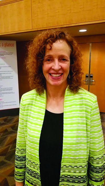
In fact, blinatumomab should be the new standard of care in these patients because it yielded better overall survival, was less toxic, and allowed more patients to proceed to transplant, said Robert A. Brodsky, MD, professor of medicine and director of the division of hematology at Johns Hopkins University, Baltimore.
Dr. Brodsky was commenting on new data presented in a late-breaking abstract (LBA1) at the annual meeting of the American Society of Hematology, for which he holds the role of secretary.
These results are “truly practice changing,” he told journalists at a press briefing.
Cure rates for B-ALL in children and adolescents and young adults (AYAs) are high, but for the small group of patients who experience relapse (about 15%), the prognosis is poor.
When relapse occurs in these patients, “it’s a real problem,” Dr. Brodsky explained. “At that point, the major emphasis is trying to get them back into full remission and get them to a transplant,” he continued, “but it’s very hard to get these patients back into remission.”
The standard treatment approach for these patients includes intensive chemotherapy. In the new study, this was compared to monotherapy with blinatumomab, which is described as a bispecific T-cell engager antibody.
The results were presented by Patrick A. Brown, MD, from the division of pediatric oncology at the Sidney Kimmel Comprehensive Cancer Center, Johns Hopkins University.
The Children’s Oncology Group Study AALL1331 trial was conducted in 208 children and AYA patients with B-ALL after a first relapse. Median follow-up was 1.4 years.
Blinatumomab was superior in achieving both disease-free survival (59.3 plus or minus 5.4% at 2 years vs. 41% plus or minus 6.2% at 2 years with chemo; P = .05) and overall survival (79.4 plus or minus 4.5% at 2 years vs. 59.2 plus or minus 6% at 2 years with chemo; P = .05).
In addition, more patients who received blinatumomab subsequently underwent transplant (79% vs. 45% with chemo; P less than .0001).
The drug was also better tolerated than chemotherapy, causing fewer and less severe toxicities, including fewer cases of grade 3+ infection, sepsis, and mucositis.
Dr. Brown concluded that, for children and AYA patients with high- or intermediate-risk first relapse of B-ALL, blinatumomab is superior to standard chemotherapy as postreinduction consolidation prior to transplant, resulting in fewer and less severe toxicities, higher rates of minimum residual disease response, greater likelihood of proceeding to hematopoietic stem cell transplant, and improved disease-free and overall survival.
Dr. Brown noted that blinatumomab already has conditional approval from the Food and Drug Administration for use in relapsed ALL in both adults and children, but that approval was based on clinical trial data in adults. This is now the definitive trial in children and AYAs, and it should support full approval for this indication, he said.
Dr. Brown has relationships with Novartis, Servier, and Jazz. Many coauthors also have relationships with pharmaceutical companies. Dr. Brodsky has relationships with Achillion, Alexion, and UpToDate.
A version of this story originally appeared on Medscape.com.
ORLANDO – In young patients who experience relapse after chemotherapy for B-cell acute lymphoblastic leukemia (B-ALL), the novel agent blinatumomab (Blincyto, Amgen) can be used instead of intensive chemotherapy to try to achieve a second remission, experts say.
In fact, blinatumomab should be the new standard of care in these patients because it yielded better overall survival, was less toxic, and allowed more patients to proceed to transplant, said Robert A. Brodsky, MD, professor of medicine and director of the division of hematology at Johns Hopkins University, Baltimore.
Dr. Brodsky was commenting on new data presented in a late-breaking abstract (LBA1) at the annual meeting of the American Society of Hematology, for which he holds the role of secretary.
These results are “truly practice changing,” he told journalists at a press briefing.
Cure rates for B-ALL in children and adolescents and young adults (AYAs) are high, but for the small group of patients who experience relapse (about 15%), the prognosis is poor.
When relapse occurs in these patients, “it’s a real problem,” Dr. Brodsky explained. “At that point, the major emphasis is trying to get them back into full remission and get them to a transplant,” he continued, “but it’s very hard to get these patients back into remission.”
The standard treatment approach for these patients includes intensive chemotherapy. In the new study, this was compared to monotherapy with blinatumomab, which is described as a bispecific T-cell engager antibody.
The results were presented by Patrick A. Brown, MD, from the division of pediatric oncology at the Sidney Kimmel Comprehensive Cancer Center, Johns Hopkins University.
The Children’s Oncology Group Study AALL1331 trial was conducted in 208 children and AYA patients with B-ALL after a first relapse. Median follow-up was 1.4 years.
Blinatumomab was superior in achieving both disease-free survival (59.3 plus or minus 5.4% at 2 years vs. 41% plus or minus 6.2% at 2 years with chemo; P = .05) and overall survival (79.4 plus or minus 4.5% at 2 years vs. 59.2 plus or minus 6% at 2 years with chemo; P = .05).
In addition, more patients who received blinatumomab subsequently underwent transplant (79% vs. 45% with chemo; P less than .0001).
The drug was also better tolerated than chemotherapy, causing fewer and less severe toxicities, including fewer cases of grade 3+ infection, sepsis, and mucositis.
Dr. Brown concluded that, for children and AYA patients with high- or intermediate-risk first relapse of B-ALL, blinatumomab is superior to standard chemotherapy as postreinduction consolidation prior to transplant, resulting in fewer and less severe toxicities, higher rates of minimum residual disease response, greater likelihood of proceeding to hematopoietic stem cell transplant, and improved disease-free and overall survival.
Dr. Brown noted that blinatumomab already has conditional approval from the Food and Drug Administration for use in relapsed ALL in both adults and children, but that approval was based on clinical trial data in adults. This is now the definitive trial in children and AYAs, and it should support full approval for this indication, he said.
Dr. Brown has relationships with Novartis, Servier, and Jazz. Many coauthors also have relationships with pharmaceutical companies. Dr. Brodsky has relationships with Achillion, Alexion, and UpToDate.
A version of this story originally appeared on Medscape.com.
‘Real-world’ data show CAR T therapies are cost effective
ORLANDO – Chimeric antigen receptor (CAR) T-cell therapy has been hailed as a major advance and a game changer, but their cost has redefined the meaning of “expensive.”
However, new “real-world” data now suggests that CAR T-cell therapy may actually be cost effective, as it may lower other expenses related to the illness.
When used in a population of older adults with non-Hodgkin lymphoma, these new data show that CAR T-cell therapy cut related expenditures, compared with health care costs prior to receiving this treatment.
“CAR T therapy was associated with fewer hospitalizations, shorter time in the hospital, fewer ED visits, and lower total health care costs,” said lead study author Karl M. Kilgore, PhD, of Avalere Health in Washington, D.C.
He presented the findings at the 2019 annual meeting of the American Society of Hematology (abstract 793).
CAR T-cell therapies were approved in the United States in 2017. First came tisagenlecleucel (Kymriah, Novartis), for the treatment of pediatric and young adult patients with acute lymphoblastic leukemia, with a price tag of $475,000. Closely following it was axicabtagene ciloleucel (Yescarta, Kite/Gliead), indicated for adult patients with relapsed/refractory aggressive B-cell non-Hodgkin lymphoma who are ineligible for autologous stem cell transplant, with a price tag of $373,000.
The formidable price tags sent shock waves through the blood cancers community, which is struggling to incorporate this novel approach because of the remarkable responses that have been seen.
Real-world experience
In the current study, Dr. Kilgore and colleagues evaluated the demographic and clinical characteristics of Medicare patients who received CAR T therapy (axicabtagene ciloleucel or tisagenlecleucel) and then compared health care utilization, costs, and outcomes pre– and post–CAR T therapy.
“The goal of this study was to look at the real use of CAR T-cell therapy and real-world data on the use of these therapies,” said Dr. Kilgore. “And to look at health care utilization.”
Data was obtained from the Centers for Medicare & Medicaid Services 100% Medicare Fee-for-Service Part A and B claims data, and patients were included in the study if they had been diagnosed with lymphoma and received CAR T therapy between Oct. 1, 2017, and Sept. 30, 2018.
A total of 177 patients met all of the inclusion criteria and were included in the analysis.
The average age was 70 years, more than half (58.8%) were male, and they were primarily white (87.6%). Nearly all patients (91.5%) had a primary diagnosis of diffuse large B-cell lymphoma (DLBCL), as well as multiple comorbidities with 74.6% having a Charlson Comorbidity Index score of 3 or less. Fewer than 5% of patients had undergone a previous autologous stem cell transplant, and 51% had one or more comorbidities that would have disqualified them from participating in CAR T clinical trials (for example, renal failure, heart failure, recent history of deep vein thrombosis/pulmonary embolism).
Just over half of participants (52%) had been treated with intravenous chemotherapy in the 6 months prior to receiving CAR T-cell therapy, and 60% received outpatient lymphodepletion.
During their index episode of care for CAR T infusion, the patients spent a median of 16 days (interquartile range, 10) in the hospital during and 45.5% required ICU care after infusion. In the 6-month period prior to CAR T-cell therapy (preindex), 51.2% had been hospitalized at least once, and almost 20% had three or more periods of hospitalization. Of that group, 27.1% were readmitted during the postindex period.
Among patients who required hospitalization, the median length of stay preindex was 7 days and 5 days post index. The number of ED visits was also lower in the post versus preindex (15.8% vs. 29.9%).
“Patients spent 17% less time in the hospital 6 months after CAR T-cell therapy than before,” he said.
While there were no deaths during the postindex period, a small percentage (less than 5%) were admitted to hospice care. It is unclear if any patients received chemotherapy during the 100-day postindex period, which would suggest disease progression. However, the authors note that claims for the period might be lagging behind for some patients.
Dr. Kilgore pointed out that half of the patients had one or more chronic conditions that, in some cases, would have excluded them from clinical trials. “But 73% remain alive at 6 months,” he said. “We have data that goes out to 21 months, and over 50% are still alive at almost 2 years.”
As for cost, the median total health care costs during the preindex period were $51,999 (mean, 58,820; standard deviation, 45,701) and $14,014 post index (mean, 23,738; SD, 29,698). This extrapolates into $9,749 pre- versus $7,121 post index for each patient per month, which is a 27% decrease in expenditures.
Dr. Kilgore explained that the total paid amounts for CAR T from all sources (Medicare and patient) varied significantly, depending on whether patients were treated in a clinical trial and whether the hospital was reimbursed under standard Medicare prospective payment system for inpatient facilities or through the PPS-exempt payment system.
Impressive survival
Commenting on the study, Sarah Rutherford, MD, a hematologist at Weill Cornell Medical College, New York, and New York–Presbyterian, believes that the key takeaway from this study is that the majority of participants – who were older and sicker than many enrolled on CAR T-cell clinical trials – did quite well.
“Diffuse large B-cell lymphoma is a disease that usually causes people to die quickly if they are refractory to multiple lines of therapy, so the 6-month survival of 75% in this patient population is impressive,” she said. “A large proportion of these patients are likely to have died had they not received CAR T-cell therapy.”
Dr. Rutherford noted that the study authors analyzed the costs associated with patients in the pre– and post–CAR T-cell setting, finding that health care costs were lower following CAR T-cell therapy in this Medicare patient subset, compared with costs prior to the therapy.
“I think CAR T-cell use is certainly justified given the lack of efficacious therapies in relapsed and refractory DLBCL patients, and this study indicates that there may be a financial benefit as well, though the actual costs associated with CAR T-cell therapy were not included in the analysis,” she told Medscape Medical News.
Also weighing in on the study, James Essell, MD, from the U.S. Oncology Network, pointed out that CAR T-cell therapy is changing the paradigm of treatment. For example, refractory lymphoma has a life expectancy of about 6 months. “But with newer data we’re seeing about 50% of patients alive at 3 years after CAR T-cell therapy and we’re thinking that will equate into a cure,” he said.
Dr. Essell explained that, in the past, the scenario would be 6 months of chemotherapy, relapse, other chemotherapy, relapse, and then CAR T, but several clinical trials are now looking at giving CAR T at first relapse. “Instead of waiting until they’ve had a transplant, which is going to be about $100,000 at least, they are going to be randomized between autologous transplant and CAR T up front,” he said.
“We are also doing clinical trials through the network for patients who are not candidates for an autologous transplant,” Dr. Essell continued. “They will go straight to CAR-T therapy and that will really increase the cure rate because these are people who weren’t eligible for any curative therapy – and now they are being given a chance.”
Whether CAR T-cell use will expand to broader populations will depend on the results of the randomized trials that are ongoing now, he added.
“That’s our hypotheses right now, but they are being studied in a wide range of hematologic cancers, and in addition, there is a lot of research in solid tumors,” Dr. Essell said. “I don’t think you’d see this mass amount of research and dollars being poured into it if people didn’t think it was going to be a game changer.”
Dr. Kilgore has disclosed research funding from Kite Pharma. Several of the other coauthors have disclosed relationships with industry, which are noted in the abstract. Dr. Essell has disclosed no relevant financial relationships.
A version of this story originally appeared on Medscape.com.
Machine learning may enable noninvasive prediction of Barrett’s esophagus
A new risk prediction panel based on machine learning may enable routine, noninvasive identification of patients with a high risk of Barrett’s esophagus, according to investigators.
The methods used in the present study could potentially be applied to other conditions to aid in diagnosis and limit low-yield invasive testing, reported lead author Avi Rosenfeld, PhD, of Jerusalem College of Technology in Israel, and colleagues.
“The currently used system for identifying patients with Barrett’s esophagus, or those at risk of esophageal adenocarcinoma, is flawed because it is based on symptoms that trigger expensive and unpleasant invasive tests,” the investigators wrote. Their report is in The Lancet Digital Health.
In an effort to improve early clinical clarity, the investigators turned to machine learning. Algorithm development and testing relied upon data from more than 1,600 patients involved in two case-control trials, BEST2 and BOOST. From BEST2, 1,299 patients were randomized in a 6:4 ratio to generate a training dataset (n = 776) and a testing dataset (n = 523). Data from all 398 patients in the BOOST trial were used for external validation. Barrett’s esophagus was common in both trials, with a prevalence of 68% and 50% in BEST2 and BOOST, respectively.
During the training phase, machine learning identified independent patient characteristics associated with a diagnosis of Barrett’s esophagus. Specific artificial intelligence techniques included information gain and correlation-based feature selection.
“Both information gain and correlation-based feature selection are filter feature selection methods and thus have the advantage of being fast, scalable, and independent of the classifier,” the investigators noted. They explained that independence from the classifier is “crucial,” as this characteristic has been associated with improved interpretability and more stable algorithms than conventional statistical approaches.
The training process revealed eight distinct diagnostic features: sex, age, waist circumference, cigarette smoking, duration of acidic taste, duration of heartburn, frequency of stomach pain, and use of antireflux medication. All of these were directly correlated with Barrett’s esophagus, except for frequency of stomach pain, which had an inverse relationship. The investigators noted that this inverse relationship may initially seem counterintuitive, but a closer look suggests that the relationship is appropriate.
“Most patients with esophageal adenocarcinoma are not identified before cancer develops despite many of them having Barrett’s esophagus,” the investigators wrote. “Indeed, 40% of patients with esophageal adenocarcinoma have not previously had symptomatic reflux and many probably had Barrett’s esophagus. Therefore, Barrett’s esophagus has been hypothesized to not be associated with severity of reflux symptoms; which fits with the model determined from our data.”
To provide a test of the model’s predictive ability, the investigators arbitrarily set sensitivity at 90%. Within this context, logistic regression was used to provide an upper estimate of the model’s predictive ability; this resulted in an area under the receiver-operator curve (AUC) of 0.87 and a specificity of 68%. In the validation phase, these figures decreased slightly. In the testing dataset, AUC was 0.86, while specificity was 65%. External validation was associated with an AUC of 0.81 and a specificity of 58%.
“We have shown that a panel with eight features, including detailed stomach and chest symptoms, can identify the presence of Barrett’s esophagus with high sensitivity and specificity in a case-control population,” the investigators concluded.
“Simple triaging of individuals might be possible on the basis of predictive panels that include variables that are widely available or easy to obtain,” they added. “Patient age and sex, together with medication and smoking history, are routinely captured in primary care systems. Additionally, asking about duration of heartburn and acidic taste, frequency of stomach pain, and measuring waist circumference should be simple for physicians. Alternatively, a patient could do a self-assessment using a web-based app and generate a personalized risk profile for having Barrett’s esophagus.”
The study was funded by the Charles Wolfson Charitable Trust and Guts UK, the National Institute for Health Research Biomedical Research Centre, Cancer Research UK, and the Wellcome/EPSRC Centre for Interventional and Surgical Sciences at University College London. Dr. Fitzgerald reported a relationship with Medtronic via licensing of the cytosponge device.
SOURCE: Rosenfeld A et al. Lancet Digital Health. 2019 Dec 5. doi: 10.1016/S2589-7500(19)30216-X.
FROM LANCET DIGITAL HEALTH
Could preventing dementia be as simple as following your mom’s advice?
SAN DIEGO – To prevent dementia, follow Mom’s advice: Get up off the couch, go play with your friends, and eat your vegetables.
After 15 years of disappointing drug trials, strong new evidence says the best way to attack Alzheimer’s disease is not to treat it once it develops, but to prevent it in the first place, Laura D. Baker, PhD, said at the Clinical Trials on Alzheimer’s Disease conference. Studies of exercise, cognitive and social stimulation, and diet show that each one can reduce the risk of dementia, and that a combination of all three may have even a more powerful and synergistic effect.
“We have become absolutely phobic of exercise,” said Dr. Baker of Wake Forest University, Winston-Salem, N.C. And it’s not just structured exercise we shirk. “We take the closest parking space, sit for hours on end, don’t even take the stairs. Yet we know from years of work that exercise has a powerful benefit on cardiovascular disease, lipid profiles, metabolic disease, stress, and mood. Now we are seeing that exercise also promotes brain health in normal aging and protects against cognitive decline and prevention.”
Get off the couch
The general benefits of exercise – chiefly aerobic exercise – are myriad, Dr. Baker said.
“Exercise increases effective neurorepair. It reduces oxidative stress. It improves insulin sensitivity and helps with maintaining normal weight. It reduces inflammation and increases normal clearance of amyloid-beta.”
A 2017 meta-analysis reviewed some of these findings. “The current review [of 16 studies] suggests that aerobic exercise may have positive effects on the right hippocampus and potentially beneficial effects on the overall and other parts of the hippocampus, the cingulate cortex, and the medial temporal areas. ... Moreover, aerobic exercise may increase functional connectivity or activation in the hippocampus, cingulate cortex, and parahippocampal gyrus regions,” wrote Mo-yi Li, PhD, of Fujian University of Traditional Chinese Medicine, Fuzhou, China, and colleagues.
Exercise increases brain-derived neurotrophic factor (BDNF), which in turn increases neuronal potentiation and synaptic plasticity. BDNF is also important in hippocampal neurogenesis; mice, after just one aerobic session, showed dramatic boosts in BDNF. A 2018 review elaborates on these findings.
Eat right
Diet mediates dementia risk through less direct, but very effective, pathways, Dr. Baker said. Diets rich in vegetables, berries, nuts, fish, lean proteins, and healthy fats improves virtually all metabolic measures. These, in turn, reduce the risk cerebrovascular disease – an important driver of vascular dementias and a contributor to Alzheimer’s disease risk as well.
The MIND diet study (Mediterranean-DASH Diet Intervention for Neurodegenerative Delay), reported in 2015 was a very successful demonstration of this concept. A combination of the Mediterranean diet and the DASH diet (Dietary Approaches to Stop Hypertension), the MIND diet stresses frequent consumption of vegetables – especially leafy greens – as well as nuts, berries, whole grains, fish, poultry, and wine or grape juice. In the large, nearly 5-year study of 923 subjects aged 58-98 years, the MIND diet was associated with significant gains in cognition – equivalent to a 7-year reversal of age. After 4.5 years, those who strictly adhered to the diet had a 53% reduction in risk for Alzheimer’s disease, and those who adhered moderately had a 35% reduction. And in a more recent Australian longitudinal study, the MIND diet was associated with a 53% reduced risk of cognitive impairment over 12 years.
Ketogenic diets also may exert a benefit. Theoretically, a state of ketosis forces the brain to burn ketones as an alternative fuel to glucose, thus boosting brain function in glucose-starved brains. A small pilot study with exploratory cognitive endpoints determined that diet-compliant subjects with mild to moderate Alzheimer’s experienced a mean 5-point improvement in the Alzheimer’s Disease Assessment Score–Cognition. They reverted to baseline scores within a month of ending the study.
Recent initial work into the gut microbiome provides some additional speculative, but interesting, data. A dysregulated microbiome can shift microbial populations toward a more inflammatory profile. Some work suggests that inflammatory cytokines then travel to the brain and induce a hyperresponse of neuron-damaging immune cells. A comprehensive review article discusses the complicated mechanisms that may be in play.
Play with your friends
Cognitive stimulation and social interaction also appear to modify dementia risk, although the data are a little more limited. But personal interaction is a key element of Dr. Baker’s ongoing EXERT trial.
The ongoing phase 3 trial randomized 300 adults with amnestic mild cognitive impairment to moderate to high intensity aerobic exercise plus one-on-one support at local YMCA gyms or a low-intensity stretching, balance, and range of motion program. In additional to cognitive testing, the trial includes brain imaging, cerebrospinal fluid sampling for biomarkers of Alzheimer’s disease, and a sleep study.
A key component is personal interaction with a trainer. “They spend a lot of one-on-one time with each person,” Dr. Baker said. “For me, that’s the crucial ingredient – that personal touch. It’s what helps people move from Point A to Point B in their behaviors.”
Virtual cognitive stimulation is also a burgeoning area of dementia prevention research right now. Numerous studies are ongoing to test whether virtual reality or other computer-based games might keep the mind sharp or even improve cognition in people at risk.
The power of three
If one lifestyle change can reduce dementia risk, what happens when all three work together?
That’s the newest question, first successfully explored in the mid-2000s, with the FINGER study (Finnish Geriatric Intervention Study to Prevent Cognitive Impairment and Disability). In FINGER, the triad of exercise, personal support at the gym, and a modified Mediterranean diet reduced Alzheimer’s disease risk and improved cognition relative to the control group.
FINGER showed that the intervention was feasible and that it was associated with cognitive preservation and reduced Alzheimer’s disease risk in a group of at-risk subjects. The active group also had a 25% greater improvement on a neuropsychological test battery relative to the control group. They also performed 150% better in processing speed, 83% better in executive function, and 40% better in short-term memory. They showed no increased risk of cognitive decline relative to the control group, which experienced a 30% increase in risk, according to lead investigator Miia Kivipelto, PhD, of the Karolinska Institute, Stockholm.
So successful was FINGER that it launched a global consortium of related studies called World Wide FINGERS. Active in six countries now, including the United States, the studies aim to discover whether such combinations of lifestyle interventions are workable across countries and cultures. World Wide FINGERS is largely supported by the Alzheimer’s Association.
Global enthusiasm for lifestyle interventions
In recognition of the importance of lifestyle changes for dementia prevention, the World Health Organization recently published “Risk reduction of cognitive decline and dementia.” The document reviews many studies and makes recommendations regarding not only exercise, diet, and cognitive stimulation, but also smoking and alcohol.
Research interest in these areas is surging, Dr. Baker said. “The [U.S.] National Institute on Aging now has 29 ongoing trials. There’s a strong commitment to investigations into how lifestyle interventions could protect brain health as we get older. Certainly, many fit and healthy people do develop Alzheimer’s. But for some, it could be medicine.”
But no matter how compliant people are, lifestyle changes will never completely rid the world of Alzheimer’s and other dementias. The view of Dr. Baker – and most other Alzheimer’s researchers – is to employ lifestyle changes to reduce risk as much as possible and not to stop when cognitive problems do present.
“We need to understand how lifestyle interventions might work in combination with pharmaceuticals,” she said. “If we can support the health of the body and the health of the mind, lifestyle interventions can be the fertilizer that would help drug therapy have its maximum effect.”
EXPERT ANALYSIS FROM CTAD 2019
Schizophrenia, bipolar disorder associated with increased risk of secondary TD
Psychiatric inpatients, particularly those with schizophrenia or bipolar disorder, have both a greater risk of having a secondary diagnosis of tardive dyskinesia and having worse illness when tardive dyskinesia is also present, according to results of a case-control study of more than 77,000 inpatients.
For the study, the investigators conducted an analysis of 77,022 adults from the Nationwide Inpatient Sample who had been admitted between January 2010 and December 2014 for mood disorders and schizophrenia; 38,382 patients in this group also had a secondary diagnosis of tardive dyskinesia (TD), reported Rikinkumar S. Patel, MD, of the department of psychiatry at Griffin Memorial Hospital in Norman, Okla., and associates. The study was published in Heliyon.
They investigators found that patients with schizophrenia and bipolar disorder were four to five times more likely to also have TD, and patients with TD were six times more likely to have severe morbidity because of a major loss of function. Compared with non-TD controls, patients with TD had a longer hospital length of stay by 6.36 days and higher cost by $20,415.
More than 60% of TD patients came from below the 50th percentile in median household income, compared with less than 40% of the non-TD group. Dr. Patel and associates also found that almost half of the patients with TD were aged 40-60 years and that the prevalence of TD in the study population increased with age.
“Our findings support the previous evidence that advanced age is a risk factor for the development of TD,” they wrote, citing research by Criscely L. Go, MD, and associates (Parkinsonism Relat Disord. 2019. 15[9]:655-9).
Dr. Patel and associates concluded that more systematic research is needed to prevent TD and “optimize inpatient outcomes in psychiatric patients with TD.”
The study authors reported having no conflicts of interest.
SOURCE: Patel RS et al. Heliyon. 2019. doi: 10.1016/j.heliyon.2019.e01745.
FROM HELIYON
ACURATE neo falls short against SAPIEN 3 for severe aortic stenosis
SAN FRANCISCO – The ACURATE neo transcatheter aortic valve replacement (TAVR) device failed to meet noninferiority, compared with the SAPIEN 3 device, in terms of the primary composite safety and efficacy endpoint at 30 days, a randomized, comparative trial showed.
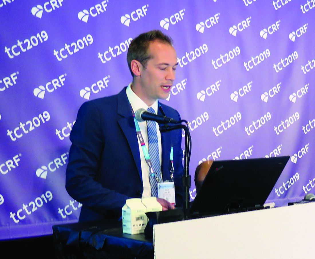
“TAVR has become an indispensable treatment option for patients with symptomatic severe aortic stenosis across all risk categories,” Jonas Lanz, MD, MSc, said at the Transcatheter Cardiovascular Therapeutics annual meeting. “The generalizability of outcomes observed in landmark trials comparing TAVR with SAVR [surgical aortic valve replacement] to other commercial TAVR systems is limited by differences in device properties and lack of head-to-head device comparisons. Iterations of the SAPIEN balloon-expandable TAVR system have been extensively investigated in several large-scale, high-quality, randomized trials and registries setting the current benchmark in terms of safety and efficacy.”
The ACURATE neo is a novel self-expanding TAVR system associated with favorable outcomes in nonrandomized studies. It has been available in Europe since 2014 but has not gained Food and Drug Administration approval.
Between February 2017 and February 2019, in a randomized trial known as SCOPE I, Dr. Lanz and colleagues at 20 European sites enrolled 739 patients with severe, symptomatic aortic stenosis at increased surgical risk. Of these, 372 were assigned to transfemoral TAVR with the ACURATE neo and 367 were assigned to the SAPIEN 3 system. The researchers designed the study to investigate noninferiority of the primary endpoint, which was a composite of safety and efficacy derived from the Valve Academic Research Consortium–2 criteria and included all-cause death, any stroke, life-threatening or disabling bleeding, major vascular complications, coronary artery obstruction requiring intervention, acute kidney injury stage 2 or higher, valve-related dysfunction requiring repeat procedure, rehospitalization for valve-related symptoms or heart failure, moderate or severe prosthetic valve regurgitation, or prosthetic valve stenosis at 30 days.
Clinical follow-up information at 30 days was available for 99% and ECG follow-up for about 98% of the total study population. The mean age of the study participants was 82 years, 57% were female, their median STS Risk Score was 3.5, and their mean transvalvular aortic gradient was 42.2 mm Hg. Dr. Lanz, with the department of cardiology at Bern (Switzerland) University Hospital, reported that the primary endpoint rate in the intention-to-treat cohort for ACURATE neo was 23.7%, compared with 16.5% with SAPIEN 3, falling short of the threshold for noninferiority (P for noninferiority = 0.42).
“I think this speaks to the fact of how high the bar is right now – how good the procedure is [and] how good the devices are,” invited discussant Michael J. Mack, MD, a cardiac surgeon at Baylor Scott & White Health, Dallas, said during the media briefing. “It’s an incredibly high bar to meet for any new device.”
Certain single components of the primary endpoint were similar in the ACURATE neo group, compared with the SAPIEN 3 group, including all-cause death (2.5% vs. 0.8%, respectively) and stroke (1.9% vs. 3%), but acute kidney injury was more common in the ACURATE neo group (3% vs. 0.8%, as was paravalvular aortic regurgitation (9.4% vs. 2.8%). “The differences between the two TAVR devices were mainly driven by moderate or severe paravalvular regurgitation and stage 2 or 3 acute kidney injury in favor of the SAPIEN 3 device,” Dr. Lanz said at the meeting sponsored by the Cardiovascular Research Foundation. “An early composite safety and efficacy endpoint proved useful in discriminating the performance of different TAVR systems.”
He pointed out that a trial called SCOPE II is underway in Europe, with a new iteration of the ACURATE neo designed to reduce the risk of paravalvular leak.
The results of SCOPE I were published online at the time of presentation (Lancet. 2019 Sep 27. doi: 10.1016/S0140-6736[19]32220-2). SCOPE I was an investigator-initiated and -conducted study funded by a dedicated research grant from Symetis in Ecublens, Switzerland (part of Boston Scientific). Dr. Lanz had no relevant disclosures.
SAN FRANCISCO – The ACURATE neo transcatheter aortic valve replacement (TAVR) device failed to meet noninferiority, compared with the SAPIEN 3 device, in terms of the primary composite safety and efficacy endpoint at 30 days, a randomized, comparative trial showed.
“TAVR has become an indispensable treatment option for patients with symptomatic severe aortic stenosis across all risk categories,” Jonas Lanz, MD, MSc, said at the Transcatheter Cardiovascular Therapeutics annual meeting. “The generalizability of outcomes observed in landmark trials comparing TAVR with SAVR [surgical aortic valve replacement] to other commercial TAVR systems is limited by differences in device properties and lack of head-to-head device comparisons. Iterations of the SAPIEN balloon-expandable TAVR system have been extensively investigated in several large-scale, high-quality, randomized trials and registries setting the current benchmark in terms of safety and efficacy.”
The ACURATE neo is a novel self-expanding TAVR system associated with favorable outcomes in nonrandomized studies. It has been available in Europe since 2014 but has not gained Food and Drug Administration approval.
Between February 2017 and February 2019, in a randomized trial known as SCOPE I, Dr. Lanz and colleagues at 20 European sites enrolled 739 patients with severe, symptomatic aortic stenosis at increased surgical risk. Of these, 372 were assigned to transfemoral TAVR with the ACURATE neo and 367 were assigned to the SAPIEN 3 system. The researchers designed the study to investigate noninferiority of the primary endpoint, which was a composite of safety and efficacy derived from the Valve Academic Research Consortium–2 criteria and included all-cause death, any stroke, life-threatening or disabling bleeding, major vascular complications, coronary artery obstruction requiring intervention, acute kidney injury stage 2 or higher, valve-related dysfunction requiring repeat procedure, rehospitalization for valve-related symptoms or heart failure, moderate or severe prosthetic valve regurgitation, or prosthetic valve stenosis at 30 days.
Clinical follow-up information at 30 days was available for 99% and ECG follow-up for about 98% of the total study population. The mean age of the study participants was 82 years, 57% were female, their median STS Risk Score was 3.5, and their mean transvalvular aortic gradient was 42.2 mm Hg. Dr. Lanz, with the department of cardiology at Bern (Switzerland) University Hospital, reported that the primary endpoint rate in the intention-to-treat cohort for ACURATE neo was 23.7%, compared with 16.5% with SAPIEN 3, falling short of the threshold for noninferiority (P for noninferiority = 0.42).
“I think this speaks to the fact of how high the bar is right now – how good the procedure is [and] how good the devices are,” invited discussant Michael J. Mack, MD, a cardiac surgeon at Baylor Scott & White Health, Dallas, said during the media briefing. “It’s an incredibly high bar to meet for any new device.”
Certain single components of the primary endpoint were similar in the ACURATE neo group, compared with the SAPIEN 3 group, including all-cause death (2.5% vs. 0.8%, respectively) and stroke (1.9% vs. 3%), but acute kidney injury was more common in the ACURATE neo group (3% vs. 0.8%, as was paravalvular aortic regurgitation (9.4% vs. 2.8%). “The differences between the two TAVR devices were mainly driven by moderate or severe paravalvular regurgitation and stage 2 or 3 acute kidney injury in favor of the SAPIEN 3 device,” Dr. Lanz said at the meeting sponsored by the Cardiovascular Research Foundation. “An early composite safety and efficacy endpoint proved useful in discriminating the performance of different TAVR systems.”
He pointed out that a trial called SCOPE II is underway in Europe, with a new iteration of the ACURATE neo designed to reduce the risk of paravalvular leak.
The results of SCOPE I were published online at the time of presentation (Lancet. 2019 Sep 27. doi: 10.1016/S0140-6736[19]32220-2). SCOPE I was an investigator-initiated and -conducted study funded by a dedicated research grant from Symetis in Ecublens, Switzerland (part of Boston Scientific). Dr. Lanz had no relevant disclosures.
REPORTING FROM TCT 2019
An off-the-shelf drug to rival CAR T cells: ‘very exciting’
ORLANDO – An investigational drug that can achieve the same results as complex cell therapy is creating a buzz at the American Society of Hematology (ASH) meeting.
For the last few years, attention at this meeting has focused on the chimeric antigen receptor (CAR) T cells, mainly “because of their incredible efficacy,” commented ASH Secretary Robert A. Brodsky, MD, professor of medicine and director of the division of hematology at Johns Hopkins University, Baltimore.
But new results with an off-the-shelf product are “very exciting,” he said, because the drug can be given immediately and appears to achieve similar results.
The new product is mosunetuzumab (Genentech/Roche), a bispecific antibody that targets both CD3 (on the surface of T cells) and CD20 (on the surface of B cells). It works by redirecting T cells to engage and eliminate malignant B cells.
“The concept here is that this monoclonal antibody engages T cells and directs their cytotoxicity against B cells – it’s basically an antibody using the patient’s own T cells to do what a CAR T cell would do,” Dr. Brodsky explained.
However, unlike CAR T cells, which are prepared for each individual patient in a complex process that involves genetic engineering that can take several weeks, mosunetuzumab is an off-the-shelf product that can be given to patients immediately (by intravenous infusion).
This is important, commented Dr. Brodsky, because very-poor-prognosis patients can deteriorate rapidly, and some may not survive while the CAR T cells are being made.
Clinical trial results
Clinical results come from a phase 1/1b trial (known as GO29781) conducted in 270 patients with poor-prognosis refractory/relapsed non-Hodgkin’s lymphoma. These patients had previously been treated with a median of three therapies; in addition, 30 patients (11%) were resistant to or had relapsed after an initial response to CAR T-cell therapy, and 77 patients (29%) had progressed after a stem cell transplant.
“These patients had no available therapy that would be expected to improve survival,” noted lead author Stephen J. Schuster, MD, of Abramson Cancer Center at the University of Pennsylvania in Philadelphia.
All patients received mosunetuzumab with an initial treatment of eight cycles. Patients who achieved complete remission (CR) stopped therapy, while patients who had a partial response or had stable disease, continued treatment for 17 cycles.
Two-thirds of patients (n = 180; 67%) had aggressive lymphomas, mainly diffuse large B-cell lymphoma (DLBCL; n = 117), while 85 patients (31%) had indolent disease, mainly follicular lymphoma (FL; n = 82). Objective responses were seen in 46 of 124 patients (37%) with aggressive lymphomas, and 24 (19%) of these patients achieved a CR.
Among patients with indolent lymphoma, objective responses were seen in 42 of 67 patients (63%), and 29 of 67 (43%) had CR.
The complete remissions appear to be long lasting, Dr. Schuster commented. With a median follow-up of 6 months since achieving CR, 17 of 24 patients (71%) with aggressive lymphoma and 24 of 29 patients (83%) with indolent lymphomas remained free of disease.
“Some patients have remained in remission without additional therapy for more than a year,” he commented.
In the subgroup of 30 patients who had previously received CAR T-cell therapy, the objective response rate was 38.9%, and CR was achieved in 4 patients (22%). These rates are similar to what was seen in patients with aggressive lymphoma who had not previously received CAR T-cell therapy, Dr. Schuster commented.
He also noted that in some of these patients, molecular testing showed that the previously administered CAR T cells increased in number. This suggests that, in addition to its ability to kill cancerous B cells, mosunetuzumab may also help augment the effect of the prior CAR T-cell treatment.
Dr. Schuster also highlighted the results of repeat treatment with mosunetuzumab. Patients who achieved CR stopped treatment – but if they relapsed, they were treated again, and the responses seen on this repeat treatment were similar to those seen with initial treatment. “This is not seen with the CAR T cells,” he noted.
Adverse events with mosunetuzumab were similar to those seen with CAR T cells, he noted, namely cytokine release syndrome, which was mostly mild and seen in 29% of patients, and neurologic toxicity, which was moderately severe in 4% patients.
Overall, the results show that “mosunetuzumab generates long-lasting responses with a very tolerable safety profile in patients with B-cell non-Hodgkin lymphomas for whom multiple prior treatments have failed and whose prognosis is poor. Of particular interest, we are seeing durable complete remissions in patients whose lymphomas progressed after CAR T,” Dr. Schuster commented in a statement.
Approached for comment, Peter Martin MD, chief of the Lymphoma Program at Weill Cornell Medicine, New York, and New York-Presbyterian, said he was excited to see these new data. “It’s good news any time we find something with the potential to save lives.”
“The more options that we have to offer to people with lymphoma the better,” he told Medscape Medical News. “There will always be scenarios where one approach might be better than another. I think there is a good chance that bispecific antibodies will have fairly broad approval in previously treated DLBCL. In many centers, it may be that bispecific antibodies are used most frequently post–CAR T cells, while in other areas people who aren’t candidates for CAR T cells or can’t receive them for whatever reason [could benefit from this new approach].”
Laurie Sehn, MD, MPH, medical oncologist at the University of British Columbia in Vancouver, Canada, and chair of the Lymphoma Tumour Group, as well as an associate editor of ASH journal Blood, also commented for Medscape Medical News.
She agreed that the new data are exciting and noted that this abstract was chosen for the plenary session. She thought the data in the 30 patients who had already been treated with CAR T cells was interesting. “This is a patient population with no other options that offer durable benefit, and mosunetuzumab clearly has clinical activity, with encouraging responses.”
Dr. Sehn also noted that toxicity seen with the drug was “far less” than has been seen with CAR T cells, and the risk of high-grade cytokine release syndrome and neurological toxicity is “very low.”
There are several other new products that are using this bispecific technology, she noted. One example is Regeneron’s REGN1979, a bispecific antibody targeting CD20 and CD3, which is also being investigated in a clinical trial in relapsed/refractory B-cell non-Hodgkin’s lymphoma, including in patients who were previously treated with CAR T cells (abstract 762).
How would it be used clinically?
In response to a question from Medscape Medical News, Dr. Schuster suggested that initial use of mosunetuzumab would be in patients who have already tried CAR T-cell therapy and had either not responded or relapsed – in lymphoma, this is about two-thirds of patients who are treated with this approach. This group of patients represents an unmet medical need, and this indication may be the quickest route to approval, he suggested.
Gary Schiller, MD, from UCLA Health, who moderated the press briefing agreed, and said this would be the quickest route to market because it would need only a phase 2 clinical trial in this specific patient population. But this would likely be only the first use for this product, and then it could be expanded to a broader patient population, he added.
Another use would for mosunetuzumab would be to enhance CAR T-cell responses by redirecting the CAR T cells to other antigens without doing any additional gene editing, Dr. Schuster commented. The idea here is to “revive” previously administered CAR T cells that have stopped working, Dr. Schiller added.
This is a chemotherapy-free approach, Dr. Schuster emphasized. “In patients who have not had a lot of chemotherapy, you can see an increase in T cells,” he commented.
Mosunetuzumab “stimulates and invigorates T cells,” and it could be useful as a pretreatment or a bridge to CAR T-cell therapy, he said.
So the product could be used before CAR T-cell therapy, and equally it could be used after CAR T-cell therapy because it could boost responses in both cases.
“Larger, randomized trials are needed to further confirm these promising data and determine whether the treatment benefit of mosunetuzumab is enhanced when it is used earlier in the course of lymphoma therapy or in combination with other agents,” he added.
Genentech says that mosunetuzumab and another bispecific antibody, CD20-TCB, are being evaluated in a robust clinical development program, both as a monotherapies and in combination with other therapies, in both aggressive and indolent non-Hodgkin’s lymphoma.
Dr. Schuster reported relationships with Celgene, Genentech, Merck, Pharmacyclics, Acerta, AbbVie, Gilead, Nordic Nanovector, Pfizer, AstraZeneca, Loxo Oncology, and Novartis. Coauthors also have multiple disclosures, and several are employees of Genentech and Roche. Dr. Sehn consults with several pharmaceutics companies, including Verastem, Roche/Genentech, Morphosys, Takeda, Janssen, Lundbeck, Amgen, Teva, and AbbVie.
A version of this story originally appeared on Medscape.com.
Employment is associated with high likelihood of declining epilepsy surgery
BALTIMORE – , according to an analysis presented at the annual meeting of the American Epilepsy Society. “Future work should confirm this finding prospectively, determine if it holds in other patient populations, and explore the decision to proceed with or decline epilepsy surgery from a patient-centered perspective,” said Vishal Mandge, MD, MPH, a clinical neurophysiology fellow at Duke University in Durham, N.C., and colleagues. “Identifying the role that factors such as the fear of losing employment due to complications from surgery and inability to take medical leave for an extended period of time play in the patient’s decision to proceed with epilepsy surgery may identify needs and suggest strategies to reduce barriers to this underutilized treatment.”
Although epilepsy surgery is known to be safe and effective, many surgical candidates with drug-resistant epilepsy decline to undergo the procedure. Prior investigations of the barriers to epilepsy surgery have focused on access to epilepsy centers that offer epilepsy surgery and patients’ reluctance to undergo presurgical evaluation. Dr. Mandge and colleagues instead set out to evaluate the association between various demographic, disease-specific, and epilepsy-evaluation variables and patients’ decision to decline surgery after they have been identified as candidates.
A retrospective case-control study
The investigators conducted a retrospective case-control study of patients who were discussed at the epilepsy surgery conference of a tertiary care hospital serving an urban New York community between Jan. 1, 2009, and June 30, 2017. They identified patients who were considered candidates for resective epilepsy surgery. Dr. Mandge and colleagues used the chi-squared test for nominal variables and analysis of variance for scale variables to evaluate these variables’ associations with a patient’s decision to decline epilepsy surgery. They also performed multivariate binary logistic regression to identify variables that predict a patient’s decision to decline surgery.
Dr. Mandge and colleagues identified 159 patients who were discussed during the study period. Of this group, 87 patients were eligible for resective epilepsy surgery after a thorough evaluation. Thirty-four (40%) of the eligible patients declined to undergo surgery. Approximately 20% of eligible patients were employed, and 70% of patients had a high school diploma or higher education.
Univariate analysis indicated that employment (odds ratio, 4.2), temporal lesion on MRI (OR, 0.35), temporal EEG localization (OR, 0.21), and temporal seizure onset zone (OR, 0.19) were independently and significantly associated with a patient’s decision to decline surgery. Multivariate logistic regression analysis indicated that current employment (OR, 7.5), the number of current antiepileptic drugs (AEDs; OR, 3.5), and concordance between seizure semiology, seizure onset on EEG, and imaging (OR, 0.08) were significantly associated with a patient’s decision to decline surgery.
Fear of unemployment may explain results

“With each additional AED, the patients were 3.5 times more likely to decline surgery, even after adjusting for other variables,” said Alexis D. Boro, MD, a neurologist at Montefiore Medical Center in New York and one of the investigators. “My suspicion is that some of this reflects the burden of taking a lot of seizure medication. While the medications are much, much safer than seizures, and looking for and dealing with side effects is a lot of what we do, people often don’t feel great when they are taking multiple seizure medications. We counsel our patients that they should generally expect to stay on some seizure medications after surgery. The reason for surgery is to stop the seizures, not to stop the medications. We are often able to reduce medications after a period of time after surgery, and for many patients, this is one of the benefits.”
The association between employment and increased likelihood of declining surgery was unexpected and may not hold everywhere, said Dr. Boro. “We had expected the opposite result because we assumed that employed patients would be concerned that a seizure at work might result in loss of work. But it may be that many of our patients who are employed are concerned about losing their jobs if they miss work for a medical procedure. Some of our patients may be concerned about sharing medical information with their employers. For some of our patients, being employed may imply limited insurance coverage.”
The study was not supported by external funding, and the investigators did not report any disclosures.
SOURCE: Mandge VA et al. AES 2019, Abstract 1.362.
BALTIMORE – , according to an analysis presented at the annual meeting of the American Epilepsy Society. “Future work should confirm this finding prospectively, determine if it holds in other patient populations, and explore the decision to proceed with or decline epilepsy surgery from a patient-centered perspective,” said Vishal Mandge, MD, MPH, a clinical neurophysiology fellow at Duke University in Durham, N.C., and colleagues. “Identifying the role that factors such as the fear of losing employment due to complications from surgery and inability to take medical leave for an extended period of time play in the patient’s decision to proceed with epilepsy surgery may identify needs and suggest strategies to reduce barriers to this underutilized treatment.”
Although epilepsy surgery is known to be safe and effective, many surgical candidates with drug-resistant epilepsy decline to undergo the procedure. Prior investigations of the barriers to epilepsy surgery have focused on access to epilepsy centers that offer epilepsy surgery and patients’ reluctance to undergo presurgical evaluation. Dr. Mandge and colleagues instead set out to evaluate the association between various demographic, disease-specific, and epilepsy-evaluation variables and patients’ decision to decline surgery after they have been identified as candidates.
A retrospective case-control study
The investigators conducted a retrospective case-control study of patients who were discussed at the epilepsy surgery conference of a tertiary care hospital serving an urban New York community between Jan. 1, 2009, and June 30, 2017. They identified patients who were considered candidates for resective epilepsy surgery. Dr. Mandge and colleagues used the chi-squared test for nominal variables and analysis of variance for scale variables to evaluate these variables’ associations with a patient’s decision to decline epilepsy surgery. They also performed multivariate binary logistic regression to identify variables that predict a patient’s decision to decline surgery.
Dr. Mandge and colleagues identified 159 patients who were discussed during the study period. Of this group, 87 patients were eligible for resective epilepsy surgery after a thorough evaluation. Thirty-four (40%) of the eligible patients declined to undergo surgery. Approximately 20% of eligible patients were employed, and 70% of patients had a high school diploma or higher education.
Univariate analysis indicated that employment (odds ratio, 4.2), temporal lesion on MRI (OR, 0.35), temporal EEG localization (OR, 0.21), and temporal seizure onset zone (OR, 0.19) were independently and significantly associated with a patient’s decision to decline surgery. Multivariate logistic regression analysis indicated that current employment (OR, 7.5), the number of current antiepileptic drugs (AEDs; OR, 3.5), and concordance between seizure semiology, seizure onset on EEG, and imaging (OR, 0.08) were significantly associated with a patient’s decision to decline surgery.
Fear of unemployment may explain results
“With each additional AED, the patients were 3.5 times more likely to decline surgery, even after adjusting for other variables,” said Alexis D. Boro, MD, a neurologist at Montefiore Medical Center in New York and one of the investigators. “My suspicion is that some of this reflects the burden of taking a lot of seizure medication. While the medications are much, much safer than seizures, and looking for and dealing with side effects is a lot of what we do, people often don’t feel great when they are taking multiple seizure medications. We counsel our patients that they should generally expect to stay on some seizure medications after surgery. The reason for surgery is to stop the seizures, not to stop the medications. We are often able to reduce medications after a period of time after surgery, and for many patients, this is one of the benefits.”
The association between employment and increased likelihood of declining surgery was unexpected and may not hold everywhere, said Dr. Boro. “We had expected the opposite result because we assumed that employed patients would be concerned that a seizure at work might result in loss of work. But it may be that many of our patients who are employed are concerned about losing their jobs if they miss work for a medical procedure. Some of our patients may be concerned about sharing medical information with their employers. For some of our patients, being employed may imply limited insurance coverage.”
The study was not supported by external funding, and the investigators did not report any disclosures.
SOURCE: Mandge VA et al. AES 2019, Abstract 1.362.
REPORTING FROM AES 2019
Sequential intercept model is really a ‘no-intercept model’
Ultimately, psychiatrists must take responsibility for complex patients.
In legal settings, the “sequential intercept model” for targeting people involved in the criminal justice system with mental illness has been proposed as an improvement for the status quo.

The model intends to divert individuals with mental illnesses at any one of five described stages in their journey through the legal system. In the first stage, a patient may be provided enough care in the community to never enter the criminal system. If that works, the patient may be diverted by first responders out of the legal system and back into treatment. Sequentially, throughout the remaining stages, the patient can be diverted by an attorney, the court, a presentencing correctional facility, the sentencing judge, a postsentencing correctional facility, or probation. The model rightfully encourages anyone in the continuum of care to take ownership of a situation and intervene.
I applaud the model for encouraging all participants to intervene in changing the course of our most challenging patients. However, I am reminded of the complexity of large systems trying to change. In practice, what I have seen is a series of half-hearted recommendations: Emergency responders who consider their role finished after giving a patient the number of the suicide hotline, attorneys who are satisfied by giving their clients an outdated list of community mental health clinics, judges who interpret their recommendations for treatment as a fait accompli, and correctional facilities that release patients with an absurdly short supply of medications and the address of an emergency room. I worry that by creating a model encouraging all to participate, we have just absolved ones who make any effort, even if inadequate.
In some ways, the sequential intercept model has similarities with modern mental health treatment teams. In many settings, a treatment team includes a series of providers who are sequentially involved in the life of a patient. A team can include a psychiatrist for psychopharmacology; a neuropsychologist for psychological testing; a social worker for psychotherapeutic strategies; another social worker to assist in obtaining social assistance; an addiction counselor for substance use disorder; another psychiatrist who monitors the administration of a single medication, like ketamine; and a pharmacist who approves the medication regimen. That’s several providers for the treatment of one patient.
As a forensic psychiatrist, I am often asked to review treatment plans of other providers. I am asked to comment on the appropriate nature of a given treatment. Often, insurance companies want to review the continued need for treatment or whether any treatment is warranted at all. Sometimes, employers want to review a treatment plan to ensure the safety of their employees. At times, courts will ask for a review and expectations from treatment of a defendant to assist in sentencing determinations. However, I have not yet been asked by anyone if the amount of care a patient is obtaining is too fragmented and without any clear leadership.
In our endless pursuit of medicalization and standardization of mental health, we have, especially in large systems, created specialization silos for the care of our patients. Many, if not most psychiatrists, do not participate in any psychotherapy; social workers and psychologists do not prescribe (for the most part); many substance abuse counselors only address sobriety and not other primary mental illness factors; and pharmacists cannot diagnose nor are they trained in psychosocial approaches. In many ways, we have defined participants not by what they do, but what they don’t do.
One also can be saddened by the enormous logistical complexity imposed on patients required to make numerous appointments, which can deprive them of time for recovery. However, my bigger concern is that the multiplicity of providers also permits the dissolution of accountability. In my experience, those large teams have an ability to deflect responsibility in ways that are unmatched by any single provider who cannot rely on putting the fault on someone else.
Sadly and ironically, those two parallel paradigms of mental illness and criminal care impose those problems on each other by averting any attempt at interception, a “no-intercept model.” Mental health programs will deny clients involved in the criminal justice system for requiring too much treatment, too little treatment, for lack of availability of one of the necessary providers, for requiring substance use treatment, or simply for being part of the criminal justice system. Accordingly, the legal system will fail to accept recommendations by mental health providers that mental health treatment is not paramount at this time and that the defendant would be better served by addressing his criminogenic risk factors. In response, the multitude of participants in the legal system will point to the mental health system for all answers.
Contrary to many if not most problems, I do not think that the solution lies somewhere in the middle, as this would require the five stages of the legal system to compromise with the nine hypothetical participants of the mental health system. For our part, as psychiatrists, we must accept that we are ultimately responsible for all levels of care. As a field, we are also responsible for educating the public and the legal system of our role and limitations in providing care as well as being available for providing such care. Correspondingly, the legal system is responsible for putting an adequate effort into diverting patients and having or obtaining adequate understanding of available and appropriate care for their defendants.
Dr. Badre is a forensic psychiatrist in San Diego and an expert in correctional mental health. He holds teaching positions at the University of California, San Diego, and the University of San Diego. He teaches medical education, psychopharmacology, ethics in psychiatry, and correctional care. Among his writings is chapter 7 in the new book “Critical Psychiatry: Controversies and Clinical Implications” (Springer, 2019).
Ultimately, psychiatrists must take responsibility for complex patients.
Ultimately, psychiatrists must take responsibility for complex patients.
In legal settings, the “sequential intercept model” for targeting people involved in the criminal justice system with mental illness has been proposed as an improvement for the status quo.
The model intends to divert individuals with mental illnesses at any one of five described stages in their journey through the legal system. In the first stage, a patient may be provided enough care in the community to never enter the criminal system. If that works, the patient may be diverted by first responders out of the legal system and back into treatment. Sequentially, throughout the remaining stages, the patient can be diverted by an attorney, the court, a presentencing correctional facility, the sentencing judge, a postsentencing correctional facility, or probation. The model rightfully encourages anyone in the continuum of care to take ownership of a situation and intervene.
I applaud the model for encouraging all participants to intervene in changing the course of our most challenging patients. However, I am reminded of the complexity of large systems trying to change. In practice, what I have seen is a series of half-hearted recommendations: Emergency responders who consider their role finished after giving a patient the number of the suicide hotline, attorneys who are satisfied by giving their clients an outdated list of community mental health clinics, judges who interpret their recommendations for treatment as a fait accompli, and correctional facilities that release patients with an absurdly short supply of medications and the address of an emergency room. I worry that by creating a model encouraging all to participate, we have just absolved ones who make any effort, even if inadequate.
In some ways, the sequential intercept model has similarities with modern mental health treatment teams. In many settings, a treatment team includes a series of providers who are sequentially involved in the life of a patient. A team can include a psychiatrist for psychopharmacology; a neuropsychologist for psychological testing; a social worker for psychotherapeutic strategies; another social worker to assist in obtaining social assistance; an addiction counselor for substance use disorder; another psychiatrist who monitors the administration of a single medication, like ketamine; and a pharmacist who approves the medication regimen. That’s several providers for the treatment of one patient.
As a forensic psychiatrist, I am often asked to review treatment plans of other providers. I am asked to comment on the appropriate nature of a given treatment. Often, insurance companies want to review the continued need for treatment or whether any treatment is warranted at all. Sometimes, employers want to review a treatment plan to ensure the safety of their employees. At times, courts will ask for a review and expectations from treatment of a defendant to assist in sentencing determinations. However, I have not yet been asked by anyone if the amount of care a patient is obtaining is too fragmented and without any clear leadership.
In our endless pursuit of medicalization and standardization of mental health, we have, especially in large systems, created specialization silos for the care of our patients. Many, if not most psychiatrists, do not participate in any psychotherapy; social workers and psychologists do not prescribe (for the most part); many substance abuse counselors only address sobriety and not other primary mental illness factors; and pharmacists cannot diagnose nor are they trained in psychosocial approaches. In many ways, we have defined participants not by what they do, but what they don’t do.
One also can be saddened by the enormous logistical complexity imposed on patients required to make numerous appointments, which can deprive them of time for recovery. However, my bigger concern is that the multiplicity of providers also permits the dissolution of accountability. In my experience, those large teams have an ability to deflect responsibility in ways that are unmatched by any single provider who cannot rely on putting the fault on someone else.
Sadly and ironically, those two parallel paradigms of mental illness and criminal care impose those problems on each other by averting any attempt at interception, a “no-intercept model.” Mental health programs will deny clients involved in the criminal justice system for requiring too much treatment, too little treatment, for lack of availability of one of the necessary providers, for requiring substance use treatment, or simply for being part of the criminal justice system. Accordingly, the legal system will fail to accept recommendations by mental health providers that mental health treatment is not paramount at this time and that the defendant would be better served by addressing his criminogenic risk factors. In response, the multitude of participants in the legal system will point to the mental health system for all answers.
Contrary to many if not most problems, I do not think that the solution lies somewhere in the middle, as this would require the five stages of the legal system to compromise with the nine hypothetical participants of the mental health system. For our part, as psychiatrists, we must accept that we are ultimately responsible for all levels of care. As a field, we are also responsible for educating the public and the legal system of our role and limitations in providing care as well as being available for providing such care. Correspondingly, the legal system is responsible for putting an adequate effort into diverting patients and having or obtaining adequate understanding of available and appropriate care for their defendants.
Dr. Badre is a forensic psychiatrist in San Diego and an expert in correctional mental health. He holds teaching positions at the University of California, San Diego, and the University of San Diego. He teaches medical education, psychopharmacology, ethics in psychiatry, and correctional care. Among his writings is chapter 7 in the new book “Critical Psychiatry: Controversies and Clinical Implications” (Springer, 2019).
ID Consult: It’s not necessarily over when measles infection clears
As I write, I imagine readers groaning at yet another measles story. But in early November 2019, in Portland, Oregon, Judy Guzman-Cottrill, DO, recently was groaning at yet another measles case.

Dr. Guzman-Cottrill, a pediatric infectious diseases specialist at Doernbecher Children’s Hospital, recently shared details provided by the local health department:
An unimmunized child developed measles while traveling outside the county. The child may have exposed others at Portland International Airport, a medical center in Vancouver, and potentially at another children’s hospital in the area.
As of Nov. 7, 2019, 1,261 cases of measles from 31 states had been reported to the Centers for Disease Control and Prevention – more cases in a single year since 1992. The case in Portland added at least one to that total, although public officials warned that additional cases could occur Nov. 18th through Dec. 9 (given the incubation period). Like the child in Oregon, most of the individuals who developed measles nationwide in 2019 were unimmunized. At press time, from Jan. 1 to Dec. 5, 2019, 1,276 individual cases of measles have been confirmed in 31 states; CDC released measles reports monthly.
The reasons for refusal of measles vaccine vary, but historically, some parents have made a calculated risk. Measles is rare. Most children are vaccinated. My child will be protected by herd immunity. In some communities, that is no longer true, as we have seen in 2019.
Other parents have decided – erroneously – that measles infection is less risky than measles vaccine. We need to be able to tell them the facts. Thirty percent of individuals who contract measles will develop at least one complication, according to the Centers for Disease Control and Prevention. One in four will be hospitalized. While death from acute measles infection is uncommon, children remain at risk for sequelae months or years after the initial infection.
For example, measles is known to suppress the immune system, an effect that lasts for months or years after the initial infection. Practically, this means that once a child recovers from acute measles infection, he or she has an increased susceptibility to other infections that may last for years. Two studies published late in 2019 described the immune “amnesia” that occurs following measles infection. Essentially, the immune system forgets how to fight other pathogens, leaving children vulnerable to potentially life-threatening infections.
Michael Mina, MD, of the Harvard T.H. Chan School of Public Health, Boston, and colleagues measured the effects of measles infection on the immune system by studying blood samples taken from 77 unimmunized children in the Netherlands before and after measles infection.1 Two months after recovery from mild measles, children had lost a median of 33 % (range, 12%-73%) of preexisting antibodies against a range of common viruses and bacteria. The median loss was 40% after severe measles (range 11% to 62%). Similar changes were not observed after measles vaccine.

A second group of researchers led by Velislava N. Petrova, PhD, of the Wellcome Sanger Institute in Cambridge, England, investigated genetic changes in 26 unvaccinated children from the Netherlands who previously had measles. They found that measles infection reduced the diversity of immune cells available to recognize and fight infections and depleted memory B cells, essentially returning the immune to a more immature state.2
Parents also need to know that children who develop measles are at risk for noninfectious complications.
Yes, SSPE is a rare, but it is not as rare as we once thought. In 2017, investigators in California described 17 cases of SSPE identified in that state between 1998 and 2005.3 The incidence of SSPE was 1 in 1,367 for children less than 5 years at the time of measles infection and 1 in 609 for children less than 12 months when they contracted the virus.
Dr. Guzman-Cottrill has seen a case of SSPE, and she hopes to never see another one. “He had been a healthy 11-year-old boy,” she recalled. “He played soccer and basketball and did well in school.” In the beginning, his symptoms were insidious and nonspecific, Dr. Guzman-Cottrill and colleagues wrote in a 2016 issue of Morbidity and Mortality Weekly Report.4 He started to struggle in school. He dozed off in the middle of meals. He started to drop things. Over a 4-month period, the boy developed progressive spasticity, became unable to eat or drink, and could no longer recognize or communicate with his family. “That’s when I met him,” Dr. Guzman-Cottrill said. “It was heartbreaking, and there was very little we could do for him except give the family a diagnosis. He eventually died in hospice care, nearly 4 years after his symptoms began.”
The boy had been infected with measles at 1 year of age while living in the Philippines. Dr. Guzman-Cottrill emphasized that this family had not refused measles immunization. The child had received a measles vaccine at 8 months of age, but a single vaccine at such a young age wasn’t enough to protect him.
We can hope for change in 2020, including improved immunization rates and a decline in measles cases. If that happens, measles will no longer be a hot topic in the news. We’ll likely never know what happens to the children infected in 2019, those who are facing the current cold and flu season with impaired immune systems. A decade or more will pass before we’ll know if anyone develops SSPE. For now, all we can do is wait … and worry.
Dr. Bryant is a pediatrician specializing in infectious diseases at the University of Louisville, Ky., and Norton Children’s Hospital, also in Louisville. Dr. Bryant had no relevant financial disclosures. Email her at [email protected].
References
1. Science. 2019 Nov 1;366:599-606.
2. Science Immunology. 2019 Nov 1;4:eaay6125.
As I write, I imagine readers groaning at yet another measles story. But in early November 2019, in Portland, Oregon, Judy Guzman-Cottrill, DO, recently was groaning at yet another measles case.
Dr. Guzman-Cottrill, a pediatric infectious diseases specialist at Doernbecher Children’s Hospital, recently shared details provided by the local health department:
An unimmunized child developed measles while traveling outside the county. The child may have exposed others at Portland International Airport, a medical center in Vancouver, and potentially at another children’s hospital in the area.
As of Nov. 7, 2019, 1,261 cases of measles from 31 states had been reported to the Centers for Disease Control and Prevention – more cases in a single year since 1992. The case in Portland added at least one to that total, although public officials warned that additional cases could occur Nov. 18th through Dec. 9 (given the incubation period). Like the child in Oregon, most of the individuals who developed measles nationwide in 2019 were unimmunized. At press time, from Jan. 1 to Dec. 5, 2019, 1,276 individual cases of measles have been confirmed in 31 states; CDC released measles reports monthly.
The reasons for refusal of measles vaccine vary, but historically, some parents have made a calculated risk. Measles is rare. Most children are vaccinated. My child will be protected by herd immunity. In some communities, that is no longer true, as we have seen in 2019.
Other parents have decided – erroneously – that measles infection is less risky than measles vaccine. We need to be able to tell them the facts. Thirty percent of individuals who contract measles will develop at least one complication, according to the Centers for Disease Control and Prevention. One in four will be hospitalized. While death from acute measles infection is uncommon, children remain at risk for sequelae months or years after the initial infection.
For example, measles is known to suppress the immune system, an effect that lasts for months or years after the initial infection. Practically, this means that once a child recovers from acute measles infection, he or she has an increased susceptibility to other infections that may last for years. Two studies published late in 2019 described the immune “amnesia” that occurs following measles infection. Essentially, the immune system forgets how to fight other pathogens, leaving children vulnerable to potentially life-threatening infections.
Michael Mina, MD, of the Harvard T.H. Chan School of Public Health, Boston, and colleagues measured the effects of measles infection on the immune system by studying blood samples taken from 77 unimmunized children in the Netherlands before and after measles infection.1 Two months after recovery from mild measles, children had lost a median of 33 % (range, 12%-73%) of preexisting antibodies against a range of common viruses and bacteria. The median loss was 40% after severe measles (range 11% to 62%). Similar changes were not observed after measles vaccine.
A second group of researchers led by Velislava N. Petrova, PhD, of the Wellcome Sanger Institute in Cambridge, England, investigated genetic changes in 26 unvaccinated children from the Netherlands who previously had measles. They found that measles infection reduced the diversity of immune cells available to recognize and fight infections and depleted memory B cells, essentially returning the immune to a more immature state.2
Parents also need to know that children who develop measles are at risk for noninfectious complications.
Yes, SSPE is a rare, but it is not as rare as we once thought. In 2017, investigators in California described 17 cases of SSPE identified in that state between 1998 and 2005.3 The incidence of SSPE was 1 in 1,367 for children less than 5 years at the time of measles infection and 1 in 609 for children less than 12 months when they contracted the virus.
Dr. Guzman-Cottrill has seen a case of SSPE, and she hopes to never see another one. “He had been a healthy 11-year-old boy,” she recalled. “He played soccer and basketball and did well in school.” In the beginning, his symptoms were insidious and nonspecific, Dr. Guzman-Cottrill and colleagues wrote in a 2016 issue of Morbidity and Mortality Weekly Report.4 He started to struggle in school. He dozed off in the middle of meals. He started to drop things. Over a 4-month period, the boy developed progressive spasticity, became unable to eat or drink, and could no longer recognize or communicate with his family. “That’s when I met him,” Dr. Guzman-Cottrill said. “It was heartbreaking, and there was very little we could do for him except give the family a diagnosis. He eventually died in hospice care, nearly 4 years after his symptoms began.”
The boy had been infected with measles at 1 year of age while living in the Philippines. Dr. Guzman-Cottrill emphasized that this family had not refused measles immunization. The child had received a measles vaccine at 8 months of age, but a single vaccine at such a young age wasn’t enough to protect him.
We can hope for change in 2020, including improved immunization rates and a decline in measles cases. If that happens, measles will no longer be a hot topic in the news. We’ll likely never know what happens to the children infected in 2019, those who are facing the current cold and flu season with impaired immune systems. A decade or more will pass before we’ll know if anyone develops SSPE. For now, all we can do is wait … and worry.
Dr. Bryant is a pediatrician specializing in infectious diseases at the University of Louisville, Ky., and Norton Children’s Hospital, also in Louisville. Dr. Bryant had no relevant financial disclosures. Email her at [email protected].
References
1. Science. 2019 Nov 1;366:599-606.
2. Science Immunology. 2019 Nov 1;4:eaay6125.
