User login
We Have a New Congressional Champion in the Fight Against CRC!
Rep. Yadira Caraveo, MD (D-CO), recently introduced the Colorectal Cancer Early Detection Act along with Reps. Donald Payne Jr. (D-NJ), Haley Stevens (D-MI), and Terri Sewell (D-AL).
The Colorectal Cancer Early Detection Act would award grants to states to promote colorectal cancer prevention and early detection efforts to individuals under age 45.

Grants would be used to:
- Screen increased risk and high-risk individuals under age 45 for colorectal cancer.
- Provide appropriate referrals for medical treatment.
- Develop and carry out a public education and awareness campaign for the detection and control of CRC.
- Improve the education and training of health providers in detecting and controlling CRC.
- Establish mechanisms through which states can monitor the quality of CRC screening procedures.
- Develop strategies to assess family history and genetic predispositions to CRC.
- Design patient and clinician decision support tools for CRC.
- Conduct surveillance to determine other risk factors for CRC in this population.
“Colorectal cancer is the second-leading cause of cancer death in the US and is increasing at an alarming rate in younger people. AGA celebrates Rep. Caraveo’s work to address this trend through education and awareness” said Barbara Jung, MD, AGA President.
We look forward to working with our congressional champions to increase screening rates and reverse the trend of early onset colorectal cancer!
Rep. Yadira Caraveo, MD (D-CO), recently introduced the Colorectal Cancer Early Detection Act along with Reps. Donald Payne Jr. (D-NJ), Haley Stevens (D-MI), and Terri Sewell (D-AL).
The Colorectal Cancer Early Detection Act would award grants to states to promote colorectal cancer prevention and early detection efforts to individuals under age 45.
Grants would be used to:
- Screen increased risk and high-risk individuals under age 45 for colorectal cancer.
- Provide appropriate referrals for medical treatment.
- Develop and carry out a public education and awareness campaign for the detection and control of CRC.
- Improve the education and training of health providers in detecting and controlling CRC.
- Establish mechanisms through which states can monitor the quality of CRC screening procedures.
- Develop strategies to assess family history and genetic predispositions to CRC.
- Design patient and clinician decision support tools for CRC.
- Conduct surveillance to determine other risk factors for CRC in this population.
“Colorectal cancer is the second-leading cause of cancer death in the US and is increasing at an alarming rate in younger people. AGA celebrates Rep. Caraveo’s work to address this trend through education and awareness” said Barbara Jung, MD, AGA President.
We look forward to working with our congressional champions to increase screening rates and reverse the trend of early onset colorectal cancer!
Pharmacogenomic Testing in Depression Treatment Decision Making: Clinical Pearls for Everyday Practice
AGA Clinical Practice Update Describes High-Quality Upper Endoscopy
The update, authored by Satish Nagula, MD, of Icahn School of Medicine at Mount Sinai, New York, NY, and colleagues, includes nine pieces of best practice advice that address procedure optimization, evaluation of suspected premalignancy, and postprocedure follow-up evaluation.

“Defining what constitutes a high-quality esophagogastroduodenoscopy (EGD) poses somewhat of a challenge because the spectrum of indications and the breadth of benign and (pre)malignant disease pathology in the upper GI tract is very broad,” the update panelists wrote in Clinical Gastroenterology and Hepatology. “Standardizing the measures defining a high-quality upper endoscopic examination is one of the first steps for assessing quality.”
Preprocedure Recommendations
Dr. Nagula and colleagues first emphasized that EGD should be performed for an appropriate indication, citing a recent meta-analysis that found 21.7% of upper endoscopy procedures were performed for an inappropriate indication. Of note, diagnostic yields were 42% higher in procedures performed for an appropriate indication.
After ensuring an appropriate indication, the update also encourages clinicians to inform patients of the various benefits, risks, and alternatives of the procedure prior to providing consent.
Intraprocedure Recommendations
During the procedure, endoscopists should take several steps to ensure optimal visualization of tissues, according to the update.
First, a high-definition (HD) white-light endoscopy system should be employed.
“Although HD imaging is a standard feature of newer-generation endoscopes, legacy standard-definition scopes remain in use,” Dr. Nagula and colleagues noted. “Moreover, to provide true HD image resolution, each component of the system (eg, the endoscope video chip, the processor, the monitor, and transmission cables) must be HD compatible.”
This HD-compatible system should be coupled with image-enhancing technology to further improve lesion detection. In Barrett’s esophagus, the panelists noted, image enhancement can improve lesion detection as much as 20%.
They predicted that AI-assisted software may boost detection rates even higher: “Computer-aided detection and computer-aided diagnosis systems for upper endoscopy are still in the early phases of development but do show similar promise for improving the detection and characterization of upper GI tract neoplasia.”
Beyond selection of best available technologies, the update encourages more fundamental strategies to improve visualization, including mucosal cleansing and insufflation, with sufficient time spent inspecting the foregut mucosa via anterograde and retroflexed views.
Where appropriate, standardized biopsy protocols should be followed to evaluate and manage foregut conditions.
Postprocedure Recommendations
After the procedure, endoscopists should offer patients management recommendations based on the endoscopic findings and, if necessary, notify them that more recommendations may be forthcoming based on histopathology results, according to the update.
Similarly, endoscopists should follow established surveillance intervals for future procedures, with modifications made as needed, based on histopathology findings.
Document, Document, Document
Throughout the update, Dr. Nagula and colleagues repeatedly emphasize the importance of documentation, from preprocedural discussions with patients through planned surveillance schedules.
However, the recommendations are clear about “weighing the practical implications” of “onerous” documentation, particularly photodocumentation requirements. For instance, the authors note that “there are some scenarios in which more rigorous photodocumentation standards during upper endoscopy should be considered, such as patients with risk factors for neoplasia,” but at the very least “photodocumentation of any suspicious abnormalities, ideally with annotations, is strongly advised.”
Moving Toward Quality Standardization for Upper Endoscopy
“These best practice advice statements are intended to improve measurable clinical, patient-reported, and economic healthcare outcomes and are not meant to put an additional burden on endoscopists,” the panelists wrote. “Ideally, future research will set threshold indicators of adherence to these best practices that optimally are associated with these aforementioned objective outcomes.”
This update was commissioned and approved by AGA. The update panelists disclosed relationships with Covidien LP, Fujifilm USA, Mahana Therapeutics, and others.
The update, authored by Satish Nagula, MD, of Icahn School of Medicine at Mount Sinai, New York, NY, and colleagues, includes nine pieces of best practice advice that address procedure optimization, evaluation of suspected premalignancy, and postprocedure follow-up evaluation.
“Defining what constitutes a high-quality esophagogastroduodenoscopy (EGD) poses somewhat of a challenge because the spectrum of indications and the breadth of benign and (pre)malignant disease pathology in the upper GI tract is very broad,” the update panelists wrote in Clinical Gastroenterology and Hepatology. “Standardizing the measures defining a high-quality upper endoscopic examination is one of the first steps for assessing quality.”
Preprocedure Recommendations
Dr. Nagula and colleagues first emphasized that EGD should be performed for an appropriate indication, citing a recent meta-analysis that found 21.7% of upper endoscopy procedures were performed for an inappropriate indication. Of note, diagnostic yields were 42% higher in procedures performed for an appropriate indication.
After ensuring an appropriate indication, the update also encourages clinicians to inform patients of the various benefits, risks, and alternatives of the procedure prior to providing consent.
Intraprocedure Recommendations
During the procedure, endoscopists should take several steps to ensure optimal visualization of tissues, according to the update.
First, a high-definition (HD) white-light endoscopy system should be employed.
“Although HD imaging is a standard feature of newer-generation endoscopes, legacy standard-definition scopes remain in use,” Dr. Nagula and colleagues noted. “Moreover, to provide true HD image resolution, each component of the system (eg, the endoscope video chip, the processor, the monitor, and transmission cables) must be HD compatible.”
This HD-compatible system should be coupled with image-enhancing technology to further improve lesion detection. In Barrett’s esophagus, the panelists noted, image enhancement can improve lesion detection as much as 20%.
They predicted that AI-assisted software may boost detection rates even higher: “Computer-aided detection and computer-aided diagnosis systems for upper endoscopy are still in the early phases of development but do show similar promise for improving the detection and characterization of upper GI tract neoplasia.”
Beyond selection of best available technologies, the update encourages more fundamental strategies to improve visualization, including mucosal cleansing and insufflation, with sufficient time spent inspecting the foregut mucosa via anterograde and retroflexed views.
Where appropriate, standardized biopsy protocols should be followed to evaluate and manage foregut conditions.
Postprocedure Recommendations
After the procedure, endoscopists should offer patients management recommendations based on the endoscopic findings and, if necessary, notify them that more recommendations may be forthcoming based on histopathology results, according to the update.
Similarly, endoscopists should follow established surveillance intervals for future procedures, with modifications made as needed, based on histopathology findings.
Document, Document, Document
Throughout the update, Dr. Nagula and colleagues repeatedly emphasize the importance of documentation, from preprocedural discussions with patients through planned surveillance schedules.
However, the recommendations are clear about “weighing the practical implications” of “onerous” documentation, particularly photodocumentation requirements. For instance, the authors note that “there are some scenarios in which more rigorous photodocumentation standards during upper endoscopy should be considered, such as patients with risk factors for neoplasia,” but at the very least “photodocumentation of any suspicious abnormalities, ideally with annotations, is strongly advised.”
Moving Toward Quality Standardization for Upper Endoscopy
“These best practice advice statements are intended to improve measurable clinical, patient-reported, and economic healthcare outcomes and are not meant to put an additional burden on endoscopists,” the panelists wrote. “Ideally, future research will set threshold indicators of adherence to these best practices that optimally are associated with these aforementioned objective outcomes.”
This update was commissioned and approved by AGA. The update panelists disclosed relationships with Covidien LP, Fujifilm USA, Mahana Therapeutics, and others.
FROM CLINICAL GASTROENTEROLOGY AND HEPATOLOGY
CGRP-Targeted Therapies for Chronic Migraine Management
Migraine attacks are classified as chronic or episodic. Chronic migraines occur at least 15 days a month, and often prove functionally debilitating. In 2018, therapies that target the calcitonin gene-related peptide (CGRP) were first introduced to help manage migraine attacks.
Dr Stephanie Nahas from Thomas Jefferson University in Philadelphia, Pennsylvania, discusses optimal approaches for incorporating these therapies, which include small molecule agents called gepants, and monoclonal antibodies. In both cases, these therapies prevent CGRP from binding to its receptor, which helps to reduce migraine symptomatology, both acutely and over time
According to Dr Nahas, the choice of therapy for an individual patient depends primarily on patient preferences. Most gepants are administered orally, and monoclonal antibodies are injected.
Dr Nahas recommends that these therapies should be considered when a previous treatment proves insufficient to reduce disease burden to the degree that allows improved functioning and quality of life for the patient.
--
Stephanie J. Nahas-Geiger, MD, MSEd, Associate Professor, Department of Neurology, Division of Headache Medicine, Thomas Jefferson University; Assistant Director, Headache Medicine Fellowship Program, Jefferson Headache Center, Philadelphia, Pennsylvania
Stephanie J. Nahas-Geiger, MD, MSEd, has disclosed the following relevant financial relationships:
Serve(d) as a consultant for: AbbVie; Eli Lilly; Lundbeck; Pfizer; Theranica; Tonix (no relationships are active)

Optimal Preventive Therapy for Episodic Migraine
Episodic migraine occurs fewer than 15 days per month but can become chronic if poorly controlled. It is estimated that preventive therapy is indicated in over one third of patients with episodic migraine. Dr Barbara Nye from Wake Forest University in Winston-Salem, North Carolina, discusses optimal approaches for managing episodic migraine. According to Dr Nye, several factors, including patient preference, clinical evidence, and insurance coverage, will help inform which treatments can be offered.
She mentions that currently approved treatments include nonspecific therapeutics such as antiseizure, antidepressant, and blood pressure medications. Newer therapies known as gepants and injectable monoclonal antibodies are also available to manage and prevent episodic migraine.
Dr Nye concludes that the appropriate therapeutic goal is a reduction in headache frequency, reduction in headache severity, and improved response to medications, as well as decreasing the level of disability that patients are experiencing.
--
Barbara L. Nye, MD, Associate Professor of Neurology, Wake Forest University; Director, Headache Fellowship, Department of Neurology, Atrium Health Wake Forest Baptist, Winston-Salem, North Carolina
Barbara L. Nye, MD, has disclosed no relevant financial relationships.

Acute Treatment of Migraine in Clinical Practice
Migraine can be divided into two broad categories: episodic, in which attacks occur between two and four times a month; and chronic, in which individuals suffer from headaches for at least half the month and experience at least eight attacks.
Acute treatment is fundamental to reducing the immediate disability of migraine attack in both types, and several effective migraine-specific therapies have been approved.
Dr Jessica Ailani, director of the Headache Center at Medstar Georgetown University Hospital, Washington, DC, discusses the benefits, potential side effects, and optimal use of migraine-specific therapies available for acute migraine, including how they can be used to build an effective treatment plan for an individual patient.
These include triptans (5-HT1B/1D receptor agonists), ergotamines (dihydroergotamine), neuromodulation devices, ditans (5-HT1F agonists), and gepants (CGRP antagonists).
--
Jessica Ailani, MD, Professor of Clinical Neurology, Director, Headache Center, Medstar Georgetown University Hospital, Washington, DC
Jessica Ailani, MD, has disclosed the following relevant financial relationships:
Serve(d) as a director, officer, partner, employee, advisor, consultant, or trustee for: AbbVie; Aeon; electroCore; Dr. Reddy; Eli-Lilly; GlaxoSmithKline (2023); Lundbeck; Linpharma; Ipsen; Merz; Miravo; Pfizer; Neurolief; Gore; Satsuma; Scilex; Theranica; Tonix
Received research grant from: Parema; Ipsen; Lundbeck
Repeat MCED Testing May ID Early-Stage and Unscreened Cancers
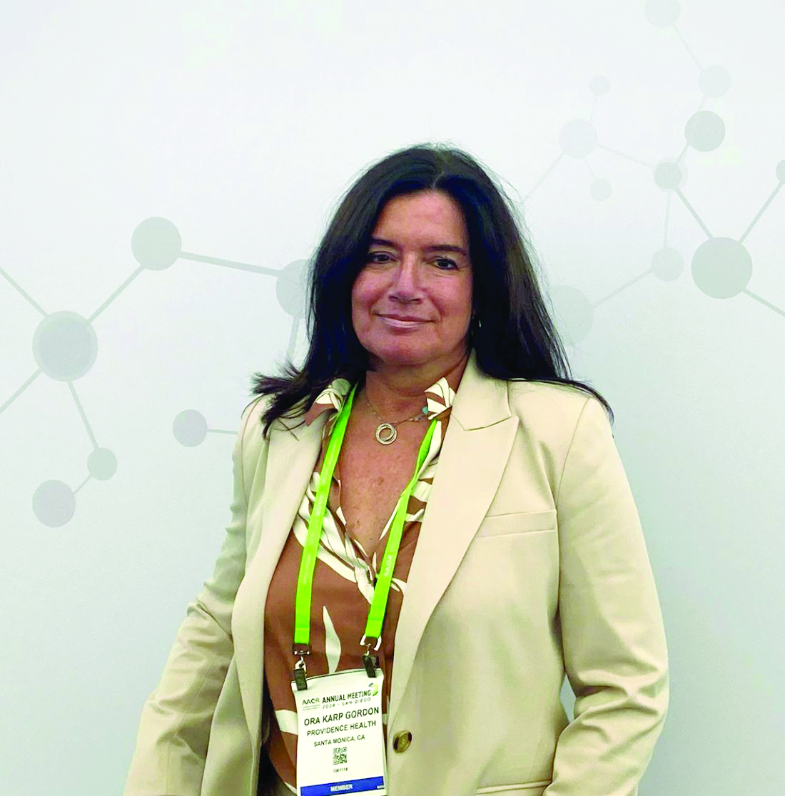
This was the conclusion of recent data presented by Ora Karp Gordon, MD, MS, during a session at the American Association for Cancer Research annual meeting.
The MCED test, known as Galleri, was made clinically available in the United States in April 2021. Developed by GRAIL LLC, the test analyzes cell-free DNA in the blood using targeted methylation analysis and machine learning to detect the presence of a cancer signal and determine its organ of origin or cancer signal origin. The initial screening of over 53,000 individuals with the Galleri test detected a cancer signal in 1.1% of participants.
The new real-world analysis examines the outcomes of repeat MCED testing in 5,794 individuals.
The study looked at individuals who initially received a ‘no cancer signal detected’ result and then underwent a second Galleri test. Over 80% of participants received their follow-up test 10-18 months after the first, with a median interval between blood draws of 12.9 months.
“The repeat tests detect those cancer cases that have reached the detection threshold since their last MCED test, which should be less than one year of incidence,” Dr. Gordon, professor at Saint John’s Cancer Institute, Santa Monica, California, said in an interview. “We are just now starting to see results from patients who get their second and even third round of screening.”
“Galleri is recommended to be used annually in addition to USPSTF [US Preventive Services Task Force]–recommended cancer screening tests, like mammography and colonoscopy,” she said.
This recommendation is based on a modeling study suggesting that annual screening would improve stage shift, diagnostic yield, and potentially mortality when compared to biennial screening, although biennial screening was still favorable compared with no screening, she explained.
Early Real-World Evidence of Repeat Testing
Among the cohort of 5,794 individuals who received repeat testing, 26 received a positive cancer signal on their second test, yielding a cancer signal detection rate of 0.45% (95% CI: 0.31%-0.66%). The cancer signal detection rate was slightly higher in men. The rate was 0.50% (95% CI: 0.32%-0.81%; 17 of 3367) in men versus 0.37% (95% CI: 0.2%-0.7%; 9 of 2427) in women.
During her presentation, Dr. Gordon highlighted that the repeat testing signal detection rate was lower than the initial 0.95% rate (95% CI: 0.87-1.0; 510 of 53,744) seen in the previous larger cohort of patients who were retested at 1 year.
She acknowledged that the lower cancer signal detection rate of repeat testing may indicate some degree of ‘early adopter’ bias, where those who return for a second test are systematically different from the general screening population. This could suggest that broader population-level screening may yield different results, she continued.
Shift Toward Unscreened Cancers
The top cancer types identified in the second round of testing were lymphoid, head and neck, bladder/urothelial, colorectal, and anal cancers. Clinicians were able to confirm clinical outcomes in 12 of 26 cases, in which cancer signals were detected. Of those 12 cases, 8 individuals received a cancer diagnosis and 4 did not have cancer. The remaining 14 of 26 cases in which cancer signals were detected are still under investigation.
“We found a shift away from USPSTF screen-detected cancers, like breast, lung, and prostate, and relative increase in unscreened urinary, head and neck, and lymphoid cancers, with 75% of cancers being those without any screening guidelines,” Dr. Gordon said in an interview.
She added that patients who choose to retest may have different cancer rates for several reasons, including bias toward a population that is health conscious and adhered to all recommended cancer screening.
“So the shift toward unscreened cancers is not unexpected and highlights the value of Galleri,” she said, but also acknowledged that “continued monitoring is needed to see if this translates in a persistent finding over time and tests.”
Shift Toward Early-Stage Cancers
Staging information was available for five cases, and Dr. Gordon highlighted in her talk that four of these confirmed cancers were stage I, including cancers of the anus, head and neck, bladder, and lymphoma. The fifth confirmed cancer with staging information was stage IV ovarian cancer.
“It is still early, and the numbers are very small, but the detection of early-stage cancers with second annual testing is very encouraging as these are the cases where MCED testing could have the greatest impact in improving outcomes through earlier treatment,” Dr. Gordon told this publication.
During an interview after the talk, Kenneth L. Kehl, MD, MPH, echoed that data must be confirmed in larger cohorts.
“The shift toward earlier stage cancers that are less detectable by standard screening methods is an interesting result, but we need to be cautious since the numbers were relatively small, and we do not have data on cancers that were diagnosed among patients whose second MCED test was also negative,” said Dr. Kehl, a medical oncologist at Dana-Farber Cancer Institute, Boston.
MCED Results Could Help Direct Diagnostic Workup
The test’s ability to predict the organ of origin was highly accurate, correctly identifying the cancer type in all eight confirmed cases. Among the eight cases with a confirmed cancer diagnosis, the accuracy of the first prediction was 100%, and diagnoses included invasive cancers across multiple tissues and organs, including anus, colon, head and neck, urothelial tract, ovary, and the lymphatic system.
“The fact that the site of origin for 100% of confirmed cancers was accurately predicted with GRAIL’s CSO by Galleri test confirms the promise that this can guide workup when a cancer signal is detected,” Dr. Gordon noted in the interview.
Looking Ahead
Dr. Kehl, who was not involved in the MCED study, noted in an interview that “further data on test characteristics beyond positive predictive value, including the sensitivity, specificity, and negative predictive value, as well as demonstration of clinical benefit — ideally in a randomized trial — will likely be required for MCED testing to become a standard public health recommendation.”
He added that challenges associated with implementing annual screening with MCED tests include the risks of both false positives and false negatives as testing becomes more widely available.
“False positives cause anxiety and lead to additional testing that may carry its own risks, and we need to understand if potentially false negative tests will be associated with less uptake of established screening strategies,” Dr. Kehl said in an interview. However, he noted that serial testing could lead to more frequent diagnoses of early-stage cancers that may be less detectable by standard methods.
Dr. Gordon reported financial relationships with GRAIL LLC and Genetic Technologies Corporation. Dr. Kehl reported no relationships with entities whose primary business is producing, marketing, selling, reselling, or distributing healthcare products used by or on patients.
FROM AACR 2024
Statins Raise Diabetes Risk, but CV Benefit Outweighs It
Statins raise the risks for increased glucose levels and the development of type 2 diabetes among people who don’t have it at baseline, but those risks are outweighed by the cardiovascular benefit, new data suggested.
The findings come from an analysis of individual participant data from a total of 23 randomized trials of statin therapy involving 154,664 individuals. In people without diabetes at baseline, statin therapy produces a dose-dependent increase in the risk for diabetes diagnosis, particularly among those whose glycemia marker levels are already at the diagnostic threshold.
Statins also tend to raise glucose levels in people who already have diabetes, but “the diabetes-related risks arising from the small changes in glycemia resulting from statin therapy are greatly outweighed by the benefits of statins on major vascular events when the direct clinical consequences of these outcomes are taken into consideration,” wrote the authors of the Cholesterol Treatment Trialists’ (CTT) Collaboration in their paper, published online in The Lancet Diabetes & Endocrinology.
Moreover, they say, “since the effect of statin therapy on measures of glycemia within an individual is small, there is likely to be little clinical benefit in measuring glucose concentrations and A1c values routinely after starting statin therapy with the aim of making comparisons to values taken before the initiation of a statin. However, people should continue to be screened for diabetes and associated risk factors and have their glycemic control monitored in accordance with current clinical guidelines.”
The CTT is co-led by Christina Reith, MBChB, PhD, and David Preiss, PhD, FRCPath, MRCP, both of the Nuffield Department of Population Health, University of Oxford, England.
In an accompanying editorial,
Dr. Gerstein and Dr. Pigeyre also said “these findings emphasize the importance of holistic care. As people at risk for cardiovascular outcomes are also at risk for type 2 diabetes, any prescription of a statin should be accompanied by promoting proven strategies to prevent or delay diabetes, such as modest weight reduction and increased physical activity. Finally, these findings emphasize the importance of always being alert for harmful adverse effects, even with the most beneficial and successful preventive therapies.”
Statins Raise Diabetes Risk, Glucose Levels Slightly
The meta-analysis of trials in the CTT Collaboration included individual participant data from 19 double-blind randomized, controlled trials with a median follow-up of 4.3 years comparing statins with placebo in a total of 123,940 participants, including 18% who had known type 2 diabetes at randomization. Also analyzed were another four double-blind trials of lower- vs higher-intensity statins involving a total of 30,724 participants followed for a median of 4.9 years, with 15% having diabetes at baseline.
In the 19 trials of low- or moderate-intensity statins vs placebo, statins resulted in a significant 10% increase in new-onset diabetes compared with placebo (rate ratio, 1.10), while high-intensity statins raised the risk by an also significant 36% (1.36). This translated to a mean absolute excess of 0.12% per year of treatment.
Compared with less intensive statin therapy, more intensive statin therapy resulted in a significant 10% proportional increase in new-onset diabetes (1.10), giving an absolute annual excess of 0.22%.
In the statin vs placebo trials, differences in A1c values from placebo were 0.06 percentage points higher for low- or moderate-intensity statins and 0.08 points greater for high-intensity statins.
Nearly two thirds (62%) of the excess cases of new-onset diabetes occurred among participants in the highest quarter of the baseline glycemia distribution for both low-intensity or moderate-intensity and high-intensity statin therapy.
And among participants who already had diabetes at baseline, there was a significant 10% relative increase in worsening glycemia (defined by adverse glycemic event, A1c increase of ≥ 0.5 percentage points, or medication escalation) with low- or moderate-intensity statins compared with placebo and a 24% relative increase in the high-intensity trials.
The Nuffield Department of Population Health has an explicit policy of not accepting any personal honoraria payments directly or indirectly from the pharmaceutical and food industries. It seeks reimbursement to the University of Oxford for the costs of travel and accommodation to participate in scientific meetings. Dr. Reith reported receiving funding to the University of Oxford from the UK National Institute for Health and Care Research Health Technology Assessment Programme and holding unpaid roles on the Clinical Data Interchange Standards Consortium as a board member and WHO as a scientific advisor. Dr. Preiss reported receiving funding to his research institution (but no personal funding) from Novartis for the ORION 4 trial of inclisiran, Novo Nordisk for the ASCEND PLUS trial of semaglutide, and Boehringer Ingelheim and Eli Lilly for the EMPA-KIDNEY trial and being a committee member for a National Institute for Health and Care Excellence guideline.
Dr. Gerstein holds the McMaster-Sanofi Population Health Institute Chair in Diabetes Research and Care. He reported research grants from Eli Lilly, AstraZeneca, Novo Nordisk, Hanmi, and Merck; continuing medical education grants to McMaster University from Eli Lilly, Abbott, Sanofi, Novo Nordisk, and Boehringer Ingelheim; honoraria for speaking from AstraZeneca, Eli Lilly, Novo Nordisk, DKSH, Zuellig Pharma, Sanofi, and Jiangsu Hanson; and consulting fees from Abbott, Eli Lilly, Novo Nordisk, Pfizer, Carbon Brand, Sanofi, Kowa, and Hanmi. Pigeyre had no disclosures.
A version of this article appeared on Medscape.com.
Early Olezarsen Results Show 50% Reduction in Triglycerides
ATLANTA — A novel antisense therapy called olezarsen reduced triglycerides (TGs) by approximately 50% with either of the two study doses relative to placebo and did so with a low relative risk for adverse events, new data from a phase 2b trial showed.
“The reduction in triglycerides was greater than that currently possible with any available therapy,” reported Brian A. Bergmark, MD, an interventional cardiologist at Brigham and Women’s Hospital, Boston.
The drug also produced meaningful improvements in multiple other lipid subfractions associated with increased cardiovascular (CV) risk, including ApoC-III, very low–density lipoprotein (VLDL) cholesterol, ApoB, and non-LDL cholesterol. High-density lipoprotein (HDL) cholesterol levels were significantly raised.
The results were presented on April 7 as a late breaker at the American College of Cardiology (ACC) Scientific Session 2024 and published online simultaneously in The New England Journal of Medicine.
No Major Subgroup Failed to Respond
The effect was seen across all the key subgroups evaluated, including women and patients with diabetes, obesity, and severe as well as moderate elevations in TGs at baseline, Dr. Bergmark reported.
Olezarsen is a N-acetylgalactosamine–conjugated antisense oligonucleotide targeting APOC3 RNA.
In this study, 154 patients at 24 sites in North America were randomized in a 1:1 ratio to 50 or 80 mg olezarsen. Those in each of these cohorts were then randomized in a 3:1 ratio to active therapy or placebo. All therapies were administered by subcutaneous injection once per month.
Patients were eligible for the trial if they had moderate hypertriglyceridemia, defined as a level of 150-499 mg/dL, and elevated CV risk or if they had severe hypertriglyceridemia (≥ 500 mg/dL) with or without other evidence of elevated CV risk. The primary endpoint was a change in TGs at 6 months. Complete follow-up was available in about 97% of patients regardless of treatment assignment.
With a slight numerical advantage for the higher dose, the TG reductions were 49.1% for the 50-mg dose and 53.1% for the 80-mg dose relative to no significant change in the placebo group (P < .001 for both olezarsen doses). The reductions in ApoC-III, an upstream driver of TG production and a CV risk factor, were 64.2% and 73.2% relative to placebo (both P < .001), respectively, Dr. Bergmark reported.
In those with moderate hypertriglyceridemia, normal TG levels, defined as < 150 mg/dL, were reached at 6 months in 85.7% and 93.3% in the 40-mg and 80-mg dose groups, respectively. Relative to these reductions, normalization was seen in only 11.8% of placebo patients (P < .001).
TG Lowering Might Not Be Best Endpoint
The primary endpoint in this trial was a change in TGs, but this target was questioned by an invited ACC discussant, Daniel Soffer, MD, who is both an adjunct professor assistant professor of medicine at Penn Medicine, Philadelphia, and current president of the National Lipid Association.
Dr. Soffer noted that highly elevated TGs are a major risk factor for acute pancreatitis, so this predicts a clinical benefit for this purpose, but he thought the other lipid subfractions are far more important for the goal of reducing atherosclerotic cardiovascular disease (ASCVD).
Indeed, he said categorically that it is not TGs that drive ASCVD risk and therefore not what is the real importance of these data. Rather, “it is the non-HDL cholesterol and ApoB lowering” that will drive the likely benefits from this therapy in CV disease.
In addition to the TG reductions, olezarsen did, in fact, produce significant reductions in many of the lipid subfractions associated with increased CV risk. While slightly more favorable in most cases with the higher dose of olezarsen, even the lower dose reduced Apo C-III from baseline by 64.2% (P < .001), VLDL by 46.2% (P < .001), remnant cholesterol by 46.6% (P < .001), ApoB by 18.2% (P < .001), and non-HDL cholesterol by 25.4% (P < .001). HDL cholesterol was increased by 39.6% (P < .001).
These favorable effects on TG and other lipid subfractions were achieved with a safety profile that was reassuring, Dr. Bergmark said. Serious adverse events leading to discontinuation occurred in 0%, 1.7%, and 1.8% of the placebo, lower-dose, and higher-dose arms, respectively. These rates did not differ significantly.
Increased Liver Enzymes Is Common
Liver enzymes were significantly elevated (P < .001) for both doses of olezarsen vs placebo, but liver enzymes > 3× the upper limit of normal did not reach significance on either dose of olezarsen relative to placebo. Low platelet counts and reductions in kidney function were observed in a minority of patients but were generally manageable, according to Dr. Bergmark. There was no impact on hemoglobin A1c levels.
Further evaluation of change in hepatic function is planned in the ongoing extension studies.
Characterizing these results as “exciting,” Neha J. Pagidipati, MD, a member of the Duke Clinical Research Institute and an assistant professor at the Duke School of Medicine, Durham, North Carolina, said that identifying a drug effective for hypertriglyceridemia is likely to be a major advance. While elevated TGs are “one of the toughest” lipid abnormalities to manage, “there is not much out there to offer for treatment.”
She, like Dr. Soffer, was encouraged by the favorable effects on multiple lipid abnormalities associated with increased CV risk, but she said the ultimate clinical utility of this or other agents that lower TGs for ASCVD requires a study showing a change in CV events.
Dr. Bergmark reported financial relationships with 15 pharmaceutical companies, including Ionis, which provided funding for the BRIDGE-TIMI 73a trial. Soffer had financial relationships with Akcea, Amgen, Amryt, AstraZeneca, Ionis, Novartis, Regeneron, and Verve. Dr. Pagidipati had financial relationships with more than 10 pharmaceutical companies but was not involved in the design of management of the BRIDGE-TIMI 73a trial.
A version of this article first appeared on Medscape.com.