User login
Will tirzepatide slow kidney function decline in type 2 diabetes?
The “twincretin” tirzepatide might become part of the “arsenal” against diabetic kidney disease, new research suggests. Notably, the drug significantly reduced the likelihood of macroalbuminuria, in a prespecified subanalysis of the SURPASS-4 clinical trial.
“Once-per-week tirzepatide compared to [daily] insulin glargine treatment resulted in a meaningful improvement in estimated glomerular filtration rate (eGFR) decline and reduced urine albumin-to-creatinine ratio (UACR) and the risk of end stage kidney disease (ESKD) – with low risk of clinically relevant hypoglycemia in participants with type 2 diabetes at high cardiovascular risk and varying degrees of chronic kidney disease (CKD),” lead investigator Hiddo J. L. Heerspink, PhD, PharmD, summarized in an email to this news organization.

The U.S. Food and Drug Administration has just approved tirzepatide (Mounjaro, Eli Lilly) – a novel, glucose-dependent insulinotropic polypeptide (GIP) combined with a glucagonlike peptide-1 (GLP-1) receptor agonist – to treat glycemia in patients with type 2 diabetes, based on five pivotal SURPASS trials.
Dr. Heerspink presented the new findings about tirzepatide’s impact on kidney function in an oral session at the annual scientific sessions of the American Diabetes Association.
40% reduced risk of kidney function decline
The main results of SURPASS-4 were published in the Lancet in October 2021, and showed that tirzepatide appeared superior to insulin glargine in lowering hemoglobin A1c in patients with type 2 diabetes at high cardiovascular risk who were inadequately controlled on oral diabetes treatments.
Now, Dr. Heerspink has shown that patients who received tirzepatide as opposed to insulin glargine were significantly less likely to have kidney function decline that included new-onset macroalbuminuria (hazard ratio, 0.59; P < .05).
“These are very large benefits and clearly indicate the potential of tirzepatide to be a very strong kidney protective drug,” said Dr. Heerspink, from the department of clinical pharmacy and pharmacology, University Medical Center Groningen (the Netherlands).
“Based on results from the SURPASS-4 trial, tirzepatide has significant kidney-protective effects in adults with type 2 diabetes with high cardiovascular risk and largely normal kidney function,” Christine Limonte, MD, chair of the session in which the analysis was presented, agreed, in an email to this news organization.
The approximate 40% reduced risk of kidney function decline in this population “is important because it suggests that this novel agent may contribute to the growing arsenal for preventing and treating diabetic kidney disease,” added Dr. Limonte, a clinical research fellow in the division of nephrology, University of Washington, Seattle.
“Over the last several years,” she noted, “sodium glucose cotransporter-2 [SGLT2] inhibitors and GLP-1 receptor agonists have been identified as having significant kidney-protective effects in type 2 diabetes, and as such are becoming first-line agents in the treatment of diabetic kidney disease.”
Additional studies are needed, she added, to assess the impacts of tirzepatide compared to these agents (particularly GLP-1 receptor agonists, which overlap in their mechanism of action).
“With the growing number of therapeutic options for diabetic kidney disease, future research should also focus on identifying combinations of agents which benefit individuals in a ‘targeted’ manner,” according to Dr. Limonte.
“Ensuring accessibility to kidney-protective agents by promoting access to health care and reducing drug costs is essential to improving outcomes in diabetic kidney disease,” she added.
Strongest reduction seen in risk of new macroalbuminuria
One in three adults with diabetes has CKD, according to a press release issued by the ADA. Therefore, there is a need for therapies to reduce the development and progression of CKD in patients with type 2 diabetes.
The prespecified analysis of SUPRESS-4 investigated potential renoprotective effects of tirzepatide.
The trial enrolled 1,995 patients with type 2 diabetes who were at increased risk of cardiovascular disease. The patients had a mean age of 63.6 years and a mean hemoglobin A1c of 8.5%.
Most patients had normal kidney function. The mean eGFR based on the Chronic Kidney Disease Epidemiology Collaboration (CKD-EPI) equation was 81.3 mL/min per 1.73 m2.
Few patients (17%) had moderately or severely reduced kidney function (eGFR <60 mL/min per 1.73 m2). Around a quarter of the patients (28%) had microalbuminuria (UACR 30-300 mg/g) and 8% had macroalbuminuria (UACR >300 mg/g).
The patients were randomized to receive a weekly injection of 5, 10, or 15 mg tirzepatide or a daily individualized injection of insulin glargine starting at 10 IU/day at bedtime, titrated to a fasting blood glucose <100 mg/dL, in addition to existing oral glucose-lowering agents. The primary outcomes in the subanalysis were:
- Endpoint 1: a composite of ≥40% decline in eGFR from baseline, renal death, progression to ESKD, and new-onset macroalbuminuria.
- Endpoint 2: the same as endpoint 1 excluding new-onset macroalbuminuria.
During a median follow up of 85 weeks and up to 104 weeks, patients who received tirzepatide versus insulin glargine were significantly less likely to reach endpoint 1 but not endpoint 2.
In addition, tirzepatide “very strongly” reduced the risk of new-onset macroalbuminuria, compared to insulin glargine, by approximately 60% in the complete study cohort (hazard ratio, 0.41; P < .05), Dr. Limonte noted.
Tirzepatide also reduced the risk of a >40% decline in eGFR, but this effect was not statistically significant, possibly because this outcome was underpowered. There were also too few kidney deaths and progressions to ESKD to meaningfully assess the effects of tirzepatide on these outcomes.
Therefore, Dr. Limonte noted, “it is likely that tirzepatide’s significant benefit on composite endpoint 1 was largely driven by this agent’s impact on reducing macroalbuminuria onset [explaining why a significant benefit was not seen with composite endpoint 2, which excluded new-onset macroalbuminuria].”
The study was funded by Eli Lilly. Dr. Heerspink disclosed that he is a consultant for AstraZeneca, Bayer AG, Boehringer Ingelheim, Chinook Therapeutics, CSL Behring, Gilead Sciences, Goldfinch Bio, Janssen Research & Development, Mitsubishi Tanabe Pharma, Mundipharma, and Traveere Pharmaceuticals, and has received research support from AstraZeneca, Boehringer Ingelheim, and Novo Nordisk.
Dr. Limonte disclosed that she receives funds from the American Kidney Fund’s Clinical Scientist in Nephrology Award.
A version of this article first appeared on Medscape.com.
The “twincretin” tirzepatide might become part of the “arsenal” against diabetic kidney disease, new research suggests. Notably, the drug significantly reduced the likelihood of macroalbuminuria, in a prespecified subanalysis of the SURPASS-4 clinical trial.
“Once-per-week tirzepatide compared to [daily] insulin glargine treatment resulted in a meaningful improvement in estimated glomerular filtration rate (eGFR) decline and reduced urine albumin-to-creatinine ratio (UACR) and the risk of end stage kidney disease (ESKD) – with low risk of clinically relevant hypoglycemia in participants with type 2 diabetes at high cardiovascular risk and varying degrees of chronic kidney disease (CKD),” lead investigator Hiddo J. L. Heerspink, PhD, PharmD, summarized in an email to this news organization.
The U.S. Food and Drug Administration has just approved tirzepatide (Mounjaro, Eli Lilly) – a novel, glucose-dependent insulinotropic polypeptide (GIP) combined with a glucagonlike peptide-1 (GLP-1) receptor agonist – to treat glycemia in patients with type 2 diabetes, based on five pivotal SURPASS trials.
Dr. Heerspink presented the new findings about tirzepatide’s impact on kidney function in an oral session at the annual scientific sessions of the American Diabetes Association.
40% reduced risk of kidney function decline
The main results of SURPASS-4 were published in the Lancet in October 2021, and showed that tirzepatide appeared superior to insulin glargine in lowering hemoglobin A1c in patients with type 2 diabetes at high cardiovascular risk who were inadequately controlled on oral diabetes treatments.
Now, Dr. Heerspink has shown that patients who received tirzepatide as opposed to insulin glargine were significantly less likely to have kidney function decline that included new-onset macroalbuminuria (hazard ratio, 0.59; P < .05).
“These are very large benefits and clearly indicate the potential of tirzepatide to be a very strong kidney protective drug,” said Dr. Heerspink, from the department of clinical pharmacy and pharmacology, University Medical Center Groningen (the Netherlands).
“Based on results from the SURPASS-4 trial, tirzepatide has significant kidney-protective effects in adults with type 2 diabetes with high cardiovascular risk and largely normal kidney function,” Christine Limonte, MD, chair of the session in which the analysis was presented, agreed, in an email to this news organization.
The approximate 40% reduced risk of kidney function decline in this population “is important because it suggests that this novel agent may contribute to the growing arsenal for preventing and treating diabetic kidney disease,” added Dr. Limonte, a clinical research fellow in the division of nephrology, University of Washington, Seattle.
“Over the last several years,” she noted, “sodium glucose cotransporter-2 [SGLT2] inhibitors and GLP-1 receptor agonists have been identified as having significant kidney-protective effects in type 2 diabetes, and as such are becoming first-line agents in the treatment of diabetic kidney disease.”
Additional studies are needed, she added, to assess the impacts of tirzepatide compared to these agents (particularly GLP-1 receptor agonists, which overlap in their mechanism of action).
“With the growing number of therapeutic options for diabetic kidney disease, future research should also focus on identifying combinations of agents which benefit individuals in a ‘targeted’ manner,” according to Dr. Limonte.
“Ensuring accessibility to kidney-protective agents by promoting access to health care and reducing drug costs is essential to improving outcomes in diabetic kidney disease,” she added.
Strongest reduction seen in risk of new macroalbuminuria
One in three adults with diabetes has CKD, according to a press release issued by the ADA. Therefore, there is a need for therapies to reduce the development and progression of CKD in patients with type 2 diabetes.
The prespecified analysis of SUPRESS-4 investigated potential renoprotective effects of tirzepatide.
The trial enrolled 1,995 patients with type 2 diabetes who were at increased risk of cardiovascular disease. The patients had a mean age of 63.6 years and a mean hemoglobin A1c of 8.5%.
Most patients had normal kidney function. The mean eGFR based on the Chronic Kidney Disease Epidemiology Collaboration (CKD-EPI) equation was 81.3 mL/min per 1.73 m2.
Few patients (17%) had moderately or severely reduced kidney function (eGFR <60 mL/min per 1.73 m2). Around a quarter of the patients (28%) had microalbuminuria (UACR 30-300 mg/g) and 8% had macroalbuminuria (UACR >300 mg/g).
The patients were randomized to receive a weekly injection of 5, 10, or 15 mg tirzepatide or a daily individualized injection of insulin glargine starting at 10 IU/day at bedtime, titrated to a fasting blood glucose <100 mg/dL, in addition to existing oral glucose-lowering agents. The primary outcomes in the subanalysis were:
- Endpoint 1: a composite of ≥40% decline in eGFR from baseline, renal death, progression to ESKD, and new-onset macroalbuminuria.
- Endpoint 2: the same as endpoint 1 excluding new-onset macroalbuminuria.
During a median follow up of 85 weeks and up to 104 weeks, patients who received tirzepatide versus insulin glargine were significantly less likely to reach endpoint 1 but not endpoint 2.
In addition, tirzepatide “very strongly” reduced the risk of new-onset macroalbuminuria, compared to insulin glargine, by approximately 60% in the complete study cohort (hazard ratio, 0.41; P < .05), Dr. Limonte noted.
Tirzepatide also reduced the risk of a >40% decline in eGFR, but this effect was not statistically significant, possibly because this outcome was underpowered. There were also too few kidney deaths and progressions to ESKD to meaningfully assess the effects of tirzepatide on these outcomes.
Therefore, Dr. Limonte noted, “it is likely that tirzepatide’s significant benefit on composite endpoint 1 was largely driven by this agent’s impact on reducing macroalbuminuria onset [explaining why a significant benefit was not seen with composite endpoint 2, which excluded new-onset macroalbuminuria].”
The study was funded by Eli Lilly. Dr. Heerspink disclosed that he is a consultant for AstraZeneca, Bayer AG, Boehringer Ingelheim, Chinook Therapeutics, CSL Behring, Gilead Sciences, Goldfinch Bio, Janssen Research & Development, Mitsubishi Tanabe Pharma, Mundipharma, and Traveere Pharmaceuticals, and has received research support from AstraZeneca, Boehringer Ingelheim, and Novo Nordisk.
Dr. Limonte disclosed that she receives funds from the American Kidney Fund’s Clinical Scientist in Nephrology Award.
A version of this article first appeared on Medscape.com.
FROM ADA 2022
Think it’s ILD? Tell it to the machines
SAN FRANCISCO – Interstitial lung disease is a difficult diagnosis to make, but a combination of artificial intelligence (AI) techniques and automated language processing could help clinicians identify the early signs of ILD and start patients on therapy, investigators say.
For example, applying an AI algorithm to spirometry readings taken from patients whose data were registered in the UK Biobank identified 27% as having ILD, and of this group, 66% had ostensibly normal lung function on spirometry but were later diagnosed with ILD, reported Marko Topalovic, PhD, from the AI company ArtiQ in Leuven, Belgium, at the American Thoracic Society’s international conference.
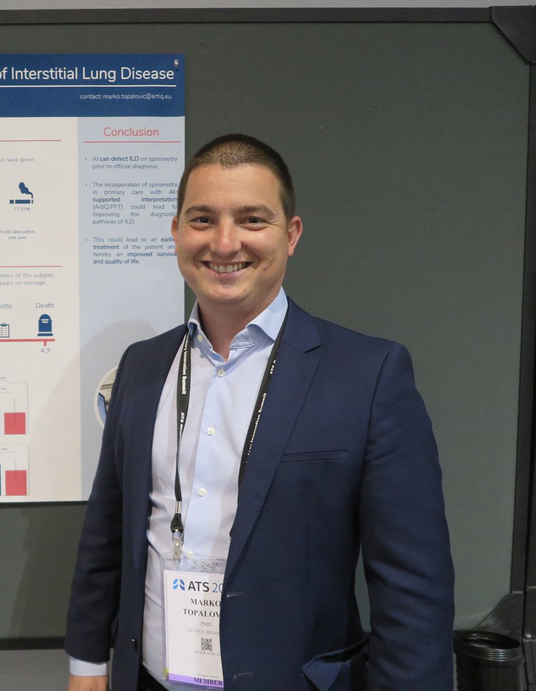
“A diagnosis of ILD is very challenging, so you have patients who are going to be misdiagnosed or have a very late diagnosis, so we aimed to apply our AI algorithm on spirometry to see whether we could detect ILD much earlier,” he said in an interview conducted during a poster discussion session.
AI detected ILD up to 6.8 years before a clinician’s diagnosis, Dr. Topalovic said.
Reading between the lines
In a separate study, investigators at the University of California, Davis, used language analysis software to scour electronic health records for words indicative of early ILD, and found that the technique dramatically shortened the median time to a pulmonary referral, compared with historical controls.
“This is a language processing program that can essentially look through the radiology reports and look for the key words that often describe interstitial lung disease, like traction, honeycomb, fibrotic, etc. With those studies being flagged, an actual pulmonologist will then further review the scan, and see whether it meets criteria for one of the interstitial lung diseases,” lead author William Leon, MD, a resident in the department of internal medicine at the University of California, Davis, said in an interview.

“We then sent the primary care doctor a message to say: ‘Hey, this patient has ILD. You need to send them to a pulmonologist,’ ” he added.
Putting it together
Philip L. Molyneaux, MRCP (UK), MBBS, BS (Hons), from Imperial College London, who comoderated the session but was not involved in the studies, speculated that combining these and other, nontechnical interventions also discussed could help to improve diagnosis of ILD and allow clinicians to prescribe therapy earlier in the disease course.
“What’s going to give you the biggest impact for patients? Everyone working individually is coming up with great advances, and if you put them all together it’s going to provide much greater benefit for our patients,” he said in an interview.
AI Spirometry details
In collaboration with colleagues at the Laboratory of Respiratory Disease at University Hospital in Leuven, Dr. Topalovic applied AI to results of spirometry performed prior to diagnosis of ILD among 109 patients registered in the UK Biobank, a repository of information on more than 500,000 volunteers.
The patients selected had ILD listed as their cause of death, had spirometry performed up to 7 years before their deaths, and did not receive a diagnosis of ILD on the day of the index spirometry.
In all 73% of patients were men, 27% women, with an average age of 64.6 years. A large majority of the sample (77.15%) had a history of smoking, and 60 of the patients (55%) died within one year of an ILD diagnosis.
The investigators plugged the spirometry data and each patients demographic information – including gender, age, height, weight, race, and smoking status – into the AI clinical decision support program, which yielded a statistical probability for each subject of having normal lung function, asthma, COPD, ILD, another obstructive disease, or another unidentifiable respiratory disease.
In 29 patients (27%) the software listed ILD as the highest probability, and of this group 19 patients (66%) had normal lung function according to standard interpretation guidelines.
Spirometry parameters among patients identified as having probable ILD were different from those where ILD was not detected. For example, forced vital capacity (FVC) was 76% of predicted among patients with likely ILD versus 87% of predicted in those who had a diagnosis later (P = .003). Similar differences were seen in the forced expiratory volume in 1 second to FVC ratio, at 0.82 vs. 0.75, respectively (P = .007).
There were no differences in mortality or in median time between spirometry and clinician diagnosis between the groups.
Language processing details
Dr. Leon and colleagues used a language analysis software package to review CT chest reports. Reports were flagged if they contained the words traction, honeycomb, fibrotic, fibrosis, reticular, or reticulation.
The CT scan accompanying each flagged reported was reviewed by a pulmonologist for the presence of ILD, and scans with ILD identified were referred to pulmonary specialists. The results of 2,198 prospective scans followed by prospective screening were compared with those of 1,690 historical controls seen in 2015 and 2016.
The investigators found that 85 incident cases of ILD were identified in the historical controls, compared with 143 in the prospective cohort, leading to 38 and 120 pulmonary referrals, respectively.
For the primary outcome of median time from CT to pulmonary referral, the authors found that it was 1.27 months for the prospective cohort, compared with not reached (censored after 18 months) in historical controls.
The hazard ratio for a pulmonary referral in the prospective versus historical cohort was 2.79, an association that was strengthened after adjusting for sex, age, race, smoking pack-years, cough, crackles, and dyspnea (HR, 4.54; both comparisons significant according to confidence intervals).
The studies were internally funded. Dr. Topalovic is CEO and cofounder of ArtiQ. Dr. Leon and Dr. Molyneaux reported no relevant conflicts of interest.
SAN FRANCISCO – Interstitial lung disease is a difficult diagnosis to make, but a combination of artificial intelligence (AI) techniques and automated language processing could help clinicians identify the early signs of ILD and start patients on therapy, investigators say.
For example, applying an AI algorithm to spirometry readings taken from patients whose data were registered in the UK Biobank identified 27% as having ILD, and of this group, 66% had ostensibly normal lung function on spirometry but were later diagnosed with ILD, reported Marko Topalovic, PhD, from the AI company ArtiQ in Leuven, Belgium, at the American Thoracic Society’s international conference.
“A diagnosis of ILD is very challenging, so you have patients who are going to be misdiagnosed or have a very late diagnosis, so we aimed to apply our AI algorithm on spirometry to see whether we could detect ILD much earlier,” he said in an interview conducted during a poster discussion session.
AI detected ILD up to 6.8 years before a clinician’s diagnosis, Dr. Topalovic said.
Reading between the lines
In a separate study, investigators at the University of California, Davis, used language analysis software to scour electronic health records for words indicative of early ILD, and found that the technique dramatically shortened the median time to a pulmonary referral, compared with historical controls.
“This is a language processing program that can essentially look through the radiology reports and look for the key words that often describe interstitial lung disease, like traction, honeycomb, fibrotic, etc. With those studies being flagged, an actual pulmonologist will then further review the scan, and see whether it meets criteria for one of the interstitial lung diseases,” lead author William Leon, MD, a resident in the department of internal medicine at the University of California, Davis, said in an interview.
“We then sent the primary care doctor a message to say: ‘Hey, this patient has ILD. You need to send them to a pulmonologist,’ ” he added.
Putting it together
Philip L. Molyneaux, MRCP (UK), MBBS, BS (Hons), from Imperial College London, who comoderated the session but was not involved in the studies, speculated that combining these and other, nontechnical interventions also discussed could help to improve diagnosis of ILD and allow clinicians to prescribe therapy earlier in the disease course.
“What’s going to give you the biggest impact for patients? Everyone working individually is coming up with great advances, and if you put them all together it’s going to provide much greater benefit for our patients,” he said in an interview.
AI Spirometry details
In collaboration with colleagues at the Laboratory of Respiratory Disease at University Hospital in Leuven, Dr. Topalovic applied AI to results of spirometry performed prior to diagnosis of ILD among 109 patients registered in the UK Biobank, a repository of information on more than 500,000 volunteers.
The patients selected had ILD listed as their cause of death, had spirometry performed up to 7 years before their deaths, and did not receive a diagnosis of ILD on the day of the index spirometry.
In all 73% of patients were men, 27% women, with an average age of 64.6 years. A large majority of the sample (77.15%) had a history of smoking, and 60 of the patients (55%) died within one year of an ILD diagnosis.
The investigators plugged the spirometry data and each patients demographic information – including gender, age, height, weight, race, and smoking status – into the AI clinical decision support program, which yielded a statistical probability for each subject of having normal lung function, asthma, COPD, ILD, another obstructive disease, or another unidentifiable respiratory disease.
In 29 patients (27%) the software listed ILD as the highest probability, and of this group 19 patients (66%) had normal lung function according to standard interpretation guidelines.
Spirometry parameters among patients identified as having probable ILD were different from those where ILD was not detected. For example, forced vital capacity (FVC) was 76% of predicted among patients with likely ILD versus 87% of predicted in those who had a diagnosis later (P = .003). Similar differences were seen in the forced expiratory volume in 1 second to FVC ratio, at 0.82 vs. 0.75, respectively (P = .007).
There were no differences in mortality or in median time between spirometry and clinician diagnosis between the groups.
Language processing details
Dr. Leon and colleagues used a language analysis software package to review CT chest reports. Reports were flagged if they contained the words traction, honeycomb, fibrotic, fibrosis, reticular, or reticulation.
The CT scan accompanying each flagged reported was reviewed by a pulmonologist for the presence of ILD, and scans with ILD identified were referred to pulmonary specialists. The results of 2,198 prospective scans followed by prospective screening were compared with those of 1,690 historical controls seen in 2015 and 2016.
The investigators found that 85 incident cases of ILD were identified in the historical controls, compared with 143 in the prospective cohort, leading to 38 and 120 pulmonary referrals, respectively.
For the primary outcome of median time from CT to pulmonary referral, the authors found that it was 1.27 months for the prospective cohort, compared with not reached (censored after 18 months) in historical controls.
The hazard ratio for a pulmonary referral in the prospective versus historical cohort was 2.79, an association that was strengthened after adjusting for sex, age, race, smoking pack-years, cough, crackles, and dyspnea (HR, 4.54; both comparisons significant according to confidence intervals).
The studies were internally funded. Dr. Topalovic is CEO and cofounder of ArtiQ. Dr. Leon and Dr. Molyneaux reported no relevant conflicts of interest.
AT ATS 2022
First evidence of disease modification with methotrexate in pre-RA
COPENHAGEN – Temporary methotrexate in clinically suspected arthralgia delays but does not prevent clinical arthritis development; however, it does lead to sustained reduction of disease burden and MRI-detected inflammation in all at-risk groups, shows the 2-year Treat Earlier study.
“These data provide the first evidence for disease modification when intervening in ‘pre-RA [rheumatoid arthritis]’ or arthralgia,” said Doortje Krijbolder, MD, of Leiden (the Netherlands) University Medical Center, who presented her study at the annual European Congress of Rheumatology.

The randomized, double-blind, study aimed to find out if giving methotrexate in the pre-arthritis phase of arthralgia (with subclinical joint inflammation) prevents the development of clinical arthritis or reduces the burden of disease.
“This is the first trial that aims to reduce the burden of disease, and as such, in pre-RA, it is important that disease modification is sustained after temporary treatment otherwise patients will start regressing with disease activity,” said Dr. Krijbolder.
She explained that methotrexate is usually initiated as first-line treatment when arthritis becomes clinically apparent with joint swelling, but “disease processes begin long before this, and only become clinically recognizable when patients develop symptoms.”
Clinically detectable arthritis development
All 236 patients included in the study had arthralgia of the small joints that, because of the character of the symptoms, was found clinically suspect for progression to RA over time.
“Importantly, these participants had not yet developed clinical arthritis that could be detected on physical joint examination, and were clinically suspected of progressing to RA. They had all undergone an MRI and subclinical joint-inflammation had been detected that was more than prevalent in symptom-free controls,” said Dr. Krijbolder.
Patients were randomized (1:1) to either a single intramuscular glucocorticoid injection (120 mg methylprednisolone [Depo Medrol]) and a 1-year course of oral methotrexate (up to 25 mg/week), or to placebo injection and placebo tablets, and were then followed for a further year without medication to see whether disease progressed.
Treatment and placebo groups were matched with an average age of 46-47 years, 62%-68% were women, they had had symptoms (joint pain) for 27-28 weeks, C-reactive protein was increased in 27%-30%, and 20%-26% were anti-citrullinated protein autoantibody (ACPA) positive.
The primary endpoint was the development of clinically detectable arthritis (fulfilling the 2010 RA-criteria or involving ≥ 2 joints) that persisted for at least 2 weeks. The main secondary endpoints were related to disease burden, including patient-reported physical functioning, along with symptoms and workability (presenteeism at work – the percentage of productivity lost caused by the joint complaints), and measured every 4 months, said Dr. Krijbolder.
She and her colleagues also followed the course of MRI-detected inflammation, which comprised the sum of tenosynovitis, synovitis, and osteitis scored with the RA-MRI Scoring (RAMRIS) method.
Analysis was carried out on an intention-to-treat basis, and two prespecified subgroup analyses were also performed to obtain a better understanding of the effect of methotrexate and glucocorticoids in participants with high risk of clinical arthritis development (positive predictive value (PPV) ≥ 70%), and in patients stratified for ACPA status.
Delays but does not prevent
There was no difference between treatment and placebo groups in the development of persistent clinical arthritis over 2 years (80% vs. 82%, hazard ratio [HR] 0.81; 95% confidence interval [CI], 0.45, 1.48]); however, in the high-risk group subanalysis, 67% in the placebo group developed persistent clinical arthritis. For the treatment group, 7%developed persistent clinical arthritis at 6 months, 27% at 12 months, 40% at 18 months, and 67% at 24 months. For the placebo group: 56% developed persistent clinical arthritis at 6 months, and 67% at 12 months, 18 months, and 24 months.
“Strikingly, in the treatment group there was a statistically significant difference between 6 and 12 months, but in year 2, this difference disappeared, suggesting a delayed arthritis development but no prevention,” said Dr. Krijbolder.
Persistent clinical arthritis became similar at 24 months, at 67% in both groups. A similar, but less pronounced, delaying effect was seen with the ACPA-positive patients, with 48% and 52% developing persistent clinical arthritis at 24 months.
In joint pain, there was a decline in the treatment group compared with the placebo group and this persisted over 2 years (–9 on scale of 0 to 100: (95%CI, –12,–4; P < .001), and a similar effect was seen in high-risk and ACPA subgroups, reported Dr. Krijbolder.
Physical functioning improved more in the treatment-group compared with the placebo group during the first months and remained better (mean between-group difference over 2 years HAQ [health assessment questionnaire] –0.1 [–0.2, –0.03; P = .004]), and morning stiffness (–12 [95%CI, –16, –8; P < .001]), and presenteeism (–8% [95%CI, –13%, –3%; P = .001]) also showed improvement over time compared with placebo.
MRI-detected joint-inflammation was also persistently improved with a mean difference over 2 years –1.4 points (95%CI, –2.0, –0.9; P < .001). “As we know that symptoms and functional impairments are associated with MRI-detected joint inflammation, it was not surprising that treatment induced a decline in joint inflammation that persisted over time, also after the stop of the treatment in the second year,” explained Dr. Krijbolder.
In the high-risk subgroup, as well as in both ACPA-positive and ACPA-negative participants, a comparable statistically significant decline in MRI-detected inflammation was found.
“It may seem counterintuitive that on the one hand we found a delaying effect of treatment on persistent clinical arthritis but no prevention, while on the other, we saw a sustained treatment effect on MRI-detected joint-inflammation and related symptoms and functional impairments,” added Dr. Krijbolder.
To help understand this, the researchers performed a post hoc analysis in high-risk participants, comparing those who did not progress with those who did progress to arthritis. “Both progressors and nonprogressors showed a sustained treatment effect for pain and MRI-detected joint-inflammation, and in nonprogressors, there was almost a complete relief of pain and they nearly returned to the normal range of MRI-detected joint-inflammation as seen in symptom-free controls,” reported Dr. Krijbolder.
“In those who progressed to arthritis, there was less pain and less MRI-detected joint inflammation in the treatment group, but also at the time when they developed clinical arthritis. So both progressors and nonprogressors benefit from treatment,” she noted.
The number of serious adverse events was the same between the groups and adverse events were as expected from methotrexate.
Dr. Krijbolder said, “the results are encouraging and will open up a new treatment landscape in pre-RA at the future, but at the moment it is too early to give recommendations for clinical practice; we definitely do not want to advocate the start of treatment in all at-risk individuals from now on.”
More research should unravel the mechanisms within the joint that contribute to the development of clinical arthritis and disease chronicity, she said. “If we understand these, we may be able to use more targeted interventions in the future and prevent the development of clinical arthritis all together.
“We are also looking forward to learning more about the long-term beneficial effects of this early treatment in our ongoing observational extension of the trial.”
Hendrik Schulze-Koops, MD, of Ludwig-Maximilians-University of Munich, who moderated the session, asked Dr. Krijbolder, “As well as giving methotrexate to the participants in the treatment group, you also gave a single glucocorticoid injection at baseline, and a placebo injection in the placebo group, can you comment on that?”

Dr. Krijbolder replied that by the first study visit of 4 months, methotrexate would have already started working, and as such from her data it was not possible to distinguish what effect arose from the Depo Medrol injection, and what was from the methotrexate.
“Maybe there even is a synergetic effect, meaning that the two medications together work even better. To learn more about this would require a novel trial with a study design that would, for example, collect data quickly after the injection, for example after 2-4 weeks, because than the effect of the methotrexate would still be limited, or, of course, a novel trial with a treatment arm that only consists of a glucocorticoid injection,” she added.
No conflicts of interest were declared.
COPENHAGEN – Temporary methotrexate in clinically suspected arthralgia delays but does not prevent clinical arthritis development; however, it does lead to sustained reduction of disease burden and MRI-detected inflammation in all at-risk groups, shows the 2-year Treat Earlier study.
“These data provide the first evidence for disease modification when intervening in ‘pre-RA [rheumatoid arthritis]’ or arthralgia,” said Doortje Krijbolder, MD, of Leiden (the Netherlands) University Medical Center, who presented her study at the annual European Congress of Rheumatology.
The randomized, double-blind, study aimed to find out if giving methotrexate in the pre-arthritis phase of arthralgia (with subclinical joint inflammation) prevents the development of clinical arthritis or reduces the burden of disease.
“This is the first trial that aims to reduce the burden of disease, and as such, in pre-RA, it is important that disease modification is sustained after temporary treatment otherwise patients will start regressing with disease activity,” said Dr. Krijbolder.
She explained that methotrexate is usually initiated as first-line treatment when arthritis becomes clinically apparent with joint swelling, but “disease processes begin long before this, and only become clinically recognizable when patients develop symptoms.”
Clinically detectable arthritis development
All 236 patients included in the study had arthralgia of the small joints that, because of the character of the symptoms, was found clinically suspect for progression to RA over time.
“Importantly, these participants had not yet developed clinical arthritis that could be detected on physical joint examination, and were clinically suspected of progressing to RA. They had all undergone an MRI and subclinical joint-inflammation had been detected that was more than prevalent in symptom-free controls,” said Dr. Krijbolder.
Patients were randomized (1:1) to either a single intramuscular glucocorticoid injection (120 mg methylprednisolone [Depo Medrol]) and a 1-year course of oral methotrexate (up to 25 mg/week), or to placebo injection and placebo tablets, and were then followed for a further year without medication to see whether disease progressed.
Treatment and placebo groups were matched with an average age of 46-47 years, 62%-68% were women, they had had symptoms (joint pain) for 27-28 weeks, C-reactive protein was increased in 27%-30%, and 20%-26% were anti-citrullinated protein autoantibody (ACPA) positive.
The primary endpoint was the development of clinically detectable arthritis (fulfilling the 2010 RA-criteria or involving ≥ 2 joints) that persisted for at least 2 weeks. The main secondary endpoints were related to disease burden, including patient-reported physical functioning, along with symptoms and workability (presenteeism at work – the percentage of productivity lost caused by the joint complaints), and measured every 4 months, said Dr. Krijbolder.
She and her colleagues also followed the course of MRI-detected inflammation, which comprised the sum of tenosynovitis, synovitis, and osteitis scored with the RA-MRI Scoring (RAMRIS) method.
Analysis was carried out on an intention-to-treat basis, and two prespecified subgroup analyses were also performed to obtain a better understanding of the effect of methotrexate and glucocorticoids in participants with high risk of clinical arthritis development (positive predictive value (PPV) ≥ 70%), and in patients stratified for ACPA status.
Delays but does not prevent
There was no difference between treatment and placebo groups in the development of persistent clinical arthritis over 2 years (80% vs. 82%, hazard ratio [HR] 0.81; 95% confidence interval [CI], 0.45, 1.48]); however, in the high-risk group subanalysis, 67% in the placebo group developed persistent clinical arthritis. For the treatment group, 7%developed persistent clinical arthritis at 6 months, 27% at 12 months, 40% at 18 months, and 67% at 24 months. For the placebo group: 56% developed persistent clinical arthritis at 6 months, and 67% at 12 months, 18 months, and 24 months.
“Strikingly, in the treatment group there was a statistically significant difference between 6 and 12 months, but in year 2, this difference disappeared, suggesting a delayed arthritis development but no prevention,” said Dr. Krijbolder.
Persistent clinical arthritis became similar at 24 months, at 67% in both groups. A similar, but less pronounced, delaying effect was seen with the ACPA-positive patients, with 48% and 52% developing persistent clinical arthritis at 24 months.
In joint pain, there was a decline in the treatment group compared with the placebo group and this persisted over 2 years (–9 on scale of 0 to 100: (95%CI, –12,–4; P < .001), and a similar effect was seen in high-risk and ACPA subgroups, reported Dr. Krijbolder.
Physical functioning improved more in the treatment-group compared with the placebo group during the first months and remained better (mean between-group difference over 2 years HAQ [health assessment questionnaire] –0.1 [–0.2, –0.03; P = .004]), and morning stiffness (–12 [95%CI, –16, –8; P < .001]), and presenteeism (–8% [95%CI, –13%, –3%; P = .001]) also showed improvement over time compared with placebo.
MRI-detected joint-inflammation was also persistently improved with a mean difference over 2 years –1.4 points (95%CI, –2.0, –0.9; P < .001). “As we know that symptoms and functional impairments are associated with MRI-detected joint inflammation, it was not surprising that treatment induced a decline in joint inflammation that persisted over time, also after the stop of the treatment in the second year,” explained Dr. Krijbolder.
In the high-risk subgroup, as well as in both ACPA-positive and ACPA-negative participants, a comparable statistically significant decline in MRI-detected inflammation was found.
“It may seem counterintuitive that on the one hand we found a delaying effect of treatment on persistent clinical arthritis but no prevention, while on the other, we saw a sustained treatment effect on MRI-detected joint-inflammation and related symptoms and functional impairments,” added Dr. Krijbolder.
To help understand this, the researchers performed a post hoc analysis in high-risk participants, comparing those who did not progress with those who did progress to arthritis. “Both progressors and nonprogressors showed a sustained treatment effect for pain and MRI-detected joint-inflammation, and in nonprogressors, there was almost a complete relief of pain and they nearly returned to the normal range of MRI-detected joint-inflammation as seen in symptom-free controls,” reported Dr. Krijbolder.
“In those who progressed to arthritis, there was less pain and less MRI-detected joint inflammation in the treatment group, but also at the time when they developed clinical arthritis. So both progressors and nonprogressors benefit from treatment,” she noted.
The number of serious adverse events was the same between the groups and adverse events were as expected from methotrexate.
Dr. Krijbolder said, “the results are encouraging and will open up a new treatment landscape in pre-RA at the future, but at the moment it is too early to give recommendations for clinical practice; we definitely do not want to advocate the start of treatment in all at-risk individuals from now on.”
More research should unravel the mechanisms within the joint that contribute to the development of clinical arthritis and disease chronicity, she said. “If we understand these, we may be able to use more targeted interventions in the future and prevent the development of clinical arthritis all together.
“We are also looking forward to learning more about the long-term beneficial effects of this early treatment in our ongoing observational extension of the trial.”
Hendrik Schulze-Koops, MD, of Ludwig-Maximilians-University of Munich, who moderated the session, asked Dr. Krijbolder, “As well as giving methotrexate to the participants in the treatment group, you also gave a single glucocorticoid injection at baseline, and a placebo injection in the placebo group, can you comment on that?”
Dr. Krijbolder replied that by the first study visit of 4 months, methotrexate would have already started working, and as such from her data it was not possible to distinguish what effect arose from the Depo Medrol injection, and what was from the methotrexate.
“Maybe there even is a synergetic effect, meaning that the two medications together work even better. To learn more about this would require a novel trial with a study design that would, for example, collect data quickly after the injection, for example after 2-4 weeks, because than the effect of the methotrexate would still be limited, or, of course, a novel trial with a treatment arm that only consists of a glucocorticoid injection,” she added.
No conflicts of interest were declared.
AT THE EULAR 2022 CONGRESS
B-cell level may affect COVID booster efficacy in MS
Patients with multiple sclerosis (MS) treated with the B-cell-depleting medication rituximab who have not yet been vaccinated against COVID-19 should get the initial vaccination as soon as possible but wait to get a booster shot until B-cell levels increase, new research suggests.
In a prospective cohort study, 90% of patients taking rituximab whose B-cell level was at least 40 cells/mcL had a sufficient antibody response to the Pfizer vaccine, whereas among those with lower levels, the antibody response was significantly lower.
Results also showed a wide variation in the length of time needed for adequate B-cell restoration. Some patients needed a year or longer for levels to become adequate.
The findings led the hospital where the study was conducted to suspend rituximab therapy until patients could be vaccinated. The findings also prompted researchers to call for new guidelines on vaccine scheduling that are based on B-cell levels and not on the current criteria of length of time since last treatment.
“It’s meaningless to just go by some recommendation covering time since the last treatment,” study investigator Joachim Burman, MD, PhD, a consultant neurologist at Uppsala University Hospital and an associate professor at Uppsala University, both in Sweden, told this news organization.
“It’s misleading and potentially harmful for patients,” Dr. Burman said.
The findings were published online in JAMA Network Open.
Finding the cutoff
Drugs such as rituximab target CD20, a protein found on the surface of B cells, resulting in B-cell depletion.
Rituximab is the most common MS therapy used in Sweden. The drug is approved in the United States to treat rheumatoid arthritis and some forms of cancer, but it is not approved for treatment of MS.
Prior research showed that antibody response to COVID-19 vaccines was lower in patients receiving B-cell therapy than in the general population. That was not altogether surprising, given the fact that studies have found a similarly weakened antibody response to other vaccines.
But before now, there was no known B-cell threshold sufficient to mount an acceptable antibody response following COVID vaccination.
Researchers enrolled 67 patients in the study. Of those patients, 60 had received rituximab treatment, and seven had not.
Approximately 6 months after the last rituximab dose, the B-cell count was lower than 10/mcL for 40% of patients. In that group, rituximab treatment duration was the only factor significantly associated with slower B-cell mobilization (median duration, 4.0 years, vs. 2.1; P = .002).
Close monitoring needed
Six weeks after vaccination with tozinameran, the mRNA vaccine manufactured by Pfizer, 28% of patients failed to generate a sufficient antibody response. Among those patients, the median B-cell count was 22/mcL, versus 51/mcL for the remainder of the cohort (P < .001).
A cutoff value of 40/mcL rendered adequate levels of anti-spike immunoglobulin G antibodies in 90% of patients and a strong response in anti-RBD antibodies in 72%.
Study participants did register an adequate T-cell response to the vaccine, suggesting at least some level of protection.
Because MS patients are at increased risk for serious illness from SARS-CoV-2 infection, the investigators recommend that patients with MS receive their initial COVID vaccines as soon as possible – but that they should hold off on receiving a booster until their B-cell counts reach 40/mcL.
Regarding when a clinician should re-vaccinate, “the results from our study strongly suggest that you should not do that right away or just follow some generic guideline,” Dr. Burman said.
“You should closely monitor the B-cell values, and re-vaccinate once those B- cells hit the level of 40 cells/mcL” he added.
Dr. Burman said he would expect that their findings would hold with the other mRNA vaccine and with any other B-cell therapy.
Too soon for B-cell measures?
Commenting for this news organization, Robert J. Fox, MD, staff neurologist at the Mellen Center for MS and vice-chair for research at the Neurological Institute at Cleveland Clinic, Ohio, said the B-cell threshold identified in the study is much higher than what is typically seen in patients who undergo treatment with ocrelizumab, an anti-CD20 B-cell therapy approved in the United States for treating MS.
“Decisions about treatment interval need to balance efficacy in treating MS with safety, including response to vaccines,” said Dr. Fox, who was not involved with the research.
“Given the unknown efficacy of these extended intervals, I don’t think we’re at the point of making management recommendations based upon B-cell counts,” he added.
And yet, Uppsala University Hospital, where the study was conducted, and other centers in Sweden decided to do just that. They suspended administering rituximab to patients with MS until the patients were vaccinated. For patients newly diagnosed with MS, therapy was initiated using another disease-modifying treatment, and for those who were due for a rituximab infusion, that treatment was delayed.
Only one patient experienced a mild MS relapse during the rituximab suspension, and that case went into remission within a week, Dr. Burman reported.
“Ever since the Bar-Or report showing that the humeral response to vaccines is markedly diminished in MS patients treated with anti-CD20 therapies, clinicians have been struggling to balance those safety concerns related to anti-CD20 monoclonal antibody treatments and the clinical benefit of this treatment class,” Dr. Fox said.
“Given the uncharted waters of the COVID pandemic, clinicians made judgments and decisions as best they could, given the paucity of data,” he noted.
“At this point, we don’t know which decisions were right or wrong, but I certainly don’t think we should judge clinicians for making decisions the best they could.”
The study was funded by the Engkvist Foundation, the Marianne and Marcus Wallenberg Foundation, and the Swedish Society for Medical Research. Dr. Burman reported no relevant financial relationships. Dr. Fox has received consulting fees from Genentech/Roche, Biogen, and other companies that promote MS therapies.
A version of this article first appeared on Medscape.com.
ECDC gives guidance on prevention and treatment of monkeypox
In a new risk-assessment document, the European Centre for Disease Prevention and Control summarizes what we currently know about monkeypox and recommends that European countries focus on the identification and management of the disease as well as contract tracing and prompt reporting of new cases of the virus.
Recent developments
From May 15 to May 23, in eight European Union member states (Belgium, France, Germany, Italy, the Netherlands, Portugal, Spain, and Sweden) a total of 85 cases of monkeypox were reported; they were acquired through autochthonous transmission. Current diagnosed cases of monkeypox have mainly been recorded in men who have sexual relations with other men, suggesting that transmission may occur during sexual intercourse, through infectious material coming into contact with mucosa or damaged skin, or via large respiratory droplets during prolonged face-to-face contact.
Andrea Ammon, MD, director of the ECDC, stated that “most current cases have presented with mild symptoms of the disease, and for the general population, the chance of diffusion is very low. However, the likelihood of a further spread of the virus through close contact, for example during sexual activities among people with multiple sexual partners, is considerably increased.”
Stella Kyriakides, European commissioner for health and food safety, added, “I am worried about the increase of cases of monkeypox in the EU and worldwide. We are currently monitoring the situation and, although, at the moment, the probability of it spreading to the general population is low, the situation is evolving. We should all remain alert, making sure that contact tracing and a sufficient diagnostic capacity are in place and guarantee that vaccines and antiviral drugs are available, as well as sufficient personal protective equipment [PPE] for health care professionals.”
Routes of transmission
Monkeypox is not easily spread among people. Person-to-person transmission occurs through close contact with infectious material, coming from skin lesions of an infected person, through air droplets in the case of prolonged face-to-face contact, and through fomites. So far, diagnosed cases suggest that transmission can occur through sexual intercourse.
The incubation period is 5-21 days, and patients are symptomatic for 2-4 weeks.
According to the ECDC, the likelihood of this infection spreading is increased among people who have more than one sexual partner. Although most current cases present with mild symptoms, monkeypox can cause severe disease in some groups (such as young children, pregnant women, and immunosuppressed people). However, the probability of severe disease cannot yet be estimated precisely.
The overall risk is considered moderate for people who have multiple sexual partners and low for the general population.
Clinical course
The disease initially presents with fever, myalgia, fatigue, and headache. Within 3 days of the onset of the prodromal symptoms, a centrifugal maculopapular rash appears on the site of primary infection and rapidly spreads to other parts of the body. The palms of the hands and bottoms of the feet are involved in cases where the rash has spread, which is a characteristic of the disease. Usually within 12 days, the lesions progress, simultaneously changing from macules to papules, blisters, pustules, and scabs before falling off. The lesions may have a central depression and be extremely itchy.
If the patient scratches them, a secondary bacterial infection may take hold (for which treatment with oral antihistamines is indicated). Lesions may also be present in the oral or ocular mucous membrane. Either before or at the same time as onset of the rash, patients may experience swelling of the lymph nodes, which usually is not seen with smallpox or chickenpox.
The onset of the rash is considered the start of the infectious period; however, people with prodromal symptoms may also transmit the virus.
Most cases in people present with mild or moderate symptoms. Complications seen in endemic countries include encephalitis, secondary bacterial skin infections, dehydration, conjunctivitis, keratitis, and pneumonia. The death rate ranges from 0% to 11% in endemic areas, with fatalities from the disease mostly occurring in younger children.
There is not a lot of information available on the disease in immunosuppressed individuals. In the 2017 Nigerian epidemic, patients with a concomitant HIV infection presented with more severe disease, with a greater number of skin lesions and genital ulcers, compared with HIV-negative individuals. No deaths were reported among seropositive patients. The main sequelae from the disease are usually disfiguring scars and permanent corneal lesions.
Treatment
No smallpox vaccines are authorized for use against monkeypox, however the third-generation smallpox vaccine Imvanex (Modified Vaccinia Ankara) has been authorized by the European Medicines Agency (EMA) for the EU market against smallpox and has demonstrated to provide protection in primates.
Old-generation smallpox vaccines have significant side effects, are no longer authorized, and should no longer be used. It is also important to note the lack of safety data for the use of Imvanex in immunocompromised people.
For this reason, National Immunization Technical Advisory Groups have been asked to develop specific guidelines for vaccination in close contacts of patients with monkeypox. The use of a smallpox vaccine for preexposure prophylaxis cannot be considered now, when taking into account the risk-benefit ratio.
In regard to treatment, tecovirimat is the only antiviral drug with an EMA-authorized indication for orthopoxvirus infection.
Brincidofovir is not authorized in the EU but has been authorized by the US Food and Drug Administration. However, availability on the European market is limited somewhat by the number of doses.
According to the ECDC, health care authorities should provide information about which groups should have priority access to treatment.
The use of antivirals for postexposure prophylaxis should be investigated further. Cidofovir is active in vitro for smallpox but has a pronounced nephrotoxicity profile that makes it unsuitable for first-line treatment.
The ECDC document also proposes an interim case definition for epidemiologic reporting. Further indications will also be provided for the management of monkeypox cases and close contacts. Those infected should remain in isolation until the scabs have fallen off and should, above all, avoid close contact with at-risk or immunosuppressed people as well as pets.
Most infected people can remain at home with supportive care.
Prevention
Close contacts for cases of monkeypox should monitor the development of their symptoms until 21 days have passed from their most recent exposure to the virus.
Health care workers should wear appropriate PPE (gloves, water-resistant gowns, FFP2 masks) during screening for suspected cases or when working with confirmed cases. Laboratory staff should also take precautions to avoid exposure in the workplace.
Close contacts of an infected person should not donate blood, organs, or bone marrow for at least 21 days from the last day of exposure.
Finally, the ECDC recommends increasing proactive communication of the risks to increase awareness and provide updates and indications to individuals who are at a greater risk, as well as to the general public. These messages should highlight that monkeypox is spread through close person-to-person contact, especially within the family unit, and also potentially through sexual intercourse. A balance, however, should be maintained between informing the individuals who are at greater risk and communicating that the virus is not easily spread and that the risk for the general population is low.
Human-to-animal transmission
A potential risk for human-to-animal transmission exists in Europe; therefore, a close collaboration is required between human and veterinary health care authorities, working together to manage domestic animals exposed to the virus and to prevent transmission of the disease to wildlife. To date, the European Food Safety Authority is not aware of any reports of animal infections (domestic or wild) within the EU.
There are still many unknown factors about this outbreak. The ECDC continues to closely monitor any developments and will update the risk assessment as soon as new data and information become available.
If human-to-animal transmission occurs and the virus spreads among animal populations, there is a risk that the disease could become an endemic in Europe. Therefore, human and veterinary health care authorities should work together closely to manage cases of domestic animals exposed to the virus and prevent transmission of the disease to wildlife.
A version of this article appeared on Medscape.com. This article was translated from Univadis Italy.
Oncologists flock to Chicago for ASCO, after 2 years online
And it appears many are eager to attend the American Society of Clinical Oncology annual meeting in person now that they can.
By early May, ASCO already had 30,000 registrations, of which 80% were in person – there were 27,000 hotel reservations.
“That’s almost identical to where we were in terms of numbers in 2019 at the same point in time,” Julie Gralow, MD, chief medical officer at ASCO, said in an interview.
These figures, which are from May 11, are likely to increase. In past years, there has been an upswing in registrations right before the meeting starts.
The annual meeting begins on Friday, June 3, and runs until Tuesday, June 7. It will be held in Chicago, yet again, in the vast McCormick Place, sections of which were transformed into field hospital wards when the pandemic hit in 2020.
But the meeting will also continue to be transmitted virtually, as it has been for the past 2 years, for those not attending in person.
“I do think that the hybrid model will move forward,” Dr. Gralow said. “We can get a lot of attendees, especially from very distant places, who can’t travel, or can’t easily travel, and we have learned how to make that experience better for them as well.”
Attendees can also change their minds if, for example, rising numbers of COVID cases as the meeting nears put them off traveling. “We are allowing people to change to virtual. So I think there may be a little bit of that, depending on what happens to COVID in different parts of the world,” Dr. Gralow commented.
For those who do attend, the organization is “doing the best that we can to keep people safe,” said Dr. Gralow, who was previously a professor of global health and is now a breast medical oncologist and clinical trialist.
To attend in person, ASCO is mandating proof of vaccination (which in the United States means two doses of the COVID vaccine). “If you prove in advance that you are vaccinated, we will send you your badge, so you don’t have to stand in line,” she added.
“As far as masks go, we are saying right now that we are complying with Chicago’s rules, which mean there is no mandatory indoor masking,” she continued. “We are recommending masking because this is a group of physicians who treat immunocompromised patients. So we are recommending that.”
This stance has gotten some push-back on Twitter from both physicians and patient advocates, with some surprised that masking is not mandatory.
“I know that ‘mask-optional’ meetings mean most will omit masks; I literally just saw this at my last meeting as one of the few masked MDs,” commented radiation oncologist Fumiko Ladd Chino, MD. She appealed to the organizers with a plea: “There’s still time to change #ASCO22 policies. We’re in it for patient health.”
Patient advocate Manju George, MVSc, PhD, a rectal cancer survivor, was also campaigning for a change in policy by setting up a letter that others could sign, adding that “ASCO leadership is being flooded with pleads from concerned HCPs.”
When asked whether it was considering a change in mask policy, ASCO replied: “As far as health and safety go, the protocols we’ve put in place meet or exceed current [World Health Organization, [Centers for Disease Control and Prevention, and city of Chicago guidelines. ASCO is also closely coordinating with both the city and the convention center and we are actively monitoring local conditions.”
“To protect the health and safety of all meeting attendees, our protocols require attendees to be fully vaccinated and self-test negative for COVID-19 within 48 hours prior to their arrival at the meeting. In addition, we expect all attendees to be masked when indoors and are encouraging regular self-testing. We fully expect members of our community to do their part to help keep everyone safe, and we’re making it easy for attendees to comply with our policies by providing medical-grade masks as well as both rapid antigen and [polymerase chain reaction] COVID-19 tests,” the organization said.
There will also be a notification system so attendees can select how they identify for closeness, with red meaning stand back, no hugs, no handshakes; yellow signifying something more intermediate; and green signaling the person is okay with contact with a handshake or a hug. This system has already been used during smaller ASCO subspecialty meetings earlier this year, and feedback from delegates was positive, Dr. Gralow commented.
Advancing equitable care
The theme of the 2022 meeting, chosen by ASCO President Everett Vokes, MD, is advancing equitable cancer care through innovation.
It builds on the theme of equity from 2021, chosen by previous president Lori Pierce, MD, which was “Equity: Every Patient. Every Day. Everywhere.”
Some of this relates to disparities in equity, commented Dr. Gralow. This is the focus of a premeeting press briefing on May 26 that will highlight a few abstracts that focus on disparities and what can be done to address them. One study (abstract 6511) focuses on telemedicine, which was increasingly used during the pandemic, but the results show not all U.S. patient populations could access the specialty care they needed in this way.
De-escalation of therapy
De-escalation of therapy is another theme running through the meeting.
“There are some cancers where we have achieved such good outcomes that it is time to look at de-escalating therapy because we know that we are probably way overtreating a component of those patients. ... So we are looking at whether we can find subpopulations where we can back off on therapy,” commented Dr. Gralow.
One example is the LUMINA trial in breast cancer (abstract LBA501), which looked at omitting radiotherapy after surgery. “In standard practice we have already been doing this, not based on solid data, but based on an accumulation of retrospective analyses and similar evidence,” commented Dr. Gralow. This trial tested the approach prospectively, lowered the age range of patients, and better defined which patients were likely to benefit.
Another example is the DYNAMIC trial in colorectal cancer (abstract LBA-100), which looks at omitting chemotherapy based on levels of circulating tumor DNA after surgery. These patients had stage 2 disease and generally do very well with surgery and adjuvant chemotherapy, Dr. Gralow stated. This trial aims to find the subset of patients who could do just as well without the chemotherapy; it may also identify those patients at the other end of the scale, who perhaps need a bit more treatment, she added.
Spotlight on innovation
The focus on innovation includes exploring drugs developed outside the United States. One example is nimotuzumab, which is already approved in China for use in nasopharyngeal cancer but is also being explored in other cancer types. At ASCO, data will be presented in patients with KRAS wild-type pancreatic cancer (abstract 4011). This study, like the other trials with nimotuzumab, was conducted in China.
This brings up an important point about the data the Food and Drug Administration requires for new drug approvals, commented Dr. Gralow.
She noted that the FDA recently rejected an application for sintilimab, a drug also developed in China, on the basis that all trial data submitted for approval were from China. The agency said it would like to see multiregional clinical trials and trials that reflect the U.S. cancer population.
Advice for attendees
A large trial in a rare cancer promises to establish a new standard of care, where previously a number of different regimens have been used in various parts of the world, and even at different hospitals within the same country. These are the results from an international trial in children and adolescents/young adults with Ewing’s sarcoma (abstract LBA-02). “I have been told by experts in the field that these results will change practice ... [and] will have a global impact,” commented Dr. Gralow.
In addition to the scientific sessions that will see new data, there are a number of educational sessions that will tackle tricky issues that clinicians sometimes face. “Microaggressions, Bias, and Equity in the Workplace” will be discussed in one session, while another promises, “Strategies to Address Moral Distress in Clinicians: What Should We Do When We Don’t Know What to Do?”
There is also a special session featuring the “Cancer Groundshot: Addressing the Global and National Inequities in Cancer Care.” This is a move spearheaded by Bishal Gyawali, MD, PhD, from Brigham and Women’s Hospital, Boston, who was reacting to the lofty goals of the presidential Cancer Moonshot, including the aim of “ending cancer as we know it.” In a blog post in 2016 he suggested “forget the moon; let’s get back to blood and flesh reality on the ground ... [and] research that can be immediately applied to every global community.” He recounts the journey from ‘Blog Post to ASCO Session’ in a recent commentary.
Dr. Gyawali also has some advice for those attending the ASCO annual meeting: Reach out to people you respect, trust that connections will happen, scrutinize the data, listen critically for jargon, and perhaps most importantly, have fun.
“There’s more to life than your job,” he wrote. “Don’t stress. Think about the bigger picture. Think about your patients. And remember, life is beautiful, even when it feels like it isn’t.”
A version of this article first appeared on Medscape.com.
Müllerian anomalies: Operative considerations

Telemedicine in cancer care: Not all patients can access
CHICAGO – for patients with cancer, but uptake of telemedicine was plagued by inequities, a retrospective study suggests.
Before March 2020, only a very small percentage of patients with cancer used telemedicine services.
By November 2021, nearly 16% of patients initiating cancer treatment were using this approach.
However, certain groups were less likely to use telemedicine, in particular, patients who were Black, uninsured, did not live in cities, and were less affluent, noted lead author Jenny S. Guadamuz, PhD, a quantitative scientist at Flatiron Health and a postdoctoral research associate at the University of Southern California, Los Angeles.
The results are concerning because they suggest that telemedicine expansion could widen cancer care disparities, Dr. Guadamuz said. Previous studies found racial disparities in care access and outcomes early on in the pandemic.
“These findings are critically important considering recent efforts to make coverage of telemedicine services permanent, instead of tied to the [Health and Human Services] public health emergency declaration,” she said. “There are also efforts to increase reimbursement rates for telemedicine services by Medicare, several Medicaid programs, and private insurers.”
This study was highlighted at a press briefing held in advance of the American Society of Clinical Oncology annual meeting, where it will be presented at a poster session (abstract 6511).
ASCO President Everett E. Vokes, MD, said telemedicine is “an important tool to communicate with patients” but that it is important to consider the “digital divide.”
He also emphasized the need “to expand and learn to use telehealth not in a crisis but as part of our regular care moving forward.” In July 2021, as telemedicine services were expanding, ASCO released practice recommendations specific to telehealth and oncology.
“Telemedicine can improve access to timely cancer care, but, as this study points out, telemedicine must be available equitably, so that every patient can access the care they need and deserve,” he said in a press statement.
Study details
For the study, Dr. Guadamuz and colleagues assessed telemedicine uptake by nearly 27,000 patients in a Flatiron electronic health record–derived deidentified database of patients who initiated treatment for any of 21 common cancers at about 280 community oncology clinics between March 2020 and November 2021.
They found that Black patients were significantly less likely than were White patients to use telemedicine (13.2% vs. 15.6%; odds ratio [OR], 0.82), as were patients without documented insurance, compared with those who were well insured (11.6% vs. 16.4%; OR, 0.68).
Those in rural and suburban areas were less likely than were those in urban areas to use telemedicine (9.7% and 13.0% vs. 17.7%; ORs, 0.50 and 0.69, respectively), and those in less affluent vs. more affluent areas were also less likely to use telemedicine (10.6% vs. 23.6%, OR, 0.39).
Dr. Guadamuz noted that the differences remained statistically significant after adjustment for clinical characteristics and that racial inequities were seen across cancer types and over time.
Future work should assess other potential characteristics associated with telemedicine inequities, evaluate whether health care delivered via telemedicine is of similar quality as in-person services, and determine the types of practice that are providing telemedicine more equitably to their patients, she concluded.
A version of this article first appeared on Medscape.com.
AT ASCO 2022
Increased social services spending ups cancer survival of Blacks
Five-year overall survival increased among non-Hispanic Black patients by 2.02% in conjunction with a 10% increase in spending. In addition, there was a decrease in racial disparities in survival between non-Hispanic Black patients and White patients for many types of cancers.
However, public welfare spending had no real impact on the overall 5-year survival for the entire cohort (0.25 % per 10% increase in spending; P = .78) or for non-Hispanic White patients (0.52% per 10% increase in spending, P = .58).
“We know from prior research that outcomes are worse for minorities,” said lead author Justin Michael Barnes, MD, from the department of radiation oncology at Washington University in St. Louis, Mo. “It’s thought that some of the differences are related to impaired access to health care for minorities, which is related to social determinants of health. This includes socioeconomic factors, educational attainment, place of residence, as well as environmental stressors.
“Our data show that greater state welfare expenditures were associated with greater 5-year survival among Black patients and decreased Black–White disparities,” said Dr. Barnes. “I think these data are thought provoking, but they certainly aren’t the end. I see these data as a proof-of-concept project.”
Dr. Barnes reported the findings at a press conference held in advance of the annual meeting of the American Society of Clinical Oncology, during which the study will be presented (Abstract 6509).
Improved 5-year survival in Black patients
For the study, Dr. Barnes and colleagues evaluated the association of 5-year overall survival and public welfare spending in 2,925,550 individuals aged 18 years and older who were diagnosed with cancer during the period 2007-2016. The cohort was drawn from the Surveillance, Epidemiology, and End Results Program. In addition, annual state spending data were obtained from the U.S. Census Bureau. The team examined survival outcomes by race and ethnicity as well as by cancer site. The investigators accounted for factors such as age, sex, metropolitan residence, state, county-level income and education, insurance status, cancer site, stage at diagnosis, and year of diagnosis.
Much of public welfare spending was related to Medicaid but also included programs that provide subsidy assistance for individuals, such as Supplemental Security Income.
As compared with White patients, the 5-year overall survival rate was 10.8% lower among non-Hispanic Black patients. But there was a 4.46% (P for interaction <.001) narrowing of the 5-year overall survival disparity in non-Hispanic Black patients in comparison with White patients per 10% increase in spending, or a 42% closure of the 10.8% disparity.
Regarding specific cancer types, increased public welfare spending was associated with a narrowing of the 5-year overall survival disparity between Black patients and non-Hispanic White patients for the following cancers: breast (a 6.15% survival increase for Black patients led to a 39% closing of the disparity), cervix (a 11.9% survival increase led to a 46% closing of the disparity), colorectum (a 4.42% survival increase led to a 48% closing of the disparity), head and neck (a 9.41% survival increase led to a 38% closing of the disparity), liver (a 7.02% survival increase led to a 49% closing of the disparity), ovary (an 8.95% survival increase led to a 41% closing of the disparity), bladder (an 8.18% survival increase led to a 44% closing of the disparity), and uterus (a 14.1% survival increase led to a 40% closing of the disparity).
“Some type of public welfare seems to be helping improve oncologic outcomes for some of our most socioeconomically at-risk patients, but we don’t know the specifics,” Dr. Barnes concluded. “Additional work is needed to identify the most influential public health expenditures. If we can do this, we can more rigorously evaluate state-level policies and their association with cancer outcomes.”
Public welfare improves outcomes
Weighing in on the data, Sarah P. Cate, MD, director, Special Surveillance and Breast Program, Mount Sinai Health System, New York, noted that racial disparities have been identified in many areas of health care and with respect to many diseases. “In the world of oncology, time to diagnosis and treatment significantly impacts overall survival,” she told this news organization. “Many studies are currently underway to investigate why certain ethnic groups have worse cancer outcomes.”
This study is important, she noted, in that it “highlights a discrete source of correcting these disparities in a large group of patients. Obviously there are multiple barriers to care, but increased public welfare spending in oncology should decrease some of these disparities.”
Julie R. Gralow, MD, ASCO’s chief medical officer and executive vice president, commented that it is known that state public welfare spending can mitigate structural racism and at least partially address social determinants of health, such as financial stability, education, place of residence, and insurance status. “This research found that states that increased their public health spending improved overall survival in Black patients with a variety of solid tumors and also resulted in a decrease in racial disparities in survival,” she said. “This important data provides clear support for the benefits of investment in public welfare spending at the state level, including Medicaid expansion.”
The study did not receive funding. Dr. Barnes and Dr. Cate have disclosed no relevant financial relationships. Dr. Gralow has relationships with Genentech, AstraZeneca Hexal, Puma BioTechnology, Roche, Novartis, Seagen, and Genomic Health.
A version of this article first appeared on Medscape.com.
FROM ASCO 2022
Long-term schizophrenia treatment may not always be necessary
NEW ORLEANS – Patients with new-onset schizophrenia often ask psychiatrist Stephen R. Marder, MD, whether they’ll need to be on medications forever to treat the disorder. Now, he said, research is showing that the answer isn’t always yes.
In many cases, “it’s an open question” whether lifelong medical treatment is needed, said Dr. Marder, a professor at the Semel Institute for Neuroscience and Human Behavior at the University of California, Los Angeles, who spoke in a presentation about schizophrenia treatment at the annual meeting of the American Psychiatric Association.

According to Dr. Marder, research about relapses suggests that there may be a subpopulation of patients who can come off antipsychotics and remain in remission or partial remission. “The problem,” he said, “is that group is very hard to identify.”
Indeed, he highlighted a 2017 study that suggested The study noted choosing the best candidates isn’t simple, as “we do not have clinical measures or biomarkers that allow us to identify them prospectively. Because relapses and delays in the treatment of psychosis have been associated with poorer outcomes, there may be risk associated with withholding or discontinuing medication.”
There are more complications. There’s some evidence that antipsychotic drugs reduce brain volume, Dr. Marder said. But on the other hand, each psychotic episode can itself be harmful. “There is clear evidence that for each psychotic episode, they can take longer to improve, and they need higher doses.”
What to do? “My suggestion for most patients is to keep them on a relatively mild dose of an antipsychotic,” Dr. Marder said, “then to have a gradual decrease in the dose. I’ve done it in many patients.”
Which drug is best over the long term – oral or long-acting injectable antipsychotics? “It’s a hard question to answer because if you rely on randomized clinical trials – with patients who signed consent and are willing to be in a study like that – the subjects are sometimes not the ones who benefit the most from the long-acting drugs. So for many of the randomized clinical trials, the data was incomplete, and it was hard to make the case.”
But if you combine meta-analyses and cohort studies, as a 2021 study did, “you come up with a really clear answer: LAIs [long-acting injectables] are superior. They lead to a superior outcomes when it comes to rehospitalization and psychotic relapse,” Dr. Marder said.
That study reported that “LAIs were more beneficial than oral antipsychotics in 60 [18.3%] of 328 comparisons, not different in 252 [76.8%] comparisons, and less beneficial in 16 [4.9%] comparisons.”
More schizophrenia treatment pearls
People with schizophrenia – including those who aren’t on medication – face three times the risk of developing type 2 diabetes as the general population, “maybe because there’s a shared genetic risk for both disorders,” Dr. Marder said. “Those of you who have a lot of schizophrenia patients, I suspect you’re monitoring if they’re treating their type 2 diabetes and their obesity.”
Which antipsychotics are the best option for these patients? He highlighted a 2020 systematic review and meta-analysis that offers helpful insight into connections between 18 drugs and factors like weight and cholesterol.
Dr. Marder added that “if somebody has an elevation in their triglycerides or [hemoglobin] A1c in one single fasting blood glucose during the first 6 weeks of treatment, even if they haven’t been rated, it suggests that they’re developing insulin resistance.” At that point, he said, it’s a good idea to reconsider the medication choice.
Also, he said, keep in mind that “there’s substantial evidence that metformin is the appropriate treatment for patients who begin to demonstrate insulin resistance. It also works sometimes for weight loss.”
Exercise in people with schizophrenia can pay important dividends. A 2016 meta-analysis suggests that “not only does exercise for people with schizophrenia lead to better cardiovascular health, it’s good for the brain and improves cognitive functioning,” Dr. Marder said. “It’s not easy sometimes to get people with schizophrenia to exercise, but it’s many times worth the effort.”
Dr. Marder reported consulting for Boehringer Ingelheim, Lundbeck, Otsuka, Roche, Neurocrine, Sunovion, Newron, Merck, and Biogen; editor of UptoDate and Schizophrenia Bulletin Open; and research support from Boehringer Ingelheim.
NEW ORLEANS – Patients with new-onset schizophrenia often ask psychiatrist Stephen R. Marder, MD, whether they’ll need to be on medications forever to treat the disorder. Now, he said, research is showing that the answer isn’t always yes.
In many cases, “it’s an open question” whether lifelong medical treatment is needed, said Dr. Marder, a professor at the Semel Institute for Neuroscience and Human Behavior at the University of California, Los Angeles, who spoke in a presentation about schizophrenia treatment at the annual meeting of the American Psychiatric Association.
According to Dr. Marder, research about relapses suggests that there may be a subpopulation of patients who can come off antipsychotics and remain in remission or partial remission. “The problem,” he said, “is that group is very hard to identify.”
Indeed, he highlighted a 2017 study that suggested The study noted choosing the best candidates isn’t simple, as “we do not have clinical measures or biomarkers that allow us to identify them prospectively. Because relapses and delays in the treatment of psychosis have been associated with poorer outcomes, there may be risk associated with withholding or discontinuing medication.”
There are more complications. There’s some evidence that antipsychotic drugs reduce brain volume, Dr. Marder said. But on the other hand, each psychotic episode can itself be harmful. “There is clear evidence that for each psychotic episode, they can take longer to improve, and they need higher doses.”
What to do? “My suggestion for most patients is to keep them on a relatively mild dose of an antipsychotic,” Dr. Marder said, “then to have a gradual decrease in the dose. I’ve done it in many patients.”
Which drug is best over the long term – oral or long-acting injectable antipsychotics? “It’s a hard question to answer because if you rely on randomized clinical trials – with patients who signed consent and are willing to be in a study like that – the subjects are sometimes not the ones who benefit the most from the long-acting drugs. So for many of the randomized clinical trials, the data was incomplete, and it was hard to make the case.”
But if you combine meta-analyses and cohort studies, as a 2021 study did, “you come up with a really clear answer: LAIs [long-acting injectables] are superior. They lead to a superior outcomes when it comes to rehospitalization and psychotic relapse,” Dr. Marder said.
That study reported that “LAIs were more beneficial than oral antipsychotics in 60 [18.3%] of 328 comparisons, not different in 252 [76.8%] comparisons, and less beneficial in 16 [4.9%] comparisons.”
More schizophrenia treatment pearls
People with schizophrenia – including those who aren’t on medication – face three times the risk of developing type 2 diabetes as the general population, “maybe because there’s a shared genetic risk for both disorders,” Dr. Marder said. “Those of you who have a lot of schizophrenia patients, I suspect you’re monitoring if they’re treating their type 2 diabetes and their obesity.”
Which antipsychotics are the best option for these patients? He highlighted a 2020 systematic review and meta-analysis that offers helpful insight into connections between 18 drugs and factors like weight and cholesterol.
Dr. Marder added that “if somebody has an elevation in their triglycerides or [hemoglobin] A1c in one single fasting blood glucose during the first 6 weeks of treatment, even if they haven’t been rated, it suggests that they’re developing insulin resistance.” At that point, he said, it’s a good idea to reconsider the medication choice.
Also, he said, keep in mind that “there’s substantial evidence that metformin is the appropriate treatment for patients who begin to demonstrate insulin resistance. It also works sometimes for weight loss.”
Exercise in people with schizophrenia can pay important dividends. A 2016 meta-analysis suggests that “not only does exercise for people with schizophrenia lead to better cardiovascular health, it’s good for the brain and improves cognitive functioning,” Dr. Marder said. “It’s not easy sometimes to get people with schizophrenia to exercise, but it’s many times worth the effort.”
Dr. Marder reported consulting for Boehringer Ingelheim, Lundbeck, Otsuka, Roche, Neurocrine, Sunovion, Newron, Merck, and Biogen; editor of UptoDate and Schizophrenia Bulletin Open; and research support from Boehringer Ingelheim.
AT APA 2022