User login
Sexual activities in seniors: Experts advise on what to ask
Sexual activity in older adults is something of a taboo, rarely discussed and largely ignored by researchers.
But failing to address human sexuality in old age can lead doctors to ask seniors the wrong questions about sex – if they ask at all.
When researchers do look at the issue, they find surprises, as Janie Steckenrider, PhD, has learned. In a new study presented at the annual scientific meeting of the Gerontological Society of America, Dr. Steckenrider, a professor of political science at Loyola Marymount University, Los Angeles, found that previous attempts to qualify the sexual activities of seniors appear to be limited largely to partnered sex – despite the fact that many older people tend to practice “solo sex,” another term for masturbation.
“Maybe they don’t have a partner, or their partner has sexual dysfunction, or has died. There could be pain involved,” Dr. Steckenrider said. “In the hierarchy of sexual activity, penetrative sex is the cultural norm. As people get older, penetrative sex becomes less important. The hierarchy shifts to include more emotional intimacy like touching and fondling.”
Of the 17 survey questionnaires Dr. Steckenrider analyzed, 11 had questions that focused exclusively on sex with a partner. Nine defined sexual activity and just five included questions about masturbation.
Take, for example, a 2018 poll by researchers at the University of Michigan, Ann Arbor, who found that 40% of people ages 65-80 said they were sexually active. Meanwhile, nearly two thirds of older adults said they were interested in sex, and more than half said sex was important to their quality of life.
But Dr. Steckenrider said this poll, like others, left the term “sexually active” undefined – raising questions about the meaning of the findings.
Sheryl A. Kingsberg, PhD, chief of behavioral medicine in the department of obstetrics and gynecology at University Hospitals Cleveland Medical Center, said she was surprised so few of the studies analyzed by Dr. Steckenrider included masturbation in their definition of sex.
“Clinical trials of potential treatments for female sexual problems, like hypoactive sexual desire disorder or painful sex, include both definitions of sexual activity and questions about masturbation, she said. “Definitions also should not assume partnered sex is male or female,” she added.
Dr. Steckenrider and Dr. Kingsberg encouraged healthcare providers to address the sexual health of their patients by asking questions about their sexual health and concerns.
“Health care professionals cannot address sexual concerns if they don’t acknowledge their patients as sexual beings and inquire about sexual problems,” Dr. Kingsberg said.
The key, according to Dr. Steckenrider, is for clinicians to ask the right questions. But which ones?
Detail is crucial.
“I think that’s far better than asking whether they are sexually active, yes or no,” she said. “Ask: ‘How often have you engaged in these types of sexual activities?’ If you are looking for frequency, and be specific about the types of sex: kissing, fondling, or masturbation.”
A version of this article first appeared on Medscape.com.
New research confirms recommendations on COVID-19 boosters in MS
, as currently recommended.
“We have shown that even MS patients whose B cells were depleted from circulation with ocrelizumab can mount immune responses to COVID-19 vaccines,” said lead study author Ilya Kister, MD, of NYU Langone’s Multiple Sclerosis Comprehensive Care Center in New York.
The findings were presented at the annual meeting of the European Committee for Treatment and Research in Multiple Sclerosis (ECTRIMS).
VIOLA study
The data stem from VIOLA, an ongoing prospective study of humoral and cellular immune responses to COVID-19 vaccines in 60 patients with MS receiving ocrelizumab at MS care centers at NYU Langone and the University of Colorado Denver.
The mean age of participants was 38 years, 73% were women, all had been taking ocrelizumab for a mean of 1.7 years, and 45% had had COVID-19 prior to vaccination.
The researchers examined antibody and cellular responses to the two-dose series of mRNA COVID-19 vaccine (80% received the Pfizer-BioNTech vaccine, 18% the Moderna vaccine, and 2% unknown) over 24 weeks. In addition, 57% of the participants received the third dose/booster.
Results showed that antibody and cellular responses to SARS-CoV-2 spike protein significantly increased after the two-dose mRNA COVID-19 vaccination, though antibody responses tended to peak between 4 and 12 weeks and declined thereafter. There was no significant decline in cellular responses at week 24.
“The third dose ‘booster’ again significantly increased antibody and cellular responses compared with the pre–third dose levels,” Dr. Kister said.
“Importantly, cellular responses remained elevated or even increased from 4 weeks to 12 weeks after third dose/booster. Overall, these data strongly support the need for a third dose in MS patients on ocrelizumab,” Dr. Kister added.
Participants with “hybrid immunity” (those who had been infected with SARS-CoV-2 and who had also been vaccinated for COVID) had markedly higher SARS-CoV-2–specific antibody and cellular responses than those of peers with vaccine-only immunity.
CDC recs
Looking ahead, Dr. Kister said the VIOLA investigators plan to present data on the durability of COVID-19 vaccines in ocrelizumab-treated patients up to 48 weeks after the third dose.
For immunocompromised patients, such as those taking ocrelizumab, the Centers for Disease Control and Prevention considers the third dose of mRNA vaccine not as a “booster” but as part of the regular vaccine series.
“In other words, all these patients should receive three doses as part of their ‘primary’ series,” Dr. Kister noted.
The CDC also recommends receiving the updated booster for COVID-19 that became available in September 2022 (the fourth dose of the vaccine).
“Our study did not evaluate the efficacy of this fourth dose; but based on our results, it is reasonable to suppose that the fourth dose would also lead to a further increase in immune defenses,” Dr. Kister said.
The VIOLA study is an investigator-initiated collaboration supported by F. Hoffmann-La Roche Ltd/Genentech Inc. Dr. Kister has reported no relevant financial relationships.
A version of this article first appeared on Medscape.com.
From ECTRIMS 2022
Testosterone ranges for young men could help classify deficiency
Normative ranges of testosterone in young men have been identified on the basis of a nationally representative data in a new study, and these data are expected to provide guidance when evaluating younger individuals presenting with signs and symptoms of potential testosterone deficiency, according to the investigators.
It has long been known that the ranges of normal testosterone differ by age, but the authors of this study contend that this is the first large-scale, population-based analysis conducted in the United States of testosterone levels among in men aged 20-44 years.
“These findings will provide valuable information that clinicians can use in the evaluation and management of young men presenting with concerns about testosterone deficiency,” reported a team of investigators led by Alex Zhu, MD, a urology resident at the University of Michigan, Ann Arbor, in the Journal of Urology.
Outside experts, however, disagree, one saying that the conclusions are “far off and irrational.”
A normative range of testosterone is particularly important for the evaluation of hypogonadism because values vary markedly between individuals and within individuals on repeat measurements over a 24-hour period. At least partially because of this variability, many guidelines, including those issued in by the Endocrine Society and the American Urological Association, recommend testosterone assays only in symptomatic individuals in order to reduce risk of detecting low relative levels that are not clinically relevant.
NHANES data provide norms
The data for this study were drawn from the National Health and Nutrition Examination Surveys (NHANES), which sample representative United States residents. The analytic cohort included 1,486 men stratified in 5-year age intervals (20-24, 25-29, 30-34, 35-39, and 40-44).
Because of the known diurnal variation in endocrine levels, only morning total testosterone levels were considered, for consistency. Individuals at risk of disturbed testosterone levels, such as those on hormonal therapy or with a history of testicular cancer, were excluded. Unlike previous analyses that have limited measurements to nonobese individuals without major comorbidities, no such restrictions were imposed in this analysis, which included a sample balanced by race.
After dividing the testosterone levels collected in the NHANES data by tertiles, the cutoff for reduced testosterone were defined as the lowest tertile for each of the five age groups studied.
Consistent with previous reports that testosterone levels decline with age, the cutoff for low testosterone declined for each increase in 5-year age interval after the age of 29 years.
Specifically, these cutoffs were, in order of advancing age, 409 ng/dL (middle tertile range, 409-558), 413 ng/dL (range, 413-575), 359 ng/dL (range, 359-498), 352 ng/dL (range, 352-478), and 350 ng/dL (range, 350-473).
As in the AUA guidelines, which define a total testosterone level below 300 ng/dL “as a reasonable cutoff in support of the diagnosis of low testosterone,” these cutoffs were established without correlation with symptoms. In younger men, like older men, testosterone levels must be within a clinical context.
“Per the AUA guidelines, clinician should consider measuring testosterone levels in patients with certain medical conditions or signs or symptoms of testosterone deficiency, such as depression, reduced motivation, infertility, reduced sex drive, and changes in erectile function,” Dr. Zhu said in an interview, adding that it is appropriate to follow the AUA guidelines “regardless of age.”
Hormone levels and symptoms not correlated
These recommendations are based on the fact that the correlation between symptomatic hypogonadism and testosterone levels is poor, meaning that other factors should be considered when considering whether symptoms relate to deficiency. However, Dr. Zhu contended that objective evidence of a low level of testosterone is useful in considering the role of hormone deficiency.
“Even if one were to choose a different cutoff, our age-specific normative testosterone ranges still provide young men and their physicians a framework for counseling,” according to Dr. Zhu. Because of the risk of nonspecific symptoms, such as fatigue and diminished physical performance, he called for “a high index of suspicion for testosterone deficiency even when evaluating younger men.”
Considering the diurnal fluctuations, the single measurement employed to calculate normative ranges is a limitation of this study, the authors acknowledged. They cited data suggested that up to 35% of men classified as hypogonadal on the basis of a single testosterone assay will not meet the same criterion even if evaluated in the subsequent 24 hours. It is for this reason that guidelines typically recommend measuring testosterone at least twice or with more than one type of assay.
Up until now, decisions about testosterone deficiency have been with a one-size-fits-all approach, but it has long been known that patient age is a variable in determining average levels of this hormone, Dr. Zhu reported. For this reason, he predicted that these data will have clinical utility.
“We believe that our new cutoffs play an important role in evaluating younger men presenting with symptoms [of testosterone deficiency],” Dr. Zhu said. “However, clinicians should still remember that these symptoms have causes other than low testosterone, so we cannot only focus only on testing testosterone.”
However, given the lack of correlation between symptoms and testosterone levels, this area remains controversial.
Value of tertile cutoffs questioned
Two independent experts challenged the methodology and conclusions of this study.
Victor Adlin, MD, an associate professor emeritus at Temple University, Philadelphia, questioned tertile levels as an approach to defining normal.
“The authors propose unusually high cut-points for a definition of low testosterone in young men,” said Dr. Adlin, whose published a comment on age-related low testosterone in response to 2020 guidelines issued by the American College of Physicians. He is concerned that these data could lead to overtreatment.
The authors “imply that [these data] would justify treatment with testosterone in many young men with symptoms such as fatigue, depression, and lack of vigor, whose relation to low testosterone is controversial,” he said in an interview. “Trials in older men have failed to show a clear response of such symptoms to testosterone therapy.”
The first author of the 2018 Endocrine Society guidelines, Shalender Bhasin, MB, BS, director of a research program in aging and metabolism at the Brigham and Women’s Hospital in Boston, was even more skeptical.
“The whole premise of generating cutoffs for a disease or condition based on the middle tertile is just so far off and irrational,” he said. A coauthor of a 2017 study designed to define harmonized testosterone reference ranges by decade of age (that he described as providing “a much larger sample size and a wider age range” than this current study), Dr. Bhasin did not see any value in the NHANES-based analysis.
Rather, he called for an effort “to dispel this ill-conceived idea that could mislead young men to think they need testosterone treatment when they are healthy.”
Dr. Zhu and Dr. Adlin reported no potential conflicts of interest. Dr. Bhasin reported financial relationships with AbbVie, Eli Lilly, Novartis, Regeneron, and Takeda.
FROM THE JOURNAL OF UROLOGY
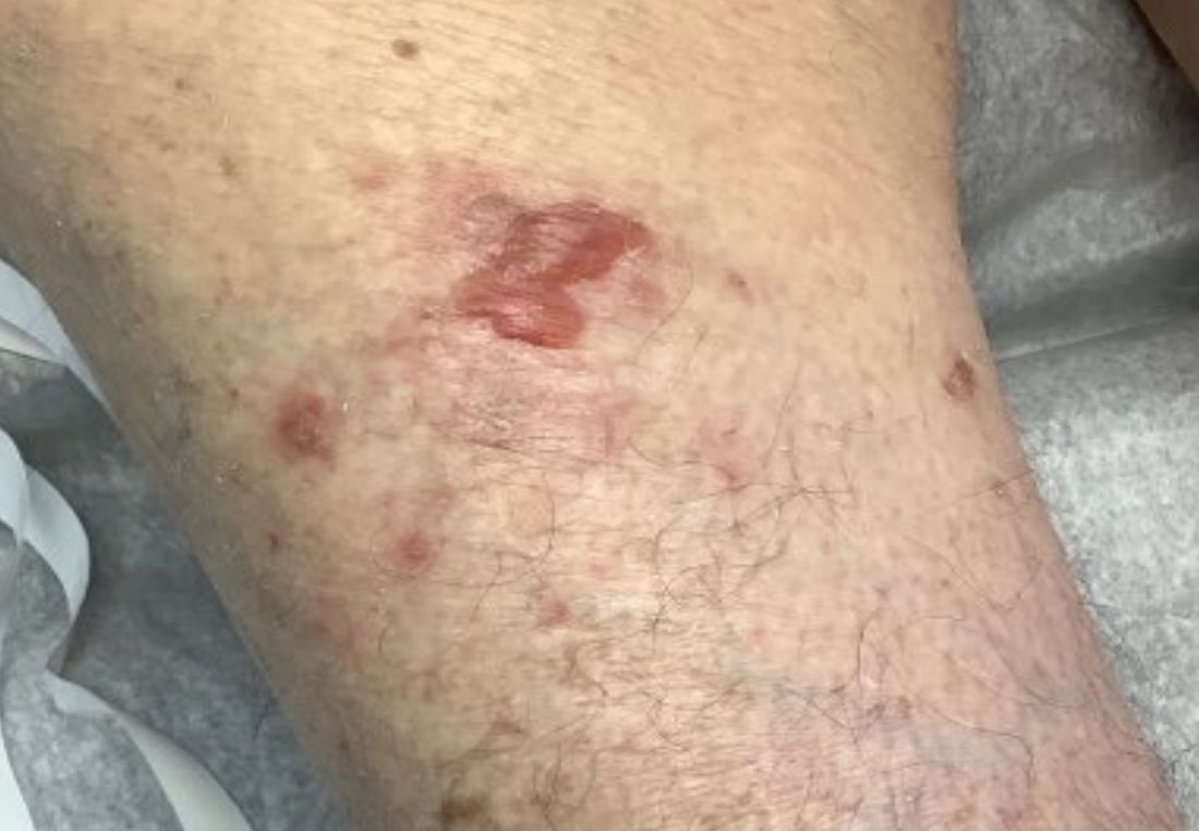
A 95-year-old White male with hypertension presented with itchy patches and bullae on the trunk and extremities
and is associated with various predisposing factors, including HLA genes, comorbidities, aging, and trigger factors such as drugs, trauma, radiation, chemotherapy, and infections. The autoimmune reaction is mediated by a dysregulation of T cells in which IgG and IgE autoantibodies form against hemidesmosomal proteins (BP180 and BP230). These autoantibodies induce neutrophil activation, recruitment, and degradation in the basement membrane of the skin.
Typically, patients present with intense pruritus followed by an urticarial or eczematous eruption. Tense blisters and bullae occur commonly on the trunk and extremities. Drug-associated bullous pemphigoid (DABP) is a common manifestation of the disease with histologic and immunologic features similar to those of the idiopathic version. Eruptions can be triggered by systemic or topical medications, and incidence of these reactions may be related to a genetic predisposition for the disease.
Some research suggests that drug-induced changes to the antigenic properties of the epidermal basement membrane result in an augmented immune response, while others point to structural modification in these zones that stimulate the immune system. Thiol- and phenol-based drugs have been largely implicated in the development of DABP because they are capable of structural modification and disruption of the dermo-epidermal junction in the basement membrane.
DABP often presents with patients taking multiple medications. Some of the most common medications are gliptins, PD-1 inhibitors, diuretics, antibiotics, anti-inflammatory drugs, and ACE-inhibitors, and other cardiovascular drugs. DABP may present with mucosal eruptions unlike its idiopathic counterpart that is mostly contained to the skin.

On this patient, two punch biopsies were taken. Histopathology revealed an eosinophil-rich subepidermal blister with a smooth epidermal undersurface consistent with bullous pemphigoid. Direct immunofluorescence was positive with a deposition of IgG and C3 at the epidermal side of salt split basement membrane zone.
Treatment for BP includes high potency topical and systemic steroids. Tetracyclines and niacinamide have been reported to improve the condition. Treatment is tailored to allow for cutaneous healing and control pruritus, but the physician must be mindful of the patient’s comorbidities and capacity for self-care. Prognosis is often better for DABP as withdrawal of the medication greatly accelerates clearance of the lesions. Worse prognosis is related to increased number of comorbidities and older age. Our patient’s BP is controlled currently with topical steroids and oral doxycycline.
This case and photo were submitted by Lucas Shapiro, BS, Nova Southeastern University College of Osteopathic Medicine, Tampa, and Dr. Bilu Martin.
Dr. Bilu Martin is a board-certified dermatologist in private practice at Premier Dermatology, MD, in Aventura, Fla. More diagnostic cases are available at mdedge.com/dermatology. To submit a case for possible publication, send an email to [email protected].
References
1. Miyamoto D et al. An Bras Dermatol. 2019 Mar-Apr;94(2):133-46.
2. Moro et al. Biomolecules. 2020 Oct 10;10(10):1432.
3. Verheyden M et al. Acta Derm Venereol. 2020 Aug 17;100(15):adv00224.
and is associated with various predisposing factors, including HLA genes, comorbidities, aging, and trigger factors such as drugs, trauma, radiation, chemotherapy, and infections. The autoimmune reaction is mediated by a dysregulation of T cells in which IgG and IgE autoantibodies form against hemidesmosomal proteins (BP180 and BP230). These autoantibodies induce neutrophil activation, recruitment, and degradation in the basement membrane of the skin.
Typically, patients present with intense pruritus followed by an urticarial or eczematous eruption. Tense blisters and bullae occur commonly on the trunk and extremities. Drug-associated bullous pemphigoid (DABP) is a common manifestation of the disease with histologic and immunologic features similar to those of the idiopathic version. Eruptions can be triggered by systemic or topical medications, and incidence of these reactions may be related to a genetic predisposition for the disease.
Some research suggests that drug-induced changes to the antigenic properties of the epidermal basement membrane result in an augmented immune response, while others point to structural modification in these zones that stimulate the immune system. Thiol- and phenol-based drugs have been largely implicated in the development of DABP because they are capable of structural modification and disruption of the dermo-epidermal junction in the basement membrane.
DABP often presents with patients taking multiple medications. Some of the most common medications are gliptins, PD-1 inhibitors, diuretics, antibiotics, anti-inflammatory drugs, and ACE-inhibitors, and other cardiovascular drugs. DABP may present with mucosal eruptions unlike its idiopathic counterpart that is mostly contained to the skin.
On this patient, two punch biopsies were taken. Histopathology revealed an eosinophil-rich subepidermal blister with a smooth epidermal undersurface consistent with bullous pemphigoid. Direct immunofluorescence was positive with a deposition of IgG and C3 at the epidermal side of salt split basement membrane zone.
Treatment for BP includes high potency topical and systemic steroids. Tetracyclines and niacinamide have been reported to improve the condition. Treatment is tailored to allow for cutaneous healing and control pruritus, but the physician must be mindful of the patient’s comorbidities and capacity for self-care. Prognosis is often better for DABP as withdrawal of the medication greatly accelerates clearance of the lesions. Worse prognosis is related to increased number of comorbidities and older age. Our patient’s BP is controlled currently with topical steroids and oral doxycycline.
This case and photo were submitted by Lucas Shapiro, BS, Nova Southeastern University College of Osteopathic Medicine, Tampa, and Dr. Bilu Martin.
Dr. Bilu Martin is a board-certified dermatologist in private practice at Premier Dermatology, MD, in Aventura, Fla. More diagnostic cases are available at mdedge.com/dermatology. To submit a case for possible publication, send an email to [email protected].
References
1. Miyamoto D et al. An Bras Dermatol. 2019 Mar-Apr;94(2):133-46.
2. Moro et al. Biomolecules. 2020 Oct 10;10(10):1432.
3. Verheyden M et al. Acta Derm Venereol. 2020 Aug 17;100(15):adv00224.
A 95-year-old White male with hypertension presented with a history of very itchy patches and bullae on the trunk and extremities.
Thyroid nodule volume reduction correlates with energy in ablation
MONTREAL – In the treatment of thyroid nodules with radiofrequency ablation (RFA), the amount of energy delivered per unit volume of the nodule strongly correlates with the extent of nodule volume reduction after 6 and 12 months, suggesting an important indicator of treatment success.
The findings “provide an objective measure or goal energy input to achieve during the [RFA] procedure rather than relying only on the subjective judgment of sonographic changes, and in turn, produce more reliable outcomes for our patients,” first author Samantha A. Wolfe, MD, said in an interview.
Dr. Wolfe, of the department of otolaryngology – head and neck surgery at Johns Hopkins University, Baltimore, presented the findings at the American Thyroid Association annual meeting.
Commenting on the study, Insoo Suh, MD, an associate professor and associate vice chair of Surgical Innovation at New York University Langone Health, agreed that “an accounting of the total amount of energy delivered can be a useful additional data point for the operator when they are determining whether an ablation is successful.”
He noted, however, that the location of a nodule can be an important factor when deciding upon amounts of RF energy.
“Some target areas are too close for comfort to critical structures, such as the trachea or the recurrent laryngeal nerve, so sound judgment would dictate that the energy be dialed down in those areas, even if the price you pay is a slightly lower volume reduction,” he explained.
Analysis of patients given RFA at Johns Hopkins
RFA utilizes RF energy for the reduction of nodule compression and aesthetic symptoms, avoiding the need for thyroid hormone replacement or surgery.
And while decisions regarding RFA treatment location and duration are commonly guided by the operator’s judgment of sonographic changes, those assessments can potentially result in inconsistent outcomes.
In observing a relationship between higher amounts of RF energy and nodule volume reduction, Dr. Wolfe and associates conducted their prospective study of nodules treated by two experienced endocrine surgeons at Johns Hopkins between June 2019 and May 2022 at 6 and 12 months in relation to the amount of total energy delivered during the treatment.
The analysis included 101 nodules, which had a median initial volume of 12.9 mL.
After 6 months, the median volume reduction ratio was 60%, and at 12 months, the median reduction was 64%.
In terms of the goal of achieving 50% or more volume reduction at 6 months, the median energy delivered was significantly higher for nodules that did reach that goal compared with those that had a volume reduction of less than 50% (2,317 vs. 1,912 J/mL, respectively; P = .01).
The figures were similar at 12 months (2,031 vs. 1254 J/mL; P < .01).
In a logistic regression analysis, the amount of energy delivered strongly increased the odds of obtaining a volume reduction ratio of at least 50% (odds ratio, 2.58; P = .048).
“Every twofold increase in energy delivered increases the odds of achieving a 50% volume ratio reduction by 2.58 times,” Dr. Wolfe explained.
Likewise, the same twofold increase in energy delivered also increased the odds of achieving a greater than 80% volume ratio reduction by 2.55 times (OR, 2.55; P = .038), she added.
Information may help to decide who needs multiple ablations
Of note, the effect was stronger with smaller nodules. Those with an initial volume of less than 20 mL had a significantly greater volume ratio reduction than nodules that were 20 mL or larger (61% vs. 48%, respectively; P = .05).
The initial volume of nodules that did, and did not, achieve a 50% volume ratio reduction at 6 months were 10.9 mL versus 19.1 mL, and the initial volumes of those that did, and did not, have at least a 50% reduction at 12 months were 10.5 mL and 41.5 mL.
“At 6 and 12 months, the successfully treated nodules had a significantly smaller immediate initial volume than those that did not,” Dr. Wolfe said.
“This information may aid in identifying patients with large nodules that are less likely to achieve a greater than 50% volume reduction ratio and may require multiple treatments,” she added.
Other factors – including the probe tip size and total energy delivered – did not significantly correlate with volume ratio reduction at 6 or 12 months.
There was also no significant difference in terms of thyroid-stimulating hormone levels among nodules that achieved at least a 50% volume reduction and those that did not.
Nodules that did not have a satisfactory volume reduction at 12 months had a relatively large median total energy value delivered during ablation (103,463 J, compared with 25,969 J among those achieving more than 50% volume ratio reduction), which Dr. Wolfe said likely reflects that those nodules had a large initial volume.
“This speaks to the importance of describing the energy utilized per unit of nodule volume rather than just a gross measurement,” she said during her presentation.
Dr. Wolfe added that in terms of strategies for getting more energy into the nodule, a key approach is time.
“Sometimes you will see sonographic changes very quickly in the nodule, and it could be tempting to consider that area ablated and move on if you only rely on sonographic changes,” she said in an interview. “However, our research shows that, by spending more time, and thus inputting more energy into the nodule, we had better volume reduction.”
Dr. Wolfe and Dr. Suh reported no relevant financial relationships.
A version of this article first appeared on Medscape.com.
AT ATA 2022
Four methods to chip away at imposter syndrome
Regardless of the setting, one of the most frequently discussed topics in health care is imposter syndrome.

Imposter syndrome was first defined by Clance and Imes as an inability to internalize success, and the tendency to attribute success to external causes such as luck, error, or knowing the appropriate individual.1 This definition is essential because most health care professionals have had a sense of doubt or questioned the full extent of their competencies in various situations. I would argue that this is normal and – within reason – helpful to the practice of medicine. The problem with true imposter syndrome is that the individual does not incorporate success in a way that builds healthy self-esteem and self-efficacy.2
Imposter syndrome has a very nasty way of interacting with burnout. Studies have shown that imposter syndrome can be associated with high levels of emotional exhaustion at work.3 In my experience, this makes clinical sense. Professionals suffering from imposter syndrome can spend a great deal of time and energy trying to maintain a particular image.4 They are acting a part 24/7. Have you ever seriously tried to act? It’s arduous work. A friend once asked me to read a role for a play because “you’d be great; you’re a natural.” By the time I was done with rehearsal, I felt like I had run a 4-by-400-meter relay, by myself, in Victoria, Tex.
And any talk of imposter syndrome must include its running mate, perfectionism. These two conditions exist together so commonly it can be a bit of a chicken or egg question as to which came first.
Imposter syndrome, perfectionism, and burnout can form a deadly triad if not recognized and addressed quickly. In medicine, perfectionism can be a coping strategy that sets up unrelenting standards. Failure to meet unrelenting standards then serves as fuel and validation for imposter syndrome and emotional exhaustion. The consequences of this cycle going unchecked over a health care professional’s career are seismic and can include downstream effects ranging from depression to suicide.
Some readers will relate to this, while others will shrug their shoulders and say that this has never happened in their professional life. I get it. However, I would now ask if you have ever felt like an imposter in your personal life. I’ll make a cup of tea and wait for you to figure out precisely what is the boundary between your personal and professional life. Okay, all done? Great. Now I’ll give you some more time to sincerely reflect if any of the traits of imposter syndrome have described you at times in your personal life. Hmmm, interesting to think about, isn’t it?
I believe that health care professionals frequently use one credit card to pay off another, but the debt remains the same. So even if things are going well at work, we may have just shifted the debt to our personal lives. (At some point in the future, I’ll share my 10 greatest father fails to date to elucidate my point.)
In my work at the GW Resiliency and Well-Being Center, I’ve gravitated toward a few methods supported by evidence that help alleviate imposter syndrome symptoms and potentially serve as protective factors against the future development of imposter syndrome.4 These include but are not limited to:
- Keep a record of small personal success that is yours alone.
- Have a mentor to share failures with.
- Use personal reflection to examine what it means to successfully reach your goals and fulfill your purpose, not a relative value unit target.
- Share experiences with each other, so you know you’re not alone.
The last method is one of my favorites because it involves connecting to others and shining a light on our shared experiences and, coincidentally, our collective strengths. Once this collective strength is realized, the circumstances of that 4-by-400-meter relay change drastically. Be safe and well, everyone.
Lorenzo Norris, MD, is a psychiatrist and chief wellness officer for the George Washington University Medical Enterprise and serves as associate dean of student affairs and administration for the George Washington University School of Medicine and Health Sciences. A version of this article first appeared on Medscape.com.
References
1. Clance PR, Imes SA. The imposter phenomenon in high achieving women: Dynamics and therapeutic intervention. Psychotherapy: Theory, Research & Practice. 1978;15(3): 241-7. doi: 10.1037/h0086006.
2. Thomas M, Bigatti S. Perfectionism, impostor phenomenon, and mental health in medicine: A literature review. Int J Med Educ. 2020 Sep 28;11:201-3. doi: 10.5116/ijme.5f54.c8f8.
3. Liu RQ et al. Impostorism and anxiety contribute to burnout among resident physicians. Med Teach. 2022 Jul;44(7):758-64. doi: 10.1080/0142159X.2022.2028751.
4. Gottlieb M et al. Impostor syndrome among physicians and physicians in training: A scoping review. Med Educ. 2020 Feb;54(2):116-24. doi: 10.1111/medu.13956.
Regardless of the setting, one of the most frequently discussed topics in health care is imposter syndrome.
Imposter syndrome was first defined by Clance and Imes as an inability to internalize success, and the tendency to attribute success to external causes such as luck, error, or knowing the appropriate individual.1 This definition is essential because most health care professionals have had a sense of doubt or questioned the full extent of their competencies in various situations. I would argue that this is normal and – within reason – helpful to the practice of medicine. The problem with true imposter syndrome is that the individual does not incorporate success in a way that builds healthy self-esteem and self-efficacy.2
Imposter syndrome has a very nasty way of interacting with burnout. Studies have shown that imposter syndrome can be associated with high levels of emotional exhaustion at work.3 In my experience, this makes clinical sense. Professionals suffering from imposter syndrome can spend a great deal of time and energy trying to maintain a particular image.4 They are acting a part 24/7. Have you ever seriously tried to act? It’s arduous work. A friend once asked me to read a role for a play because “you’d be great; you’re a natural.” By the time I was done with rehearsal, I felt like I had run a 4-by-400-meter relay, by myself, in Victoria, Tex.
And any talk of imposter syndrome must include its running mate, perfectionism. These two conditions exist together so commonly it can be a bit of a chicken or egg question as to which came first.
Imposter syndrome, perfectionism, and burnout can form a deadly triad if not recognized and addressed quickly. In medicine, perfectionism can be a coping strategy that sets up unrelenting standards. Failure to meet unrelenting standards then serves as fuel and validation for imposter syndrome and emotional exhaustion. The consequences of this cycle going unchecked over a health care professional’s career are seismic and can include downstream effects ranging from depression to suicide.
Some readers will relate to this, while others will shrug their shoulders and say that this has never happened in their professional life. I get it. However, I would now ask if you have ever felt like an imposter in your personal life. I’ll make a cup of tea and wait for you to figure out precisely what is the boundary between your personal and professional life. Okay, all done? Great. Now I’ll give you some more time to sincerely reflect if any of the traits of imposter syndrome have described you at times in your personal life. Hmmm, interesting to think about, isn’t it?
I believe that health care professionals frequently use one credit card to pay off another, but the debt remains the same. So even if things are going well at work, we may have just shifted the debt to our personal lives. (At some point in the future, I’ll share my 10 greatest father fails to date to elucidate my point.)
In my work at the GW Resiliency and Well-Being Center, I’ve gravitated toward a few methods supported by evidence that help alleviate imposter syndrome symptoms and potentially serve as protective factors against the future development of imposter syndrome.4 These include but are not limited to:
- Keep a record of small personal success that is yours alone.
- Have a mentor to share failures with.
- Use personal reflection to examine what it means to successfully reach your goals and fulfill your purpose, not a relative value unit target.
- Share experiences with each other, so you know you’re not alone.
The last method is one of my favorites because it involves connecting to others and shining a light on our shared experiences and, coincidentally, our collective strengths. Once this collective strength is realized, the circumstances of that 4-by-400-meter relay change drastically. Be safe and well, everyone.
Lorenzo Norris, MD, is a psychiatrist and chief wellness officer for the George Washington University Medical Enterprise and serves as associate dean of student affairs and administration for the George Washington University School of Medicine and Health Sciences. A version of this article first appeared on Medscape.com.
References
1. Clance PR, Imes SA. The imposter phenomenon in high achieving women: Dynamics and therapeutic intervention. Psychotherapy: Theory, Research & Practice. 1978;15(3): 241-7. doi: 10.1037/h0086006.
2. Thomas M, Bigatti S. Perfectionism, impostor phenomenon, and mental health in medicine: A literature review. Int J Med Educ. 2020 Sep 28;11:201-3. doi: 10.5116/ijme.5f54.c8f8.
3. Liu RQ et al. Impostorism and anxiety contribute to burnout among resident physicians. Med Teach. 2022 Jul;44(7):758-64. doi: 10.1080/0142159X.2022.2028751.
4. Gottlieb M et al. Impostor syndrome among physicians and physicians in training: A scoping review. Med Educ. 2020 Feb;54(2):116-24. doi: 10.1111/medu.13956.
Single dose of psilocybin for major depression tied to short-term remission
, new research shows.
In the largest study of psilocybin for TRD to date, results of the phase 2b randomized, double-blind trial show participants in the 25-mg dose group experienced a significant reduction in depressive symptoms for at least 3 weeks vs. patients in the 10-mg or 1-mg group, which served as the control group.
Investigators found that 29% of participants who received the 25-mg dose were in remission 3 weeks after the treatment and 37% had at least a 50% drop in depression scores. However, at the 3-month mark, only 20% of those on the 25-mg dose experienced significant improvement.
The change from baseline to week 3 in the Montgomery–Åsberg Depression Rating Scale (MADRS) total score was significantly better with a 25-mg dose than with a 1-mg dose; there was no significant difference between the 10-mg dose and the 1-mg dose, the investigators reported.
The response rate was high for those receiving the 25-mg dose, lead investigator Guy Goodwin, MD, DPhil, told reporters attending a press briefing.
“It’s important to understand that response rates in these patients are usually somewhere between 10% and 20%, and we are seeing remission rates at three weeks of 30%,” he said.
Dr. Goodwin is chief medical officer of COMPASS Pathways, the company that funded the trial and created COMP360, the synthetic formulation of psilocybin used in the trial, and professor emeritus of psychiatry at the University of Oxford, England.
Based on the results of the trial it was announced that a phase 3 trial will launch in December.
The study was published online in the New England Journal of Medicine.
Further research planned
Psilocybin has been under investigation for TRD for some time, including one study that compared it with the antidepressant escitalopram (Lexapro) with promising results.
In the current study the researchers sought to find an acceptable, efficacious dose and the safety of a synthetic formulation of the drug administered in combination with psychological support.
The multicenter study was conducted at 22 sites in 10 countries and included 233 participants with TRD and evaluated the safety and efficacy of one of three doses. The study’s primary endpoint was change from baseline to 3 weeks in MADRS scores in patients with TRD. The scale runs from 0 to 60 with higher scores indicating more severe depression.
Participants were randomly assigned to receive 25 mg of psilocybin (n = 79), 10 mg (n = 75) or 1 mg (n = 79). Those taking medications discontinued them at least 2 weeks before the baseline visit. The mean MADRS score was 32 or 33 in each study group.
There was a 3- to 6-week run-up period to the study in which each participant met with a study therapist about three times to build trust and prepare for the psychedelic experience.
On the day of psilocybin administration, each participant listened to a tailored music playlist and wore eye shades while reclining in a comfortable chair to direct attention inwardly.
The psychotherapy sessions lasted 6-8 hours, and two therapists were always present. The following day, participants returned for an “integration” session with the therapists that was designed to help the participants explore insights from their session.
MADRS scores were measured at baseline, the day following psilocybin administration, and at weeks 1, 3, 6, 9, and 12.
Participants were asked to stay off standard antidepressant treatment during the first 3 weeks of the trial but could be restarted at any time if deemed necessary by a trial investigator.
Mean changes from baseline to week 3 in MADRS scores were −12.0 for 25-mg, −7.9 for 10-mg, and −5.4 for 1-mg groups. The difference between the 25-mg group and 1-mg group was −6.6 (95% confidence interval [CI], −10.2 to −2.9; P < .001 and between the 10-mg group and 1-mg group was −2.5 (95% CI, −6.2 to 1.2; P = .18).
The investigators reported that in the 25-mg group, the incidences of response and remission at 3 weeks, but not sustained response at 12 weeks, were generally supportive of the primary results.
Up to 84% of those who received the 25-mg dosage reported adverse events, with the occurrence dropping slightly with each dosage group. The most frequent adverse events included headache, nausea, dizziness, and fatigue, and occurred only on administration day.
Among those who received the 25-mg dose of psilocybin, two participants reported suicidal thoughts during the 3 weeks following treatment, and 3 months post treatment, three patients exhibited suicidal behavior.
Dr. Goodwin noted that these participants had a prior history of suicidal behavior. Two participants in the 10-mg group also had suicidal thoughts. However, the investigators also noted that suicidal ideation, behavior, or self-injury occurred in all dose groups.
The researchers noted that longer and larger trials, including comparisons with existing depression treatments, are needed to determine the safety and efficacy of psilocybin for TRD.
Intriguing, sobering
In an accompanying editorial, Bertha Madras, PhD, McLean Hospital, Belmont, Mass., and Harvard Medical School, Boston, noted “the findings are both intriguing and sobering. The highest dose (25 mg), but not the intermediate dose (10 mg), resulted in significantly lower levels of depressive symptoms after 3 weeks than the lowest dose (1 mg, which served as a control), but the 37% incidence of response with the 25-mg dose was numerically lower than that in large trials of conventional antidepressants and less robust than in a trial showing similar efficacies of psilocybin and a selective serotonin reuptake inhibitor.”
Also sobering, she noted, were the high percentages of adverse events in the 25-mg group and suicidal ideation and behavior. Dr. Madras also wondered if “legalization and commercialization [of psychedelics] are allied with the medical movement, psychedelic shops and ‘clinics’ could proliferate even for vulnerable populations, and rigorously designed medical protocols will be compromised.
“Nevertheless,” she concluded, “it is provocative that these agents show some short-term benefit for depression in selected populations.”
Dr. Goodwin is CMO of Compass Pathways, which funded the study. He and several coauthors disclosed relationships with industry. Dr. Madras reports no relevant financial relationships.
A version of this article first appeared on Medscape.com.
FROM THE NEW ENGLAND JOURNAL OF MEDICINE

STEP TEENS: Semaglutide ‘gives hope’ to adolescents with obesity
Attendees at ObesityWeek® 2022 listened with much excitement to the results of the STEP TEENS phase 3 trial of once-weekly subcutaneous semaglutide 2.4 mg (Wegovy) in adolescents aged 12 up to 18 years old with obesity.
When a session panel member said that clinical trials of weight-loss medications for adolescents with obesity should henceforth stop using placebo controls – implying that comparison with the once-weekly injection semaglutide would be more informative – the audience applauded.
The results were also simultaneously published in the New England Journal of Medicine to coincide with the presentation.
The research “gives hope” to adolescents with obesity, their parents, and their doctors, the trial’s principal investigator, Daniel Weghuber, MD, said in an interview.
“Many of them have been struggling for such a long time – both the parents and the kids themselves,” said Dr. Weghuber, from the department of pediatrics, Paracelsus Medical University, Salzburg, Austria.
“It’s not an issue of lack of willpower,” he stressed. “That’s a major misunderstanding.”
“This drug [semaglutide] seems to enable people who are living with obesity to adhere to the recommendations that they may have been following for years and years but were [still] not able to achieve their goal,” he said. It “enables people to achieve their goals.”
Asked about any potential negative impact on normal growth, Dr. Weghuber pointed out that the average weight of study participants was 107 kg (236 lb). “I’m really not afraid of a 15-year-old with 107 kg losing 10%, 15%, 20%” of their weight, he said. There was no indication of a problem regarding normal growth or development in the study.
, he summarized.
Senior study author, Silva Arslanian, MD, who holds the Richard L. Day Endowed Chair in Pediatrics at the University of Pittsburgh, agreed. “The results are amazing,” said Dr. Arslanian in a press release issued by the University of Pittsburgh. “For a person who is 5 foot, 5 inches tall and weighs 240 pounds, the average reduction in BMI equates to shedding about 40 pounds.”
‘Mind-blowing, awesome’ results
The session at ObesityWeek® 2022, the annual meeting of the Obesity Society, was chaired by Aaron S. Kelly, PhD, professor of pediatrics and codirector of the center for pediatric obesity medicine at the University of Minnesota, Minneapolis.
Dr. Kelly led the SCALE TEENS clinical trial of liraglutide (Saxenda), also a glucagon-like peptide (GLP-1) agonist like semaglutide, for adolescents aged 12 up to 18 years with obesity, which assigned 125 participants to the daily injectable liraglutide group and 126 to the placebo group. SCALE TEENS was presented and published in May 2020, leading to the approval of liraglutide for obesity in this age group, in December 2020.
Dr. Kelly called on two experts who were not involved in the research to offer their comments, starting with Claudia K. Fox, MD, MPH.
“These results are mind-blowing,” said Dr. Fox, who is associate professor of pediatrics and codirector of the center for pediatric obesity medicine at the University of Minnesota.
“We are getting close to bariatric surgery results” in these adolescent patients with obesity, added Dr. Fox, who is an American Board of Obesity Medicine diplomate. To have 40% of patients attain normal weight, “that’s massive” and “life-changing,” she said. And improvement in quality of life is what families care most about. “I am super excited,” she commented.
Next, Dr. Kelly called on Sarah C. Armstrong, MD, director of the Duke Children’s Healthy Lifestyles Program, Duke University, Durham, N.C.
Dr. Armstrong is a member of the executive committee for the American Academy of Pediatrics Section on Obesity and a coauthor of the upcoming clinical practice guidelines that are being published.
Looking at more than 16,000 abstracts at the meeting shows that “watchful waiting is not effective,” Dr. Armstrong said.
200 teens with obesity, only 1 with overweight
Obesity affects almost one in five children and adolescents worldwide. The chronic disease is linked with decreased life expectancy and higher risk of developing serious health problems such as type 2 diabetes, heart disease, nonalcoholic fatty liver disease, sleep apnea, and certain cancers. Teenagers with obesity are also more likely to have depression, anxiety, poor self-esteem, and other psychological issues.
STEP TEENS enrolled 201 adolescents aged 12 up to 18 years with obesity (body mass index [BMI] ≥ 95th percentile) or overweight (BMI ≥ 85th percentile) plus at least one weight-related comorbidity.
Only one recruited patient fit the latter category; the rest had obesity.
Most patients (62%) were female. They had a mean age of 15.4 years, a mean BMI of 37 kg/m2, and a mean waist circumference of 110 cm (43 inches).
Patients were randomized 2:1 to receive a once-weekly 2.4-mg subcutaneous injection of semaglutide or placebo for 68 weeks, plus lifestyle intervention.
Dr. Weghuber noted that 89.6% of patients in the semaglutide group completed treatment.
The primary endpoint, mean change in BMI from baseline to week 68, was −16.1% with semaglutide and +0.6% with placebo (estimated difference, −16.7 percentage points; P < .001).
A second confirmatory endpoint, at least 5% weight loss at week 68, was met by 73% of patients in the semaglutide group versus 18% of patients in the placebo group (P < .001).
Reductions in body weight and improvements in waist circumference, A1c, lipids (except HDL cholesterol), and the liver enzyme alanine aminotransferase were greater with semaglutide than placebo.
The Impact of Weight on Quality of Life – Kids (IWQOL-Kids) questionnaire total score as well as scores for body esteem, family relation, physical comfort, and social life were better in the semaglutide group.
However, the incidence of gastrointestinal adverse events was greater with semaglutide than placebo (62% versus 42%).
Five participants (4%) in the semaglutide group and none in the placebo group developed gallstones (cholelithiasis).
Serious adverse events were reported in 11% of patients in the semaglutide group and 9% of patients in the placebo group.
‘Big change’ coming in guidelines for obesity in teens
Commenting on the upcoming new recommendations for adolescents, Dr. Armstrong noted “there’s going to be a strong recommendation” for therapy in the new guidelines for pediatric obesity. “That’s a big change,” she said.
In the lively question-and-answer session that followed, a clinician wanted to know what explained the very high rate of study completion during the COVID-19 pandemic (when STEP TEENS was conducted). “What can we learn?” he asked.
“The bottom line is the relationship” and “close communication” between study investigators and patients, Dr. Weghuber replied.
“The fast track is likely to lead to approval in adolescents,” another member of the audience noted. He wanted to know if the company is planning a trial of semaglutide in younger children.
They are, Dr. Weghuber replied, and one with liraglutide is already underway.
The SCALE KIDS clinical trial of liraglutide is randomizing 78 participants aged 6 up to 12 years for 56 weeks of treatment and 26 weeks of follow-up, with an estimated primary completion date of July 7, 2023.
The last words went to Dr. Fox. The current results “are indeed very awesome,” she said, yet “thousands of providers are hesitant” to prescribe medications for adolescents with obesity.
The trial was funded by Novo Nordisk. Dr. Weghuber has reported being a consultant for Novo Nordisk and member of the Global Pediatric Obesity Expert Panel for the company. Disclosures for the other authors are listed with the article. Dr. Kelly has reported receiving donated drugs from AstraZeneca and travel support from Novo Nordisk and serving as an unpaid consultant for Novo Nordisk, Orexigen Therapeutics, VIVUS, and WW (formerly Weight Watchers).
A version of this article first appeared on Medscape.com.
FROM OBESITYWEEK® 2022
Fitness trackers: Useful in sleep medicine?
Who doesn’t love data, especially their own? With that thought in mind, over the years I have owned several activity trackers, including at least two Fitbits, and I frequently check my iPhone to see how far I’ve walked or how many steps I have taken. My most recent acquisition is an Oura (smart ring, third generation), which includes my first sleep tracker.
Sleep trackers are not unique to the Oura Ring; they are included on many of the newer activity trackers and smart watches, but the design and breakdown of daily sleep, activity, and readiness scores are hallmarks of Oura Rings.
The ring generates data for different phases of sleep, movements, oxygen saturation, disturbances in breathing, heart rate, and heart rate variability. I began to wonder how useful this information would be clinically and whether it might be helpful in either the diagnosis or treatment of sleep disorders.
David Neubauer, MD, is a psychiatrist at the Johns Hopkins Sleep Disorders Center. “Sleep tracking devices are more than just toys but less than medical devices. They do have clinical utility and might show findings that warrant further medical workup,” Dr. Neubauer said. “It is impressive that these devices estimate sleep as well as they do, but there is a problem with how they divide sleep stages that can lead people to believe their sleep is worse than it really is.”
For more than 50 years, he explained, sleep researchers and clinicians have categorized sleep as non–rapid eye movement (NREM) sleep stages 1-4 and REM sleep. More recently, sleep was reorganized to N1, N2, and N3 (which combines the older stages 3 and 4, representing “deep sleep” or “slow wave sleep”) and REM sleep. We normally spend more time in N2 than the other stages. However, the device companies often categorize their sleep estimates as “light sleep,” “deep sleep,” or “REM.” With “light sleep,” they are lumping together N1 and N2 sleep, and this is misleading, said Dr. Neubauer. “Understandably, people often think that there is something wrong if their tracker reports they are spending a lot of time in light sleep, when actually their sleep may be entirely normal.”
Sleep tracker validity
A study by Massimiliano de Zambotti, PhD, and colleagues, “The Sleep of the Ring: Comparison of the ŌURA Sleep Tracker Against Polysomnography”, looked at sleep patterns of 41 adolescents and young adults and concluded that the second-generation tracker was accurate in terms of total sleep but underestimated time spent in N3 stage sleep by approximately 20 minutes while overestimating time spent in REM sleep by 17 minutes. They concluded that the ring had potential to be clinically useful but that further studies and validation were needed.
A larger study of the newest, third-generation Oura tracker, conducted by Altini and Kinnunen at Oura Health, found that the added sensors with the newer-generation ring led to improved accuracy, but they noted that the study was done with a healthy population and might not generalize to clinical populations.
Fernando Goes, MD, and Matthew Reid, PhD, both at Johns Hopkins, are working on a multicenter study using the Oura Ring and the mindLAMP app to look at the impact of sleep on mood in people with mood disorders as well as healthy controls. Dr. Reid said that “validation of sleep stages takes a hit when the ring is used in people with insomnia. We find it useful for total sleep time, but when you look at sleep architecture, the concordance is only 60%. And oxygen saturation measures are less accurate in people with dark skin.”
Clinical uses for sleep trackers
More accurate information might prove reassuring to patients. Dr. Goes added, “One use, for example, might be to help patients to limit or come off of long-term hypnotics with a more benign intervention that incorporates passive monitoring such as that in the Oura Ring. Some patients worry excessively about not being able to sleep, and sleep monitoring data can be helpful to reduce some of these concerns so patients can focus on safer interventions, such as cognitive behavioral therapy for insomnia.” Dr. Reid believes that wearable trackers have potential usefulness in monitoring sleep in patients with insomnia. “In insomnia, sleep state misperception is common. They are hyper-aroused, and they perceive that they are awake when in fact they are sleeping.”
Dr. Goes mentioned another use for sleep trackers in clinical settings: “In our inpatient units, the nurses open the door to look in on patients every hour to monitor and document if they are sleeping. If they look in and the patient isn’t moving, they will ask the patient to raise their hand, which of course is not going to help someone to fall back asleep.” Wearable devices might provide data on sleep without the risk of waking patients every hour through the night.
Not medical devices
However, Dr. Neubauer emphasized that current sleep trackers are not medical devices, saying “they may be measuring the same parameters that are measured with medical devices, for example pulse oximetry or sleep states, but there’s no simple answer yet to the question of whether the devices provide reliable data for clinical decision-making.”
Dr. Neubauer is skeptical about the accuracy of some of the measures the device provides. “I would not use the information from a consumer device to rule out obstructive sleep apnea based on good oxygen saturation numbers. So much depends on the history – snoring, gasping awakenings, reports from bed partners, and daytime sleepiness. These devices do not measure respiratory effort or nasal airflow as sleep studies do. But big drops in oxygen saturation from a consumer device certainly warrant attention for further evaluation.” Dr. Neubauer also noted that the parameters on sleep trackers do not differentiate between central or obstructive sleep apnea and that insurers won’t pay for continuous positive airway pressure to treat sleep apnea without a sleep study.
I enjoy looking at the data, even knowing that they are not entirely accurate. and we may find more clinical uses for these devices. For now, I’m off to get more exercise, at the suggestion of my tracker!
Dinah Miller, MD, is assistant professor of psychiatry and behavioral sciences, Johns Hopkins Medicine, Baltimore.
A version of this article first appeared on Medscape.com.

Microplastics and health risks: What do we really know?
You eat a credit card’s worth of plastic in a week. That may bother you. But does it harm you?
The answer depends on who you ask. Awareness of microplastics in general is certainly increasing; the most recent news is the detection of microplastics in human breast milk. Other research has suggested that we may be consuming up to 5 grams of plastic each week from our food, water, and certain consumer products.
The World Health Organization has been releasing reports on microplastics and human health since 2019. Their most recent report was released in late August 2022.
“Although the limited data provide little evidence that nano- and microplastic particles have adverse effects in humans, there is increasing public awareness and an overwhelming consensus among all stakeholders that plastics do not belong in the environment, and measures should be taken to mitigate exposure,” the WHO said at the time.
The WHO can’t go beyond what the data shows, of course. If microplastics are wreaking long-term havoc in our bodies as we speak, science hasn’t connected the dots enough to definitively say “this is the problem.”
But some researchers are willing to speculate – and, at the very least, the risks are becoming impossible to ignore. Dick Vethaak, PhD, a microplastics researcher and emeritus professor of ecotoxicology at Vrije Universiteit, Amsterdam, is blunt, calling them “a plastic time bomb.”
The plastic problem
Every piece of plastic that has ever been created is still on our planet today, apart from what has been burned. Past estimates show we only recycle about 9% of all plastic, leaving 9 billion tons in our landfills, oceans, and ecosystems. For context, that amount is 1,500 times heavier than the Great Pyramid of Khufu.
New data is even more dire. A 2022 report from Greenpeace showed a 5% U.S. recycling rate in 2021, with a large portion of what consumers think of as “recycled” still winding up in garbage piles or bodies of water.
And this plastic doesn’t disappear. Instead, it breaks down into smaller and smaller pieces known as microplastics and nanoplastics.
Microplastics have been confirmed in human blood, lung tissue, colons, placentas, stool, and breast milk. But how they impact our health is still unknown.
To assess risk, we must ask: “How hazardous is the material?” said Flemming Cassee, PhD, professor of inhalation toxicology at Utrecht (the Netherlands) University and coauthor of the WHO’s recent microplastics report.
There are three potential hazards of microplastics: their physical presence in our bodies, what they’re made of, and what they carry. To determine the extent of these risks, we need to know how much we’re exposed to, said Dr. Cassee.
The first initiative to research the impact of microplastics on human health came from the European Union in 2018. Although microplastics were around before then, we were unable to detect them, said Dr. Cassee.
That’s the real problem: warned Dr. Vethaak.
What, exactly, are microplastics?
Microplastics are plastic particles between 5 mm and 100 nm in diameter, or the width of a pencil eraser and something 10 times thinner than a human hair. Anything smaller than that is known as a nanoplastic.
“Microplastics include a wide range of different materials, different sizes, different shapes, different densities, and different colors,” said Evangelos Danopoulos, PhD, a microplastics researcher at Hull York (England) Medical School.
“Primary” microplastics are manufactured to be small and used in things like cosmetics and paints. “Secondary” microplastics result from the breaking down of larger plastic materials, like water bottles and plastic bags.
Secondary microplastics are more diverse than primary microplastics and can take forms ranging from fibers shed from synthetic clothing (like polyester) to pieces of a plastic spoon left in our rivers, lakes, and oceans. Any plastic in the environment will eventually become a secondary microplastic as natural forces such as wind, water currents, and UV radiation break it down into smaller and smaller pieces.
Plastic is a diverse material. Heather Leslie, PhD, senior researcher in Vrije Universiteit’s department of environment and health, likens it to spaghetti with sauce. The noodles are the long polymer backbone that all plastic shares. The sauces are “the pigments, the antioxidants, the flame retardants, etc., that make it functional,” she said.
What makes microplastics dangerous?
There are more than 10,000 different chemicals, or “sauces,” used to alter a plastic’s physical characteristics – making it softer, more rigid, or more flexible, said Hanna Dusza, PhD, of the Institute for Risk Assessment Sciences at Utrecht University.
As plastics degrade and become microplastics, these chemicals likely remain. Recent research has shown that microplastics leach these chemicals locally in human tissues, or other areas of accumulation, said Dr. Dusza. Some 2,400 of the 10,000 chemical additives were classified as substances of potential concern, meeting the European Union’s criteria for persistence, bioaccumulation, or toxicity.
Many of these chemicals also act as endocrine-disrupting compounds, or toxicants that imitate hormones when they enter the body. Hormones are active at very low concentrations in your bloodstream, explained Dr. Leslie. To your body, some chemical additives in plastic resemble hormones, so the body responds.
“Sometimes even a low dose of some of these additives can cause unwanted effects,” said Dr. Leslie.
Bisphenol A (BPA), for example, is one of the more infamous endocrine disruptors. It is used as an additive to make plastics more rigid and can be found in any number of plastic products, though areas of concern have been plastic water bottles, baby bottles, and the protective coatings in canned foods.
BPA may mimic estrogen, the female sex hormone essential for reproduction, neurodevelopment, and bone density. In men, estrogen regulates sperm count, sex drive, and erectile function. BPA exposure has been linked with – but not proven to cause – multiple cancer types, ADHD, obesity, and low sperm count. Most everyone has some amount of BPA circulating within their blood, but microplastics may retain BPA as they degrade, potentially increasing our exposure, leading to its unwanted consequences, said Dr. Dusza.
And BPA is just one of those 2,400 substances of “potential concern.”
The inflammation problem
A potentially larger health issue emerges from our bodies yet again doing what they are supposed to do when encountering microplastics. Particles can trigger an immune response when they enter your bloodstream, explains Nienke Vrisekoop, PhD, assistant professor at UMC Utrecht.
White blood cells have no issue breaking down things like bacteria, but microplastics cannot be degraded. When a white blood cell engulfs a certain mass of microplastics – either many small particles or a singular large one – it dies, releasing its enzymes and causing local inflammation.
Meanwhile, the plastic particle remains. So more white blood cells attack.
“This triggers continual activation that can result in various adverse effects, including oxidative stress and the release of cytokines that trigger inflammatory reactions, said Dr. Vethaak.
And “chronic inflammation is the prelude to chronic diseases,” said Dr. Leslie. “Every chronic disease, like cancer, heart disease, and even neuropsychiatric diseases like Parkinson’s or major depression, begins with inflammation.”
Meanwhile, inhaling microplastic particles can lead to respiratory diseases and cancer.
“The smallest particles – less than one-tenth of a micrometer – penetrate deep into the lungs and even into the bloodstream, causing damage to the heart, blood vessels and brain,” said Dr. Vetaak. “The only direct evidence comes from workers in the textile and plastic industries that had been exposed to very high amounts of plastic fibrous dust.”
Microplastics as carriers
Microplastics can also pick up harmful substances and deliver them into your body.
“When they’re in an environment, they basically can suck up [chemicals] like a sponge,” said Dr. Dusza. “These chemicals are known environmental pollutants, like pesticides, fluorinated compounds, flame retardants, and so on.”
Once in the body, these chemicals can be released, potentially leading to cancer, chronic inflammation, or other unknown effects.
Particles can also act as a vector for microbes, bacteria, and viruses. A September 2022 study found that infectious viruses can survive for 3 days in fresh water by “hitchhiking” on microplastics. Their porous nature provides microbes with a perfect environment in which to live and reproduce, said Dr. Dusza. If you ingest the plastics, you ingest the microbes.
How to minimize exposure
There is no way to avoid microplastics. They’re in the air we breathe, the products we use, the water we drink, and the food we eat.
Dr. Danopoulos reviewed 72 studies to quantify our consumption of microplastics in drinking water, salt, and seafood.
“We are exposed to millions of microplastics every year, and I was only looking at three food sources, so there are really a lot more,” he said. “Once plastic waste is mismanaged and it enters the environment, there is very little we can do to extract it.”
That said, we can take steps to lower our exposure and keep the problem from getting worse.
Water filtration is one option, though it is not perfect. Research has shown that municipal water treatment can be effective. An October 2021 study found that two methods – electrocoagulation-electroflotation and membrane filtration – can be 100% effective in removing microplastics from treated water. The problem? Not all municipal water treatment uses these methods – and you would have to investigate to find out if your locality does.
As for at-home filtration methods, they can be effective but can also be dicey. Some consumer brands claim they remove microplastics, but how well depends on not just the type of filter but the size of the particles in the water. Meanwhile, how do you know if a filter is working on your water without testing it, something few people will do? Best not to take a brand’s claims on face-value, but look for independent testing on at-home brands.
A longer-term project: Reduce our risk by reusing and recycling plastic waste. Limiting our consumption of plastic, especially single use plastic, decreases the amount available to become micro- and nanoplastics.
We must all learn to not treat plastic as waste, but rather as a renewable material, said Dr. Cassee. But if that seems like a tall order, it’s because it is.
“You’re a human being and you have a voice and there are a lot of other humans out there with voices,” said Dr. Leslie.
“You sign a petition in your community. You talk about it with your friends at the pub. If you’re a teacher, you discuss it in your class. You call your elected representatives and tell them what you think and how you want them to vote on bills.”
When people start working together, you can really amplify that voice, said Dr. Leslie.
What’s the bottom line right now, today?
Numerous sources have declared microplastics do not impact human health. But that’s largely because no direct evidence of this exists yet.
Even the WHO in its report suggests that progress must happen if we’re to fully understand the scope of the problem.
“Strengthening of the evidence necessary for reliable characterization and quantification of the risks to human health posed by [nano- and microplastics] will require active participation by all stakeholders,” it said.
All researchers interviewed for this article agree we don’t have enough evidence to draw any definite conclusions. But “if you look at the wrong endpoints, things will look safe, until you look at the endpoint where it’s really causing the problem,” said Dr. Leslie.
We must research our blind spots and continually ask: Where could we be wrong?
“It is a problem; it’s not going to go away,” said Dr. Danopoulos. “It’s going to get worse, and will continue to get worse, not by something that we are doing now but by something we did 5 years ago.”
Perhaps the question to be asked, then, is the hardest to answer: Are we willing to wait for the science?
A version of this article first appeared on WebMD.com.