User login
Clinical Psychiatry News is the online destination and multimedia properties of Clinica Psychiatry News, the independent news publication for psychiatrists. Since 1971, Clinical Psychiatry News has been the leading source of news and commentary about clinical developments in psychiatry as well as health care policy and regulations that affect the physician's practice.
Dear Drupal User: You're seeing this because you're logged in to Drupal, and not redirected to MDedge.com/psychiatry.
Depression
adolescent depression
adolescent major depressive disorder
adolescent schizophrenia
adolescent with major depressive disorder
animals
autism
baby
brexpiprazole
child
child bipolar
child depression
child schizophrenia
children with bipolar disorder
children with depression
children with major depressive disorder
compulsive behaviors
cure
elderly bipolar
elderly depression
elderly major depressive disorder
elderly schizophrenia
elderly with dementia
first break
first episode
gambling
gaming
geriatric depression
geriatric major depressive disorder
geriatric schizophrenia
infant
ketamine
kid
major depressive disorder
major depressive disorder in adolescents
major depressive disorder in children
parenting
pediatric
pediatric bipolar
pediatric depression
pediatric major depressive disorder
pediatric schizophrenia
pregnancy
pregnant
rexulti
skin care
suicide
teen
wine
section[contains(@class, 'nav-hidden')]
footer[@id='footer']
div[contains(@class, 'pane-pub-article-cpn')]
div[contains(@class, 'pane-pub-home-cpn')]
div[contains(@class, 'pane-pub-topic-cpn')]
div[contains(@class, 'panel-panel-inner')]
div[contains(@class, 'pane-node-field-article-topics')]
section[contains(@class, 'footer-nav-section-wrapper')]
More data needed to better understand COVID-19 skin manifestations
Qing Zhao, MD, Xiaokai Fang, MD, and their colleagues at the Shandong Provincial Hospital for Skin Diseases & Shandong Provincial Institute of Dermatology and Venereology, in Jinan, China, reported the results of a literature review of 44 articles published through May 2020 that included 507 patients with cutaneous manifestations of COVID-19. The review was published in the Journal of The European Academy of Dermatology and Venereology.
Nearly all of the patients (96%) were from Europe, and more than half were women (60%), with an average age of 49 years. Most patients had multiple skin symptoms, with the most common being erythema (44%), chilblain-like lesions (20%), urticaria-like lesions (16%), vesicular manifestations (13%), livedo/necrosis (6%), and petechiae (almost 2%). The authors described erythema as being present in specific sites, such as the trunk, extremities, flexural regions, face, and mucous membranes. Slightly less than half of all patients had significant pruritus.
Data on systemic COVID-19 symptoms were available for 431 patients and included fever in about two-thirds of patients and cough in almost 70%, with dyspnea in almost half of patients. Almost 60% had fatigue, and almost 60% had asthenia. Information about the onset of skin symptoms was available in 88 patients; of these patients, lesions were seen an average of almost 10 days after systemic symptoms appeared and, in almost 15%, were the first symptoms noted.
Histopathologic exams were done for only 23 patients and, in all cases, showed “inflammatory features without specific pathological changes, such as lymphocyte infiltration.” In one study, reverse transcription polymerase chain reaction testing of skin biopsy specimens tested negative for SARS-CoV-2.
Expression of ACE2, the receptor of SARS-CoV-2, in the skin was evaluated in six of the studies. “Higher ACE2 expression was identified in keratinocytes, mainly in differentiating keratinocytes and basal cells compared to the other cells of skin tissues,” the authors wrote. These results were confirmed with immunohistochemistry, which, they said, found “ACE2-positive keratinocytes in the stratum basal, the stratum spinosum, and the stratum granulosum of epiderma.” They added that this provides evidence “for percutaneous infection or the entry of virus into patients through skin tissues,” but cautioned that more research is needed.
The authors acknowledged that there are still many unanswered questions about COVID-19, and that more clinical data and research are needed, to improve the understanding of the cutaneous manifestations associated with COVID-19.

In an interview, Alisa N. Femia, MD, director of inpatient dermatology in the department of dermatology at New York University, said that the cutaneous signs described in the review align well with what she has seen in patients with COVID-19.
At this point, it is unclear whether cutaneous manifestations of COVID-19 are a result of SARS-CoV-2 invading the skin or an immune response related to SARS-CoV-2, noted Dr. Femia, who was not involved in the research. One method of entry could be through transmitting virus present on the skin to another part of the body where infection is more likely.
While it is possible COVID-19 could be contracted through the skin, she noted, it is much more likely an individual would be infected by SARS-CoV-2 through more traditionally understood means of transmission, such as through respiratory droplets in person-to-person contact. “I think we are far away from drawing that conclusion, that one could touch a surface or a person who has COVID and contract it through their skin,” Dr. Femia said. “The skin has a lot of other ways to protect against that from occurring,” she added.
“SAR-CoV-2 obviously enters through the ACE2 receptor, which is fairly ubiquitous, and it has been seen in keratinocytes,” she said. “But the skin is one of our biggest barriers ... and further, studies to date have shown that that receptor is expressed in relatively low levels of the keratinocytes.”
Pathogenesis of different cutaneous manifestations may be different, Dr. Femia said. For example, urticaria and morbilliform eruption were described by the authors of the review as more benign eruptions, but pathogenesis may differ from that of so-called COVID toes and from the pathogenesis of purpura and ulcerations seen in patients with more severe disease, she noted. It is plausible, she added, that purpura and ulcerations may be a “direct invasion of SARS-CoV-2 into endothelial cells,” which creates secondary processes “that ultimately destroy the skin.”
Urticaria and morbilliform eruptions, on the other hand, “are more simply that the immune system is recognizing COVID, and in doing so, is also recognizing some antigens in the skin and creating a hypersensitive response to the skin” and has “nothing to do with the SARS-CoV-2 virus actually being in that location,” she said.
It is important to differentiate between patients who have skin manifestations attributed to COVID-19 and those with manifestations independent of COVID-19, which is difficult, Dr. Femia noted. A patient with COVID-19 and a cutaneous manifestation may be having a reaction to a medication. “It’s important to have a critical eye and to remember that, when we see these manifestations, we should always be investigating whether there was an alternative cause so that we can better learn what exactly we should be attributing to this infection,” she said

Adam Friedman, MD, professor and interim chair of dermatology at George Washington University, Washington, said the authors of the review had presented interesting work, but made some “assumptions that need to be proven.” Dr. Friedman also was not involved in the research, but agreed in an interview with the assessment that it is unlikely SARS-CoV-2 would penetrate the skin. While some viruses – such as the poxvirus that causes molluscum contagiosum and the herpes simplex virus – invade keratinocytes specifically, there is a particular clinical phenotype that results that is associated with changes in the epidermis. However, “the skin manifestations of COVID-19 do not fit with direct skin invasion, [but] rather the immune response to systemic disease,” he said.
“[I]n terms of systemic invasion through the skin, it is possible, but this study certainly doesn’t show that. The presence/expression of ACE2 in the epidermis doesn’t translate to route of infection,” Dr. Friedman said..
The study received financial support from Shandong First Medical University, the Innovation Project of Shandong Academy of Medical Sciences and the Shandong Province Taishan Scholar Project. The authors report no relevant financial disclosures. Dr. Femia and Dr. Friedman had no relevant financial disclosures.
SOURCE: Zhao Q et al. J Eur Acad Dermatol Venereol. 2020 Jun 28. doi: 10.1111/jdv.16778.
Qing Zhao, MD, Xiaokai Fang, MD, and their colleagues at the Shandong Provincial Hospital for Skin Diseases & Shandong Provincial Institute of Dermatology and Venereology, in Jinan, China, reported the results of a literature review of 44 articles published through May 2020 that included 507 patients with cutaneous manifestations of COVID-19. The review was published in the Journal of The European Academy of Dermatology and Venereology.
Nearly all of the patients (96%) were from Europe, and more than half were women (60%), with an average age of 49 years. Most patients had multiple skin symptoms, with the most common being erythema (44%), chilblain-like lesions (20%), urticaria-like lesions (16%), vesicular manifestations (13%), livedo/necrosis (6%), and petechiae (almost 2%). The authors described erythema as being present in specific sites, such as the trunk, extremities, flexural regions, face, and mucous membranes. Slightly less than half of all patients had significant pruritus.
Data on systemic COVID-19 symptoms were available for 431 patients and included fever in about two-thirds of patients and cough in almost 70%, with dyspnea in almost half of patients. Almost 60% had fatigue, and almost 60% had asthenia. Information about the onset of skin symptoms was available in 88 patients; of these patients, lesions were seen an average of almost 10 days after systemic symptoms appeared and, in almost 15%, were the first symptoms noted.
Histopathologic exams were done for only 23 patients and, in all cases, showed “inflammatory features without specific pathological changes, such as lymphocyte infiltration.” In one study, reverse transcription polymerase chain reaction testing of skin biopsy specimens tested negative for SARS-CoV-2.
Expression of ACE2, the receptor of SARS-CoV-2, in the skin was evaluated in six of the studies. “Higher ACE2 expression was identified in keratinocytes, mainly in differentiating keratinocytes and basal cells compared to the other cells of skin tissues,” the authors wrote. These results were confirmed with immunohistochemistry, which, they said, found “ACE2-positive keratinocytes in the stratum basal, the stratum spinosum, and the stratum granulosum of epiderma.” They added that this provides evidence “for percutaneous infection or the entry of virus into patients through skin tissues,” but cautioned that more research is needed.
The authors acknowledged that there are still many unanswered questions about COVID-19, and that more clinical data and research are needed, to improve the understanding of the cutaneous manifestations associated with COVID-19.
In an interview, Alisa N. Femia, MD, director of inpatient dermatology in the department of dermatology at New York University, said that the cutaneous signs described in the review align well with what she has seen in patients with COVID-19.
At this point, it is unclear whether cutaneous manifestations of COVID-19 are a result of SARS-CoV-2 invading the skin or an immune response related to SARS-CoV-2, noted Dr. Femia, who was not involved in the research. One method of entry could be through transmitting virus present on the skin to another part of the body where infection is more likely.
While it is possible COVID-19 could be contracted through the skin, she noted, it is much more likely an individual would be infected by SARS-CoV-2 through more traditionally understood means of transmission, such as through respiratory droplets in person-to-person contact. “I think we are far away from drawing that conclusion, that one could touch a surface or a person who has COVID and contract it through their skin,” Dr. Femia said. “The skin has a lot of other ways to protect against that from occurring,” she added.
“SAR-CoV-2 obviously enters through the ACE2 receptor, which is fairly ubiquitous, and it has been seen in keratinocytes,” she said. “But the skin is one of our biggest barriers ... and further, studies to date have shown that that receptor is expressed in relatively low levels of the keratinocytes.”
Pathogenesis of different cutaneous manifestations may be different, Dr. Femia said. For example, urticaria and morbilliform eruption were described by the authors of the review as more benign eruptions, but pathogenesis may differ from that of so-called COVID toes and from the pathogenesis of purpura and ulcerations seen in patients with more severe disease, she noted. It is plausible, she added, that purpura and ulcerations may be a “direct invasion of SARS-CoV-2 into endothelial cells,” which creates secondary processes “that ultimately destroy the skin.”
Urticaria and morbilliform eruptions, on the other hand, “are more simply that the immune system is recognizing COVID, and in doing so, is also recognizing some antigens in the skin and creating a hypersensitive response to the skin” and has “nothing to do with the SARS-CoV-2 virus actually being in that location,” she said.
It is important to differentiate between patients who have skin manifestations attributed to COVID-19 and those with manifestations independent of COVID-19, which is difficult, Dr. Femia noted. A patient with COVID-19 and a cutaneous manifestation may be having a reaction to a medication. “It’s important to have a critical eye and to remember that, when we see these manifestations, we should always be investigating whether there was an alternative cause so that we can better learn what exactly we should be attributing to this infection,” she said
Adam Friedman, MD, professor and interim chair of dermatology at George Washington University, Washington, said the authors of the review had presented interesting work, but made some “assumptions that need to be proven.” Dr. Friedman also was not involved in the research, but agreed in an interview with the assessment that it is unlikely SARS-CoV-2 would penetrate the skin. While some viruses – such as the poxvirus that causes molluscum contagiosum and the herpes simplex virus – invade keratinocytes specifically, there is a particular clinical phenotype that results that is associated with changes in the epidermis. However, “the skin manifestations of COVID-19 do not fit with direct skin invasion, [but] rather the immune response to systemic disease,” he said.
“[I]n terms of systemic invasion through the skin, it is possible, but this study certainly doesn’t show that. The presence/expression of ACE2 in the epidermis doesn’t translate to route of infection,” Dr. Friedman said..
The study received financial support from Shandong First Medical University, the Innovation Project of Shandong Academy of Medical Sciences and the Shandong Province Taishan Scholar Project. The authors report no relevant financial disclosures. Dr. Femia and Dr. Friedman had no relevant financial disclosures.
SOURCE: Zhao Q et al. J Eur Acad Dermatol Venereol. 2020 Jun 28. doi: 10.1111/jdv.16778.
FROM THE JOURNAL OF THE EUROPEAN ACADEMY OF DERMATOLOGY AND VENEREOLOGY
Are you SARS-CoV-2 vaccine hesitant?
When the pandemic was just emerging from its infancy and we were just beginning to think about social distancing, I was sitting around enjoying an adult beverage and some gluten free (not my choice) snacks with some friends. A retired nurse who had just celebrated her 80th birthday said, “I can’t wait until they’ve developed a vaccine.” A former electrical engineer sitting just short of 2 meters to her left responded, “Don’t save me a place near the front of the line for something that is being developed in a program called Warp Speed.”

How do you feel about the potential SARS-CoV-2 vaccine? Are you going to roll up your sleeve as soon as the vaccine becomes available in your community? What are you going to suggest to your patients, your children? I suspect many of you will answer, “It depends.”
Will it make any difference to you which biochemical-immune-bending strategy is being used to make the vaccine? All of them will probably be the result of a clever sounding but novel technique, all of them with a track record that is measured in months and not years. Will you be swayed by how large the trials were? Or how long the follow-up lasted? How effective must the vaccine be to convince you that it is worth receiving or recommending? Do you have the tools and experience to make a decision like that? I know I don’t. And should you and I even be put in a position to make that decision?
In the past, you and I may have relied on the Centers for Disease Control and Prevention for advice. But given the somewhat murky and stormy relationship between the CDC and the president, the vaccine recommendation may be issued by the White House and not the CDC.

For those of us who were practicing medicine during the Swine Flu fiasco of 1976, the pace and the politics surrounding the development of a SARS-CoV-2 vaccine has a discomforting déjà vu quality about it. The fact that like this year 1976 was an election year that infused the development process with a sense of urgency above and beyond any of the concerns about the pandemic that never happened. Although causality was never proven, there was a surge in Guillain-Barré syndrome cases that had been linked temporally to the vaccine.
Of course, our pandemic is real, and it would be imprudent to wait a year or more to watch for long-term vaccine sequelae. However, I am more than a little concerned that fast tracking the development process may result in unfortunate consequences in the short term that could have been avoided with a more measured approach to trialing the vaccines.
The sad reality is that as a nation we tend to be impatient. We are drawn to quick fixes that come in a vial or a capsule. We are learning that simple measures like mask wearing and social distancing can make a difference in slowing the spread of the virus. It would be tragic to rush a vaccine into production that at best turns out to simply be an expensive alternative to the measures that we know work or at worst injures more of us than it saves.
Dr. Wilkoff practiced primary care pediatrics in Brunswick, Maine, for nearly 40 years. He has authored several books on behavioral pediatrics, including “How to Say No to Your Toddler.” Email him at [email protected].
When the pandemic was just emerging from its infancy and we were just beginning to think about social distancing, I was sitting around enjoying an adult beverage and some gluten free (not my choice) snacks with some friends. A retired nurse who had just celebrated her 80th birthday said, “I can’t wait until they’ve developed a vaccine.” A former electrical engineer sitting just short of 2 meters to her left responded, “Don’t save me a place near the front of the line for something that is being developed in a program called Warp Speed.”
How do you feel about the potential SARS-CoV-2 vaccine? Are you going to roll up your sleeve as soon as the vaccine becomes available in your community? What are you going to suggest to your patients, your children? I suspect many of you will answer, “It depends.”
Will it make any difference to you which biochemical-immune-bending strategy is being used to make the vaccine? All of them will probably be the result of a clever sounding but novel technique, all of them with a track record that is measured in months and not years. Will you be swayed by how large the trials were? Or how long the follow-up lasted? How effective must the vaccine be to convince you that it is worth receiving or recommending? Do you have the tools and experience to make a decision like that? I know I don’t. And should you and I even be put in a position to make that decision?
In the past, you and I may have relied on the Centers for Disease Control and Prevention for advice. But given the somewhat murky and stormy relationship between the CDC and the president, the vaccine recommendation may be issued by the White House and not the CDC.
For those of us who were practicing medicine during the Swine Flu fiasco of 1976, the pace and the politics surrounding the development of a SARS-CoV-2 vaccine has a discomforting déjà vu quality about it. The fact that like this year 1976 was an election year that infused the development process with a sense of urgency above and beyond any of the concerns about the pandemic that never happened. Although causality was never proven, there was a surge in Guillain-Barré syndrome cases that had been linked temporally to the vaccine.
Of course, our pandemic is real, and it would be imprudent to wait a year or more to watch for long-term vaccine sequelae. However, I am more than a little concerned that fast tracking the development process may result in unfortunate consequences in the short term that could have been avoided with a more measured approach to trialing the vaccines.
The sad reality is that as a nation we tend to be impatient. We are drawn to quick fixes that come in a vial or a capsule. We are learning that simple measures like mask wearing and social distancing can make a difference in slowing the spread of the virus. It would be tragic to rush a vaccine into production that at best turns out to simply be an expensive alternative to the measures that we know work or at worst injures more of us than it saves.
Dr. Wilkoff practiced primary care pediatrics in Brunswick, Maine, for nearly 40 years. He has authored several books on behavioral pediatrics, including “How to Say No to Your Toddler.” Email him at [email protected].
COVID-19 bits and pieces
It turns out that a pandemic, at least this COVID-19 version, can be a challenge for folks like me who are seldom at a loss for words. The pandemic has so overwhelmed every corner of our lives that it is hard to think of another topic on which to pontificate and still not tromp on someone’s political toes. One can always write about the pandemic itself, and I’ve tried that, but as the curtain is gradually being pulled back on this crafty little germ one runs the risk of making an observation today that will be disproved in a week or 2. However, I can’t suppress my urge to write, and so I have decided to share a few brief random observations. Of course they are related to the pandemic. And of course I realize that there is a better than fifty percent chance that they will be proved wrong by the time you read my next Letters from Maine.
Under the radar

Two of the many mysteries about SARS-CoV-2 involve young children who as a group appear to be less easily infected than adults and even when infected seem to be less likely to spread the disease to other people, particularly adults. One explanation posited by some researchers in France is that young children are less likely to have symptoms such as cough and are less powerful speakers and so might be less likely to spew out a significant number of infected aerosolized droplets (“How to Reopen Schools: What Science and Other Countries Teach Us.” By Pam Belluck, Apoorva Mandavill, and Benedict Carey. New York Times, July 11, 2020). While there are probably several factors to explain this observation, one may be that young children are short, seldom taller than an adult waistline. I suspect the majority of aerosols they emit fall and inactivate harmlessly to the floor several feet below an adult’s nose and mouth. Regardless of the explanation, it appears to be good news for the opening of schools, at least for the early grades.
Forget the deep cleaning
There has been a glut of news stories about reopening schools, and many of these stories are accompanied by images of school custodians with buckets, mops, spray bottles, and sponges scouring desks and walls. The most recent image in our local newspaper was of someone scrubbing the underside of a desk. I know it’s taking the World Health Organization an unconscionable period of time to acknowledge that SARS-CoV-2 is airborne, but the rest of us should have gotten the message long ago and been directing our attention to air handling and ventilation. The urge to scrub and deep clean is a hard habit to break, but this nasty bug is not like influenza or a flesh eating bacteria in which deep cleaning might help. A better image to attach to a story on school reopening would be one of a custodian with a screwdriver struggling to pry open a classroom window that had been painted shut a decade ago.
Managing the inevitable
Middlebury College in Vermont and Bowdoin College here in Brunswick, Maine, are similar in many respects because they are small and situated in relatively isolated small New England towns with good track records for pandemic management. Middlebury has elected to invite all its 2,750 students back to campus, whereas Bowdoin has decided to allow only incoming first years and transfer students (for a total of about 600) to return. Both schools will institute similar testing and social distancing protocols and restrict students from access to their respective towns (“A Tale of 2 Colleges.” By Bill Burger. Inside Higher Ed, June 29,2020). It will be an interesting experiment. I’m voting for Middlebury and not because my son and daughter-in-law are alums, but because I think Middlebury seems to have acknowledged that no matter how diligent one is in creating a SARS-CoV-2–free environment at the outset, these are college kids and there will be some cases on both campuses. It is on how those inevitable realities are managed and contained that an institution should be judged.
Patience
Unfortunately, We always have been a restless and impatient population eager to get moving and it has driven us to greatness. Hopefully, patience will be a lesson that we will learn, along with many others.
Dr. Wilkoff practiced primary care pediatrics in Brunswick, Maine, for nearly 40 years. He has authored several books on behavioral pediatrics, including “How to Say No to Your Toddler.” Email him at [email protected].
It turns out that a pandemic, at least this COVID-19 version, can be a challenge for folks like me who are seldom at a loss for words. The pandemic has so overwhelmed every corner of our lives that it is hard to think of another topic on which to pontificate and still not tromp on someone’s political toes. One can always write about the pandemic itself, and I’ve tried that, but as the curtain is gradually being pulled back on this crafty little germ one runs the risk of making an observation today that will be disproved in a week or 2. However, I can’t suppress my urge to write, and so I have decided to share a few brief random observations. Of course they are related to the pandemic. And of course I realize that there is a better than fifty percent chance that they will be proved wrong by the time you read my next Letters from Maine.
Under the radar
Two of the many mysteries about SARS-CoV-2 involve young children who as a group appear to be less easily infected than adults and even when infected seem to be less likely to spread the disease to other people, particularly adults. One explanation posited by some researchers in France is that young children are less likely to have symptoms such as cough and are less powerful speakers and so might be less likely to spew out a significant number of infected aerosolized droplets (“How to Reopen Schools: What Science and Other Countries Teach Us.” By Pam Belluck, Apoorva Mandavill, and Benedict Carey. New York Times, July 11, 2020). While there are probably several factors to explain this observation, one may be that young children are short, seldom taller than an adult waistline. I suspect the majority of aerosols they emit fall and inactivate harmlessly to the floor several feet below an adult’s nose and mouth. Regardless of the explanation, it appears to be good news for the opening of schools, at least for the early grades.
Forget the deep cleaning
There has been a glut of news stories about reopening schools, and many of these stories are accompanied by images of school custodians with buckets, mops, spray bottles, and sponges scouring desks and walls. The most recent image in our local newspaper was of someone scrubbing the underside of a desk. I know it’s taking the World Health Organization an unconscionable period of time to acknowledge that SARS-CoV-2 is airborne, but the rest of us should have gotten the message long ago and been directing our attention to air handling and ventilation. The urge to scrub and deep clean is a hard habit to break, but this nasty bug is not like influenza or a flesh eating bacteria in which deep cleaning might help. A better image to attach to a story on school reopening would be one of a custodian with a screwdriver struggling to pry open a classroom window that had been painted shut a decade ago.
Managing the inevitable
Middlebury College in Vermont and Bowdoin College here in Brunswick, Maine, are similar in many respects because they are small and situated in relatively isolated small New England towns with good track records for pandemic management. Middlebury has elected to invite all its 2,750 students back to campus, whereas Bowdoin has decided to allow only incoming first years and transfer students (for a total of about 600) to return. Both schools will institute similar testing and social distancing protocols and restrict students from access to their respective towns (“A Tale of 2 Colleges.” By Bill Burger. Inside Higher Ed, June 29,2020). It will be an interesting experiment. I’m voting for Middlebury and not because my son and daughter-in-law are alums, but because I think Middlebury seems to have acknowledged that no matter how diligent one is in creating a SARS-CoV-2–free environment at the outset, these are college kids and there will be some cases on both campuses. It is on how those inevitable realities are managed and contained that an institution should be judged.
Patience
Unfortunately, We always have been a restless and impatient population eager to get moving and it has driven us to greatness. Hopefully, patience will be a lesson that we will learn, along with many others.
Dr. Wilkoff practiced primary care pediatrics in Brunswick, Maine, for nearly 40 years. He has authored several books on behavioral pediatrics, including “How to Say No to Your Toddler.” Email him at [email protected].

Higher death rate seen in cancer patients with nosocomial COVID-19
, according to researchers.
In an observational study of patients with COVID-19 and cancer, 19% of patients had COVID-19 acquired during a non-COVID-related hospital stay, and 81% had community-acquired COVID-19.
At a median follow-up of 23 days, the overall mortality rate was 28%. However, the all-cause mortality rate in patients with nosocomial COVID-19 was more than double that of patients with community-acquired COVID-19, at 47% and 23%, respectively.
Arielle Elkrief, MD, of the University of Montreal, reported these results during the AACR virtual meeting: COVID-19 and Cancer.
“This is the first report that describes a high rate of hospital-acquired COVID-19 in patients with cancer, at a rate of 19%,” Dr. Elkrief said. “This was associated with high mortality in both univariate and multivariate analyses.”
The study included 250 adults and 3 children with COVID-19 and cancer who were identified between March 3 and May 23, 2020. They ranged in age from 4 to 95 years, but the median age was 73 years.
All patients had either laboratory-confirmed (95%) or presumed COVID-19 (5%) and invasive cancer. The most common cancer types were similar to those seen in the general population. Lung and breast cancer were the most common, followed by lymphoma, prostate cancer, and colorectal cancer. Most patients were on active anticancer therapy, most often chemotherapy.
Most patients (n = 236) were residents of Quebec, but 17 patients were residents of British Columbia.
“It is important to note that Quebec was one of the most heavily affected areas in North America at the time of the study,” Dr. Elkrief said.
Outcomes by group
There were 206 patients (81%) who had community-acquired COVID-19 and 47 (19%) who had nosocomial COVID-19. The two groups were similar with respect to sex, performance status, and cancer stage. A small trend toward more patients on active therapy was seen in the nosocomial group, but the difference did not reach statistical significance.
The median overall survival was 27 days in the nosocomial group and 71 days in the community-acquired group (hazard ratio, 2.2; P = .002).
A multivariate analysis showed that nosocomial infection was “strongly and independently associated with death,” Dr. Elkrief said. “Other risk factors for poor prognosis included age, poor [performance] status, and advanced stage of cancer.”
There were no significant differences between the hospital-acquired and community-acquired groups for other outcomes, including oxygen requirements (43% and 47%, respectively), ICU admission (13% and 11%), need for mechanical ventilation (6% and 5%), or length of stay (median, 9.5 days and 8.5 days).
The low rate of ICU admission, considering the mortality rate of 28%, “could reflect that patients with cancer are less likely to be admitted to the ICU,” Dr. Elkrief noted.
Applying the findings to practice
The findings reinforce the importance of adherence to stringent infection control guidelines to protect vulnerable patients, such as those with cancer, Dr. Elkrief said.
In ambulatory settings, this means decreasing in-person visits through increased use of teleconsultations, and for those who need to be seen in person, screening for symptoms or use of polymerase chain reaction testing should be used when resources are available, she said.
“Similar principles apply to chemotherapy treatment units,” Dr. Elkrief said. She added that staff must avoid cross-contamination between COVID and COVID-free zones, and that “dedicated personnel and equipment should be maintained and separate between these two zones.
“Adequate protective personal equipment and strict hand hygiene protocols are also of utmost importance,” Dr. Elkrief said. “The threat of COVID-19 is not behind us, and so we continue to enforce these strategies to protect our patients.”
Session moderator Gypsyamber D’Souza, PhD, an infectious disease epidemiologist at Johns Hopkins University in Baltimore, raised the question of whether the high nosocomial infection and death rate in this study was related to patients having more severe disease because of underlying comorbidities.
Dr. Elkrief explained that the overall mortality rate was indeed higher than the 13% reported in other studies, and it may reflect an overrepresentation of hospitalized or more severely ill patients in the cohort.
However, the investigators made every effort to include all patients with both cancer and COVID-19 by using systematic screening of inpatient and outpatients lists and registries.
Further, the multivariate analysis included both inpatients and outpatients and adjusted for known negative prognostic factors for COVID-19 outcomes. These included increasing age, poor performance status, and different comorbidities.
The finding that nosocomial infection was an independent predictor of death “pushed us to look at nosocomial infection as a new independent risk factor,” Dr. Elkrief said.
Dr. Elkrief reported grant support from AstraZeneca. Dr. D’Souza did not report any disclosures.
SOURCE: Elkrief A et al. AACR: COVID and Cancer, Abstract S12-01.
FROM AACR: COVID-19 AND CANCER
Low vitamin D linked to increased COVID-19 risk
Low plasma vitamin D levels emerged as an independent risk factor for COVID-19 infection and hospitalization in a large, population-based study.
Participants positive for COVID-19 were 50% more likely to have low vs normal 25(OH)D levels in a multivariate analysis that controlled for other confounders, for example.
The take home message for physicians is to “test patients’ vitamin D levels and keep them optimal for the overall health – as well as for a better immunoresponse to COVID-19,” senior author Milana Frenkel-Morgenstern, PhD, head of the Cancer Genomics and BioComputing of Complex Diseases Lab at Bar-Ilan University in Ramat Gan, Israel, said in an interview.
The study was published online July 23 in The FEBS Journal.
Previous and ongoing studies are evaluating a potential role for vitamin D to prevent or minimize the severity of SARS-CoV-2 infection, building on years of research addressing vitamin D for other viral respiratory infections. The evidence to date regarding COVID-19, primarily observational studies, has yielded mixed results.
Multiple experts weighed in on the controversy in a previous report. Many point out the limitations of observational data, particularly when it comes to ruling out other factors that could affect the severity of COVID-19 infection. In addition, in a video report, JoAnn E. Manson, MD, DrPH, of Harvard Medical School in Boston, cited an observational study from three South Asian hospitals that found more severe COVID-19 patients had lower vitamin D levels, as well as other “compelling evidence” suggesting an association.
Dr. Frenkel-Morgenstern and colleagues studied data for 7,807 people, of whom 10.1% were COVID-19 positive. They assessed electronic health records for demographics, potential confounders, and outcomes between February 1 and April 30.
Participants positive for COVID-19 tended to be younger and were more likely to be men and live in a lower socioeconomic area, compared with the participants who were negative for COVID-19, in a univariate analysis.
Key findings
A higher proportion of COVID-19–positive patients had low plasma 25(OH)D concentrations, about 90% versus 85% of participants who were negative for COVID-19. The difference was statistically significant (P < .001). Furthermore, the increased likelihood for low vitamin D levels among those positive for COVID-19 held in a multivariate analysis that controlled for demographics and psychiatric and somatic disorders (adjusted odds ratio, 1.50). The difference remained statistically significant (P < .001).
The study also was noteworthy for what it did not find among participants with COVID-19. For example, the prevalence of dementia, cardiovascular disease, chronic lung disorders, and hypertension were significantly higher among the COVID-19 negative participants.
“Severe social contacts restrictions that were imposed on all the population and were even more emphasized in this highly vulnerable population” could explain these findings, the researchers noted.
“We assume that following the Israeli Ministry of Health instructions, patients with chronic medical conditions significantly reduced their social contacts” and thereby reduced their infection risk.
In contrast to previous reports, obesity was not a significant factor associated with increased likelihood for COVID-19 infection or hospitalization in the current study.
The researchers also linked low plasma 25(OH)D level to an increased likelihood of hospitalization for COVID-19 infection (crude OR, 2.09; P < .05).
After controlling for demographics and chronic disorders, the aOR decreased to 1.95 (P = .061) in a multivariate analysis. The only factor that remained statistically significant for hospitalization was age over 50 years (aOR, 2.71; P < .001).
Implications and future plans
The large number of participants and the “real world,” population-based design are strengths of the study. Considering potential confounders is another strength, the researchers noted. The retrospective database design was a limitation.
Going forward, Dr. Frenkel-Morgenstern and colleagues will “try to decipher the potential role of vitamin D in prevention and/or treatment of COVID-19” through three additional studies, she said. Also, they would like to conduct a meta-analysis to combine data from different countries to further explore the potential role of vitamin D in COVID-19.
“A compelling case”
“This is a strong study – large, adjusted for confounders, consistent with the biology and other clinical studies of vitamin D, infections, and COVID-19,” Wayne Jonas, MD, a practicing family physician and executive director of Samueli Integrative Health Programs, said in an interview.
Because the research was retrospective and observational, a causative link between vitamin D levels and COVID-19 risk cannot be interpreted from the findings. “That would need a prospective, randomized study,” said Dr. Jonas, who was not involved with the current study.
However, “the study makes a compelling case for possibly screening vitamin D levels for judging risk of COVID infection and hospitalization,” Dr. Jonas said, “and the compelling need for a large, randomized vitamin D supplement study to see if it can help prevent infection.”
“Given that vitamin D is largely safe, such a study could be done quickly and on healthy people with minimal risk for harm,” he added.
More confounders likely?
“I think the study is of interest,” Naveed Sattar, PhD, professor of metabolic medicine at the University of Glasgow, who also was not affiliated with the research, said in an interview.
“Whilst the authors adjusted for some confounders, there is a strong potential for residual confounding,” said Dr. Sattar, a coauthor of a UK Biobank study that did not find an association between vitamin D stages and COVID-19 infection in multivariate models.
For example, Dr. Sattar said, “Robust adjustment for social class is important since both Vitamin D levels and COVID-19 severity are both strongly associated with social class.” Further, it remains unknown when and what time of year the vitamin D concentrations were measured in the current study.
“In the end, only a robust randomized trial can tell us whether vitamin D supplementation helps lessen COVID-19 severity,” Dr. Sattar added. “I am not hopeful we will find this is the case – but I am glad some such trials are [ongoing].”
Dr. Frenkel-Morgenstern received a COVID-19 Data Sciences Institute grant to support this work. Dr. Frenkel-Morgenstern, Dr. Jonas, and Dr. Sattar have disclosed no relevant financial relationships.
A version of this article originally appeared on Medscape.com.
Aerobic exercise may up brain-training benefits in schizophrenia
Recent research has shown that social cognition training can benefit patients with schizophrenia, and a new study suggests that adding regular aerobic exercise sessions substantially increases the improvements in a dose-response manner.

In a randomized controlled trial (RCT) in 47 patients with schizophrenia, improvement in cognition tripled after adding an aerobic exercise program to cognitive training (CT) compared with CT alone.
Investigators, led by Keith H. Nuechterlein, PhD, professor of psychology, University of California, Los Angeles, note that there is “increasing evidence” to support the use of aerobic exercise to improve cognition and functioning in schizophrenia.
However, the “extent to which these gains are dependent on the amount of aerobic exercise completed remains unclear, although variability in adherence to intended exercise regiments is evident,” they write.
They also point out that
The findings were scheduled to be presented at the Congress of the Schizophrenia International Research Society (SIRS) 2020, but the meeting was canceled because of the coronavirus pandemic.
Body Circuit Training
In the study, 47 patients with first-episode schizophrenia were randomly assigned to receive 6 months of CT alone or 6 months of CT plus exercise (CT+E).
All participants underwent 4 hours per week of computerized CT with BrainHQ and SocialVille programs (PositScience).
Patients in the CT+E group also took part in total body circuit training. Two aerobic exercise sessions per week were held at the clinic and two were to be completed at home. The goal was 150 minutes of exercise per week in total.
Exercise intensity was titrated to the individual, at a target of 60% to 80% of heart rate reserve.
Both the CT and CT+E groups showed cognitive gains on the MATRICS Consensus Cognitive Battery (MCCB) test, as well as work/school functioning gains on the Global Assessment Scale: Role.
However, results showed that the improvements in the CT+E group were three times greater than those shown in the CT group (P < .02 for the MCCB overall composite score).
Cognitive Gain Predictors
Because there were also substantial differences in the magnitude of cognitive improvement between the CT+E patients, the investigators sought to identify predictors of cognitive gain.
They found that patients in the CT+E group completed, on average, 85% of their in-clinic exercise sessions but only 39% of their home exercise sessions.
Those who completed a higher overall proportion of the exercise sessions had the largest cognitive gains (P = .03). This relationship was even stronger for patients who completed home exercise sessions (P = .02).
“Thus, aerobic exercise showed a dose-response relationship to cognitive improvement,” the researchers report.
To improve completion rates for home sessions, the investigators tried paying the patients $5 for each session completed, which was “helpful” but did not iron out the variability in adherence.
They also tried assigning points for completing the most exercise sessions in the desired heart rate. They awarded a monthly winner and divided the patients into two completion groups. However, there were “mixed” results.
“Development of systematic incentive strategies to encourage regular aerobic exercise will be critical to successful dissemination of exercise programs as part of the treatment of schizophrenia,” the researchers write.
They add that “pilot work with smartphone reminder systems is underway.”
Effective, but Intensity Is Key
Commenting on the study for Medscape Medical News, David Kimhy, PhD, program leader for New Interventions in Schizophrenia, Department of Psychiatry, Icahn School of Medicine at Mount Sinai, New York City, said the results are consistent with previous research.
Aerobic exercise is “highly effective in improving neurocognitive functioning” in patients with schizophrenia, said Kimhy, who was not involved in the research.
“Many individuals with schizophrenia tend to have a highly sedentary lifestyle resulting in poor aerobic fitness,” he said. “Thus, aerobic fitness may represent one of the few modifiable risk factors for ameliorating poor neurocognitive functioning.”
He noted that those benefits are in addition to “the many cardiovascular and health benefits aerobic exercise provide, which are nearly nonexistent for cognitive training and pharmacological interventions.”
However, even if patients do take part in exercise sessions, “an important issue is in-session fidelity with training goals, as individuals may attend scheduled sessions but exercise very lightly,” Kimhy noted.
He pointed out that the proportion of time these patients exercise at their designated target training intensity is highly correlated with neurocognitive improvement. Consequently, “exercising with a trainer may increase both attendance and in-session training fidelity.”
Overall, although the current study suggests that in-clinic exercise sessions can be advantageous, “the recent COVID-19 pandemic made such options very challenging,” Kimhy said.
“To address this issue, our research group and others are currently examining employment of aerobic exercise training at home, connected with trainers via live two-way telehealth video calls,” he added.
Plasticity-Based Training
Two recent studies also indicate that remotely administered training programs can improve social cognition.
In the first study, published online July 2 in Schizophrenia Bulletin, 147 outpatients with schizophrenia were randomly assigned to complete 40 sessions of either SocialVille plasticity-based social cognition training or computer-based games such as crossword puzzles and solitaire.
“To develop these social cognition training exercises, we analyzed a tremendous amount of prior research about how the brain processes social information,” lead author Mor Nahum, PhD, School of Occupational Therapy, Hebrew University, Jerusalem, Israel, said in a press release.
“It turns out that social cognition requires fast and accurate brain information processing, so we developed exercises that trained the brain to process social stimuli, like faces and emotions, quickly and accurately,” Nahum added.
The interventions were conducted at home, with 55 participants completing the cognitive training and 53 completing the computer game sessions. (The remaining 39 either dropped out or withdrew.)
An average of 28 hours of social cognition training over 3 months was associated with a significant improvement on social cognitive composite scores compared with computer games (P < .001), but not on the UCSD Performance-Based Skills Assessment.
Further analysis suggested that more time spent on the cognitive training was associated with greater improvements in social cognition and social functioning, as well as on a motivation subscale.
The results “provide support for the efficacy of a remote, plasticity-based social cognitive training program,” the investigators write.
Such programs “may serve as a cost-effective adjunct to existing psychosocial treatments,” they add.
Auditory vs Visual Training
In the other study, published online May 21 in Schizophrenia Research, investigators led by Rogerio Panizzutti, MD, PhD, Instituto de Ciencias Biomedicas, Federal University of Rio de Janeiro, Brazil, randomly assigned 79 patients with schizophrenia to 40 hours of auditory or visual computerized training.
The exercises were dynamically equivalent between the two types of training, and their difficulty increased as the training progressed.
Both groups showed improvements in reasoning, problem-solving, and reported symptoms. However, the group receiving visual training also had greater improvement in global cognition and attention than the group receiving auditory training.
All studies were supported by Posit Science Corporation. The study authors and Kimhy have disclosed no relevant financial relationships.
This article first appeared on Medscape.com.
Recent research has shown that social cognition training can benefit patients with schizophrenia, and a new study suggests that adding regular aerobic exercise sessions substantially increases the improvements in a dose-response manner.
In a randomized controlled trial (RCT) in 47 patients with schizophrenia, improvement in cognition tripled after adding an aerobic exercise program to cognitive training (CT) compared with CT alone.
Investigators, led by Keith H. Nuechterlein, PhD, professor of psychology, University of California, Los Angeles, note that there is “increasing evidence” to support the use of aerobic exercise to improve cognition and functioning in schizophrenia.
However, the “extent to which these gains are dependent on the amount of aerobic exercise completed remains unclear, although variability in adherence to intended exercise regiments is evident,” they write.
They also point out that
The findings were scheduled to be presented at the Congress of the Schizophrenia International Research Society (SIRS) 2020, but the meeting was canceled because of the coronavirus pandemic.
Body Circuit Training
In the study, 47 patients with first-episode schizophrenia were randomly assigned to receive 6 months of CT alone or 6 months of CT plus exercise (CT+E).
All participants underwent 4 hours per week of computerized CT with BrainHQ and SocialVille programs (PositScience).
Patients in the CT+E group also took part in total body circuit training. Two aerobic exercise sessions per week were held at the clinic and two were to be completed at home. The goal was 150 minutes of exercise per week in total.
Exercise intensity was titrated to the individual, at a target of 60% to 80% of heart rate reserve.
Both the CT and CT+E groups showed cognitive gains on the MATRICS Consensus Cognitive Battery (MCCB) test, as well as work/school functioning gains on the Global Assessment Scale: Role.
However, results showed that the improvements in the CT+E group were three times greater than those shown in the CT group (P < .02 for the MCCB overall composite score).
Cognitive Gain Predictors
Because there were also substantial differences in the magnitude of cognitive improvement between the CT+E patients, the investigators sought to identify predictors of cognitive gain.
They found that patients in the CT+E group completed, on average, 85% of their in-clinic exercise sessions but only 39% of their home exercise sessions.
Those who completed a higher overall proportion of the exercise sessions had the largest cognitive gains (P = .03). This relationship was even stronger for patients who completed home exercise sessions (P = .02).
“Thus, aerobic exercise showed a dose-response relationship to cognitive improvement,” the researchers report.
To improve completion rates for home sessions, the investigators tried paying the patients $5 for each session completed, which was “helpful” but did not iron out the variability in adherence.
They also tried assigning points for completing the most exercise sessions in the desired heart rate. They awarded a monthly winner and divided the patients into two completion groups. However, there were “mixed” results.
“Development of systematic incentive strategies to encourage regular aerobic exercise will be critical to successful dissemination of exercise programs as part of the treatment of schizophrenia,” the researchers write.
They add that “pilot work with smartphone reminder systems is underway.”
Effective, but Intensity Is Key
Commenting on the study for Medscape Medical News, David Kimhy, PhD, program leader for New Interventions in Schizophrenia, Department of Psychiatry, Icahn School of Medicine at Mount Sinai, New York City, said the results are consistent with previous research.
Aerobic exercise is “highly effective in improving neurocognitive functioning” in patients with schizophrenia, said Kimhy, who was not involved in the research.
“Many individuals with schizophrenia tend to have a highly sedentary lifestyle resulting in poor aerobic fitness,” he said. “Thus, aerobic fitness may represent one of the few modifiable risk factors for ameliorating poor neurocognitive functioning.”
He noted that those benefits are in addition to “the many cardiovascular and health benefits aerobic exercise provide, which are nearly nonexistent for cognitive training and pharmacological interventions.”
However, even if patients do take part in exercise sessions, “an important issue is in-session fidelity with training goals, as individuals may attend scheduled sessions but exercise very lightly,” Kimhy noted.
He pointed out that the proportion of time these patients exercise at their designated target training intensity is highly correlated with neurocognitive improvement. Consequently, “exercising with a trainer may increase both attendance and in-session training fidelity.”
Overall, although the current study suggests that in-clinic exercise sessions can be advantageous, “the recent COVID-19 pandemic made such options very challenging,” Kimhy said.
“To address this issue, our research group and others are currently examining employment of aerobic exercise training at home, connected with trainers via live two-way telehealth video calls,” he added.
Plasticity-Based Training
Two recent studies also indicate that remotely administered training programs can improve social cognition.
In the first study, published online July 2 in Schizophrenia Bulletin, 147 outpatients with schizophrenia were randomly assigned to complete 40 sessions of either SocialVille plasticity-based social cognition training or computer-based games such as crossword puzzles and solitaire.
“To develop these social cognition training exercises, we analyzed a tremendous amount of prior research about how the brain processes social information,” lead author Mor Nahum, PhD, School of Occupational Therapy, Hebrew University, Jerusalem, Israel, said in a press release.
“It turns out that social cognition requires fast and accurate brain information processing, so we developed exercises that trained the brain to process social stimuli, like faces and emotions, quickly and accurately,” Nahum added.
The interventions were conducted at home, with 55 participants completing the cognitive training and 53 completing the computer game sessions. (The remaining 39 either dropped out or withdrew.)
An average of 28 hours of social cognition training over 3 months was associated with a significant improvement on social cognitive composite scores compared with computer games (P < .001), but not on the UCSD Performance-Based Skills Assessment.
Further analysis suggested that more time spent on the cognitive training was associated with greater improvements in social cognition and social functioning, as well as on a motivation subscale.
The results “provide support for the efficacy of a remote, plasticity-based social cognitive training program,” the investigators write.
Such programs “may serve as a cost-effective adjunct to existing psychosocial treatments,” they add.
Auditory vs Visual Training
In the other study, published online May 21 in Schizophrenia Research, investigators led by Rogerio Panizzutti, MD, PhD, Instituto de Ciencias Biomedicas, Federal University of Rio de Janeiro, Brazil, randomly assigned 79 patients with schizophrenia to 40 hours of auditory or visual computerized training.
The exercises were dynamically equivalent between the two types of training, and their difficulty increased as the training progressed.
Both groups showed improvements in reasoning, problem-solving, and reported symptoms. However, the group receiving visual training also had greater improvement in global cognition and attention than the group receiving auditory training.
All studies were supported by Posit Science Corporation. The study authors and Kimhy have disclosed no relevant financial relationships.
This article first appeared on Medscape.com.
Microbiome research ‘opening doors’ to new Alzheimer’s disease treatments
Research into the microbiome is yielding some positive new potential treatment options for Alzheimer’s disease, according to George T. Grossberg, MD.

“I think the growing focus on the gut-brain axis is opening doors to new Alzheimer’s disease and other brain disorders, and I think the first of a possible future generation of compounds for prevention or treatment of Alzheimer’s disease may indeed be emerging,” Dr. Grossberg said at a virtual meeting presented by Current Psychiatry and the American Academy of Clinical Psychiatrists.
Focus on the microbiome and microbiota is “a really hot, really new, really emerging area,” said Dr. Grossberg, professor in the department of psychiatry & behavioral neuroscience at Saint Louis University. But the microbiota, which is the microorganisms within a specific organ such as the colon, is sometimes confused with the microbiome – which is defined as all of the bacteria, viruses, fungi and other microorganisms within a habitat as well as their genomes and the environment around them. “These are often used interchangeably, but they’re not the same,” Dr. Grossberg said at the meeting, presented by Global Academy for Medical Education.
A person’s microbiome is unique to them, and nearly all of the microbiome is contained in the gut. A reduction in diversity of the microbiota in the digestive system has been linked to a wide variety of diseases, Dr. Grossberg explained. Conversely, a microbial imbalance or dysbiosis has been implicated in anxiety and/or depression, dementia, and certain cancers, he noted.
Bacteria that positively affect the microbiome come from two main genera: Lactobacillus and Bifidobacterium. Factors such as diet, medications, geography, stage of life, birthing process, infant feeding method, and stress can all affect a person’s microbiome. “We’re all beginning to understand that trying to manage or trying to diversify, trying to manipulate the microbiota may have a lot of remote effects – even effects on weight or diabetes, or other disorders,” Dr. Grossberg said.
Fecal microbiota transplantation (FMT), or the process of administering a donor’s fecal matter into a recipient’s intestinal tract, has proved beneficial in improving the health of patients suffering from recurrent Clostridioides difficile infection. A recent Harvard Health Letter, written by Jessica Allegretti, MD, MPH, observed that FMT is standard of care for patients with C. diff, and the procedure has a success rate of between 80% and 90%.
“It shows us very directly, in a very practical way, how addressing the dysbiosis – the imbalance of the gut microbiome – by infusing healthy bacteria may make a potential lifesaving difference,” Dr. Grossberg said.
Research is beginning to show that the link between gut microbiota and health extends to Alzheimer’s disease as well. Within the last few years, “we’ve started to understand that the microbial diversity in Alzheimer’s disease versus healthy age-matched controls is decreased,” Dr. Grossberg said.
In a study published by Nicholas M. Vogt and colleagues, there was decreased fecal microbial diversity among individuals with Alzheimer’s, compared with healthy individuals matched for age. Another study by Ping Liu, PhD, and colleagues found that patients with Alzheimer’s disease had decreased fecal microbial diversity, compared with individuals who had pre-onset amnestic mild cognitive impairment and normal cognition.
Dr. Grossberg noted that, while these studies do not prove that less fecal microbial diversity is responsible for mild cognitive impairment or Alzheimer’s disease, “it makes us think that, maybe, there’s a contributing factor.”
“What happens with the dysbiosis of the gut microbiome is increased permeability of the epithelial area of the gut, which can then lead to the gut-brain axis dysregulation and may in fact allow the selective entry of bacteria into the central nervous system because the blood-brain barrier comes to be dysfunctional,” he said.
Early evidence suggests that the gut-brain axis can affect cognition. In an animal model study, transferring the microbiota of a mouse with Alzheimer’s disease to one that had been bred to be germ-free resulted in cognitive decline – but there was no cognitive decline for germ-free mice that received a microbiota transplant from a mouse in a healthy control group. Results from another animal study showed that transferring healthy microbiota from a mouse model into a mouse with Alzheimer’s disease reduces amyloid and tau pathology. “The conclusions of these studies seems to be that microbiota mediated intestinal and systemic immune changes or aberrations seem to contribute to the pathogenesis of Alzheimer’s disease in these mouse models,” Dr. Grossberg said. “Consequently, restoring the gut microbial homeostasis may have beneficial effects on Alzheimer’s disease treatment.”
Periodontal disease also might be linked to Alzheimer’s disease, Dr. Grossberg said. Several studies have shown gingipains secreted from Porphyromonas gingivalis, which contribute to inflammation in the brain, have been found in cadavers of patients with Alzheimer’s disease (Sci Adv. 2019 Jan 23;5[1]:eaau3333). “There’s reason to think that the same changes may be occurring in the human brain with periodontal disease,” he said.
The relationship also might extend to the gut microbiota and the central nervous system. “There seems to be a direct communication, a direct relationship between normal gut physiology and healthy central nervous system functioning, and then, when you have abnormal gut function, it may result in a variety of abnormal central nervous system functions,” Dr. Grossberg said.
Studies that have examined a relationship between Alzheimer’s disease and gut microbiota have highlighted the potential of probiotics and prebiotics as a method of restoring the gut microbiota (Aging [Albany NY]. 2020 Mar 31; 12[6]:5539-50). Probiotics are popularly sold in health food aisles of grocery stores, and prebiotics are available in foods such as yogurts, tempeh, sauerkraut, and kimchi, as well as in drinks such as Kombucha tea. The effectiveness of probiotics and prebiotics also are being examined in randomized, controlled trials in patients with mild cognitive decline and mild Alzheimer’s disease, Dr. Grossberg said. One therapy, Sodium oligomannate, a marine algae–derived oral oligosaccharide, has shown effectiveness in remodeling gut microbiota and has been approved in China to treat patients with mild or moderate Alzheimer’s disease. Currently, no approved gut microbiota therapies are approved in the United States to treat Alzheimer’s disease; however, encouraging use of a prebiotic, a probiotic, or a Mediterranean diet is something clinicians might want to consider for their patients.
“The fact that we’re studying these things has really led to the notion that it may not be a bad idea for people to consume these healthy bacteria in later life, either as a way to prevent or delay, or to treat Alzheimer’s disease,” Dr. Grossberg said. “There’s really no downside.”
Global Academy and this news organization are owned by the same parent company. Dr. Grossberg reported that he is a consultant for Acadia, Alkahest, Avanir, Axsome, Biogen, BioXcel, Karuna, Lundbeck, Otsuka, Roche, and Takeda; receives research support from the National Institute on Aging, Janssen, and Roche; performs safety monitoring for EryDel, Merck, and Newron; and serves on data monitoring committees for Avanex and ITI Therapeutics.
Research into the microbiome is yielding some positive new potential treatment options for Alzheimer’s disease, according to George T. Grossberg, MD.
“I think the growing focus on the gut-brain axis is opening doors to new Alzheimer’s disease and other brain disorders, and I think the first of a possible future generation of compounds for prevention or treatment of Alzheimer’s disease may indeed be emerging,” Dr. Grossberg said at a virtual meeting presented by Current Psychiatry and the American Academy of Clinical Psychiatrists.
Focus on the microbiome and microbiota is “a really hot, really new, really emerging area,” said Dr. Grossberg, professor in the department of psychiatry & behavioral neuroscience at Saint Louis University. But the microbiota, which is the microorganisms within a specific organ such as the colon, is sometimes confused with the microbiome – which is defined as all of the bacteria, viruses, fungi and other microorganisms within a habitat as well as their genomes and the environment around them. “These are often used interchangeably, but they’re not the same,” Dr. Grossberg said at the meeting, presented by Global Academy for Medical Education.
A person’s microbiome is unique to them, and nearly all of the microbiome is contained in the gut. A reduction in diversity of the microbiota in the digestive system has been linked to a wide variety of diseases, Dr. Grossberg explained. Conversely, a microbial imbalance or dysbiosis has been implicated in anxiety and/or depression, dementia, and certain cancers, he noted.
Bacteria that positively affect the microbiome come from two main genera: Lactobacillus and Bifidobacterium. Factors such as diet, medications, geography, stage of life, birthing process, infant feeding method, and stress can all affect a person’s microbiome. “We’re all beginning to understand that trying to manage or trying to diversify, trying to manipulate the microbiota may have a lot of remote effects – even effects on weight or diabetes, or other disorders,” Dr. Grossberg said.
Fecal microbiota transplantation (FMT), or the process of administering a donor’s fecal matter into a recipient’s intestinal tract, has proved beneficial in improving the health of patients suffering from recurrent Clostridioides difficile infection. A recent Harvard Health Letter, written by Jessica Allegretti, MD, MPH, observed that FMT is standard of care for patients with C. diff, and the procedure has a success rate of between 80% and 90%.
“It shows us very directly, in a very practical way, how addressing the dysbiosis – the imbalance of the gut microbiome – by infusing healthy bacteria may make a potential lifesaving difference,” Dr. Grossberg said.
Research is beginning to show that the link between gut microbiota and health extends to Alzheimer’s disease as well. Within the last few years, “we’ve started to understand that the microbial diversity in Alzheimer’s disease versus healthy age-matched controls is decreased,” Dr. Grossberg said.
In a study published by Nicholas M. Vogt and colleagues, there was decreased fecal microbial diversity among individuals with Alzheimer’s, compared with healthy individuals matched for age. Another study by Ping Liu, PhD, and colleagues found that patients with Alzheimer’s disease had decreased fecal microbial diversity, compared with individuals who had pre-onset amnestic mild cognitive impairment and normal cognition.
Dr. Grossberg noted that, while these studies do not prove that less fecal microbial diversity is responsible for mild cognitive impairment or Alzheimer’s disease, “it makes us think that, maybe, there’s a contributing factor.”
“What happens with the dysbiosis of the gut microbiome is increased permeability of the epithelial area of the gut, which can then lead to the gut-brain axis dysregulation and may in fact allow the selective entry of bacteria into the central nervous system because the blood-brain barrier comes to be dysfunctional,” he said.
Early evidence suggests that the gut-brain axis can affect cognition. In an animal model study, transferring the microbiota of a mouse with Alzheimer’s disease to one that had been bred to be germ-free resulted in cognitive decline – but there was no cognitive decline for germ-free mice that received a microbiota transplant from a mouse in a healthy control group. Results from another animal study showed that transferring healthy microbiota from a mouse model into a mouse with Alzheimer’s disease reduces amyloid and tau pathology. “The conclusions of these studies seems to be that microbiota mediated intestinal and systemic immune changes or aberrations seem to contribute to the pathogenesis of Alzheimer’s disease in these mouse models,” Dr. Grossberg said. “Consequently, restoring the gut microbial homeostasis may have beneficial effects on Alzheimer’s disease treatment.”
Periodontal disease also might be linked to Alzheimer’s disease, Dr. Grossberg said. Several studies have shown gingipains secreted from Porphyromonas gingivalis, which contribute to inflammation in the brain, have been found in cadavers of patients with Alzheimer’s disease (Sci Adv. 2019 Jan 23;5[1]:eaau3333). “There’s reason to think that the same changes may be occurring in the human brain with periodontal disease,” he said.
The relationship also might extend to the gut microbiota and the central nervous system. “There seems to be a direct communication, a direct relationship between normal gut physiology and healthy central nervous system functioning, and then, when you have abnormal gut function, it may result in a variety of abnormal central nervous system functions,” Dr. Grossberg said.
Studies that have examined a relationship between Alzheimer’s disease and gut microbiota have highlighted the potential of probiotics and prebiotics as a method of restoring the gut microbiota (Aging [Albany NY]. 2020 Mar 31; 12[6]:5539-50). Probiotics are popularly sold in health food aisles of grocery stores, and prebiotics are available in foods such as yogurts, tempeh, sauerkraut, and kimchi, as well as in drinks such as Kombucha tea. The effectiveness of probiotics and prebiotics also are being examined in randomized, controlled trials in patients with mild cognitive decline and mild Alzheimer’s disease, Dr. Grossberg said. One therapy, Sodium oligomannate, a marine algae–derived oral oligosaccharide, has shown effectiveness in remodeling gut microbiota and has been approved in China to treat patients with mild or moderate Alzheimer’s disease. Currently, no approved gut microbiota therapies are approved in the United States to treat Alzheimer’s disease; however, encouraging use of a prebiotic, a probiotic, or a Mediterranean diet is something clinicians might want to consider for their patients.
“The fact that we’re studying these things has really led to the notion that it may not be a bad idea for people to consume these healthy bacteria in later life, either as a way to prevent or delay, or to treat Alzheimer’s disease,” Dr. Grossberg said. “There’s really no downside.”
Global Academy and this news organization are owned by the same parent company. Dr. Grossberg reported that he is a consultant for Acadia, Alkahest, Avanir, Axsome, Biogen, BioXcel, Karuna, Lundbeck, Otsuka, Roche, and Takeda; receives research support from the National Institute on Aging, Janssen, and Roche; performs safety monitoring for EryDel, Merck, and Newron; and serves on data monitoring committees for Avanex and ITI Therapeutics.
FROM CP/AACP PSYCHIATRY UPDATE
A better tau blood test for diagnosing Alzheimer’s disease?
.
In one new development, experts at the University of California, San Francisco (UCSF) compared phosphorylated-tau181 (P-tau181) to a related form of tau called P-tau217 to determine which can best identify individuals with Alzheimer’s disease.
Results showed that the two biomarkers were similar overall, but P-tau 217 had a slight edge in terms of accuracy. Importantly, both tau isoforms distinguished frontotemporal lobar degeneration (FTLD).
“These new blood tests for P-tau are going to be really exciting because they will improve our ability to simply and inexpensively assess whether someone is at high risk for having Alzheimer’s disease,” said study author Adam L. Boxer, MD, PhD, professor in UCSF’s department of neurology.
With the approval of the first disease-modifying therapy for Alzheimer’s disease possibly around the corner, developing an accurate diagnostic blood test for this condition is even more urgent, added Dr. Boxer, who is also director of UCSF’s Neurosciences Clinical Research Unit and AD and FTD Clinical Trials Program.
The findings were presented at the virtual annual meeting of the Alzheimer’s Association International Conference.
Important implications
Currently, the only approved Alzheimer’s disease biomarkers are expensive positron emission tomography (PET) scans using agents that detect tau or amyloid, another hallmark Alzheimer’s disease protein, and cerebrospinal fluid levels of amyloid and tau, the measurement of which entails invasive lumbar puncture procedures. This limits the ability to easily confirm the underlying cause of dementia or cognitive impairment, which “obviously has important prognostic and therapeutic implications,” said Dr. Boxer.
Having a plasma biomarker, especially for tau, would be extremely useful. Patients with increased tau in the brain tend to exhibit Alzheimer’s disease symptoms while those with amyloid plaques do not always have clear signs, at least not immediately. “We think that P-tau is probably a better measure because it is much more closely related to symptoms of disease,” said Dr. Boxer.
Earlier this year, he and colleagues published a study in Nature Medicine showing that P-tau181 is more than three times as high in individuals with Alzheimer’s disease compared with healthy elderly people. It also differentiated Alzheimer’s disease from frontotemporal dementia (FTD). “We found that P-tau 181 was almost as good as a PET scan or lumbar puncture at identifying individuals with Alzheimer’s disease pathology in the brain,” said Dr. Boxer.
They next wanted to assess how well P-tau 217 held up as a possible biomarker.
The new retrospective study was composed of 210 participants: 37 who acted as healthy controls, 99 who had FTLD, 39 who had Alzheimer’s disease, and 35 who had mild cognitive impairment.
More accurate test
Results showed that plasma P-tau217 was increased 5.7-fold in the participants with Alzheimer’s disease compared with the healthy controls group, and increased fivefold compared with those who had FTLD (both comparisons, P < .001).
The increase in plasma P-tau181 was lower. It was increased only 4.5-times in participants with Alzheimer’s disease compared with the healthy controls and 3.8-times relative to those with FTLD (both, P < .001). In addition, P-tau217 was potentially superior in predicting whether a person had a tau positive FTP-PET brain scan.
“This newer P-tau 217 test produces very similar results to the previous test we published [on P-tau181], but might be incrementally better or slightly more accurate, and even more closely related to the signal you get with a tau PET scan,” Dr. Boxer said.
The researchers are now examining these issues in a larger group of participants (N = 617). Results for those analyses are expected to be published soon. In addition to tau and amyloid markers, the researchers are examining another potential biomarker of neurodegeneration: the triple protein neurofilament light chain.
It’s too early to say which biomarker or biomarkers will prove to be the most useful in diagnosing Alzheimer’s disease, Dr. Boxer noted. “It’s an open question whether it will be necessary to measure multiple P-taus plus beta amyloid plus neurofilament, or maybe just measuring one P-tau level will be sufficient,” he said.
Upcoming therapy?
Having a test that verifies Alzheimer’s disease is becoming all the more important now that a therapy might soon be available. Massachusetts-based biotech company Biogen has submitted aducanumab, a monoclonal antibody that targets amyloid-beta (Abeta), to the Food and Drug Administration for approval. Should that move forward, aducanumab would be the first disease-modifying therapy for Alzheimer’s disease.
“If that’s the case, it will be even more important to have simple ways to screen people, to see if they might eventually be eligible for treatment,” said Dr. Boxer. Even if the drug isn’t approved, many patients simply want to know what is causing their cognitive problems, he added. Knowing they have Alzheimer’s disease might impact their life planning. If they have mild symptoms, interventions such as exercise and reducing cardiovascular risk could improve their overall health and quality of life, he said.
If individuals have another type of dementia, such as FTLD, that, too, might determine a different approach. Some forms of FTLD are caused by “completely different biological processes,” which are now being studied, Dr. Boxer said. So knowing that patients have this condition would allow them to participate in relevant clinical trials.
Exciting aspect
Having a tau blood test will also help those in underserviced and minority communities who can’t easily access memory specialists, Dr. Boxer noted. “It might allow them to access care, and get help much more easily, and that is a really exciting aspect of this new technology,” he said. It’s not clear when such blood tests will be on the market, although many companies are “scrambling” to make them available, said Dr. Boxer.
P-tau217 also holds promise as a marker for early Alzheimer’s disease pathology, according to another study presented at AAIC 2020. A Swedish research team measured P-tau217 in more than 1,000 participants, including those who were unimpaired and those with mild cognitive impairment, Alzheimer’s disease dementia, or non-Alzheimer’s disease neurodegenerative diseases.
Results showed that plasma P-tau217 levels increase in early stages of Alzheimer’s disease when insoluble tau aggregates are not yet detectable with PET. They also predict subsequent increases in tau-PET, as well as conversion to Alzheimer’s disease dementia.
‘Incredible breakthrough’
Commenting on the research, Howard Fillit, MD, founding executive director and chief science officer of the Alzheimer’s Drug Discovery Foundation, called the study amazing and “an incredible breakthrough.
“Researchers are able to detect disease up to 20 years before symptoms. The blood test has very good characteristics in terms of sensitivity and specificity. It correlates with the spinal fluid, it’s better than the PET imaging, it correlates with the amyloid test, and the results are being confirmed in many different cohorts,” said Dr. Fillit, who was not involved with the research.
A tau blood test, especially for P-tau 217, has the potential to be as important to determining dementia risk as cholesterol is to gauging heart disease risk, he added.
Having a tau blood test will “make our clinical trials much more precise and more efficient and reduce costs tremendously,” Dr. Fillit said, adding that he thinks tau blood tests might come to market as early as within a year.
Also commenting on the research, Rebecca M. Edelmayer, PhD, director of scientific engagement for the Alzheimer’s Association, said the new studies illustrate the rapid progress being made “in the blood biomarker space.”
Even 5 years ago, researchers would “never have thought” that blood biomarkers could be used as a tool to detect brain changes related to Alzheimer’s disease, said Dr. Edelmayer.
These new studies are “filling a gap in our understanding around tau” in Alzheimer’s disease and other neurodegenerative diseases, she said. “Being able to distinguish between diseases is going to be very, very crucial for clinicians in the future,” she added.
Dr. Edelmayer foresees that in the future there will be a panel of blood biomarkers in addition to imaging tests to help clinicians make an accurate diagnosis.
The study was supported by the National Institutes of Health and the Tau Research Consortium. Dr. Boxer disclosed that the blood p-tau test was done as part of a research collaboration between UCSF and Eli Lilly. Dr. Fillit and Dr. Edelmayer have disclosed no relevant financial relationships.
A version of this article originally appeared on Medscape.com.
FROM AAIC 2020
Flu and pneumonia vaccination tied to lower dementia risk
In a cohort study of more than 9,000 older adults, receiving a single influenza vaccination was associated with a 17% lower prevalence of Alzheimer’s disease compared with not receiving the vaccine. In addition, for those who were vaccinated more than once over the years, there was an additional 13% reduction in Alzheimer’s disease incidence.
In another study, which included more than 5,000 older participants, being vaccinated against pneumonia between the ages of 65 and 75 reduced the risk of developing Alzheimer’s disease by 30%.
The subject of vaccines “is obviously very topical with the COVID-19 pandemic,” said Rebecca M. Edelmayer, PhD, director of scientific engagement for the Alzheimer’s Association. “While these are very preliminary data, these studies do suggest that with vaccination against both respiratory illnesses, there is the potential to lower risk for developing cognitive decline and dementia,” said Dr. Edelmayer, who was not involved in the research.
The findings of both studies were presented at the virtual annual meeting of the Alzheimer’s Association International Conference.
Lower Alzheimer’s disease prevalence
The influenza vaccine study was presented by Albert Amran, a fourth-year medical student at McGovern Medical School at the University of Texas Health Science Center at Houston. The researchers used electronic health record data to create a propensity-matched cohort of 9,066 vaccinated and unvaccinated adults ages 60 and older.
Influenza vaccination, increased frequency of administration, and younger age at time of vaccination were all associated with reduced incidence of Alzheimer’s disease, Mr. Amran reported.
Being vaccinated for influenza was significantly linked to a lower prevalence of Alzheimer’s disease (odds ratio [OR], 0.83; P < .0001) in comparison with not being vaccinated. Receiving more than one vaccination over the years was associated with an additional reduction in AD incidence (OR, 0.87; P = .0342). The protection appeared to be strongest for those who received their first vaccination at a younger age, for example, at age 60 versus 70.
Mr. Amran and research colleagues have two theories as to why influenza vaccination may protect the brain.
One is that vaccination may aid the immune system as people age. “As people get older, their immune systems become less able to control infection. We’ve seen this with the ongoing pandemic, with older people at much higher risk for dying. Giving people the vaccine once a year may help keep the immune system in shape,” Mr. Amran said.
Another theory is that the prevention of influenza itself may be relevant. “Flu infections can be extremely deadly in older patients. Maybe the results of our study will give another reason for people to get vaccinated,” Mr. Amran said.
Pneumonia vaccine
The other study was presented by Svetlana Ukraintseva, PhD, of Duke University, Durham, N.C.
Dr. Ukraintseva and colleagues investigated associations between pneumococcal vaccine, with and without an accompanying influenza vaccine, and the risk for Alzheimer’s disease among 5,146 participants in the Cardiovascular Health Study. Covariates included sex, race, birth cohort, education, smoking, and a known genetic risk factor for Alzheimer’s disease: the rs2075650 G allele in the TOMM40 gene.
In a logistic model with all covariates, vaccination against pneumonia between ages 65 and 75 was significantly associated with reduced risk of developing AD (OR, 0.70; P < .04). The largest reduction in Alzheimer’s disease risk (OR, 0.62; P < .04) was among those vaccinated against pneumonia who were noncarriers of the rs2075650 G allele.
Total number of vaccinations against pneumonia and influenza between ages 65 and 75 was also associated with a lower risk for Alzheimer’s disease (OR, 0.88; P < .01). However, the effect was not evident for the influenza vaccination alone.
“The fact that very different pathogens – viral, bacterial, fungal – have been linked to Alzheimer’s disease indicates a possibility that compromised host immunity may play a role in Alzheimer’s disease through increasing overall brain’s vulnerability to various microbes,” said Dr. Ukraintseva.
The current findings support further investigation of pneumococcal vaccine as a “reasonable candidate for repurposing in personalized AD prevention,” she noted. “These results also support the important role of boosting overall immune robustness/resilience in preventing Alzheimer’s disease,” Dr. Ukraintseva added.
Her group is currently working on confirming the findings in another population.
Brain protective?
“Neither study can prove that the benefit is directly related to the vaccine itself, but what they can indicate is that potentially, vaccines are a way to protect your health and brain,” Dr. Edelmayer said.
In a statement, Maria Carrillo, PhD, chief science officer for the Alzheimer’s Association, noted that more research is needed.
The new data call “for further studies in large, diverse clinical trials to inform whether vaccinations as a public health strategy decrease our risk for developing dementia as we age,” Dr. Carillo said.
Funding for the influenza vaccine study was provided by the Christopher Sarofim Family Professorship in Biomedical Informatics and Bioengineering, a UT STARs Award, the Cancer Prevention and Research Institute of Texas, and the National Institutes of Health. Funding for the pneumonia study was provided by the National Institute on Aging. Dr. Amran, Dr. Ukraintseva, Dr. Edelmayer, and Dr. Carrillo have reported no relevant financial relationships.
A version of this article originally appeared on Medscape.com.
FROM AAIC 2020
Pandemic-related stress causing health issues in many Americans
Over the last 2 months, more than half of Americans have experienced some sort of adverse effect caused by stress related to the COVID-19 pandemic, according to a survey from the Kaiser Family Foundation (KFF).
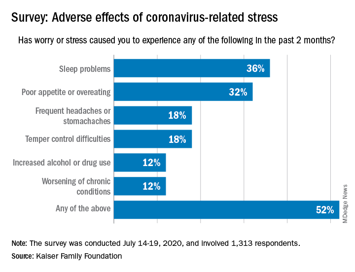
More than a third (36%) of the 1,313 respondents said they either had difficulty sleeping, falling asleep, or sleeping too much, KFF said in its latest Health Tracking Poll, conducted July 14-19, 2020. That was followed by poor appetite or overeating, which was mentioned by 32% of those surveyed.
Other adverse effects included frequent headaches or stomachaches (18%), temper-control issues (18%), increased drug or alcohol use (12%), and worsening of chronic conditions such as diabetes or hypertension (12%). Altogether, 52% of Americans have had at least one of these issues in the past 2 months, Liz Hamel and associates at KFF reported.
breaking down to 26% reporting a major impact and 28% reporting a minor impact (figures have been rounded), they said.
“As life with the coronavirus pandemic wears on, Americans increasingly say it is taking a negative toll on their mental health,” the investigators wrote. Earlier polls showed that pandemic-related stress was having an impact on mental health for 39% of respondents in May, compared with 45% in early April and 32% in March.
In the July poll, Black adults were much more likely to report a negative mental health impact (68%) than were Hispanics or Whites, who were both at 51%. Age was also a factor: The youngest group of respondents (ages 18-29 years) had the highest negative-impact rate (62%), and the oldest group (65 years and older) had the lowest (47%), they said.
When it came to reporting the adverse effects of stress or worry, however, the situation was somewhat different. Hispanics had the highest rate of such effects at 63%, while Blacks had a rate of 57% and 47% of Whites reported issues with sleep, eating, temper, and other problems, Ms. Hamel and associates reported.
Over the last 2 months, more than half of Americans have experienced some sort of adverse effect caused by stress related to the COVID-19 pandemic, according to a survey from the Kaiser Family Foundation (KFF).
More than a third (36%) of the 1,313 respondents said they either had difficulty sleeping, falling asleep, or sleeping too much, KFF said in its latest Health Tracking Poll, conducted July 14-19, 2020. That was followed by poor appetite or overeating, which was mentioned by 32% of those surveyed.
Other adverse effects included frequent headaches or stomachaches (18%), temper-control issues (18%), increased drug or alcohol use (12%), and worsening of chronic conditions such as diabetes or hypertension (12%). Altogether, 52% of Americans have had at least one of these issues in the past 2 months, Liz Hamel and associates at KFF reported.
breaking down to 26% reporting a major impact and 28% reporting a minor impact (figures have been rounded), they said.
“As life with the coronavirus pandemic wears on, Americans increasingly say it is taking a negative toll on their mental health,” the investigators wrote. Earlier polls showed that pandemic-related stress was having an impact on mental health for 39% of respondents in May, compared with 45% in early April and 32% in March.
In the July poll, Black adults were much more likely to report a negative mental health impact (68%) than were Hispanics or Whites, who were both at 51%. Age was also a factor: The youngest group of respondents (ages 18-29 years) had the highest negative-impact rate (62%), and the oldest group (65 years and older) had the lowest (47%), they said.
When it came to reporting the adverse effects of stress or worry, however, the situation was somewhat different. Hispanics had the highest rate of such effects at 63%, while Blacks had a rate of 57% and 47% of Whites reported issues with sleep, eating, temper, and other problems, Ms. Hamel and associates reported.