User login
Preventing clots: Don’t let the complex overshadow the simple

Along with massive pulmonary embolism, the most feared thromboembolic event is the clot that migrates to the brain, resulting in life-altering stroke. We assess this risk in a semiquantitative manner in patients with atrial fibrillation using the CHADS2 score, hoping to maximize the benefits of anticoagulation while reducing the risks. We recognize that patients at the greatest risk of stroke in this setting are those with a history of a prior stroke. Also, patients bedridden with a recent cerebrovascular accident (CVA) seem to be hypercoagulable, potentially adding risk to recent injury. Thus, we try to start anticoagulation as soon as feasible after the diagnosis of a possible thrombotic event.
But the decision to start or resume anticoagulation is especially agonizing in a patient who has suffered an intracerebral hemorrhage. In this issue of the Journal, Drs. Joshua Goldstein and Steven Greenberg and Dr. Franklin Michota provide a thoughtful discussion of the issues we need to consider in these patients.
While not contributing to the prevention of additional CVAs or other arterial thrombotic events, a modality often underused in the prevention of thrombotic disease is the application (not just the ordering) of compressive leg stockings to bedridden hospitalized patients who cannot, for any reason, be provided pharmacologic anticoagulation therapy. I just completed a stint of hospital consultation, and I was pleased to see the widespread integration of prophylactic anticoagulation therapy, but somewhat dismayed by the number of compressive stockings I watched pumping with vigor, but to no one’s benefit, as they were draped over a bed rail.
As we struggle with complex clinical decisions, we need to also be attentive to the simple and the seemingly mundane: using the foam dispenser at the door, offering the verbal greeting and patient touch at the bedside, and rewrapping the pneumatic stockings that have somehow migrated between mattress and footboard.
Intracerebral hemorrhage: Pick your poison
Anticoagulants have been helping patients at risk of thrombosis since the late 1930s.1,2 Although the indications for these agents are many, the development of anticoagulants beyond oral vitamin K antagonists and parenteral heparin has been slow. In the United States, the vitamin K antagonist warfarin (Coumadin) is still the only oral anticoagulant available.
The major complication of anticoagulant therapy is bleeding, and vitamin K antagonists have proven challenging to use in clinical practice.1,3 They have a narrow therapeutic window, they vary considerably in dose-response from patient to patient, and they are subject to significant interactions with other drugs and with foods. For these reasons, therapy must be monitored with laboratory testing, and good patient compliance and patient education are essential. Yet even with these measures, life-threatening hemorrhage still can occur.
In this issue of the Cleveland Clinic Journal of Medicine, Goldstein and Greenberg4 review warfarin-related intracerebral hemorrhage (ICH) and provide a framework for considering whether to resume anticoagulant therapy.
WHAT TO DO IN THE ACUTE PHASE
Goldstein and Greenberg divide the difficult clinical question of what to do after ICH into the acute phase and the chronic phase.
What to do in the acute phase appears straightforward, as the risk of hematoma expansion in the hours immediately after warfarin-related ICH outweighs the risk of arterial or venous thromboembolism. Anticoagulant reversal should be the primary consideration in the first 24 hours, and, assuming the patient does not have acute (< 4-week-old) deep vein thrombosis, intermittent pneumatic compression should be applied to the lower extremities to reduce the risk of venous thromboembolism associated with ICH.5
Prophylactic anticoagulation with subcutaneous fixed-dose heparin or low-molecular-weight heparin is recommended starting 72 hours after ICH is diagnosed, provided the patient is not underweight (< 50 kg), has relatively normal renal function (creatinine clearance > 30 mL/minute/1.73 m2) and normal platelet function, and does not have coagulopathy. 6 If any one of these criteria is not met, the risk of bleeding can be higher, even with only prophylactic doses of anticoagulant drugs. Prophylactic anticoagulation should be continued until hospital and rehabilitation discharge, typically 1 to 2 weeks after ICH, depending on the severity of the patient’s neurologic impairment.
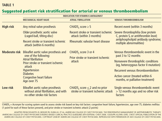
If a patient with warfarin-related ICH has concomitant acute proximal deep vein thrombosis or pulmonary embolism (ie, < 4 weeks old), then caval interruption therapy would be indicated.7 Although retrievable inferior vena cava filters are increasingly preferred over permanent filters, it is important to recognize the relative lack of both longitudinal and prospective data on retrievable devices. Given that provoked venous thromboembolism requires a minimum of 3 months of anticoagulation, and retrievable filters generally need to be removed before 3 months, a retrievable filter should be chosen only if the clinician has already decided that oral anticoagulation will be restarted in the next 3 to 4 weeks after filter removal.
WHAT TO DO IN THE CHRONIC PHASE
A more difficult question in patients with warfarin-related ICH arises in the chronic phase: should oral anticoagulation be resumed at all?
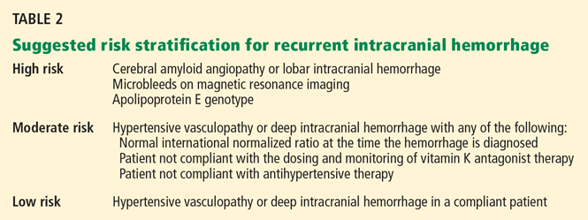
Since the risk of ICH is related to the intensity of anticoagulation, a lower target international normalized ratio may be the best compromise, depending on the patient. Alternatively, antiplatelet therapy alone may offer some benefit with less risk of ICH.
THE NEWER ORAL ANTICOAGULANTS
As Goldstein and Greenberg mention, the ongoing development of new and potentially safer oral anticoagulants may affect how we approach these risk-benefit equations.
Three new oral anticoagulants—dabigatran (Pradaxa), apixaban, and rivaroxaban (Xarelto)—are being tested for various anticoagulant indications, and several phase III studies have recently closed or are nearing completion.
Dabigatran is an oral direct thrombin inhibitor currently available in Europe and Canada.
In the Randomized Evaluation of Long-term Anticoagulant Therapy (RE-LY) trial, the efficacy and safety of two different doses of dabigatran (110 mg twice daily or 150 mg twice daily) relative to warfarin were studied in more than 18,000 patients with atrial fibrillation. 9 The primary outcome measure was stroke or systemic embolism. Dabigatran 110 mg was not inferior to warfarin in terms of the primary outcome, while dabigatran 150 mg was superior. The rate of major bleeding was 3.36% per year in the warfarin group vs 2.71% in the 110-mg group (P = .003) and 3.11% in the 150-mg group (P not significant).
Additional safety data on this drug are available from the 2,500-patient RE-COVER trial.10 Dabigatran was not inferior to warfarin in the treatment of acute venous thromboembolism, with a similar rate of major bleeding and a lower rate of combined major plus nonmajor bleeding.
Apixaban, an oral direct factor Xa inhibitor, is in a phase III trial in patients with atrial fibrillation—Apixaban for Reduction in Stroke and Other Thromboembolic Events in Atrial Fibrillation (ARISTOTLE)11—comparing apixaban vs warfarin. Another phase III trial, AVERROES,12 was stopped early after a predefined interim analysis by the independent data-monitoring committee found clear evidence of benefit in the apixaban group.13 The AVERROES results were presented at the 2010 European Society of Cardiology Congress, August 28–September 1, Stockholm, Sweden.14
Rivaroxaban, another promising oral direct factor Xa inhibitor, is currently available in Europe and Canada for the prevention of thrombosis in orthopedic surgery patients. Rivaroxaban is also in large phase III trials for the treatment of acute venous thromboembolism15–17 and for the prevention of stroke in atrial fibrillation.18
Newer agents have drawbacks, too
These new agents need no laboratory monitoring, and they do not appear to be subject to the dose variability and the interactions with drugs and foods seen with vitamin K antagonists. As a result, they may pose less risk of anticoagulant-related ICH.
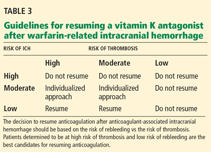
The decision to resume anticoagulation after anticoagulant-associated intracranial hemorrhage should be based on the risk of rebleeding vs the risk of thrombosis. Patients determined to be at high risk of thrombosis and low risk of rebleeding are the best candidates for resuming anticoagulation.
Still, for patients who suffer an anticoagulant- or warfarin-related ICH, these new anticoagulants are not likely to simplify the issue of restarting anticoagulant therapy. Unlike vitamin K antagonists, dabigatran and the direct factor Xa inhibitors have no known antidote for their anticoagulant effects. Animal data suggest that factor Xa concentrates may help,19 but for patients at risk of a second anticoagulant-related ICH, this does not provide much reassurance.
As with all clinical decisions in medicine, the potential benefits of any therapy should outweigh the risks. In the case of warfarin-related ICH, resuming anticoagulant therapy requires careful consideration of many factors, including patient preferences and tolerance of different levels of risk. As new and perhaps safer anticoagulants become available, clinicians may face such difficult questions less and less. But in the meantime, doctors and their patients are left to pick their poison.
- Ansell J, Hirsh J, Hylek E, Jacobson A, Crowther M, Palareti G; American College of Chest Physicians. Pharmacology and management of the vitamin K antagonists: American College of Chest Physicians Evidence-Based Clinical Practice Guidelines (8th Edition). Chest 2008; 133(suppl 6):160S–198S.
- Hirsh J, Bauer KA, Donati MB, Gould M, Samama MM, Weitz JI; American College of Chest Physicians. Parenteral anticoagulants: American College of Chest Physicians Evidence-Based Clinical Practice Guidelines (8th Edition). Chest 2008; 133(suppl 6):141S–159S.
- Schulman S, Beyth RJ, Kearon C, Levine MN; American College of Chest Physicians. Hemorrhagic complications of anticoagulant and thrombolytic treatment: American College of Chest Physicians Evidence-Based Clinical Practice Guidelines (8th edition). Chest 2008; 133(suppl 6):257S–298S.
- Goldstein JN, Greenberg SM. Should anticoagulation be resumed after intracerebral hemorrhage? Cleve Clin J Med 2010; 77:791–799.
- Geerts WH, Bergqvist D, Pineo GF, et al; American College of Chest Physicians. Prevention of venous thromboembolism: American College of Chest Physicians Evidence-Based Clinical Practice Guidelines (8th Edition). Chest 2008; 133(suppl 6):381S–453S.
- Michota F, Merli G. Anticoagulation in special patient populations: are special dosing considerations required? Cleve Clin J Med 2005; 72(suppl 1):S37–S42.
- Kearon C, Kahn SR, Agnelli G, Goldhaber S, Raskob GE, Comerota AJ; American College of Chest Physicians. Antithrombotic therapy for venous thromboembolic disease: American College of Chest Physicians Evidence-Based Clinical Practice Guidelines (8th Edition). Chest 2008; 133(suppl 6):454S–545S.
- Douketis JD, Berger PB, Dunn AS, et al; American College of Chest Physicians. The perioperative management of antithrombotic therapy: American College of Chest Physicians Evidence-Based Clinical Practice Guidelines (8th Edition). Chest 2008; 133(suppl 6):299S–339S.
- Connolly SJ, Ezekowitz MD, Yusuf S, et al; RE-LY Steering Committee and Investigators. Dabigatran versus warfarin in patients with atrial fibrillation. N Engl J Med 2009; 361:1139–1151.
- Schulman S, Kearon C, Kakkar AK, et al; RE-COVER Study Group. Dabigatran versus warfarin in the treatment of acute venous thromboembolism. N Engl J Med 2009; 361:2342–2352.
- Lopes RD, Alexander JH, Al-Khatib SM; ARISTOTLE Investigators. Apixaban for Reduction in Stroke and Other Thromboembolic Events in Atrial Fibrillation (ARISTOTLE) trial: design and rationale. Am Heart J 2010; 159:331–339.
- Eikelboom JW, O’Donnell M, Yusuf S, et al. Rationale and design of AVERROES: apixaban versus acetylsalicylic acid to prevent stroke in atrial fibrillation patients who have failed or are unsuitable for vitamin K antagonist treatment. Am Heart J 2010; 159:348–353.
- Pfizer/Bristol-Myers Squibb. AVERROES study of investigational agent apixaban closes early due to clear evidence of efficacy, June 9, 2010. www.theheart.org/article/1087291.do. Accessed September 26, 2010.
- Connolly SJ, Arnesen H. AVERROES: Apixaban versus acetylsalicylic acid. http://www.escardio.org/congresses/esc-2010/congress-reports/Pages/708-3-AVERROES.aspx. Accessed September 7, 2010.
- Once-daily oral direct factor Xa inhibitor rivaroxaban in the long-term prevention of recurrent symptomatic venous thromboembolism in patients with symptomatic deep-vein thrombosis or pulmonary embolism. The Einstein-Extension Study. http://clinicaltrials.gov/ct2/show/NCT00439725. Accessed September 26, 2010.
- Oral direct factor Xa inhibitor rivaroxaban in patients with acute symptomatic deep-vein thrombosis without symptomatic pulmonary embolism: Einstein-DVT Evaluation. http://clinicaltrials.gov/ct2/show/NCT00440193. Accessed September 26, 2010.
- Oral direct factor Xa inhibitor rivaroxaban in patients with acute symptomatic pulmonary embolism with or without symptomatic deep-vein thrombosis: Einstein-PE Evaluation. http://clinicaltrials.gov/ct2/show/NCT00439777. Accessed September 26, 2010.
- ROCKET AF Study Investigators. Rivaroxaban once-daily, oral, direct factor Xa inhibition compared with vitamin K antagonism for prevention of stroke and embolism trial in atrial fibrillation: rationale and design of the ROCKET AF study. Am Heart J 2010; 159:340–347.
- Weitz JI, Hirsh J, Samama MM; American College of Chest Physicians. New antithrombotic drugs: American College of Chest Physicians Evidence-Based Clinical Practice Guidelines (8th Edition). Chest 2008; 133(suppl 6):234S–256S.
Anticoagulants have been helping patients at risk of thrombosis since the late 1930s.1,2 Although the indications for these agents are many, the development of anticoagulants beyond oral vitamin K antagonists and parenteral heparin has been slow. In the United States, the vitamin K antagonist warfarin (Coumadin) is still the only oral anticoagulant available.
The major complication of anticoagulant therapy is bleeding, and vitamin K antagonists have proven challenging to use in clinical practice.1,3 They have a narrow therapeutic window, they vary considerably in dose-response from patient to patient, and they are subject to significant interactions with other drugs and with foods. For these reasons, therapy must be monitored with laboratory testing, and good patient compliance and patient education are essential. Yet even with these measures, life-threatening hemorrhage still can occur.
In this issue of the Cleveland Clinic Journal of Medicine, Goldstein and Greenberg4 review warfarin-related intracerebral hemorrhage (ICH) and provide a framework for considering whether to resume anticoagulant therapy.
WHAT TO DO IN THE ACUTE PHASE
Goldstein and Greenberg divide the difficult clinical question of what to do after ICH into the acute phase and the chronic phase.
What to do in the acute phase appears straightforward, as the risk of hematoma expansion in the hours immediately after warfarin-related ICH outweighs the risk of arterial or venous thromboembolism. Anticoagulant reversal should be the primary consideration in the first 24 hours, and, assuming the patient does not have acute (< 4-week-old) deep vein thrombosis, intermittent pneumatic compression should be applied to the lower extremities to reduce the risk of venous thromboembolism associated with ICH.5
Prophylactic anticoagulation with subcutaneous fixed-dose heparin or low-molecular-weight heparin is recommended starting 72 hours after ICH is diagnosed, provided the patient is not underweight (< 50 kg), has relatively normal renal function (creatinine clearance > 30 mL/minute/1.73 m2) and normal platelet function, and does not have coagulopathy. 6 If any one of these criteria is not met, the risk of bleeding can be higher, even with only prophylactic doses of anticoagulant drugs. Prophylactic anticoagulation should be continued until hospital and rehabilitation discharge, typically 1 to 2 weeks after ICH, depending on the severity of the patient’s neurologic impairment.
If a patient with warfarin-related ICH has concomitant acute proximal deep vein thrombosis or pulmonary embolism (ie, < 4 weeks old), then caval interruption therapy would be indicated.7 Although retrievable inferior vena cava filters are increasingly preferred over permanent filters, it is important to recognize the relative lack of both longitudinal and prospective data on retrievable devices. Given that provoked venous thromboembolism requires a minimum of 3 months of anticoagulation, and retrievable filters generally need to be removed before 3 months, a retrievable filter should be chosen only if the clinician has already decided that oral anticoagulation will be restarted in the next 3 to 4 weeks after filter removal.
WHAT TO DO IN THE CHRONIC PHASE
A more difficult question in patients with warfarin-related ICH arises in the chronic phase: should oral anticoagulation be resumed at all?
Since the risk of ICH is related to the intensity of anticoagulation, a lower target international normalized ratio may be the best compromise, depending on the patient. Alternatively, antiplatelet therapy alone may offer some benefit with less risk of ICH.
THE NEWER ORAL ANTICOAGULANTS
As Goldstein and Greenberg mention, the ongoing development of new and potentially safer oral anticoagulants may affect how we approach these risk-benefit equations.
Three new oral anticoagulants—dabigatran (Pradaxa), apixaban, and rivaroxaban (Xarelto)—are being tested for various anticoagulant indications, and several phase III studies have recently closed or are nearing completion.
Dabigatran is an oral direct thrombin inhibitor currently available in Europe and Canada.
In the Randomized Evaluation of Long-term Anticoagulant Therapy (RE-LY) trial, the efficacy and safety of two different doses of dabigatran (110 mg twice daily or 150 mg twice daily) relative to warfarin were studied in more than 18,000 patients with atrial fibrillation. 9 The primary outcome measure was stroke or systemic embolism. Dabigatran 110 mg was not inferior to warfarin in terms of the primary outcome, while dabigatran 150 mg was superior. The rate of major bleeding was 3.36% per year in the warfarin group vs 2.71% in the 110-mg group (P = .003) and 3.11% in the 150-mg group (P not significant).
Additional safety data on this drug are available from the 2,500-patient RE-COVER trial.10 Dabigatran was not inferior to warfarin in the treatment of acute venous thromboembolism, with a similar rate of major bleeding and a lower rate of combined major plus nonmajor bleeding.
Apixaban, an oral direct factor Xa inhibitor, is in a phase III trial in patients with atrial fibrillation—Apixaban for Reduction in Stroke and Other Thromboembolic Events in Atrial Fibrillation (ARISTOTLE)11—comparing apixaban vs warfarin. Another phase III trial, AVERROES,12 was stopped early after a predefined interim analysis by the independent data-monitoring committee found clear evidence of benefit in the apixaban group.13 The AVERROES results were presented at the 2010 European Society of Cardiology Congress, August 28–September 1, Stockholm, Sweden.14
Rivaroxaban, another promising oral direct factor Xa inhibitor, is currently available in Europe and Canada for the prevention of thrombosis in orthopedic surgery patients. Rivaroxaban is also in large phase III trials for the treatment of acute venous thromboembolism15–17 and for the prevention of stroke in atrial fibrillation.18
Newer agents have drawbacks, too
These new agents need no laboratory monitoring, and they do not appear to be subject to the dose variability and the interactions with drugs and foods seen with vitamin K antagonists. As a result, they may pose less risk of anticoagulant-related ICH.
The decision to resume anticoagulation after anticoagulant-associated intracranial hemorrhage should be based on the risk of rebleeding vs the risk of thrombosis. Patients determined to be at high risk of thrombosis and low risk of rebleeding are the best candidates for resuming anticoagulation.
Still, for patients who suffer an anticoagulant- or warfarin-related ICH, these new anticoagulants are not likely to simplify the issue of restarting anticoagulant therapy. Unlike vitamin K antagonists, dabigatran and the direct factor Xa inhibitors have no known antidote for their anticoagulant effects. Animal data suggest that factor Xa concentrates may help,19 but for patients at risk of a second anticoagulant-related ICH, this does not provide much reassurance.
As with all clinical decisions in medicine, the potential benefits of any therapy should outweigh the risks. In the case of warfarin-related ICH, resuming anticoagulant therapy requires careful consideration of many factors, including patient preferences and tolerance of different levels of risk. As new and perhaps safer anticoagulants become available, clinicians may face such difficult questions less and less. But in the meantime, doctors and their patients are left to pick their poison.
- Ansell J, Hirsh J, Hylek E, Jacobson A, Crowther M, Palareti G; American College of Chest Physicians. Pharmacology and management of the vitamin K antagonists: American College of Chest Physicians Evidence-Based Clinical Practice Guidelines (8th Edition). Chest 2008; 133(suppl 6):160S–198S.
- Hirsh J, Bauer KA, Donati MB, Gould M, Samama MM, Weitz JI; American College of Chest Physicians. Parenteral anticoagulants: American College of Chest Physicians Evidence-Based Clinical Practice Guidelines (8th Edition). Chest 2008; 133(suppl 6):141S–159S.
- Schulman S, Beyth RJ, Kearon C, Levine MN; American College of Chest Physicians. Hemorrhagic complications of anticoagulant and thrombolytic treatment: American College of Chest Physicians Evidence-Based Clinical Practice Guidelines (8th edition). Chest 2008; 133(suppl 6):257S–298S.
- Goldstein JN, Greenberg SM. Should anticoagulation be resumed after intracerebral hemorrhage? Cleve Clin J Med 2010; 77:791–799.
- Geerts WH, Bergqvist D, Pineo GF, et al; American College of Chest Physicians. Prevention of venous thromboembolism: American College of Chest Physicians Evidence-Based Clinical Practice Guidelines (8th Edition). Chest 2008; 133(suppl 6):381S–453S.
- Michota F, Merli G. Anticoagulation in special patient populations: are special dosing considerations required? Cleve Clin J Med 2005; 72(suppl 1):S37–S42.
- Kearon C, Kahn SR, Agnelli G, Goldhaber S, Raskob GE, Comerota AJ; American College of Chest Physicians. Antithrombotic therapy for venous thromboembolic disease: American College of Chest Physicians Evidence-Based Clinical Practice Guidelines (8th Edition). Chest 2008; 133(suppl 6):454S–545S.
- Douketis JD, Berger PB, Dunn AS, et al; American College of Chest Physicians. The perioperative management of antithrombotic therapy: American College of Chest Physicians Evidence-Based Clinical Practice Guidelines (8th Edition). Chest 2008; 133(suppl 6):299S–339S.
- Connolly SJ, Ezekowitz MD, Yusuf S, et al; RE-LY Steering Committee and Investigators. Dabigatran versus warfarin in patients with atrial fibrillation. N Engl J Med 2009; 361:1139–1151.
- Schulman S, Kearon C, Kakkar AK, et al; RE-COVER Study Group. Dabigatran versus warfarin in the treatment of acute venous thromboembolism. N Engl J Med 2009; 361:2342–2352.
- Lopes RD, Alexander JH, Al-Khatib SM; ARISTOTLE Investigators. Apixaban for Reduction in Stroke and Other Thromboembolic Events in Atrial Fibrillation (ARISTOTLE) trial: design and rationale. Am Heart J 2010; 159:331–339.
- Eikelboom JW, O’Donnell M, Yusuf S, et al. Rationale and design of AVERROES: apixaban versus acetylsalicylic acid to prevent stroke in atrial fibrillation patients who have failed or are unsuitable for vitamin K antagonist treatment. Am Heart J 2010; 159:348–353.
- Pfizer/Bristol-Myers Squibb. AVERROES study of investigational agent apixaban closes early due to clear evidence of efficacy, June 9, 2010. www.theheart.org/article/1087291.do. Accessed September 26, 2010.
- Connolly SJ, Arnesen H. AVERROES: Apixaban versus acetylsalicylic acid. http://www.escardio.org/congresses/esc-2010/congress-reports/Pages/708-3-AVERROES.aspx. Accessed September 7, 2010.
- Once-daily oral direct factor Xa inhibitor rivaroxaban in the long-term prevention of recurrent symptomatic venous thromboembolism in patients with symptomatic deep-vein thrombosis or pulmonary embolism. The Einstein-Extension Study. http://clinicaltrials.gov/ct2/show/NCT00439725. Accessed September 26, 2010.
- Oral direct factor Xa inhibitor rivaroxaban in patients with acute symptomatic deep-vein thrombosis without symptomatic pulmonary embolism: Einstein-DVT Evaluation. http://clinicaltrials.gov/ct2/show/NCT00440193. Accessed September 26, 2010.
- Oral direct factor Xa inhibitor rivaroxaban in patients with acute symptomatic pulmonary embolism with or without symptomatic deep-vein thrombosis: Einstein-PE Evaluation. http://clinicaltrials.gov/ct2/show/NCT00439777. Accessed September 26, 2010.
- ROCKET AF Study Investigators. Rivaroxaban once-daily, oral, direct factor Xa inhibition compared with vitamin K antagonism for prevention of stroke and embolism trial in atrial fibrillation: rationale and design of the ROCKET AF study. Am Heart J 2010; 159:340–347.
- Weitz JI, Hirsh J, Samama MM; American College of Chest Physicians. New antithrombotic drugs: American College of Chest Physicians Evidence-Based Clinical Practice Guidelines (8th Edition). Chest 2008; 133(suppl 6):234S–256S.
Revision Extensor Mechanism Allografting After Total Knee Arthroplasty
Augmented Demineralized Bone Matrix: A Potential Alternative for Posterolateral Lumbar Spinal Fusion
Remodeling and Repair of Orthopedic Tissue: Role of Mechanical Loading and Biologics Part I: Tendon and Ligament; Meniscus
Part I: Tendon and Ligament; Meniscus
Part I: Tendon and Ligament; Meniscus
Part I: Tendon and Ligament; Meniscus
What's Eating You? Rhipicephalus Ticks
Pediatric Molluscum Contagiosum: Reflections on the Last Challenging Poxvirus Infection, Part 1
Birth control prescription blamed for stroke...Removal of mole without follow-up leads to death...more
Birth control prescription blamed for stroke
A 29-YEAR-OLD WOMAN SUFFERED A BLOOD CLOT in her leg. Her family physician advised her to start taking aspirin, which she did, and counseled her to use birth control that didn’t contain estrogen. She was taking norgestimate/ ethinyl estradiol at the time of the clot.
The woman subsequently went to an obstetrician-gynecologist (ob-gyn), whom she said she told about her family physician’s advice to avoid estrogen-containing birth control medication. The ob-gyn prescribed and inserted an etonogestrel/ethinyl estradiol vaginal ring.
A few months later the patient was hospitalized with severe headaches. She had blood clots in her brain and had suffered a stroke, which affected her speech and executive functions.
PLAINTIFF’S CLAIM The ob-gyn was negligent in prescribing the vaginal ring.
THE DEFENSE The cause of the first clot was an injury; the vaginal ring didn’t cause the second clot and stroke.
VERDICT $523,000 Georgia verdict.
COMMENT A comprehensive history, and clear documentation of communicating the potential risks of therapy, might have prevented this judgment.
Elevated PSA without referral delays diagnosis
ROUTINE BLOOD WORK before orthopedic surgery revealed an elevated prostate-specific antigen (PSA) of 7.4 in a 53-year-old man. A medical assistant who was directed to refer the patient to a urologist didn’t do so. Widespread metastatic prostate cancer was diagnosed 18 months later.
PLAINTIFF’S CLAIM Diagnosing the cancer 18 months earlier would have given the patient a >50% chance of 5-year survival. Because of the delay, he was terminal. The clinic was negligent in having no written procedure or system for tracking adverse lab test results.
THE DEFENSE The patient already had metastatic disease when the PSA level was discovered and would have required the same treatment.
VERDICT $1 million Washington settlement.
COMMENT A clear system for tracking test results is imperative in today’s litigious society.
Removal of mole without follow-up leads to death
A MOLE ON THE UPPER BACK prompted a 26-year-old man to visit a dermatologist, who performed a complete excision. The pathologist who examined the excised tissue suggested that the patient return for follow-up. During the next 6 months, the patient saw the dermatologist twice but didn’t receive proper follow-up.
Two years later, the patient noticed a suspicious area on his back near the scar from the excision. A hospital biopsy resulted in a diagnosis of metastatic melanoma. A review of the slides from the original biopsy found “melanoma, superficial spreading type, invasive to a depth of a minimum of 1.0 mm anatomic level IV, extending to inked deep resection margin.”
The patient underwent a wide local excision and was given a diagnosis of stage III melanoma. The patient underwent neck and back radiation and high-dose treatment with alpha interferon, followed by high-dose interleukin-2 and chemotherapy. Nevertheless, the patient died.
PLAINTIFF’S CLAIM The dermatologist’s office had no system to contact the patient when he didn’t return. The chances for cure would have been between 73% and 94% if the melanoma had been diagnosed at the time of the original excision.
THE DEFENSE No information about the defense is available.
VERDICT $1.7 million Massachusetts settlement.
COMMENT Failure to follow up on abnormal results is a potentially preventable cause of malpractice. Do you have a mechanism to track such testing?
Suggestive symptoms, but no Dx until it was too late
A 42-YEAR-OLD WOMAN went to the hospital in February for chest pain, dizziness, and shortness of breath. The emergency room physician diagnosed sinusitis and bronchitis and discharged the patient in stable condition. In April, the woman visited her primary care physician complaining of fatigue and shortness of breath. She claimed that her physician knew about the February emergency room visit. Later in April, she again went to her physician with shortness of breath; in July, she reported an irregular heart rhythm.
In October, the patient was found unresponsive after suffering cardiorespiratory arrest, hypoxic ischemic brain injury, and static encephalopathy. She has since been in a vegetative state.
PLAINTIFF’S CLAIM The patient had gone to her primary care physician many times during the 2 years before her emergency room visit with complaints suggesting an underlying cardiac condition, including shortness of breath, dizziness, light-headedness, vertigo, chest tightness, fatigue, and an irregular heart rhythm. The defendants were negligent in failing to diagnose the patient’s condition and provide proper treatment, failing to order proper diagnostic testing, and failing to perform a cardiac workup.
THE DEFENSE No negligence occurred.
VERDICT $6.3 million Florida verdict.
COMMENT Comprehensive documentation, including your medical decision making, can help prevent multimillion dollar judgments.
A serendipitous finding—to no avail
A FALL ON THE ICE sent a 74-year-old woman to the hospital with a fractured ankle. A preoperative chest radiograph taken before open reduction and internal fixation to repair the fracture showed a 2-cm nodular opacity in the right upper hemithorax. The radiologist recommended a computed tomography scan to rule out lung cancer, but the treating internists didn’t order a scan or refer the patient for biopsy.
The nodule appeared again on a second radiograph taken 2 days later. The patient wasn’t informed, and the attending internist at the time didn’t order follow-up testing or refer the patient to a specialist. The attending physicians continued to treat the patient without further testing or referral for the nodule.
Two years after the fracture, the patient was admitted to the hospital with complaints of sweating and shortness of breath. A chest radiograph showed pneumonia and the previously noted nodule. The patient was diagnosed with metastatic, inoperable small-cell lung cancer. She died after receiving extensive chemotherapy and radiation.
PLAINTIFF’S CLAIM The doctors were negligent in failing to diagnose and treat the lung cancer in a timely manner.
THE DEFENSE No information about the defense is available.
VERDICT $325,000 Michigan settlement.
COMMENT Could this happen to you? How many times have you serendipitously noted an abnormal result that was not followed up adequately?
Alert! The 2011 ICD-9 code set is already in force
This year, ObGyn-related additions and revisions to the International Classification of Diseases, Clinical Modification (ICD-9-CM), involve tinkering with existing codes and adding some new code categories. The latter development means that more information will be required of you to code to the highest level of specificity.
On the obstetrics side, there are now specific codes for placental status for multiple gestations and some revised terminology.
In gynecology, changes include new codes for congenital anomalies of the cervix, vagina, and uterus; reporting an expanded list of a history of dysplasia; and reporting the insertion and removal of an intrauterine device for contraception.
In addition, new codes have been established for fecal incontinence and for reporting a body mass index >40.
Last, changes to the alphabetical index of codes have been put in place that will help you select the most appropriate code.
The new and revised ICD-9-CM codes were added to the national code set on October 1, 2010. As in previous years, there is no grace period for failing to use the new code set!
Changes to obstetric codes
PLACENTAL/AMNIOTIC SAC SPECIFICATION FOR MULTIPLE-GESTATION PREGNANCY
Multiple-gestation pregnancies are classified as monochorionic/monoamniotic, monochorionic/diamniotic, and dichorionic/diamniotic. Until now, however, you’ve had no way to report this additional information to a payer.
For fiscal year 2011, you are able to be more specific, which can increase your ability to report medical support care for a higher-risk pregnancy or an expanded treatment plan.
Because the current category of multiple-gestation codes (651) did not allow for expansion to include this information, a new code category, V91 (multiple gestation placenta status), was created for that purpose. The V91 category has distinct codes for twin gestation, triplet gestation, quadruplet gestation, and other “unspecified” gestations to denote placental/amniotic sac status.
Be aware that use of the V91 codes is optional, and that they can be reported only as a secondary diagnosis, with a category 651.xx (multiple gestation, etc.) code as primary. As I noted, however, the new codes may provide better information to the payer—and that might result in additional reimbursement for your care of such pregnancies.
The new codes for a twin pregnancy are:
V91.00 Twin gestation, unspecified number of placentae, unspecified number of amniotic sacs
V91.01 Twin gestation, monochorionic/monoamniotic (one placenta, one amniotic sac)
V91.02 Twin gestation, monochorionic/diamniotic (one placenta, two amniotic sacs)
V91.03 Twin gestation, dichorionic/diamniotic (two placentae, two amniotic sacs)
V91.09 Twin gestation, unable to determine number of placentae and number of amniotic sacs
There are similar V codes for triplet gestations (V91.10–V91.19), quadruplet gestations (V91.20–V91.29), and other unspecified multiple gestations (V91.91– V91.99).
RECURRENT PREGNANCY LOSS
The term “habitual aborter” has been replaced for 2011 with the more clinically accurate term “recurrent pregnancy loss.” This change is noted in both the ICD-9 alphabetical index and in the code definitions in the tabular section. The codes affected by this terminology change are:
629.81 Recurrent pregnancy loss without current pregnancy
646.3x Recurrent pregnancy loss (affecting the current pregnancy)
INDEX AND INSTRUCTIONAL CHANGES
These OB changes took effect on October 1, 2010:
- Periurethral trauma should be reported using 664.8x (other specified trauma to perineum and vulva), not 665.5x (other injury to pelvic organs).
- If you report puerperal sepsis (670.2x), you must report an additional code to identify severe sepsis (995.92) and any associated acute organ dysfunction, if applicable.
- If your diagnosis is superficial thrombosis (671.2x), an additional code—either 453.6, 453.71, or 453.81—should be reported to further explain the type of thrombophlebitis.
- If your patient has either asymptomatic, inactive, or a history of genital herpes that is complicating her current pregnancy, report 647.6x (other viral diseases).
- If you report pneumonia as complicating pregnancy, assign code 648.9x (other current conditions classifiable elsewhere).
Changes to gyn codes
CONGENITAL ANOMALIES OF THE UTERUS, CERVIX, AND VAGINA
Before October 1, 2010, of the seven distinct types of uterine anomalies, only a didelphus uterus (752.2, doubling of the uterus) and a diethylstilbestrol-related anomaly (760.76 [noxious influences affecting fetus or newborn via placenta or breast milk; diethylstilbestrol (DES)]) had specific codes. All other uterine anomalies were coded to “other” or “unspecified” codes that could include many different conditions.
Although vaginal and cervical anomalies may be less common, the only codes available before October 1, 2010, were ones that described an unspecified anomaly (753.40), imperforate hymen (752.42), or an embryonic cyst (752.41).
A higher level of specificity in coding, however, can make all the difference in receiving adequate reimbursement and preventing denials. For example, if you perform a Pap smear on a patient who has two cervices, a code that specifies a duplicate cervix can clearly tell the payer that billing for both is not a duplicate service or billing error.
Changes to codes in this area of care take the form of expanding existing codes. Code 752.3 (other anomalies of uterus) has been expanded to seven distinct five-digit codes to capture the seven anomalies of the uterus:
752.31 Agenesis of uterus
752.32 Hypoplasia of uterus
752.33 Unicornuate uterus (This code would be reported if the unicornuate uterus did or did not have a separate uterine horn, or if the uterus had only one functioning horn.)
752.34 Bicornuate uterus
752.35 Septate uterus (This code would be reported whether the septate was complete or partial.)
752.36 Arcuate uterus
752.39 Other anomalies of uterus (This code category includes aplasia or any other Müllerian anomaly of the uterus that is not otherwise or elsewhere classified.)
New codes have been added to the 752.4 code category (anomalies of cervix, vagina, and external female genitalia) to expand the options. Before October 1, 2010, any of these conditions would have been coded as 752.49, an “other” category.
752.43 Cervical agenesis
752.44 Cervical duplication
752.45 Vaginal agenesis (This code can also be reported for vaginal hypoplasia.)
752.46 Transverse vaginal septum
752.47 Longitudinal vaginal septum
For a patient who has a history of one of these anomalies, you would report new code V13.62, (personal history of other [corrected] congenital malformations of genitourinary system) if this history was a factor in her current care.
Inserting and removing an IUD are integral services that most ObGyn practices provide, so it is imperative that your encounter forms reflect two new codes, to avoid denials for an invalid diagnosis code. A reminder: Code V45.51 (intrauterine contraceptive device) is a status code. It indicates that a patient has an IUD in place but you should never use it as a diagnosis code when the purpose of the visit is for you to check on the device and assess how it is working.
Changes in this area are:
Before October 1, 2010:
V25.1 Insertion
V25.42 Checking, reinsertion and/or removal After October 1, 2010:
V25.11 Encounter for insertion of intrauterine contraceptive device
V25.12 Encounter for removal of intrauterine contraceptive device
V25.13 Encounter for removal and reinsertion of intrauterine contraceptive device
V25.42 Encounter for routine checking of intrauterine contraceptive device
Regrettably, the number of patients who have a very high body mass index (BMI) is increasing. When surgery is planned, reporting this information in your coding can help establish 1) the medical need for significant additional work during the procedure or 2) health risks in support of therapy.
Code V85.4 (Body mass index 40 and over, adult) has been expanded to five new codes. They should be reported secondary to the type of obesity (i.e., codes 278.0x [overweight and obesity]).
V85.41 Body Mass Index 40.0–44.9, adult
V85.42 Body Mass Index 45.0–49.9, adult
V85.43 Body Mass Index 50.0–59.9, adult
V85.44 Body Mass Index 60.0–69.9, adult
V85.45 Body Mass Index 70 and over, adult
Fecal incontinence can present as problematic symptoms—fecal smearing, fecal urgency, incomplete defecation—but, until now, you only had one code to report any of these problems.
For that reason, 787.6 (incontinence of feces) has been expanded into four new five-digit codes. In addition, a new code has been added to report fecal impaction, which, in the past, was reported as 560.39, an “other” category code that was not specific to this problem.
New codes are:
560.32 Fecal impaction
787.60 Full incontinence of feces
787.61 Incomplete defecation
787.62 Fecal smearing
787.63 Fecal urgency
PERSONAL HISTORY OF DYSPLASIA
New codes have been added to complete the personal history codes for dysplasia. In addition to the existing code for cervical dysplasia history (V13.22), you can now report:
V13.23 Personal history of vaginal dysplasia
V13.34 Personal history of vulvar dysplasia
INDEX AND INSTRUCTIONAL CHANGES
These changes take effect October 1, 2010:
- Clarification that an abnormal Pap result indicated non-atypical endometrial cells should be reported using 795.09.
- Clearly indicate whether a fistula between the uterus and another organ is congenital (752.39) or noncongenital (619.0-619.9).
- Precocious menstruation should be coded as 259.1, not as a menstrual disorder.
- The terminology in the index and tabular sections has been revised to more clearly differentiate long-term from prophylactic use of medications. This change affects only code category titles and lookup terms, not existing code numbers.
We want to hear from you! Tell us what you think.
Is it time to drug test your chronic pain patient?
• When you initiate opioid therapy for chronic pain, inform the patient that routine monitoring includes random urine drug testing (UDT). C
• Consider UDT not only for patients at high risk for abuse of prescription opioids, but for lower-risk individuals receiving opioid therapy, as well. C
• Use caution in interpreting the results of UDT; testing cannot reliably detect some opioids, and a negative result is not necessarily an indication of noncompliance. C
Strength of recommendation (SOR)
A Good-quality patient-oriented evidence
B Inconsistent or limited-quality patient-oriented evidence
C Consensus, usual practice, opinion, disease-oriented evidence, case series
CASE 1 Marilyn H, a 54-year-old woman with multiple chronic conditions, visits a “new” primary care physician to establish care following the retirement of her previous physician. She has poorly controlled diabetes, hypertension, and a lung nodule recently noted on a chest radiograph. Marilyn requests prescriptions for hydrocodone and alprazolam, stating that she has taken both drugs for years for chronic back pain and anxiety.
CASE 2 Don F, 38, has been on oxycodone/acetaminophen (Percocet) for 3 years for back pain resulting from a car accident. He has a remote history of amphetamine abuse, but reports that he has been clean for 10 years. Since the initiation of his pain medication, there have been no problems, and no “lost” prescriptions or requests for early refills.
If you were Marilyn and Don’s physician, would you order urine drug testing (UDT)?
Management of opioid therapy is a challenge for many family physicians, particularly when treating noncancer pain. Seemingly contradictory messages from various medical associations are part of the problem. Organizations such as the Joint Commission on Accreditation of Healthcare Organizations and the American Medical Association emphasize the importance of appropriate pain management, while regulatory bodies like the US Drug Enforcement Agency and many state medical boards warn of inappropriate prescribing and diversion of controlled substances.1-4
In the realm of opioid management guidelines, however, patient monitoring is a common theme. It’s not hard to understand why. The Centers for Disease Control and Prevention reports that, between 1999 and 2005, the incidence of unintentional drug overdose more than doubled—a consequence of increasing abuse of prescription opioid analgesics.5 Prescription medications are now the second most commonly abused drug category (marijuana is first6), accounting for more cases of abuse than heroin, cocaine, and hallucinogens combined.7 In primary care and pain management settings, estimates are that more than 1 in 4 chronic pain patients misuse opioids or illicit drugs.8
Thus, physicians who prescribe controlled substances face increasing pressure to prevent opioid diversion. That pressure is reflected in the US Federation of State Medical Boards (FSMB)’s Model Policy for the Use of Controlled Substances for the Treatment of Pain, which was updated in 2004.4 This policy underscores physicians’ responsibility to closely monitor patients being treated with opioids for chronic pain.
Inexpensive and noninvasive. To this end, UDT can be a valuable tool. It is the most widely used and acceptable form of drug testing, because it is inexpensive and noninvasive, and because most drugs can be detected in urine for 1 to 3 days.9,10 But many primary care physicians are unfamiliar with the complexities of UDT, and often fail to use it.11 One study found that only 8% of family physicians employed UDT for patients on chronic opioid therapy.12
When introduced and used appropriately, UDT can not only help detect misuse of controlled substances, but may strengthen the doctor-patient relationship, as well. For that to happen, however, the physician who orders urine testing must know which patients to test, when to test, and what urine testing can (and cannot) reveal.
Consider testing all patients treated for chronic pain
The FSMB urges physicians to consider a written agreement with any patient receiving chronic opioid therapy who has a history of, or is at high risk for, substance abuse.4 (Red flags are listed in TABLE 1.13-15) The document should state that the patient is responsible for providing urine and serum specimens for drug monitoring upon request. The 2009 guidelines from the American Pain Society and the American Academy of Pain Medicine also address the role of drug monitoring, strongly recommending periodic urine screens for high-risk patients on chronic opioid therapy.16
Evidence suggests that predictors of aberrant behavior are not completely reliable, however, and that a substantial number of individuals using illicit substances will be missed if clinicians restrict urine testing to those they deem to be at high risk.17 Thus, UDT may be a valuable tool for low-risk patients on chronic opioid therapy, as well. Written agreements governing opioid therapy may also be useful for low-risk patients.
TABLE 1
Aberrant drug-related behavior: Red flags13-15
| Use of opioids for non-analgesic indications |
| Lack of control (related to drug use or to patient behavior) |
| Compulsive use of medications |
| Continued use of drugs despite harm/lack of benefit |
| Cravings |
| Escalation of drug use |
| Selling/altering prescriptions |
| Theft or diversion |
| Request for early refills |
| Claims of “lost” prescriptions |
| Reluctance to try nonpharmacologic options |
| Use of multiple prescribers or pharmacies |
| Odd stories regarding need for medication |
| Reporting vague medical history or textbook symptoms |
| Unwillingness to name regular physician |
| No interest in a physical exam, diagnostic testing, or providing past records |
| Request for specific drug(s) |
| Extensive (or very limited) understanding of medications |
| Calling or arriving after hours or when regular doctor is unavailable |
| Insistence on being seen urgently (eg, because of being late for another appointment) |
When to test, what to test for
No guidelines specify when to test, but testing upon initiation of chronic opioid treatment, followed by random testing, is the most widely used strategy. Unobserved specimen collection is generally acceptable,13 provided the specimens are requested at random rather than routinely at every visit.
Initial testing is done using an immuno-assay drug panel.13,18 TABLE 2 lists the drugs most commonly included in a standard urine test. However, the drug panel can vary from 1 laboratory to another, as can the lower limits of drug detection. No-threshold testing is mentioned in pain management literature, but is not often available in clinical practice.
Before initiating UDT, it is important to know which drugs the laboratory you use routinely tests for and what its lower limits are. The simplest way to find out is to ask lab personnel.
CASE 1 At her first visit, Marilyn H’s new physician focuses on controlling her blood sugar and blood pressure, ordering follow-up testing of the lung nodule, and refilling her hydrocodone and alprazolam prescriptions. The physician requests the patient’s medical records and orders a urine drug screen per clinic protocol, testing for benzodiazepines as well as for opioids. He gives his patient prescriptions for a 1-month supply of both drugs while the UDT results are pending.
The lab report comes in the following day, and indicates that Marilyn tested positive for cocaine but negative for other substances, including narcotics and benzodiazepines. The clinic immediately notifies the pharmacy to confiscate the patient’s new prescriptions when she presents them and calls Marilyn, advising her that she will not be given any further prescriptions for controlled substances.
The physician refers the patient to a pain clinic, gives her the number of a substance abuse treatment center, and encourages her to follow up at the clinic for other medical issues. Marilyn fails to keep her appointment at the pain clinic and does not respond to a subsequent call.
TABLE 2
A standard urine drug test panel13,18
| Amphetamines |
| Cocaine |
| Marijuana (THC) |
| Opiates (morphine and codeine) |
| Phencyclidine |
| THC, tetrahydrocannabinol. |
Talking to patients about drug testing
Physicians are often concerned about patients’ feelings about drug testing—worrying that patients may not feel trusted or respected by a doctor who asks them to submit to UDT. Others may fear that the mere mention of urine testing will encourage patients to misuse prescription opioids, that patients will view UDT as a punitive measure, or that those being tested will believe that the physician is more concerned with self-protection than with providing optimal care to the patient.
Making UDT routine. One way to circumvent such possibilities is to implement a systematic approach to drug testing. We recommend that physicians discuss the role of UDT in the initial education session with patients being started on a course of opiates. Describing UDT as simply another routine monitoring parameter—akin to the measure of microalbuminuria for patients with diabetes—can decrease or eliminate the stigma associated with drug testing.
CASE 2 A new policy encouraging UDT for all patients on chronic controlled substances has just been implemented at the clinic where Don F is being treated. His physician tells him about the policy, and a urine test is ordered at his next visit. The test comes back negative for all substances, including opioids.
When presented with the results over the phone, Don insists that he regularly takes his prescription medication, and makes a same-day appointment to discuss the results with his physician.
Interpreting test results— what UDT can (and can’t) reveal
To avoid eroding trust by falsely accusing a patient of diversion or use of an illicit substance, it’s important to familiarize yourself with testing limitations. Factors that can affect the results, and may interfere with the ability of UDT to provide a definitive picture, include:
Lab variability and technical limitations. Some urine drug panels may not have a lower limit sufficient to detect small quantities of opioids. Others may not detect certain substances, notably the semisynthetic and synthetic opioids (TABLE 3).19 Oxycodone is a prime example of a commonly prescribed semisynthetic opioid that does not appear on many urine test panels.8
In addition, individual drugs within a class may not be identified on UDT panels. When confirmation of a specific drug or metabolite is needed, serum testing must be performed.8
TABLE 3
Classifying opioids19
| Natural | Semisynthetic | Synthetic |
|---|---|---|
| Codeine | Hydrocodone | Fentanyl |
| Morphine | Hydromorphone | Meperidine |
| Oxycodone | Methadone | |
| Propoxyphene |
Differences in metabolism. Genetic differences in metabolism can also skew the results of UDT. Codeine, which relies on hepatic metabolism via cytochrome P450 2D6 for conversion to morphine, is the classic example; cytochrome P450 2D6 is a polymorphic enzyme, meaning that it manifests with different activity levels in different people. Patients who are poor metabolizers (an estimated 5%-10% of Caucasians, 1%-5% of Asians, 2%-7% of African Americans, and 2%-6% of Hispanics20) will convert very little codeine to morphine; conversely, those who are rapid metabolizers will convert extensive amounts. A rapid metabolizer taking codeine as prescribed may therefore have a negative UDT; in an average metabolizer taking the same dose, both codeine and morphine will be detected.20
Drugs’ half-lives. Opioids with a short half-life (TABLE 4)9,21 may not appear in the urine if the test is done several hours after the last dose. On the other hand, some opioids may have an extended half-life in patients with liver or kidney disease, and may appear in the urine longer than would be expected.9
False-positive results. Substances that may cause false positives for opioids on a urine test include dextromethorphan, papaverine, poppy seeds and oil, quinine, quinolones, rifampin, and verapamil.10
False-negative results. Many of the problems already discussed can lead to false-negative results, including the panel’s failure to detect semisynthetic and synthetic opioids, rapid metabolism (most notably, of codeine), the timing of the test relative to the dose, and adulteration of the specimen. Thus, a negative test result in a patient on opioid therapy does not necessarily mean that he or she is noncompliant—and certainly is not proof of diversion.
Because of the variables that affect UDT outcomes, unanticipated results should be reviewed with the patient and possibly, with the lab, and viewed within the therapeutic context. When more definitive information is needed, serum testing may be performed as follow-up. While serum testing can detect drugs and their metabolites ingested within hours, it is not widely used on initial screening because it is a more invasive procedure with higher associated costs.13
CASE 2 Upon further discussion with Don F, the physician orders a serum oxycodone test, which shows a level of 10 ng/mL. The physician notes that serum testing is more appropriate than UDT for Don because of the inconsistent detection of oxycodone in urine.
TABLE 4
Pharmacokinetics of common opioids: Time detectable in urine9,21
| Drug (half-life) | Time detectable in urine | Comment |
|---|---|---|
| Codeine (2.5-3 h) | 48 h | Pharmacogenetic-dependent effects may affect detection |
| Fentanyl Transdermal (17 h) Submucosal (7 h) | Not usually detected in urine (lack of metabolites) | Excretion of transdermal fentanyl can last days |
| Hydromorphone IR (2.3 h) ER (18.6 h) | 2-4 d | Significant interpatient variability |
| Methadone (8-59 h) | 3 d | |
| Morphine (1.5-2 h) | 48-72 h | 90% eliminated within 24 h |
| Oxycodone IR (3.2 h) ER (4.5 h) | Often not detected in urine | High-fat meals may increase serum concentrations of ER formulation |
| Propoxyphene Parent drug (6-12 h) Metabolite (30-36 h) | 6-48 h | |
| ER, extended release; IR, immediate release. | ||
CORRESPONDENCE Sarah McBane, PharmD, CDC, BCPS, University of California, San Diego, Skaggs School of Pharmacy and Pharmaceutical Sciences, 9500 Gilman Drive, La Jolla, CA 92023; [email protected]
1. Health Care Issues. Joint Commission on Accreditation of Health Care Organizations. Available at: http://www.jointcommission.org/. Accessed March 5, 2010.
2. Standards, Laws, and Regulations Addressing Pain Medications and Medical Practice: Report 6 of the Council on Science and Public Health. American Medical Association. June 2007. Available at: http://www.ama-assn.org/ama/pub/physician-resources/medical-science/council-science-public-health.shtml. Accessed July 16, 2009.
3. US Department of Justice Drug Enforcement Administration Office of Diversion Control. Available at: http://www.deadiversion.usdoj.gov/. Accessed March 5, 2010.
4. Federation of State Medical Boards. Model Policy for the Use of Controlled Substances for the Treatment of Pain. May 2004. Available at: http://www.fsmb.org/pdf/2004_grpol_Controlled_Substances.pdf. Accessed March 5, 2010.
5. Centers for Disease Control and Prevention. Prescription Drug Overdose: State Health Agencies Respond. Available at: http://www.cdc.gov/HomeandRecreationalSafety/pubs/RXReport_web-a.pdf. Accessed May 15, 2010.
6. Drug Enforcement Agency Office of Diversion Control NFLIS Special Report: Controlled Substance Prescription Drugs 2001-2005. November 2006. Available at: http://www.deadiversion.usdoj.gov/nflis/2006rx_drugs_report.pdf. Accessed May 15, 2010.
7. Department of Health and Human Services Office of Substance Abuse and Mental Health Services Administration. Results from the 2007 National Survey on Drug Use and Health: national findings. Available at: http://www.oas.samhsa.gov/nsduh/2k7nsduh/2k7Results.pdf. Accessed May 7, 2010.
8. Reisfield G, Salazar E, Bertholf R. Rational use and interpretation of urine drug testing in chronic opioid therapy. Ann Clin Lab Sci. 2007;37:301-314.
9. Clinical Pharmacology [online]. Tampa, FL: Gold Standard Inc; 2010. Accessed March 5, 2010.
10. Drug abuse urine tests: false-positive results. Pharmacist’s Letter/Prescriber’s Letter. 2005;21:210314.-
11. Reisfield GM, Webb FJ, Bertholf RL, et al. Family physicians’ proficiency in urine drug test interpretation. J Opioid Manag. 2007;3:333-337.
12. Adams N, Plane M, Fleming M, et al. Opioids and the treatment of chronic pain in a primary care sample. J Pain Symptom Manage. 2001;22:791-796.
13. Gourlay D, Caplan Y, Heit H. Urine drug testing in clinical practice: dispelling the myths and designing strategies. San Francisco, CA: California Academy of Family Physicians; 2006.
14. Cole BE. Recognizing and preventing medication diversion. Fam Pract Manag. 2001;8:37-41.
15. Jackman R, Purvis J, Mallett B. Chronic nonmalignant pain in primary care. Am Fam Physician. 2008;78:1155-1162.
16. Chou R, Fanciullo G, Fine P, et al. Clinical guidelines for the use of chronic opioid therapy in chronic noncancer pain. J Pain. 2009;10:113-130.
17. Chou R, Fanciullo G, Fine P, et al. Opioids for chronic noncancer pain: prediction and identification of aberrant drug-related behaviors: a review of the evidence for an American Pain Society and American Academy of Pain Medicine Clinical Practice Guideline. J Pain. 2009;10:131-146.
18. Moeller K, Lee K, Kissack J. Urine drug screening: practical guide for clinicians. Mayo Clin Proc. 2008;83:66-76.
19. Amabile C, Bowman B. Overview of oral modified-release opioid products for the management of chronic pain. Ann Pharmaco-therapy. 2006;40:1327-1335.
20. Zhou S. Polymorphism of human cytochrome P450 2D6 and its clinical significance. Clin Pharmacokinet. 2009;48:689-723.
21. Drug Facts and Comparisons [online]. 2010. Available at: http://www.factsandcomparisons.com/. Accessed March 5, 2010.
| Drug testing conversations: Finding the right words |
Sarah McBane, PharmD, CDE, BCPS
University of California, San Diego, Skaggs School of Pharmacy and Pharmaceutical Sciences, La Jolla
[email protected]
Nancy Weigle, MD
Duke University Medical Center, Durham, NC
The authors reported no potential conflict of interest relevant to this article.
• When you initiate opioid therapy for chronic pain, inform the patient that routine monitoring includes random urine drug testing (UDT). C
• Consider UDT not only for patients at high risk for abuse of prescription opioids, but for lower-risk individuals receiving opioid therapy, as well. C
• Use caution in interpreting the results of UDT; testing cannot reliably detect some opioids, and a negative result is not necessarily an indication of noncompliance. C
Strength of recommendation (SOR)
A Good-quality patient-oriented evidence
B Inconsistent or limited-quality patient-oriented evidence
C Consensus, usual practice, opinion, disease-oriented evidence, case series
CASE 1 Marilyn H, a 54-year-old woman with multiple chronic conditions, visits a “new” primary care physician to establish care following the retirement of her previous physician. She has poorly controlled diabetes, hypertension, and a lung nodule recently noted on a chest radiograph. Marilyn requests prescriptions for hydrocodone and alprazolam, stating that she has taken both drugs for years for chronic back pain and anxiety.
CASE 2 Don F, 38, has been on oxycodone/acetaminophen (Percocet) for 3 years for back pain resulting from a car accident. He has a remote history of amphetamine abuse, but reports that he has been clean for 10 years. Since the initiation of his pain medication, there have been no problems, and no “lost” prescriptions or requests for early refills.
If you were Marilyn and Don’s physician, would you order urine drug testing (UDT)?
Management of opioid therapy is a challenge for many family physicians, particularly when treating noncancer pain. Seemingly contradictory messages from various medical associations are part of the problem. Organizations such as the Joint Commission on Accreditation of Healthcare Organizations and the American Medical Association emphasize the importance of appropriate pain management, while regulatory bodies like the US Drug Enforcement Agency and many state medical boards warn of inappropriate prescribing and diversion of controlled substances.1-4
In the realm of opioid management guidelines, however, patient monitoring is a common theme. It’s not hard to understand why. The Centers for Disease Control and Prevention reports that, between 1999 and 2005, the incidence of unintentional drug overdose more than doubled—a consequence of increasing abuse of prescription opioid analgesics.5 Prescription medications are now the second most commonly abused drug category (marijuana is first6), accounting for more cases of abuse than heroin, cocaine, and hallucinogens combined.7 In primary care and pain management settings, estimates are that more than 1 in 4 chronic pain patients misuse opioids or illicit drugs.8
Thus, physicians who prescribe controlled substances face increasing pressure to prevent opioid diversion. That pressure is reflected in the US Federation of State Medical Boards (FSMB)’s Model Policy for the Use of Controlled Substances for the Treatment of Pain, which was updated in 2004.4 This policy underscores physicians’ responsibility to closely monitor patients being treated with opioids for chronic pain.
Inexpensive and noninvasive. To this end, UDT can be a valuable tool. It is the most widely used and acceptable form of drug testing, because it is inexpensive and noninvasive, and because most drugs can be detected in urine for 1 to 3 days.9,10 But many primary care physicians are unfamiliar with the complexities of UDT, and often fail to use it.11 One study found that only 8% of family physicians employed UDT for patients on chronic opioid therapy.12
When introduced and used appropriately, UDT can not only help detect misuse of controlled substances, but may strengthen the doctor-patient relationship, as well. For that to happen, however, the physician who orders urine testing must know which patients to test, when to test, and what urine testing can (and cannot) reveal.
Consider testing all patients treated for chronic pain
The FSMB urges physicians to consider a written agreement with any patient receiving chronic opioid therapy who has a history of, or is at high risk for, substance abuse.4 (Red flags are listed in TABLE 1.13-15) The document should state that the patient is responsible for providing urine and serum specimens for drug monitoring upon request. The 2009 guidelines from the American Pain Society and the American Academy of Pain Medicine also address the role of drug monitoring, strongly recommending periodic urine screens for high-risk patients on chronic opioid therapy.16
Evidence suggests that predictors of aberrant behavior are not completely reliable, however, and that a substantial number of individuals using illicit substances will be missed if clinicians restrict urine testing to those they deem to be at high risk.17 Thus, UDT may be a valuable tool for low-risk patients on chronic opioid therapy, as well. Written agreements governing opioid therapy may also be useful for low-risk patients.
TABLE 1
Aberrant drug-related behavior: Red flags13-15
| Use of opioids for non-analgesic indications |
| Lack of control (related to drug use or to patient behavior) |
| Compulsive use of medications |
| Continued use of drugs despite harm/lack of benefit |
| Cravings |
| Escalation of drug use |
| Selling/altering prescriptions |
| Theft or diversion |
| Request for early refills |
| Claims of “lost” prescriptions |
| Reluctance to try nonpharmacologic options |
| Use of multiple prescribers or pharmacies |
| Odd stories regarding need for medication |
| Reporting vague medical history or textbook symptoms |
| Unwillingness to name regular physician |
| No interest in a physical exam, diagnostic testing, or providing past records |
| Request for specific drug(s) |
| Extensive (or very limited) understanding of medications |
| Calling or arriving after hours or when regular doctor is unavailable |
| Insistence on being seen urgently (eg, because of being late for another appointment) |
When to test, what to test for
No guidelines specify when to test, but testing upon initiation of chronic opioid treatment, followed by random testing, is the most widely used strategy. Unobserved specimen collection is generally acceptable,13 provided the specimens are requested at random rather than routinely at every visit.
Initial testing is done using an immuno-assay drug panel.13,18 TABLE 2 lists the drugs most commonly included in a standard urine test. However, the drug panel can vary from 1 laboratory to another, as can the lower limits of drug detection. No-threshold testing is mentioned in pain management literature, but is not often available in clinical practice.
Before initiating UDT, it is important to know which drugs the laboratory you use routinely tests for and what its lower limits are. The simplest way to find out is to ask lab personnel.
CASE 1 At her first visit, Marilyn H’s new physician focuses on controlling her blood sugar and blood pressure, ordering follow-up testing of the lung nodule, and refilling her hydrocodone and alprazolam prescriptions. The physician requests the patient’s medical records and orders a urine drug screen per clinic protocol, testing for benzodiazepines as well as for opioids. He gives his patient prescriptions for a 1-month supply of both drugs while the UDT results are pending.
The lab report comes in the following day, and indicates that Marilyn tested positive for cocaine but negative for other substances, including narcotics and benzodiazepines. The clinic immediately notifies the pharmacy to confiscate the patient’s new prescriptions when she presents them and calls Marilyn, advising her that she will not be given any further prescriptions for controlled substances.
The physician refers the patient to a pain clinic, gives her the number of a substance abuse treatment center, and encourages her to follow up at the clinic for other medical issues. Marilyn fails to keep her appointment at the pain clinic and does not respond to a subsequent call.
TABLE 2
A standard urine drug test panel13,18
| Amphetamines |
| Cocaine |
| Marijuana (THC) |
| Opiates (morphine and codeine) |
| Phencyclidine |
| THC, tetrahydrocannabinol. |
Talking to patients about drug testing
Physicians are often concerned about patients’ feelings about drug testing—worrying that patients may not feel trusted or respected by a doctor who asks them to submit to UDT. Others may fear that the mere mention of urine testing will encourage patients to misuse prescription opioids, that patients will view UDT as a punitive measure, or that those being tested will believe that the physician is more concerned with self-protection than with providing optimal care to the patient.
Making UDT routine. One way to circumvent such possibilities is to implement a systematic approach to drug testing. We recommend that physicians discuss the role of UDT in the initial education session with patients being started on a course of opiates. Describing UDT as simply another routine monitoring parameter—akin to the measure of microalbuminuria for patients with diabetes—can decrease or eliminate the stigma associated with drug testing.
CASE 2 A new policy encouraging UDT for all patients on chronic controlled substances has just been implemented at the clinic where Don F is being treated. His physician tells him about the policy, and a urine test is ordered at his next visit. The test comes back negative for all substances, including opioids.
When presented with the results over the phone, Don insists that he regularly takes his prescription medication, and makes a same-day appointment to discuss the results with his physician.
Interpreting test results— what UDT can (and can’t) reveal
To avoid eroding trust by falsely accusing a patient of diversion or use of an illicit substance, it’s important to familiarize yourself with testing limitations. Factors that can affect the results, and may interfere with the ability of UDT to provide a definitive picture, include:
Lab variability and technical limitations. Some urine drug panels may not have a lower limit sufficient to detect small quantities of opioids. Others may not detect certain substances, notably the semisynthetic and synthetic opioids (TABLE 3).19 Oxycodone is a prime example of a commonly prescribed semisynthetic opioid that does not appear on many urine test panels.8
In addition, individual drugs within a class may not be identified on UDT panels. When confirmation of a specific drug or metabolite is needed, serum testing must be performed.8
TABLE 3
Classifying opioids19
| Natural | Semisynthetic | Synthetic |
|---|---|---|
| Codeine | Hydrocodone | Fentanyl |
| Morphine | Hydromorphone | Meperidine |
| Oxycodone | Methadone | |
| Propoxyphene |
Differences in metabolism. Genetic differences in metabolism can also skew the results of UDT. Codeine, which relies on hepatic metabolism via cytochrome P450 2D6 for conversion to morphine, is the classic example; cytochrome P450 2D6 is a polymorphic enzyme, meaning that it manifests with different activity levels in different people. Patients who are poor metabolizers (an estimated 5%-10% of Caucasians, 1%-5% of Asians, 2%-7% of African Americans, and 2%-6% of Hispanics20) will convert very little codeine to morphine; conversely, those who are rapid metabolizers will convert extensive amounts. A rapid metabolizer taking codeine as prescribed may therefore have a negative UDT; in an average metabolizer taking the same dose, both codeine and morphine will be detected.20
Drugs’ half-lives. Opioids with a short half-life (TABLE 4)9,21 may not appear in the urine if the test is done several hours after the last dose. On the other hand, some opioids may have an extended half-life in patients with liver or kidney disease, and may appear in the urine longer than would be expected.9
False-positive results. Substances that may cause false positives for opioids on a urine test include dextromethorphan, papaverine, poppy seeds and oil, quinine, quinolones, rifampin, and verapamil.10
False-negative results. Many of the problems already discussed can lead to false-negative results, including the panel’s failure to detect semisynthetic and synthetic opioids, rapid metabolism (most notably, of codeine), the timing of the test relative to the dose, and adulteration of the specimen. Thus, a negative test result in a patient on opioid therapy does not necessarily mean that he or she is noncompliant—and certainly is not proof of diversion.
Because of the variables that affect UDT outcomes, unanticipated results should be reviewed with the patient and possibly, with the lab, and viewed within the therapeutic context. When more definitive information is needed, serum testing may be performed as follow-up. While serum testing can detect drugs and their metabolites ingested within hours, it is not widely used on initial screening because it is a more invasive procedure with higher associated costs.13
CASE 2 Upon further discussion with Don F, the physician orders a serum oxycodone test, which shows a level of 10 ng/mL. The physician notes that serum testing is more appropriate than UDT for Don because of the inconsistent detection of oxycodone in urine.
TABLE 4
Pharmacokinetics of common opioids: Time detectable in urine9,21
| Drug (half-life) | Time detectable in urine | Comment |
|---|---|---|
| Codeine (2.5-3 h) | 48 h | Pharmacogenetic-dependent effects may affect detection |
| Fentanyl Transdermal (17 h) Submucosal (7 h) | Not usually detected in urine (lack of metabolites) | Excretion of transdermal fentanyl can last days |
| Hydromorphone IR (2.3 h) ER (18.6 h) | 2-4 d | Significant interpatient variability |
| Methadone (8-59 h) | 3 d | |
| Morphine (1.5-2 h) | 48-72 h | 90% eliminated within 24 h |
| Oxycodone IR (3.2 h) ER (4.5 h) | Often not detected in urine | High-fat meals may increase serum concentrations of ER formulation |
| Propoxyphene Parent drug (6-12 h) Metabolite (30-36 h) | 6-48 h | |
| ER, extended release; IR, immediate release. | ||
CORRESPONDENCE Sarah McBane, PharmD, CDC, BCPS, University of California, San Diego, Skaggs School of Pharmacy and Pharmaceutical Sciences, 9500 Gilman Drive, La Jolla, CA 92023; [email protected]
1. Health Care Issues. Joint Commission on Accreditation of Health Care Organizations. Available at: http://www.jointcommission.org/. Accessed March 5, 2010.
2. Standards, Laws, and Regulations Addressing Pain Medications and Medical Practice: Report 6 of the Council on Science and Public Health. American Medical Association. June 2007. Available at: http://www.ama-assn.org/ama/pub/physician-resources/medical-science/council-science-public-health.shtml. Accessed July 16, 2009.
3. US Department of Justice Drug Enforcement Administration Office of Diversion Control. Available at: http://www.deadiversion.usdoj.gov/. Accessed March 5, 2010.
4. Federation of State Medical Boards. Model Policy for the Use of Controlled Substances for the Treatment of Pain. May 2004. Available at: http://www.fsmb.org/pdf/2004_grpol_Controlled_Substances.pdf. Accessed March 5, 2010.
5. Centers for Disease Control and Prevention. Prescription Drug Overdose: State Health Agencies Respond. Available at: http://www.cdc.gov/HomeandRecreationalSafety/pubs/RXReport_web-a.pdf. Accessed May 15, 2010.
6. Drug Enforcement Agency Office of Diversion Control NFLIS Special Report: Controlled Substance Prescription Drugs 2001-2005. November 2006. Available at: http://www.deadiversion.usdoj.gov/nflis/2006rx_drugs_report.pdf. Accessed May 15, 2010.
7. Department of Health and Human Services Office of Substance Abuse and Mental Health Services Administration. Results from the 2007 National Survey on Drug Use and Health: national findings. Available at: http://www.oas.samhsa.gov/nsduh/2k7nsduh/2k7Results.pdf. Accessed May 7, 2010.
8. Reisfield G, Salazar E, Bertholf R. Rational use and interpretation of urine drug testing in chronic opioid therapy. Ann Clin Lab Sci. 2007;37:301-314.
9. Clinical Pharmacology [online]. Tampa, FL: Gold Standard Inc; 2010. Accessed March 5, 2010.
10. Drug abuse urine tests: false-positive results. Pharmacist’s Letter/Prescriber’s Letter. 2005;21:210314.-
11. Reisfield GM, Webb FJ, Bertholf RL, et al. Family physicians’ proficiency in urine drug test interpretation. J Opioid Manag. 2007;3:333-337.
12. Adams N, Plane M, Fleming M, et al. Opioids and the treatment of chronic pain in a primary care sample. J Pain Symptom Manage. 2001;22:791-796.
13. Gourlay D, Caplan Y, Heit H. Urine drug testing in clinical practice: dispelling the myths and designing strategies. San Francisco, CA: California Academy of Family Physicians; 2006.
14. Cole BE. Recognizing and preventing medication diversion. Fam Pract Manag. 2001;8:37-41.
15. Jackman R, Purvis J, Mallett B. Chronic nonmalignant pain in primary care. Am Fam Physician. 2008;78:1155-1162.
16. Chou R, Fanciullo G, Fine P, et al. Clinical guidelines for the use of chronic opioid therapy in chronic noncancer pain. J Pain. 2009;10:113-130.
17. Chou R, Fanciullo G, Fine P, et al. Opioids for chronic noncancer pain: prediction and identification of aberrant drug-related behaviors: a review of the evidence for an American Pain Society and American Academy of Pain Medicine Clinical Practice Guideline. J Pain. 2009;10:131-146.
18. Moeller K, Lee K, Kissack J. Urine drug screening: practical guide for clinicians. Mayo Clin Proc. 2008;83:66-76.
19. Amabile C, Bowman B. Overview of oral modified-release opioid products for the management of chronic pain. Ann Pharmaco-therapy. 2006;40:1327-1335.
20. Zhou S. Polymorphism of human cytochrome P450 2D6 and its clinical significance. Clin Pharmacokinet. 2009;48:689-723.
21. Drug Facts and Comparisons [online]. 2010. Available at: http://www.factsandcomparisons.com/. Accessed March 5, 2010.