User login
Update in hospital medicine: Studies likely to affect inpatient practice in 2011
A number of studies published in the last few years will likely affect the way we practice medicine in the hospital. Here, we will use a hypothetical case scenario to focus on the issues of anticoagulants, patient safety, quality improvement, critical care, transitions of care, and perioperative medicine.
AN ELDERLY MAN WITH NEW-ONSET ATRIAL FIBRILLATION
P.G. is an 80-year-old man with a history of hypertension and type 2 diabetes mellitus who is admitted with new-onset atrial fibrillation. In the hospital, his heart rate is brought under control with intravenous metoprolol (Lopressor). On discharge, he will be followed by his primary care physician (PCP). He does not have access to an anticoagulation clinic.
1. What are this patient’s options for stroke prevention?
- Aspirin 81 mg daily and clopidogrel (Plavix) 75 mg daily
- Warfarin (Coumadin) with a target international normalized ratio (INR) of 2.0 to 3.0
- Aspirin mg daily by itself
- Dabigatran (Pradaxa) 150 mg daily
A new oral anticoagulant agent
In deciding what type of anticoagulation to give to a patient with atrial fibrillation, it is useful to look at the CHADS2 score (1 point each for congestive heart failure, hypertension, age 75 or older, and diabetes mellitus; 2 points for prior stroke or transient ischemic attack. This patient has a CHADS2 score of 3, indicating that he should receive warfarin. An alternative is dabigatran, the first new anticoagulant agent in more than 50 years.
In a multicenter, international trial, Connolly et al1 randomized 18,113 patients (mean age 71, 64% men) to receive dabigatran 110 mg twice daily, dabigatran 150 mg twice daily, or warfarin with a target INR of 2.0 to 3.0. In this noninferiority trial, dabigatran was given in a blinded manner, but the use of warfarin was open-label. Patients were eligible if they had atrial fibrillation at screening or within the previous 6 months and were at risk of stroke—ie, if they had at least one of the following: a history of stroke or transient ischemic attack, a left ventricular ejection fraction of less than 40%, symptoms of congestive heart failure (New York Heart Association class II or higher), and an age of 75 or older or an age of 65 to 74 with diabetes mellitus, hypertension, or coronary artery disease.
At a mean follow-up of 2 years, the rate of stroke or systolic embolism was 1.69% per year in the warfarin group compared with 1.1% in the higher-dose dabigatran group (relative risk 0.66, 95% confidence interval [CI] 0.53–0.82, P < .001). The rates of major hemorrhage were similar between these two groups. Comparing lower-dose dabigatran and warfarin, the rates of stroke or systolic embolism were not significantly different, but the rate of major bleeding was significantly lower with lower-dose dabigatran.
In a trial in patients with acute venous thromboembolism, Schulman et al2 found that dabigatran was not inferior to warfarin in preventing venous thromboembolism.
Guidelines from the American College of Cardiology Foundation and the American Heart Association now endorse dabigatran as an alternative to warfarin for patients with atrial fibrillation.3 However, the guidelines state that it should be reserved for those patients who:
- Do not have a prosthetic heart valve or hemodynamically significant valve disease
- Have good kidney function (dabigatran is cleared by the kidney; the creatinine clearance rate should be greater than 30 mL/min for patients to receive dabigatran 150 mg twice a day, and at least 15 mL/min to receive 75 mg twice a day)
- Do not have severe hepatic dysfunction (which would impair baseline clotting function).
They note that other factors to consider are whether the patient:
- Can comply with the twice-daily dosing required
- Can afford the drug
- Has access to an anticoagulation management program (which would argue in favor of using warfarin).
Dabigatran is not yet approved to prevent venous thromboembolism.
CASE CONTINUED: HE GETS AN INFECTION
P.G. is started on dabigatran 150 mg by mouth twice a day.
While in the hospital he develops shortness of breath and needs intravenous furosemide (Lasix). Because he has bad veins, a percutaneous intravenous central catheter (PICC) line is placed. However, 2 days later, his temperature is 101.5°F, and his systolic blood pressure is 70 mm Hg. He is transferred to the medical intensive care unit (ICU) for treatment of sepsis. The anticoagulant is held, the PICC line is removed, and a new central catheter is inserted.
2. Which of the following directions is incorrect?
- Wash your hands before inserting the catheter. The accompanying nurse is required to directly observe this procedure or, if this step is not observed, to confirm that the physician did it.
- Before inserting the catheter, clean the patient’s skin with chlorhexidine antiseptic.
- Place sterile drapes over the entire patient.
- Wear any mask, hat, gown, and gloves available.
- Put a sterile dressing over the catheter.
A checklist can prevent infections when inserting central catheters
A checklist developed at Johns Hopkins Hospital consists of the five statements above, except for the second to last one—you should wear a sterile mask, hat, gown and gloves. This is important to ensure that sterility is not broken at any point during the procedure.
Pronovost et al4 launched a multicenter initiative at 90 ICUs, predominantly in the state of Michigan, to implement interventions to improve staff culture and teamwork and to translate research into practice by increasing the extent to which these five evidence-based recommendations were applied. The mean rate of catheter-related blood stream infections at baseline was 7.7%; this dropped to 2.8% during the implementation period, 2.3% in the first 3 months after implementation, 1.3% in months 16 through 18, and 1.1% in months 34 through 36, demonstrating that the gains from this quality-improvement project were sustainable.
If this intervention and collaborative model were implemented in all ICUs across the United States and if similar success rates were achieved, substantial and sustained reductions could be made in the 82,000 infections, 28,000 deaths, and $2.3 billion in costs attributed to these infections annually.
CASE CONTINUED: HE IS RESUSCITATED
P.G. is started on a 1-L fluid bolus but he remains hypotensive, necessitating a norepinephrine drip. He does well for about 6 hours, but in the middle of the night he develops ventricular tachycardia and ventricular fibrillation, and a code is called. He is successfully resuscitated, but the family is looking for prognostic information.
3. What are P.G.’s chances of surviving and leaving the hospital?
- 5%
- 8%
- 15%
- 23%
A registry of cardiopulmonary resuscitation
Tian et al5 evaluated outcomes in the largest registry of cardiopulmonary resuscitation to date. In this analysis, 49,656 adult patients with a first cardiopulmonary arrest occurring in an ICU between January 1, 2000, and August 26, 2008, were evaluated for their outcomes on pressors vs those not on pressors.
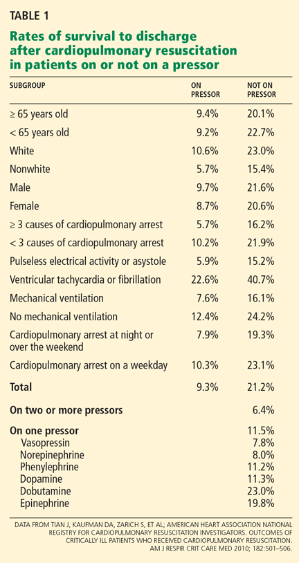
Other independent predictors of a lower survival rate were nonwhite race, mechanical ventilation, having three or more immediate causes of cardiopulmonary arrest, age 65 years or older, and cardiopulmonary arrest occurring at night or over the weekend.
Fortunately, for our patient, survival rates were higher for patients with ventricular tachycardia or fibrillation than with other causes of cardiopulmonary arrest: 22.6% for those on pressors (like our patient) and 40.7% for those on no pressors.
CASE CONTINUED: HE RECOVERS AND GOES HOME
P.G. makes a remarkable recovery and is now ready to go home. It is the weekend, and you are unable to schedule a follow-up appointment before his discharge, so you ask him to make an appointment with his PCP.
4. What is the likelihood that P.G. will be readmitted within 1 month?
- 5%
- 12%
- 20%
- 25%
- 30%
The importance of follow-up with a primary care physician
Misky et al,6 in a small study, attempted to identify the characteristics and outcomes of discharged patients who lack timely follow-up with a PCP. They prospectively enrolled 65 patients admitted to University of Colorado Hospital, an urban 425-bed tertiary care center, collecting information about patient demographics, diagnosis, payer source, and PCPs. After discharge, they called the patients to determine their PCP follow-up and readmission status. Thirty-day readmission rates and hospital length of stay were compared in patients with and without timely PCP follow-up (ie, within 4 weeks).
Patients lacking timely PCP follow-up were 10 times more likely to be readmitted (odds ratio [OR] = 9.9, P = .04): the rate was 21% in patients lacking timely PCP follow-up vs 3% in patients with timely PCP follow-up, P = .03. Lack of insurance was associated with lower rates of timely PCP follow-up: 29% vs 56% (P = .06), but did not independently increase the readmission rate or length of stay (OR = 1.0, P = .96). Index hospital length of stay was longer in patients lacking timely PCP follow-up: 4.4 days vs 6.3 days, P = 0.11.
Comment. Nearly half of the patients in this study, who were discharged from a large urban academic center, lacked timely follow-up with a PCP, resulting in higher rates of readmission and a nonsignificant trend toward longer length of stay. Timely follow-up is necessary for vulnerable patients.
Since the lack of timely PCP follow-up results in higher readmission rates and possibly a longer length of stay, a PCP appointment at discharge should perhaps be considered a core quality measure. This would be problematic in our American health care system, in which many patients lack health insurance and do not have a PCP.
A MAN UNDERGOING GASTRIC BYPASS SURGERY
A 55-year-old morbidly obese man (body mass index 45 kg/m2) with a history of type 2 diabetes mellitus, chronic renal insufficiency (serum creatinine level 2.1 mg/dL), hypercholesterolemia, and previous stroke is scheduled for gastric bypass surgery. His functional capacity is low, but he is able to do his activities of daily living. He reports having dyspnea on exertion and intermittently at rest, but no chest pain. His medications include insulin, atorvastatin (Lipitor), aspirin, and atenolol (Tenormin). He is afebrile; his blood pressure is 130/80 mm Hg, pulse 75, and oxygen saturation 97% on room air. His baseline electrocardiogram shows no Q waves.
5. Which of the following is an appropriate next step before proceeding to surgery?
- Echocardiography
- Cardiac catheterization
- Dobutamine stress echocardiography or adenosine thallium scanning
- No cardiac testing is necessary before surgery
Is cardiac testing necessary before noncardiac surgery?
Wijeysundera et al7 performed a retrospective cohort study of patients who underwent elective surgery at acute care hospitals in Ontario, Canada, in the years 1994 through 2004. The aim was to determine the association of noninvasive cardiac stress testing before surgery with survival rates and length of hospital stay. Included were 271,082 patients, of whom 23,991 (8.9%) underwent stress testing less than 6 months before surgery. These patients were matched with 46,120 who did not undergo testing.
One year after surgery, fewer patients who underwent stress testing had died: 1,622 (7.0%) vs 1,738 (7.5%); hazard ratio 0.92, 95% CI 0.86–0.99, P = .03. The number needed to treat (ie, to be tested) to prevent one death was 221. The tested patients also had a shorter mean hospital stay: 8.72 vs 8.96 days, a difference of 0.24 days (95% CI −0.07 to −0.43; P < .001).
However, the elderly patients (ie, older than 66 years) who underwent testing were more likely to be on beta-blockers and statins than those who did not undergo testing, which may be a confounding factor.
Furthermore, the benefit was all in the patients at intermediate or high risk. The authors performed a subgroup analysis, dividing the patients on the basis of their Revised Cardiac Risk Index (RCRI; 1 point each for ischemic heart disease, congestive heart failure, cerebrovascular disease, diabetes, renal insufficiency, and high-risk surgery).8 Patients with an RCRI of 0 points (indicating low risk) actually had a higher risk of death with testing than without testing: hazard ratio 1.35 (95% CI 1.03–1.74), number needed to harm 179—ie, for every 179 low-risk patients tested, one excess death occurred. Those with an RCRI of 1 or 2 points (indicating intermediate risk) had a hazard ratio of 0.92 with testing (95% CI 085–0.99), and those with an RCRI of 3 to 6 points (indicating high risk) had a hazard ratio of 0.80 with testing (95% CI 0.67- 0.97; number needed to treat = 38).
Comment. These findings indicate that cardiac stress testing should be done selectively before noncardiac surgery, and primarily for patients at high risk (with an RCRI of 3 or higher) and in some patients at intermediate risk, but not in patients at low risk, in whom it may be harmful. Stress testing may change patient management because a positive stress test allows one to start a beta-blocker or a statin, use more aggressive intraoperative and postoperative care, and identify patients who have indications for revascularization.
- Connolly SJ, Ezekowitz MD, Yusuf S, et al; RE-LY Steering Committee and Investigators. Dabigatran versus warfarin in patients with atrial fibrillation. N Engl J Med 2009; 361:1139–1151.
- Schulman S, Kearon C, Kakkar AK, et al; RE-COVER Study Group. Dabigatran versus warfarin in the treatment of acute venous thromboembolism. N Engl J Med 2009; 361:2342–2352.
- Wann LS, Curtis AB, Ellenbogen KA, et al. 2011 ACCF/AHA/HRS focused update on the management of patients with atrial fibrillation (update on dabigatran): A report of the American College of Cardiology Foundation/American Heart Association Task Force on Practice Guidelines. Circulation 2011; 123:1144–1150.
- Pronovost PJ, Goeschel CA, Colantuoni E, et al. Sustaining reductions in catheter-related bloodstream infections in Michigan intensive care units: observational study. BMJ 2010; 340:c309.
- Tian J, Kaufman DA, Zarich S, et al; American Heart Association National Registry for Cardiopulmonary Resuscitation Investigators. Outcomes of critically ill patients who received cardiopulmonary resuscitation. Am J Respir Crit Care Med 2010; 182:501–506.
- Misky GJ, Wald HL, Coleman EA. Post-hospitalization transitions: examining the effects of timing of primary care provider follow-up. J Hosp Med 2010; 5:392–397.
- Wijeysundera DN, Beattie WS, Austin PC, Hux JE, Laupacis A. Non-invasive cardiac stress testing before elective major non-cardiac surgery: population based cohort study. BMJ 2010; 340:b5526.
- Lee TH, Marcantonio ER, Mangione CM, et al. Derivation and prospective validation of a simple index for prediction of cardiac risk of major noncardiac surgery. Circulation 1999; 100:1043–1049.
A number of studies published in the last few years will likely affect the way we practice medicine in the hospital. Here, we will use a hypothetical case scenario to focus on the issues of anticoagulants, patient safety, quality improvement, critical care, transitions of care, and perioperative medicine.
AN ELDERLY MAN WITH NEW-ONSET ATRIAL FIBRILLATION
P.G. is an 80-year-old man with a history of hypertension and type 2 diabetes mellitus who is admitted with new-onset atrial fibrillation. In the hospital, his heart rate is brought under control with intravenous metoprolol (Lopressor). On discharge, he will be followed by his primary care physician (PCP). He does not have access to an anticoagulation clinic.
1. What are this patient’s options for stroke prevention?
- Aspirin 81 mg daily and clopidogrel (Plavix) 75 mg daily
- Warfarin (Coumadin) with a target international normalized ratio (INR) of 2.0 to 3.0
- Aspirin mg daily by itself
- Dabigatran (Pradaxa) 150 mg daily
A new oral anticoagulant agent
In deciding what type of anticoagulation to give to a patient with atrial fibrillation, it is useful to look at the CHADS2 score (1 point each for congestive heart failure, hypertension, age 75 or older, and diabetes mellitus; 2 points for prior stroke or transient ischemic attack. This patient has a CHADS2 score of 3, indicating that he should receive warfarin. An alternative is dabigatran, the first new anticoagulant agent in more than 50 years.
In a multicenter, international trial, Connolly et al1 randomized 18,113 patients (mean age 71, 64% men) to receive dabigatran 110 mg twice daily, dabigatran 150 mg twice daily, or warfarin with a target INR of 2.0 to 3.0. In this noninferiority trial, dabigatran was given in a blinded manner, but the use of warfarin was open-label. Patients were eligible if they had atrial fibrillation at screening or within the previous 6 months and were at risk of stroke—ie, if they had at least one of the following: a history of stroke or transient ischemic attack, a left ventricular ejection fraction of less than 40%, symptoms of congestive heart failure (New York Heart Association class II or higher), and an age of 75 or older or an age of 65 to 74 with diabetes mellitus, hypertension, or coronary artery disease.
At a mean follow-up of 2 years, the rate of stroke or systolic embolism was 1.69% per year in the warfarin group compared with 1.1% in the higher-dose dabigatran group (relative risk 0.66, 95% confidence interval [CI] 0.53–0.82, P < .001). The rates of major hemorrhage were similar between these two groups. Comparing lower-dose dabigatran and warfarin, the rates of stroke or systolic embolism were not significantly different, but the rate of major bleeding was significantly lower with lower-dose dabigatran.
In a trial in patients with acute venous thromboembolism, Schulman et al2 found that dabigatran was not inferior to warfarin in preventing venous thromboembolism.
Guidelines from the American College of Cardiology Foundation and the American Heart Association now endorse dabigatran as an alternative to warfarin for patients with atrial fibrillation.3 However, the guidelines state that it should be reserved for those patients who:
- Do not have a prosthetic heart valve or hemodynamically significant valve disease
- Have good kidney function (dabigatran is cleared by the kidney; the creatinine clearance rate should be greater than 30 mL/min for patients to receive dabigatran 150 mg twice a day, and at least 15 mL/min to receive 75 mg twice a day)
- Do not have severe hepatic dysfunction (which would impair baseline clotting function).
They note that other factors to consider are whether the patient:
- Can comply with the twice-daily dosing required
- Can afford the drug
- Has access to an anticoagulation management program (which would argue in favor of using warfarin).
Dabigatran is not yet approved to prevent venous thromboembolism.
CASE CONTINUED: HE GETS AN INFECTION
P.G. is started on dabigatran 150 mg by mouth twice a day.
While in the hospital he develops shortness of breath and needs intravenous furosemide (Lasix). Because he has bad veins, a percutaneous intravenous central catheter (PICC) line is placed. However, 2 days later, his temperature is 101.5°F, and his systolic blood pressure is 70 mm Hg. He is transferred to the medical intensive care unit (ICU) for treatment of sepsis. The anticoagulant is held, the PICC line is removed, and a new central catheter is inserted.
2. Which of the following directions is incorrect?
- Wash your hands before inserting the catheter. The accompanying nurse is required to directly observe this procedure or, if this step is not observed, to confirm that the physician did it.
- Before inserting the catheter, clean the patient’s skin with chlorhexidine antiseptic.
- Place sterile drapes over the entire patient.
- Wear any mask, hat, gown, and gloves available.
- Put a sterile dressing over the catheter.
A checklist can prevent infections when inserting central catheters
A checklist developed at Johns Hopkins Hospital consists of the five statements above, except for the second to last one—you should wear a sterile mask, hat, gown and gloves. This is important to ensure that sterility is not broken at any point during the procedure.
Pronovost et al4 launched a multicenter initiative at 90 ICUs, predominantly in the state of Michigan, to implement interventions to improve staff culture and teamwork and to translate research into practice by increasing the extent to which these five evidence-based recommendations were applied. The mean rate of catheter-related blood stream infections at baseline was 7.7%; this dropped to 2.8% during the implementation period, 2.3% in the first 3 months after implementation, 1.3% in months 16 through 18, and 1.1% in months 34 through 36, demonstrating that the gains from this quality-improvement project were sustainable.
If this intervention and collaborative model were implemented in all ICUs across the United States and if similar success rates were achieved, substantial and sustained reductions could be made in the 82,000 infections, 28,000 deaths, and $2.3 billion in costs attributed to these infections annually.
CASE CONTINUED: HE IS RESUSCITATED
P.G. is started on a 1-L fluid bolus but he remains hypotensive, necessitating a norepinephrine drip. He does well for about 6 hours, but in the middle of the night he develops ventricular tachycardia and ventricular fibrillation, and a code is called. He is successfully resuscitated, but the family is looking for prognostic information.
3. What are P.G.’s chances of surviving and leaving the hospital?
- 5%
- 8%
- 15%
- 23%
A registry of cardiopulmonary resuscitation
Tian et al5 evaluated outcomes in the largest registry of cardiopulmonary resuscitation to date. In this analysis, 49,656 adult patients with a first cardiopulmonary arrest occurring in an ICU between January 1, 2000, and August 26, 2008, were evaluated for their outcomes on pressors vs those not on pressors.
Other independent predictors of a lower survival rate were nonwhite race, mechanical ventilation, having three or more immediate causes of cardiopulmonary arrest, age 65 years or older, and cardiopulmonary arrest occurring at night or over the weekend.
Fortunately, for our patient, survival rates were higher for patients with ventricular tachycardia or fibrillation than with other causes of cardiopulmonary arrest: 22.6% for those on pressors (like our patient) and 40.7% for those on no pressors.
CASE CONTINUED: HE RECOVERS AND GOES HOME
P.G. makes a remarkable recovery and is now ready to go home. It is the weekend, and you are unable to schedule a follow-up appointment before his discharge, so you ask him to make an appointment with his PCP.
4. What is the likelihood that P.G. will be readmitted within 1 month?
- 5%
- 12%
- 20%
- 25%
- 30%
The importance of follow-up with a primary care physician
Misky et al,6 in a small study, attempted to identify the characteristics and outcomes of discharged patients who lack timely follow-up with a PCP. They prospectively enrolled 65 patients admitted to University of Colorado Hospital, an urban 425-bed tertiary care center, collecting information about patient demographics, diagnosis, payer source, and PCPs. After discharge, they called the patients to determine their PCP follow-up and readmission status. Thirty-day readmission rates and hospital length of stay were compared in patients with and without timely PCP follow-up (ie, within 4 weeks).
Patients lacking timely PCP follow-up were 10 times more likely to be readmitted (odds ratio [OR] = 9.9, P = .04): the rate was 21% in patients lacking timely PCP follow-up vs 3% in patients with timely PCP follow-up, P = .03. Lack of insurance was associated with lower rates of timely PCP follow-up: 29% vs 56% (P = .06), but did not independently increase the readmission rate or length of stay (OR = 1.0, P = .96). Index hospital length of stay was longer in patients lacking timely PCP follow-up: 4.4 days vs 6.3 days, P = 0.11.
Comment. Nearly half of the patients in this study, who were discharged from a large urban academic center, lacked timely follow-up with a PCP, resulting in higher rates of readmission and a nonsignificant trend toward longer length of stay. Timely follow-up is necessary for vulnerable patients.
Since the lack of timely PCP follow-up results in higher readmission rates and possibly a longer length of stay, a PCP appointment at discharge should perhaps be considered a core quality measure. This would be problematic in our American health care system, in which many patients lack health insurance and do not have a PCP.
A MAN UNDERGOING GASTRIC BYPASS SURGERY
A 55-year-old morbidly obese man (body mass index 45 kg/m2) with a history of type 2 diabetes mellitus, chronic renal insufficiency (serum creatinine level 2.1 mg/dL), hypercholesterolemia, and previous stroke is scheduled for gastric bypass surgery. His functional capacity is low, but he is able to do his activities of daily living. He reports having dyspnea on exertion and intermittently at rest, but no chest pain. His medications include insulin, atorvastatin (Lipitor), aspirin, and atenolol (Tenormin). He is afebrile; his blood pressure is 130/80 mm Hg, pulse 75, and oxygen saturation 97% on room air. His baseline electrocardiogram shows no Q waves.
5. Which of the following is an appropriate next step before proceeding to surgery?
- Echocardiography
- Cardiac catheterization
- Dobutamine stress echocardiography or adenosine thallium scanning
- No cardiac testing is necessary before surgery
Is cardiac testing necessary before noncardiac surgery?
Wijeysundera et al7 performed a retrospective cohort study of patients who underwent elective surgery at acute care hospitals in Ontario, Canada, in the years 1994 through 2004. The aim was to determine the association of noninvasive cardiac stress testing before surgery with survival rates and length of hospital stay. Included were 271,082 patients, of whom 23,991 (8.9%) underwent stress testing less than 6 months before surgery. These patients were matched with 46,120 who did not undergo testing.
One year after surgery, fewer patients who underwent stress testing had died: 1,622 (7.0%) vs 1,738 (7.5%); hazard ratio 0.92, 95% CI 0.86–0.99, P = .03. The number needed to treat (ie, to be tested) to prevent one death was 221. The tested patients also had a shorter mean hospital stay: 8.72 vs 8.96 days, a difference of 0.24 days (95% CI −0.07 to −0.43; P < .001).
However, the elderly patients (ie, older than 66 years) who underwent testing were more likely to be on beta-blockers and statins than those who did not undergo testing, which may be a confounding factor.
Furthermore, the benefit was all in the patients at intermediate or high risk. The authors performed a subgroup analysis, dividing the patients on the basis of their Revised Cardiac Risk Index (RCRI; 1 point each for ischemic heart disease, congestive heart failure, cerebrovascular disease, diabetes, renal insufficiency, and high-risk surgery).8 Patients with an RCRI of 0 points (indicating low risk) actually had a higher risk of death with testing than without testing: hazard ratio 1.35 (95% CI 1.03–1.74), number needed to harm 179—ie, for every 179 low-risk patients tested, one excess death occurred. Those with an RCRI of 1 or 2 points (indicating intermediate risk) had a hazard ratio of 0.92 with testing (95% CI 085–0.99), and those with an RCRI of 3 to 6 points (indicating high risk) had a hazard ratio of 0.80 with testing (95% CI 0.67- 0.97; number needed to treat = 38).
Comment. These findings indicate that cardiac stress testing should be done selectively before noncardiac surgery, and primarily for patients at high risk (with an RCRI of 3 or higher) and in some patients at intermediate risk, but not in patients at low risk, in whom it may be harmful. Stress testing may change patient management because a positive stress test allows one to start a beta-blocker or a statin, use more aggressive intraoperative and postoperative care, and identify patients who have indications for revascularization.
- Connolly SJ, Ezekowitz MD, Yusuf S, et al; RE-LY Steering Committee and Investigators. Dabigatran versus warfarin in patients with atrial fibrillation. N Engl J Med 2009; 361:1139–1151.
- Schulman S, Kearon C, Kakkar AK, et al; RE-COVER Study Group. Dabigatran versus warfarin in the treatment of acute venous thromboembolism. N Engl J Med 2009; 361:2342–2352.
- Wann LS, Curtis AB, Ellenbogen KA, et al. 2011 ACCF/AHA/HRS focused update on the management of patients with atrial fibrillation (update on dabigatran): A report of the American College of Cardiology Foundation/American Heart Association Task Force on Practice Guidelines. Circulation 2011; 123:1144–1150.
- Pronovost PJ, Goeschel CA, Colantuoni E, et al. Sustaining reductions in catheter-related bloodstream infections in Michigan intensive care units: observational study. BMJ 2010; 340:c309.
- Tian J, Kaufman DA, Zarich S, et al; American Heart Association National Registry for Cardiopulmonary Resuscitation Investigators. Outcomes of critically ill patients who received cardiopulmonary resuscitation. Am J Respir Crit Care Med 2010; 182:501–506.
- Misky GJ, Wald HL, Coleman EA. Post-hospitalization transitions: examining the effects of timing of primary care provider follow-up. J Hosp Med 2010; 5:392–397.
- Wijeysundera DN, Beattie WS, Austin PC, Hux JE, Laupacis A. Non-invasive cardiac stress testing before elective major non-cardiac surgery: population based cohort study. BMJ 2010; 340:b5526.
- Lee TH, Marcantonio ER, Mangione CM, et al. Derivation and prospective validation of a simple index for prediction of cardiac risk of major noncardiac surgery. Circulation 1999; 100:1043–1049.
KEY POINTS
- Dabigatran (Pradaxa) will likely start to replace warfarin (Coumadin) both to prevent stroke in patients with atrial fibrillation and to prevent recurrent venous thromboembolism.
- Using a checklist during insertion of central venous catheters can decrease the rate of catheter-related bloodstream infections in the intensive care unit.
- The overall survival rate of patients who undergo cardiopulmonary resuscitation in the intensive care unit is approximately 16%; the rate is lower in patients who are receiving pressor drugs and higher in those with ventricular tachycardia or ventricular fibrillation.
- Patients lacking follow-up with a primary care physician within 30 days of discharge are at high risk of readmission and have a trend for longer length of hospital stay.
- Preoperative stress testing for patients undergoing noncardiac surgery should be done selectively, ie, in patients at high risk.
When good drugs turn weirdly bad

Many of the adverse effects of the small-molecule drugs such as azathioprine (Imuran) and methotrexate are those expected from chemical toxicity or inhibition of proliferation, eg, aminotransferase elevation, leukopenia, and alopecia. Mycophenolate mofetil (CellCept) uniquely can cause profound anemia, cyclophosphamide (Cytoxan) elicits cystitis, and many of these drugs trigger virus-associated malignancies. In perhaps 8% of patients, azathioprine causes a systemic hypersensitivity reaction with high fevers, variable rash, leukocytosis, and elevated aminotransferase levels shortly after it is started. Yet we are often slow to recognize this syndrome, as we tend to search for an infection and forget that even immunosuppressive drugs can cause systemic allergic-type reactions. A similar syndrome following initiation of phenytoin (Dilantin) would be recognized far more rapidly.
But the biologic agents, which target specific components of the immune system, resulting in focal immunosuppression and a disturbance in the homeostatic balance of the immune system, elicit some of the more challenging and sometimes paradoxical side effects. Interferon alfa, which has antiviral effects, is also used as an immunomodulator to treat Behçet disease and as part of regimens that treat specific malignancies. Perhaps because it up-regulates the expression of major histocompatibility complex class II molecules on antigen-presenting cells, interferon therapy also triggers several organ-specific autoimmune syndromes, including autoimmune thrombocytopenia, hypothyroidism, hemolytic anemia, hepatitis, and psoriasis.
Even more challenging to understand and sometimes to treat are the inflammatory effects of anti-tumor necrosis factor agents. Drugs of this class can evoke a demyelinating syndrome similar to multiple sclerosis. Further, even though they are used to treat psoriasis, they can also provoke a psoriasiform, often palmar and pustular, reaction.
So as we continue to adopt targeted immunologic therapies and revel in their efficacy, we need to remain humbled by what we don’t yet fully understand about the complexity of what the 19th century physiologist Claude Bernard termed the milieu intérieur (homeostasis) and keep in mind that even the most specific of drugs can have untoward biologic effects by weirdly disrupting our finely balanced immune system.
Rash from hepatitis C treatment
A 54-year-old woman with hepatitis C virus infection presents with generalized rash, pruritus, and fever over the past week. The rash appeared on her left arm after she received her fifth weekly injection of pegylated interferon alfa 2b, in combination with daily oral ribavirin (Copegus, Rebetol). Over the course of 3 days, it spread to her face and the rest of her body.


Q: What is the most likely clinical diagnosis?
- Stevens-Johnson syndrome
- Mixed cryoglobulinemia
- Acute eczematous drug eruption
- Lichen planus
A: Acute eczematous drug eruption is the most likely diagnosis.
The clinical presentation and laboratory findings suggest (the latter by exclusion) that our patient had an allergic drug reaction to the interferon or to the ribavirin therapy, or to both. Although this combination is a standard treatment for chronic hepatitis C, some patients experience adverse reactions that lead to its discontinuation. Local injection-site reactions are the most prevalent, affecting up to 12% of patients, whereas eczematous dermatoses manifest less commonly, occurring in up to 5% of patients.1
While awaiting the results of skin biopsy, a careful evaluation of the clinical features of the physical examination and an appropriate laboratory evaluation can rule out other important conditions in the differential diagnosis.
The absence of mucous membrane involvement steers the diagnosis away from Stevens-Johnson syndrome, a life-threatening hypersensitivity condition often triggered by drugs, malignant tumors, and viral infections, which may also affect internal organs. In this condition, skin biopsy specimens would be distinguished by subepidermal bullae and epidermal cell necrosis—neither of which was seen in our patient.
Mixed cryoglobulinemia should always be considered in hepatitis C patients because of the strong association between this infection and the development of cryoglobulins. The rash usually is purpuric, but it may be pleomorphic.2,3 This vasculitis often manifests with excess cryoglobulins, elevated rheumatoid factor, and low titers of complement in the blood due to consumption by immune complexes. Tissue biopsy would usually show typical vascular changes if performed on fresh lesions.4,5 The normal levels of these components in our patient coupled with the appearance of her skin makes cryoglobulinemia a less likely cause.
Furthermore, hepatitis C infection, whether or not treated with interferon and ribavirin, can cause an onset or recurrence of other dermatologic conditions, notably lichen planus, psoriasis, vitiligo, and systemic lupus erythematosus.1–4
In lichen planus, the rash is often described as flat-topped, pruritic, and violaceous. It may involve the extremities, the genitalia, and the oral cavity.4,5 The difference in quality of the rash compared with the rash in our patient makes lichen planus less likely.
Exclusion of the other conditions in the differential diagnosis, in addition to results from a definitive punch biopsy, solidified the diagnosis in our patient. Skin biopsy of the patient’s lower-extremity lesions revealed spongiotic dermatitis with lymphocytes, neutrophils, and few eosinophils—a finding characteristic of an acute eczematous drug eruption. Improvement of her rash after discontinuation of interferon and ribavirin further supported this conclusion, although it was unclear whether one or both agents were responsible.
OUTCOME
Management of acute eczematous drug eruption entails stopping the offending drug and alleviating the symptoms. Our patient’s non-life-threatening rash improved dramatically with cessation of interferon and ribavirin. She received a single dose of a systemic corticosteroid initially, out of concern for a severe medication-induced reaction (ie, Stevens-Johnson syndrome), but she was otherwise maintained with diphenhydramine (Benadryl) and a multivitamin ointment for the rash throughout her 9-day hospital stay. Her pruritus was well controlled with hydroxyzine (Atarax, Vistaril). At discharge, she was referred back to her hepatologist for further treatment of her hepatitis C, possibly with interferon and ribavirin again.
TAKE-HOME MESSAGE
Adverse reactions to interferon and ribavirin treatment in hepatitis C patients can manifest dermatologically, and the combination therapy should be discontinued to prevent further insult. A broad variety of conditions in the differential diagnosis should be taken into account, but dermatologic conditions that occur or recur specifically in hepatitis C patients should be considered as well.
- Dereure O, Raison-Peyron N, Larrey D, Blanc F, Guilhou JJ. Diffuse inflammatory lesions in patients treated with interferon alfa and ribavirin for hepatitis C: a series of 20 patients. Br J Dermatol 2002; 147:1142–1146.
- Ferri C, Zignego AL, Pileri SA. Cryoglobulins. J Clin Pathol 2002; 55:4–13.
- Faurie P, Broussolle C, Zoulim F, Trepo C, Sève P. Sarcoidosis and hepatitis C: clinical description of 11 cases. Eur J Gastroenterol Hepatol 2010; 22:967–972.
- Shengyuan L, Songpo Y, Wen W, Wenjing T, Haitao Z, Binyou W. Hepatitis C virus and lichen planus: a reciprocal association determined by a meta-analysis. Arch Dermatol 2009; 145:1040–1047.
- Aamir S, Ullah Z, Iqbal Z, Khan AA, Yaqub F, Malik K. Cutaneous manifestations of interferon alfa and ribavirin for hepatitis C. J Pak Assoc Dermatol 2008; 18:14–20.
A 54-year-old woman with hepatitis C virus infection presents with generalized rash, pruritus, and fever over the past week. The rash appeared on her left arm after she received her fifth weekly injection of pegylated interferon alfa 2b, in combination with daily oral ribavirin (Copegus, Rebetol). Over the course of 3 days, it spread to her face and the rest of her body.
Q: What is the most likely clinical diagnosis?
- Stevens-Johnson syndrome
- Mixed cryoglobulinemia
- Acute eczematous drug eruption
- Lichen planus
A: Acute eczematous drug eruption is the most likely diagnosis.
The clinical presentation and laboratory findings suggest (the latter by exclusion) that our patient had an allergic drug reaction to the interferon or to the ribavirin therapy, or to both. Although this combination is a standard treatment for chronic hepatitis C, some patients experience adverse reactions that lead to its discontinuation. Local injection-site reactions are the most prevalent, affecting up to 12% of patients, whereas eczematous dermatoses manifest less commonly, occurring in up to 5% of patients.1
While awaiting the results of skin biopsy, a careful evaluation of the clinical features of the physical examination and an appropriate laboratory evaluation can rule out other important conditions in the differential diagnosis.
The absence of mucous membrane involvement steers the diagnosis away from Stevens-Johnson syndrome, a life-threatening hypersensitivity condition often triggered by drugs, malignant tumors, and viral infections, which may also affect internal organs. In this condition, skin biopsy specimens would be distinguished by subepidermal bullae and epidermal cell necrosis—neither of which was seen in our patient.
Mixed cryoglobulinemia should always be considered in hepatitis C patients because of the strong association between this infection and the development of cryoglobulins. The rash usually is purpuric, but it may be pleomorphic.2,3 This vasculitis often manifests with excess cryoglobulins, elevated rheumatoid factor, and low titers of complement in the blood due to consumption by immune complexes. Tissue biopsy would usually show typical vascular changes if performed on fresh lesions.4,5 The normal levels of these components in our patient coupled with the appearance of her skin makes cryoglobulinemia a less likely cause.
Furthermore, hepatitis C infection, whether or not treated with interferon and ribavirin, can cause an onset or recurrence of other dermatologic conditions, notably lichen planus, psoriasis, vitiligo, and systemic lupus erythematosus.1–4
In lichen planus, the rash is often described as flat-topped, pruritic, and violaceous. It may involve the extremities, the genitalia, and the oral cavity.4,5 The difference in quality of the rash compared with the rash in our patient makes lichen planus less likely.
Exclusion of the other conditions in the differential diagnosis, in addition to results from a definitive punch biopsy, solidified the diagnosis in our patient. Skin biopsy of the patient’s lower-extremity lesions revealed spongiotic dermatitis with lymphocytes, neutrophils, and few eosinophils—a finding characteristic of an acute eczematous drug eruption. Improvement of her rash after discontinuation of interferon and ribavirin further supported this conclusion, although it was unclear whether one or both agents were responsible.
OUTCOME
Management of acute eczematous drug eruption entails stopping the offending drug and alleviating the symptoms. Our patient’s non-life-threatening rash improved dramatically with cessation of interferon and ribavirin. She received a single dose of a systemic corticosteroid initially, out of concern for a severe medication-induced reaction (ie, Stevens-Johnson syndrome), but she was otherwise maintained with diphenhydramine (Benadryl) and a multivitamin ointment for the rash throughout her 9-day hospital stay. Her pruritus was well controlled with hydroxyzine (Atarax, Vistaril). At discharge, she was referred back to her hepatologist for further treatment of her hepatitis C, possibly with interferon and ribavirin again.
TAKE-HOME MESSAGE
Adverse reactions to interferon and ribavirin treatment in hepatitis C patients can manifest dermatologically, and the combination therapy should be discontinued to prevent further insult. A broad variety of conditions in the differential diagnosis should be taken into account, but dermatologic conditions that occur or recur specifically in hepatitis C patients should be considered as well.
- Dereure O, Raison-Peyron N, Larrey D, Blanc F, Guilhou JJ. Diffuse inflammatory lesions in patients treated with interferon alfa and ribavirin for hepatitis C: a series of 20 patients. Br J Dermatol 2002; 147:1142–1146.
- Ferri C, Zignego AL, Pileri SA. Cryoglobulins. J Clin Pathol 2002; 55:4–13.
- Faurie P, Broussolle C, Zoulim F, Trepo C, Sève P. Sarcoidosis and hepatitis C: clinical description of 11 cases. Eur J Gastroenterol Hepatol 2010; 22:967–972.
- Shengyuan L, Songpo Y, Wen W, Wenjing T, Haitao Z, Binyou W. Hepatitis C virus and lichen planus: a reciprocal association determined by a meta-analysis. Arch Dermatol 2009; 145:1040–1047.
- Aamir S, Ullah Z, Iqbal Z, Khan AA, Yaqub F, Malik K. Cutaneous manifestations of interferon alfa and ribavirin for hepatitis C. J Pak Assoc Dermatol 2008; 18:14–20.

Managing cancer pain: Frequently asked questions
Some 90% of patients with cancer experience pain during their illness.1 The pain usually worsens as the disease progresses, and patients may experience different types of pain.
Persistent pain decreases function, appetite, and sleep, induces fear, causes depression, and generally lowers the quality of life.2 Persistent pain is demoralizing and debilitating for patients and their caregivers.3
Adequate pain control is important to ensure that patients can function productively, maintain social relationships, and improve their quality of life.2 Yet 86% of practicing physicians surveyed believed that most cancer patients with pain were undermedicated,2 and most felt that pain management is unsuccessful in more than half of patients who seek help.3
The critical importance of pain management has been emphasized by the World Health Organization (WHO), by international and national professional organizations, and by government agencies. All practitioners who care for cancer patients need to be well educated in managing cancer pain, a key part of which is to educate patients about the process and what to expect. This results in better pain control.4
Although much has been written on the management of cancer pain in a referral setting, little has been published on how to manage it in primary care. In this article, we discuss common questions faced by generalists. We emphasize the use of opioids, perhaps the most challenging aspect of cancer pain management. We also discuss when consultation with a specialist in pain management or a palliative medicine specialist is especially helpful.
WHAT ARE THE DIFFERENT TYPES OF PAIN SYNDROMES?
Pain is classified in several ways1–6:
Nociceptive vs neuropathic. Nociceptive pain comprises somatic and visceral components and is the result of continued tissue injury.4 Neuropathic pain is due to injury to the peripheral and central nervous systems and occurs within an area of sensory or motor deficit.
Continuous vs intermittent. Continuous pain, even if controlled, can have breakthroughs, ie, flares of pain above the controlled baseline level. Intermittent pain is a pain flare without chronic baseline pain. Intermittent pain is further divided into incident pain (ie, on movement) and end-of-dose failure (ie, pain occurring just before the next scheduled opioid dose).5 Pain specialists continue to debate the meaning and the use of these terms.
Malignant vs nonmalignant. Cancer pain is multifactorial,1 being induced by the disease itself, by the treatment of cancer, and by pain unrelated to cancer or its treatment (eg, osteoarthritis or diabetic neuropathy).2
Familiarity with the causes and the types of pain, including pain related to cancer, is important, as this influences treatment decisions.
HOW IS PAIN ASSESSED?
The assessment of pain is vital in managing it.
Since pain is inherently subjective, the patient’s self-report is the gold standard.4 Characteristics of the pain along with a physical examination, laboratory testing, and imaging studies can define the pathophysiology of the pain and influence the decision to undertake further assessment or specific therapies.
Patients and physicians can use various scales, such as a visual analog scale, a numerical rating scale, a graphic scale, a verbal scale, a word descriptor scale, and a functional pain scale. A verbal scale can be used if the patient is alert, or a nonverbal scale if the patient has impaired cognition or speaks a different language. Intensity is the most common dimension evaluated in cancer pain, primarily via a numerical or visual analog scale. A numerical scale score of 0 to 10 has been found to be as effective as a visual analog scale (0 to 100 mm),7,8 and the numerical rating scale is generally preferred as a measure of pain intensity.9
There are no clear guidelines for selecting one scale over another.7 A clinically meaningful response (ie, meaningful to patients) is at least a two-point decrease on the 10-point numerical scale or a 13-mm decrease on the 100-mm visual analog scale. A decrease in the percentage of the pain relates to global improvement better than an absolute reduction on the numerical scale.
WHAT PROBLEMS ARE ENCOUNTERED IN MANAGING CANCER PAIN?
WHAT ARE THE DIFFERENT WAYS TO MANAGE CANCER PAIN?
Pain should be treated promptly and aggressively, because if untreated it can lead to delays in healing, changes in the central nervous system (eg, sensitization, plasticity), chronic stress, family stress, depression, job loss, and even suicide.12–14
Comprehensive pain management improves outcomes and includes the rational use of opioids and adjuvant analgesics, physical rehabilitation, cognitive behavioral (non-drug) therapies, family counseling, interventional procedures (kyphoplasty, nerve blocks, local injections, spinal analgesia), and complementary therapies such as acupuncture.12 Adjuvant analgesics include antidepressants, anticonvulsants, and local anesthetics.
HOW DO OPIOIDS RELIEVE CANCER PAIN?
Opioids bind to receptors in tissues throughout the body, including in the central and peripheral nervous systems15 and the digestive tract. The binding of an opioid to an opioid receptor—including mu, kappa, and delta receptors and orphan receptor-like ligand-1—initiates a cascade of intracellular reactions. Due to the nature of different interactions of opioids with each of these receptors, individuals vary in their response to opioids.15
WHAT ARE THE CHARACTERISTICS OF COMMON OPIOIDS?
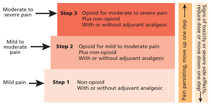
- Step 1. Mild pain calls for a nonopioid analgesic with or without an adjuvant (more about adjuvants below).
- Step 2. Mild or moderate pain that persists or increases calls for a weak opioid such as codeine, tramadol (Ultram), or hydrocodone, with or without a nonopioid and with or without an adjuvant.
- Step 3. Severe pain calls for a strong opioid with or without a nonopioid, and with or without an adjuvant.
Morphine, the prototypical opioid, is well studied and versatile, as it can be given orally, parenterally, rectally, or intraspinally. It is readily available in the United States and Western Europe but not in some parts of the world, such as Asia and Africa. It is also cost-effective.
Hydromorphone (Dilaudid) is similar to morphine in terms of versatility, cost, and effectiveness in pain management. An extended-release form (Exalgo) is now available in the United States.
Oxycodone is readily available in both slow-release (eg, OxyContin) and immediate-release (eg, Oxy-IR) preparations and is also cost-effective. However, there is no parenteral formulation in the United States.
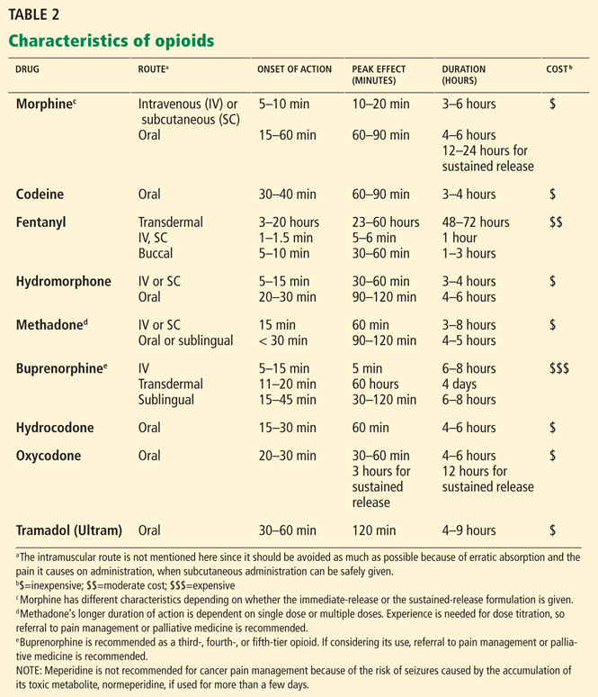
Methadone is inexpensive and can be used as a long-acting or an immediate-release opioid. However, it should be used with caution in patients with a prolonged QTc interval: in general, a QTc interval of 430 to 450 msec is not a contraindication, but there is a risk of torsades de pointes when the QTc is greater than 500 msec. The physician should also look for drug interactions when prescribing methadone, which is metabolized in the liver via the cytochrome P450 3A4 system. Methadone use can also lead to respiratory depression, prolonged QTc interval, and sudden death.
Buprenorphine can be used as a third- or fourth-tier opioid for patients with both kidney and liver failure. It can be given sublingually or parenterally. It may not be readily available, may not be covered by insurance, and is expensive.
Selecting an opioid to try first
The following are some general considerations when selecting an opioid to try first:
- Does the patient have a history of organ failure? Has the patient had a therapeutic response to, or adverse effects from, a particular opioid in the past?
- Which route would best fit the patient’s needs? (Oral is always preferable.)
- How often will breakthrough dosing be required? (In general, the breakthrough dose is administered at the drug’s half-life, but it can be administered between 1 and 4 hours.)
- How much will it cost? (Consider the cost, insurance coverage, and co-pays.)
Table 2 shows different characteristics of commonly used opioids, including route of administration, onset of action, peak effect, and duration of action.1
WHAT ARE THE EQUIANALGESIC DOSES OF COMMONLY USED OPIOIDS?
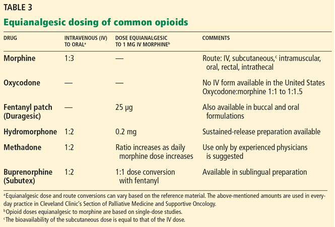
Table 3 lists equianalgesic doses and route conversions of commonly used opioids.18–20
WHAT ARE THE PRINCIPLES BEHIND OPIOID DOSING?
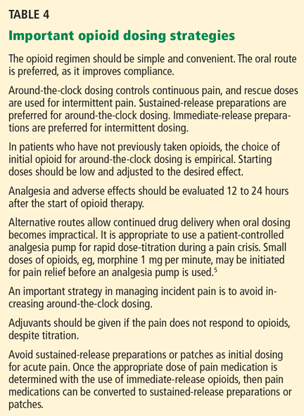
Table 4 shows important strategies for opioid dosing. An in-depth discussion of specific opioid dosing strategies is beyond the scope of this article.5
WHAT ARE THE COMMON ADVERSE EFFECTS OF OPIOIDS?
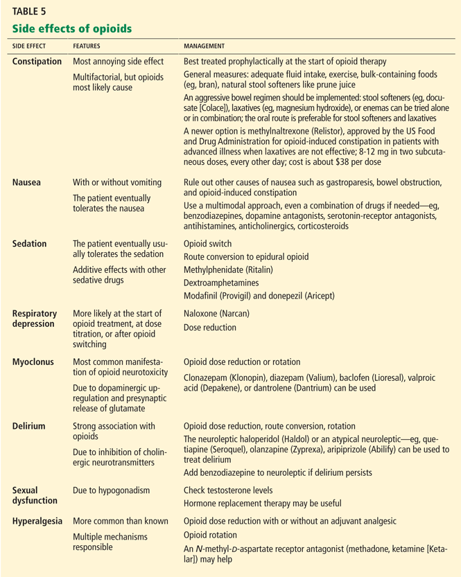
Adverse effects are among the most common reasons for failure of opioids to relieve pain. If these effects are not anticipated and treated prophylactically, patients may avoid taking their opioid drugs or may complain that they are “allergic” to them. In reality, true allergy to any of the opioids is rare. Patients comply better if they are taught to expect that most adverse effects are either preventable or manageable.21 A simple strategy includes reducing the opioid dose by 25% to 50%, using different opioids (“rotation”), changing the route of administration, and directly treating adverse effects.21,22
WHAT IS OPIOID ROTATION AND HOW IS IT DONE?
Opioid rotation involves changing to a different drug using the same administration route, with the aim of improving the analgesic response or reducing adverse effects.16 It may be useful in widening the therapeutic window, ie, establishing a more advantageous relationship between analgesia and toxicity.16 This strategy applies, for example, to patients who have an adverse reaction to morphine, and who may need rotation to fentanyl or methadone.
The major indication for switching opioids is poorly controlled pain with unacceptable adverse effects due to opioid toxicity, the rapid development of tolerance, refractory pain, or difficult pain syndromes.24 A recent prospective study showed that 42% of patients underwent opioid rotation, and the two most common reasons were inadequate analgesia and severe adverse effects.25 Opioid rotation resulted in relief of confusion (72%), nausea and vomiting (68%), and drowsiness (53%).25
Before trying opioid rotation, review the patient’s pain syndromes and the use of an adjuvant analgesic, and assess for evidence of opioid toxicity or contributing abnormal biochemical factors such as hydration status.24,26 Most opioids are mu-receptor agonists and may exhibit cross-tolerance, a phenomenon in which the alternative drug does not have the expected effects because of similar pharmacologic action of the first drug. Because the degree of cross-tolerance may change as opioid doses are escalated, it is advisable to proceed with caution when switching from one opioid to another in patients who are receiving very high doses. Opioid rotation generally would be ineffective if there is complete analgesic cross-tolerance between opioids.
The common equivalency conversion tables are based either on studies in patients who received low doses of opioids or on single-dose studies.16,24 By substituting opioids and using lower doses than expected according to the equivalency conversion tables (generally a 25% to 30% decrease), it is possible in most cases to reduce or relieve the symptoms of opioid toxicity and to manage patients highly tolerant to previous opioids while improving analgesia.24
Alternatives to opioid rotation are route conversion (oral to parenteral or spinal), addition of an adjuvant analgesic, and opioid dose reduction.
WHAT IS OPIOID TOXICITY AND HOW IS IT MANAGED?
Opioid overdose is commonly the result of an error in pain assessment, opioid prescribing, or dose administration. Opioid overdose classically presents as sedation or respiratory depression. The combination of coma, reduced respiratory rate, and pinpoint pupils is highly suggestive of opioid toxicity, and treatment should be initiated promptly.
This scenario, however, is the extreme example of opioid overdose, and it is rare when a patient is given the correct opioid dose titrated gradually over a period of time. The more common scenario is when a patient’s pain has finally been managed and the patient is resting comfortably with slow respirations. This would not warrant naloxone (Narcan) administration but rather close observation and monitoring of vital signs.
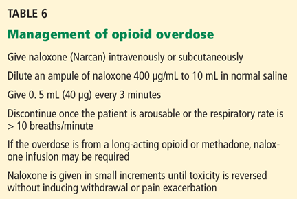
Naloxone has antagonist activity at all of the receptor sites.27 It is important to be alert for acute opioid withdrawal in patients taking high-dose opioids for a long time.27 There are no guidelines as to the route of administration and the dosing of naloxone. Table 6 summarizes the management of opioid overdose using naloxone.5
WHAT IS THE ROLE OF ADJUVANTS?
An adjuvant analgesic is any drug with a primary indication other than pain, but with analgesic properties in some painful conditions. Adjuvants are best used when a patient cannot obtain satisfactory pain relief from an opioid.28 Antidepressants, anticonvulsants, neuroleptics, antiarrhythmics, antihistamines, N-methyl-d-aspartate (NMDA) receptor antagonists, steroids, muscle relaxants, bisphosphonates, and radiopharmaceuticals can be adjuvant agents.29
Adjuvants are generally used to complement the analgesic effects of opioids to achieve optimal pain control with a minimum of adverse effects.28 The following scenarios should prompt the use of adjuvants in clinical practice28:
- The toxic limit of a primary pain medication has been reached.
- The therapeutic benefit of the primary pain medication has reached a plateau.
- The primary analgesic could not be used because of substance-abuse behavior, multiple organ failure, allergy, etc.
- The patient has multiple pain syndromes.
- The patient has additional symptoms unrelated to pain, eg, insomnia or depression.
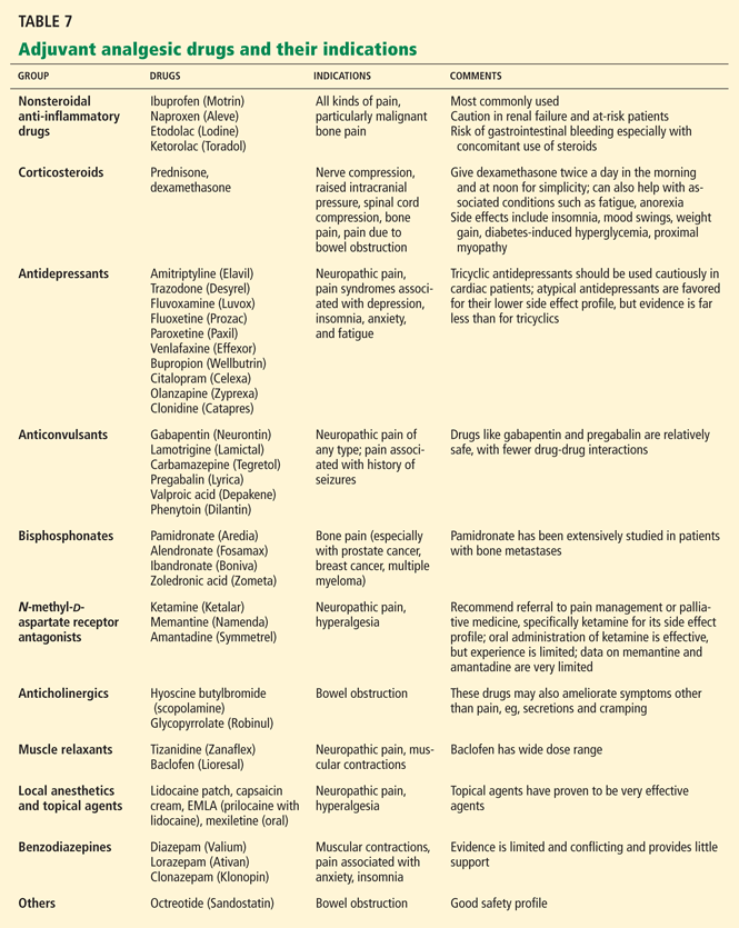
Table 7 lists adjuvants with specific indications and points to remember when prescribing them.28,29
WHAT IS THE ROLE OF NSAIDs FOR CANCER PAIN?
Nonsteroidal anti-inflammatory drugs (NSAIDs) have a well-established role in treating cancer-related pain, either on their own for mild pain or in combination with opioids for moderate to severe pain, leading to additive analgesia. Using NSAIDs as adjuvants is common practice in certain cancer pain syndromes, such as malignant bone pain, although there is considerable variation in response.31
NSAIDs have long been known to inhibit peripheral prostaglandin synthesis, but recently they have also been suggested to have a central action. The central effect is related to NMDA receptor-induced activation of the nitric oxide system.31
NSAIDs have ceiling effects, and there is no therapeutic advantage to increasing the dose beyond that which is recommended.
Ketorolac (Toradol), indomethacin (Indocin), and diclofenac (Voltaren) have potent analgesic activity, whereas the “oxicam” NSAIDs show predominantly anti-inflammatory effects.30
No NSAID is clearly superior for a particular type of pain. Certain NSAIDs block the NMDA receptor and inhibit cyclo-oxygenase-1 and cyclo-oxygenase-2. There is a poor correlation between the analgesic effects of NSAIDs and cyclo-oxygenase inhibition. There is no evidence to support the use of selective cyclo-oxygenase-2 inhibitors for cancer pain, and these agents have no advantage over nonselective NSAIDs on the basis of limited gastrointestinal toxicity.32
In cancer pain, NSAIDs may delay the development of tolerance and allow lower doses of opioids to be used, with fewer central nervous system side effects.31,32 Despite the extensive use of NSAIDs, relatively few randomized studies have documented their efficacy in cancer pain compared with other chronic pain syndromes. Data on safe and effective doses from studies of nonmalignant pain may not apply to cancer pain, since cancer patients often have several serious conditions and are on multiple medications. In addition, the potential for adverse effects of NSAIDs (gastrointestinal bleeding, renal failure, thrombosis) may be greater in patients with advanced cancer.
In conclusion, NSAIDs may help if used judiciously in somatic pain and visceral pain, and perhaps even in neuropathic pain.31
HOW IS CANCER PAIN MANAGED IN PATIENTS WITH ORGAN FAILURE?
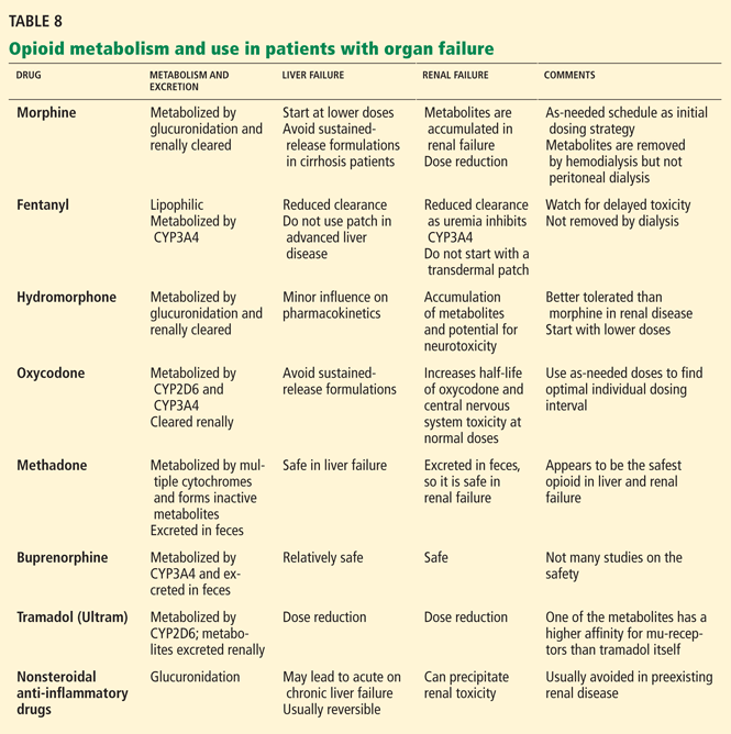
- Opioids that can be used in liver failure or cirrhosis: morphine, hydromorphone, methadone, levorphanol, buprenorphine.
- Opioids that can be used in renal failure: methadone, fentanyl, and buprenorphine are safest; oxycodone and hydromorphone are moderately safe; morphine is the least safe.35,36
- Opioids that can be used in both kidney and liver failure: methadone, buprenorphine.
HOW CAN PROBLEMS RELATED TO SUBSTANCE ABUSE BE AVOIDED?
Substance abuse is less a problem in managing cancer pain than in chronic nonmalignant pain. Prescribing opioids safely is challenging, and very little has been published on substance abuse and the management of cancer pain. However, in the absence of practice guidelines, the best approach is to establish a dosing structure, control prescription refills, and monitor the patient.
Abuse is the misuse of an opioid via self-titration or altering the dosing schedule or route of administration. Patients who misuse opioids—ie, take them differently than prescribed—are not necessarily addicted.
Addiction is the abuse of a drug associated with psychological dependence, despite harm.
Diversion can occur without addiction and is done for financial gain, and this is the worst offense as it may harm others.
Pseudoaddiction is abnormal, demanding, often hostile behavior resulting from uncontrolled pain; once the pain is controlled, the behavior resolves.
Behaviors such as forging prescriptions, stealing or borrowing drugs, frequently “losing” prescriptions, and resisting changes to medication despite adverse effects are more predictive of addiction than are behaviors such as aggressive complaining about the need for more drugs, drug-hoarding, and unsanctioned dose escalations or other forms of noncompliance, as the latter three are more likely to indicate poorly controlled pain.37
Predictors of opioid abuse include a family history or a personal history of alcohol or drug abuse (including prescription drugs); a history of psychiatric illness (including anxiety disorder); male sex; nonwhite race; a history of driving under the influence of alcohol or drugs; a record of drug-related convictions; lost or stolen prescriptions; and using supplemental sources to obtain opioids.38 Socioeconomic status and disability level were not found to be significant predictors.38
Different scales are available to predict the risk of aberrant drug behavior in patients on chronic opioid therapy. Of the many available, the Screener and Opioid Assessment for Patients With Pain and the Current Opioid Misuse Measure assess all the key factors.38
After an assessment, the next step is monitoring. Unfortunately, no specific method has been validated. In one study, urine toxicology testing was more effective at identifying problems than monitoring patient behavior alone, and monitoring behavior alone would have resulted in missing about half of the patients with a problem.39 The same study showed that even in the absence of aberrant drug-related behavior based on predictors, a significant number of urine toxicology screens were positive.39
A negative urine screen for the patient’s opioid suggests diversion. The clinician should order a screen for the prescribed opioid because a general screen may not detect nonmorphine opioids. A general screen may detect polysubstance abuse, which is common in individuals with addiction.
Table 9 summarizes a strategy to manage opioid therapy in patients with history of substance abuse.40
WHAT IS THE ROLE OF COMPLEMENTARY AND ALTERNATIVE THERAPIES?
Complementary and alternative medicine therapies are commonly used by cancer patients, with an average prevalence rate of 31%.41–43 As the names suggest, they have been used both as an alternative to and as a complement to conventional medicine. Practitioners of complementary and alternative medicine emphasize its holistic, individualistic, empowering, and educational nature.
Patients do not routinely ask their physicians about these therapies,44 and physicians often have only a limited knowledge of them.45 Surveys of North American physicians showed that they view certain of these therapies as legitimate and effective.46,47
The role of complementary and alternative medicine in cancer pain has been the subject of debate, as relatively little is known about adverse effects and drug interactions. Nevertheless, the American Cancer Society and the National Comprehensive Cancer Network guidelines on cancer pain recommend nonpharmacologic treatment be added for patients who report a pain score of 4 or greater on a 10-point scale after analgesic adjustment.48,49
Most studies of complementary and alternative therapies for cancer pain are of poor quality, with significant shortcomings in methodology and study design and with no clear definition of outcomes.50
Acupuncture is probably the most studied of these therapies, but clinical trials so far have not shown it to be an effective adjunct analgesic for cancer pain.51 A placebo-controlled, blinded randomized trial using auricular acupuncture showed a pain score decrease of 36% from baseline at 2 months compared with controls.52
Studies involving cognitive therapy, supportive psychotherapy, and hypnosis showed modest benefit.53,54 Two trials involving relaxation and imagery reduced cancer pain compared with controls.55,56
Studies of massage therapy have shown mixed results; two studies reported a significant reduction in pain immediately after intervention, and no study found pain relief after 4 weeks.57–60 Studies involving Reiki and touch therapy were inconclusive.60,61
Music therapy has been used to treat patients physically, psychologically, socially, emotionally, and spiritually, with evidence still equivocal. A large prospective observational study involving 200 patients conducted by Gallagher et al62 showed pain was reduced by 30% after music therapy intervention. The same study showed a reduction in depression and anxiety.62 Music therapy could be used as a component of a multimodal approach to pain.
Herbal preparations are often used to treat cancer and symptoms by patients and naturalists. Some herbal medicines are known to cause toxicity in cancer patients. Examples are PC-SPES, mistletoe, and saw palmetto.63
At this juncture, there is some evidence that some complementary and alternative therapies can relieve cancer pain, and the most promising therapy seems to be related to mind-body medicine (eg, biofeedback, relaxation techniques). But before we can legitimately integrate these therapies into the management of cancer pain, we need large randomized controlled trials to determine if they are effective in patients on chronic high-dose opioids and if they decrease the need for opioids.
- Laird B, Colvin L, Fallon M. Management of cancer pain: basic principles and neuropathic cancer pain. Eur J Cancer 2008; 44:1078–1082.
- Chang HM. Cancer pain management. Med Clin North Am 1999; 83:711–736,
- Stannard C, Johnson M. Chronic pain management—can we do better? An interview-based survey in primary care. Curr Med Res Opin 2003; 19:703–706.
- Portenoy RK, Lesage P. Management of cancer pain. Lancet 1999; 353:1695–1700.
- Walsh D, Rivera NI, Davis MP, Lagman R, Legrand SB. Strategies for pain management: Cleveland Clinic Foundation guidelines for opioid dosing for cancer pain. Support Cancer Ther 2004; 1:157–164.
- Foley KM. Acute and chronic pain syndromes. In:Doyle D, Hanks G, Cherny N, Calman K, editors. Oxford Textbook of Palliative Medicine. 3rd ed. Oxford, UK: Oxford University Press; 2005:298–316.
- Jensen MP. The validity and reliability of pain measures in adults with cancer. J Pain 2003; 4:2–21.
- Jensen MP, Turner JA, Romano JM. What is the maximum number of levels needed in pain intensity measurement? Pain 1994; 58:387–392.
- Preston CC, Colman AM. Optimal number of response categories in rating scales: reliability, validity, discriminating power, and respondent p. Acta Psychol (Amst) 2000; 104:1–15.
- Peretti-Watel P, Bendiane MK, Obadia Y, Favre R, Lapiana JM, Moatti JP; South-Eastern France Palliative Care Group. The prescription of opioid analgesics to terminal cancer patients: impact of physicians’ general attitudes and contextual factors. Palliat Support Care 2003; 1:345–352.
- Jacobsen R, Liubarskiene Z, Møldrup C, Christrup L, Sjøgren P, Samsanaviciene J. Barriers to cancer pain management: a review of empirical research. Medicina (Kaunas) 2009; 45:427–433.
- Wiedemer NL, Harden PS, Arndt IO, Gallagher RM. The opioid renewal clinic: a primary care, managed approach to opioid therapy in chronic pain patients at risk for substance abuse. Pain Med 2007; 8:573–584.
- Rome HP, Rome JD. Limbically augmented pain syndrome (LAPS): kindling, corticolimbic sensitization, and the convergence of affective and sensory symptoms in chronic pain disorders. Pain Med 2000; 1:7–23.
- Fishbain DA, Rosomoff HL, Rosomoff RS. Drug abuse, dependence, and addiction in chronic pain patients. Clin J Pain 1992; 8:77–85.
- Murányi M, Radák Z. Pain and opioids. Orv Hetil 2008; 149:2363–2370.
- Vadalouca A, Moka E, Argyra E, Sikioti P, Siafaka I. Opioid rotation in patients with cancer: a review of the current literature. J Opioid Manag 2008; 4:213–250.
- Galvagno SM, Correll DJ, Narang S. Safe oral equianalgesic opioid dosing for patients with moderate-to-severe pain. www.hcplive.com/publications/Resident-and-Staff/2007/2007-04/2007-04_06. Accessed May 25, 2011.
- Walsh D. Pharmacological management of cancer pain. Semin Oncol 2000; 27:45–63.
- Shaheen PE, Walsh D, Lasheen W, Davis MP, Lagman RL. Opioid equianalgesic tables: are they all equally dangerous? J Pain Symptom Manage 2009; 38:409–417.
- Pereira J, Lawlor P, Vigano A, Dorgan M, Bruera E. Equianalgesic dose ratios for opioids. a critical review and proposals for long-term dosing. J Pain Symptom Manage 2001; 22:672–687.
- Harris JD. Management of expected and unexpected opioid-related side effects. Clin J Pain 2008; 24(suppl 10):S8–S13.
- Cherny N, Ripamonti C, Pereira J; Expert Working Group of the European Association of Palliative Care Network. Strategies to manage the adverse effects of oral morphine: an evidence-based report. J Clin Oncol 2001; 19:2542–2554.
- Harris JD, Kotob F. Management of opioid-related side effects. In:de Leon-Casasola OA, ed. Cancer Pain: Pharmacological, Interventional and Palliative Care. Philadelphia: Elsevier Inc; 2006:207–230.
- Mercadante S. Opioid rotation for cancer pain: rationale and clinical aspects. Cancer 1999; 86:1856–1866.
- Cheema B, Lagman RL, Walsh D, et al. A prospective study of opioid rotation in pain due to advanced cancer. J Cancer Pain & Symp Palliat 2006; 2:39–46.
- Schug SA, Zech D, Grond S, Jung H, Meuser T, Stobbe B. A long-term survey of morphine in cancer pain patients. J Pain Symptom Manage 1992; 7:259–266.
- Clarke SF, Dargan PI, Jones AL. Naloxone in opioid poisoning: walking the tightrope. Emerg Med J 2005; 22:612–616.
- Knotkova H, Pappagallo M. Adjuvant analgesics. Med Clin North Am 2007; 91:113–124.
- Lussier D, Huskey AG, Portenoy RK. Adjuvant analgesics in cancer pain management. Oncologist 2004; 9:571–591.
- Johnson JR, Burnell-Nugent M, Lossignol D, Ganae-Motan ED, Potts R, Fallon MT. Multicenter, double-blind, randomized, placebo-controlled, parallel-group study of the efficacy, safety, and tolerability of THC:CBD extract and THC extract in patients with intractable cancerrelated pain. J Pain Symptom Manage 2010; 39:167–179.
- Mercadante S. The use of anti-inflammatory drugs in cancer pain. Cancer Treat Rev 2001; 27:51–61.
- Davis MP, Walsh D, Lagman R, LeGrand SB. Controversies in pharmacotherapy of pain management. Lancet Oncol 2005; 6:696–704.
- Klotz U. Tramadol—the impact of its pharmacokinetic and pharmacodynamic properties on the clinical management of pain. Arzneimittelforschung 2003; 53:681–687.
- Davis MP, Lasheen W, Gamier P. Practical guide to opioids and their complications in managing cancer pain. What oncologists need to know. Oncology (Williston Park) 2007; 21:1229–1238.
- Dean M. Opioids in renal failure and dialysis patients. J Pain Symptom Manage 2004; 28:497–504.
- Davis MP. Buprenorphine in cancer pain. Support Care Cancer 2005; 13:878–887.
- Portenoy RK. Opioid therapy for chronic nonmalignant pain: a review of the critical issues. J Pain Symptom Manage 1996; 11:203–217.
- Turk DC, Swanson KS, Gatchel RJ. Predicting opioid misuse by chronic pain patients: a systematic review and literature synthesis. Clin J Pain 2008; 24:497–508.
- Katz NP, Sherburne S, Beach M, et al. Behavioral monitoring and urine toxicology testing in patients receiving long-term opioid therapy. Anesth Analg 2003; 97:1097–1102,
- Passik SD, Kirsh KL. Managing pain in patients with aberrant drug-taking behaviors. J Support Oncol 2005; 3:83–86.
- Ernst E, Cassileth BR. The prevalence of complementary/alternative medicine in cancer: a systematic review. Cancer 1998; 83:777–782.
- Eisenberg DM, Davis RB, Ettner SL, et al. Trends in alternative medicine use in the United States, 1990–1997: results of a follow-up national survey. JAMA 1998; 280:1569–1575.
- Richardson MA, Sanders T, Palmer JL, Greisinger A, Singletary SE. Complementary/alternative medicine use in a comprehensive cancer center and the implications for oncology. J Clin Oncol 2000; 18:2505–2514.
- Adler SR, Fosket JR. Disclosing complementary and alternative medicine use in the medical encounter: a qualitative study in women with breast cancer. J Fam Pract 1999; 48:453–458.
- Newell S, Sanson-Fisher RW. Australian oncologists’ self-reported knowledge and attitudes about non-traditional therapies used by cancer patients. Med J Aust 2000; 172:110–113.
- Berman BM, Singh BK, Lao L, Singh BB, Ferentz KS, Hartnoll SM. Physicians’ attitudes toward complementary or alternative medicine: a regional survey. J Am Board Fam Pract 1995; 8:361–366.
- Verhoef MJ, Sutherland LR. General practitioners’ assessment of and interest in alternative medicine in Canada. Soc Sci Med 1995; 41:511–515.
- American Cancer Society: Treatment guidelines for patients. Version 1. http://www.cancer.org/downloads/CRI/NCCN_pain.pdf.
- Benedetti C, Brock C, Cleeland C, et al; National Comprehensive Cancer Network. NCCN Practice Guidelines for Cancer Pain. Oncology (Williston Park) 2000; 14:135–150.
- Bardia A, Barton DL, Prokop LJ, Bauer BA, Moynihan TJ. Efficacy of complementary and alternative medicine therapies in relieving cancer pain: a systematic review. J Clin Oncol 2006; 24:5457–5464.
- Lee H, Schmidt K, Ernst E. Acupuncture for the relief of cancer-related pain—a systematic review. Eur J Pain 2005; 9:437–444.
- Alimi D, Rubino C, Pichard-Léandri E, Fermand-Brulé S, Dubreuil-Lemaire ML, Hill C. Analgesic effect of auricular acupuncture for cancer pain: a randomized, blinded, controlled trial. J Clin Oncol 2003; 21:4120–4126.
- Spiegel D, Bloom JR. Group therapy and hypnosis reduce metastatic breast carcinoma pain. Psychosom Med 1983; 45:333–339.
- Goodwin PJ, Leszcz M, Ennis M, et al. The effect of group psychosocial support on survival in metastatic breast cancer. N Engl J Med 2001; 345:1719–1726.
- Syrjala KL, Donaldson GW, Davis MW, Kippes ME, Carr JE. Relaxation and imagery and cognitive-behavioral training reduce pain during cancer treatment: a controlled clinical trial. Pain 1995; 63:189–198.
- Sloman R, Brown P, Aldana E, Chee E. The use of relaxation for the promotion of comfort and pain relief in persons with advanced cancer. Contemp Nurse 1994; 3:6–12.
- Weinrich SP, Weinrich MC. The effect of massage on pain in cancer patients. Appl Nurs Res 1990; 3:140–145.
- Wilkie DJ, Kampbell J, Cutshall S, et al. Effects of massage on pain intensity, analgesics and quality of life in patients with cancer pain: a pilot study of a randomized clinical trial conducted within hospice care delivery. Hosp J 2000; 15:31–53.
- Soden K, Vincent K, Craske S, Lucas C, Ashley S. A randomized controlled trial of aromatherapy massage in a hospice setting. Palliat Med 2004; 18:87–92.
- Post-White J, Kinney ME, Savik K, Gau JB, Wilcox C, Lerner I. Therapeutic massage and healing touch improve symptoms in cancer. Integr Cancer Ther 2003; 2:332–344.
- Olson K, Hanson J, Michaud M. A phase II trial of Reiki for the management of pain in advanced cancer patients. J Pain Symptom Manage 2003; 26:990–997.
- Gallagher LM, Lagman R, Walsh D, Davis MP, Legrand SB. The clinical effects of music therapy in palliative medicine. Support Care Cancer 2006; 14:859–866.
- Olaku O, White JD. Herbal therapy use by cancer patients: a literature review on case reports. Eur J Cancer 2011; 47:508–514.
Some 90% of patients with cancer experience pain during their illness.1 The pain usually worsens as the disease progresses, and patients may experience different types of pain.
Persistent pain decreases function, appetite, and sleep, induces fear, causes depression, and generally lowers the quality of life.2 Persistent pain is demoralizing and debilitating for patients and their caregivers.3
Adequate pain control is important to ensure that patients can function productively, maintain social relationships, and improve their quality of life.2 Yet 86% of practicing physicians surveyed believed that most cancer patients with pain were undermedicated,2 and most felt that pain management is unsuccessful in more than half of patients who seek help.3
The critical importance of pain management has been emphasized by the World Health Organization (WHO), by international and national professional organizations, and by government agencies. All practitioners who care for cancer patients need to be well educated in managing cancer pain, a key part of which is to educate patients about the process and what to expect. This results in better pain control.4
Although much has been written on the management of cancer pain in a referral setting, little has been published on how to manage it in primary care. In this article, we discuss common questions faced by generalists. We emphasize the use of opioids, perhaps the most challenging aspect of cancer pain management. We also discuss when consultation with a specialist in pain management or a palliative medicine specialist is especially helpful.
WHAT ARE THE DIFFERENT TYPES OF PAIN SYNDROMES?
Pain is classified in several ways1–6:
Nociceptive vs neuropathic. Nociceptive pain comprises somatic and visceral components and is the result of continued tissue injury.4 Neuropathic pain is due to injury to the peripheral and central nervous systems and occurs within an area of sensory or motor deficit.
Continuous vs intermittent. Continuous pain, even if controlled, can have breakthroughs, ie, flares of pain above the controlled baseline level. Intermittent pain is a pain flare without chronic baseline pain. Intermittent pain is further divided into incident pain (ie, on movement) and end-of-dose failure (ie, pain occurring just before the next scheduled opioid dose).5 Pain specialists continue to debate the meaning and the use of these terms.
Malignant vs nonmalignant. Cancer pain is multifactorial,1 being induced by the disease itself, by the treatment of cancer, and by pain unrelated to cancer or its treatment (eg, osteoarthritis or diabetic neuropathy).2
Familiarity with the causes and the types of pain, including pain related to cancer, is important, as this influences treatment decisions.
HOW IS PAIN ASSESSED?
The assessment of pain is vital in managing it.
Since pain is inherently subjective, the patient’s self-report is the gold standard.4 Characteristics of the pain along with a physical examination, laboratory testing, and imaging studies can define the pathophysiology of the pain and influence the decision to undertake further assessment or specific therapies.
Patients and physicians can use various scales, such as a visual analog scale, a numerical rating scale, a graphic scale, a verbal scale, a word descriptor scale, and a functional pain scale. A verbal scale can be used if the patient is alert, or a nonverbal scale if the patient has impaired cognition or speaks a different language. Intensity is the most common dimension evaluated in cancer pain, primarily via a numerical or visual analog scale. A numerical scale score of 0 to 10 has been found to be as effective as a visual analog scale (0 to 100 mm),7,8 and the numerical rating scale is generally preferred as a measure of pain intensity.9
There are no clear guidelines for selecting one scale over another.7 A clinically meaningful response (ie, meaningful to patients) is at least a two-point decrease on the 10-point numerical scale or a 13-mm decrease on the 100-mm visual analog scale. A decrease in the percentage of the pain relates to global improvement better than an absolute reduction on the numerical scale.
WHAT PROBLEMS ARE ENCOUNTERED IN MANAGING CANCER PAIN?
WHAT ARE THE DIFFERENT WAYS TO MANAGE CANCER PAIN?
Pain should be treated promptly and aggressively, because if untreated it can lead to delays in healing, changes in the central nervous system (eg, sensitization, plasticity), chronic stress, family stress, depression, job loss, and even suicide.12–14
Comprehensive pain management improves outcomes and includes the rational use of opioids and adjuvant analgesics, physical rehabilitation, cognitive behavioral (non-drug) therapies, family counseling, interventional procedures (kyphoplasty, nerve blocks, local injections, spinal analgesia), and complementary therapies such as acupuncture.12 Adjuvant analgesics include antidepressants, anticonvulsants, and local anesthetics.
HOW DO OPIOIDS RELIEVE CANCER PAIN?
Opioids bind to receptors in tissues throughout the body, including in the central and peripheral nervous systems15 and the digestive tract. The binding of an opioid to an opioid receptor—including mu, kappa, and delta receptors and orphan receptor-like ligand-1—initiates a cascade of intracellular reactions. Due to the nature of different interactions of opioids with each of these receptors, individuals vary in their response to opioids.15
WHAT ARE THE CHARACTERISTICS OF COMMON OPIOIDS?
- Step 1. Mild pain calls for a nonopioid analgesic with or without an adjuvant (more about adjuvants below).
- Step 2. Mild or moderate pain that persists or increases calls for a weak opioid such as codeine, tramadol (Ultram), or hydrocodone, with or without a nonopioid and with or without an adjuvant.
- Step 3. Severe pain calls for a strong opioid with or without a nonopioid, and with or without an adjuvant.
Morphine, the prototypical opioid, is well studied and versatile, as it can be given orally, parenterally, rectally, or intraspinally. It is readily available in the United States and Western Europe but not in some parts of the world, such as Asia and Africa. It is also cost-effective.
Hydromorphone (Dilaudid) is similar to morphine in terms of versatility, cost, and effectiveness in pain management. An extended-release form (Exalgo) is now available in the United States.
Oxycodone is readily available in both slow-release (eg, OxyContin) and immediate-release (eg, Oxy-IR) preparations and is also cost-effective. However, there is no parenteral formulation in the United States.
Methadone is inexpensive and can be used as a long-acting or an immediate-release opioid. However, it should be used with caution in patients with a prolonged QTc interval: in general, a QTc interval of 430 to 450 msec is not a contraindication, but there is a risk of torsades de pointes when the QTc is greater than 500 msec. The physician should also look for drug interactions when prescribing methadone, which is metabolized in the liver via the cytochrome P450 3A4 system. Methadone use can also lead to respiratory depression, prolonged QTc interval, and sudden death.
Buprenorphine can be used as a third- or fourth-tier opioid for patients with both kidney and liver failure. It can be given sublingually or parenterally. It may not be readily available, may not be covered by insurance, and is expensive.
Selecting an opioid to try first
The following are some general considerations when selecting an opioid to try first:
- Does the patient have a history of organ failure? Has the patient had a therapeutic response to, or adverse effects from, a particular opioid in the past?
- Which route would best fit the patient’s needs? (Oral is always preferable.)
- How often will breakthrough dosing be required? (In general, the breakthrough dose is administered at the drug’s half-life, but it can be administered between 1 and 4 hours.)
- How much will it cost? (Consider the cost, insurance coverage, and co-pays.)
Table 2 shows different characteristics of commonly used opioids, including route of administration, onset of action, peak effect, and duration of action.1
WHAT ARE THE EQUIANALGESIC DOSES OF COMMONLY USED OPIOIDS?
Table 3 lists equianalgesic doses and route conversions of commonly used opioids.18–20
WHAT ARE THE PRINCIPLES BEHIND OPIOID DOSING?
Table 4 shows important strategies for opioid dosing. An in-depth discussion of specific opioid dosing strategies is beyond the scope of this article.5
WHAT ARE THE COMMON ADVERSE EFFECTS OF OPIOIDS?
Adverse effects are among the most common reasons for failure of opioids to relieve pain. If these effects are not anticipated and treated prophylactically, patients may avoid taking their opioid drugs or may complain that they are “allergic” to them. In reality, true allergy to any of the opioids is rare. Patients comply better if they are taught to expect that most adverse effects are either preventable or manageable.21 A simple strategy includes reducing the opioid dose by 25% to 50%, using different opioids (“rotation”), changing the route of administration, and directly treating adverse effects.21,22
WHAT IS OPIOID ROTATION AND HOW IS IT DONE?
Opioid rotation involves changing to a different drug using the same administration route, with the aim of improving the analgesic response or reducing adverse effects.16 It may be useful in widening the therapeutic window, ie, establishing a more advantageous relationship between analgesia and toxicity.16 This strategy applies, for example, to patients who have an adverse reaction to morphine, and who may need rotation to fentanyl or methadone.
The major indication for switching opioids is poorly controlled pain with unacceptable adverse effects due to opioid toxicity, the rapid development of tolerance, refractory pain, or difficult pain syndromes.24 A recent prospective study showed that 42% of patients underwent opioid rotation, and the two most common reasons were inadequate analgesia and severe adverse effects.25 Opioid rotation resulted in relief of confusion (72%), nausea and vomiting (68%), and drowsiness (53%).25
Before trying opioid rotation, review the patient’s pain syndromes and the use of an adjuvant analgesic, and assess for evidence of opioid toxicity or contributing abnormal biochemical factors such as hydration status.24,26 Most opioids are mu-receptor agonists and may exhibit cross-tolerance, a phenomenon in which the alternative drug does not have the expected effects because of similar pharmacologic action of the first drug. Because the degree of cross-tolerance may change as opioid doses are escalated, it is advisable to proceed with caution when switching from one opioid to another in patients who are receiving very high doses. Opioid rotation generally would be ineffective if there is complete analgesic cross-tolerance between opioids.
The common equivalency conversion tables are based either on studies in patients who received low doses of opioids or on single-dose studies.16,24 By substituting opioids and using lower doses than expected according to the equivalency conversion tables (generally a 25% to 30% decrease), it is possible in most cases to reduce or relieve the symptoms of opioid toxicity and to manage patients highly tolerant to previous opioids while improving analgesia.24
Alternatives to opioid rotation are route conversion (oral to parenteral or spinal), addition of an adjuvant analgesic, and opioid dose reduction.
WHAT IS OPIOID TOXICITY AND HOW IS IT MANAGED?
Opioid overdose is commonly the result of an error in pain assessment, opioid prescribing, or dose administration. Opioid overdose classically presents as sedation or respiratory depression. The combination of coma, reduced respiratory rate, and pinpoint pupils is highly suggestive of opioid toxicity, and treatment should be initiated promptly.
This scenario, however, is the extreme example of opioid overdose, and it is rare when a patient is given the correct opioid dose titrated gradually over a period of time. The more common scenario is when a patient’s pain has finally been managed and the patient is resting comfortably with slow respirations. This would not warrant naloxone (Narcan) administration but rather close observation and monitoring of vital signs.
Naloxone has antagonist activity at all of the receptor sites.27 It is important to be alert for acute opioid withdrawal in patients taking high-dose opioids for a long time.27 There are no guidelines as to the route of administration and the dosing of naloxone. Table 6 summarizes the management of opioid overdose using naloxone.5
WHAT IS THE ROLE OF ADJUVANTS?
An adjuvant analgesic is any drug with a primary indication other than pain, but with analgesic properties in some painful conditions. Adjuvants are best used when a patient cannot obtain satisfactory pain relief from an opioid.28 Antidepressants, anticonvulsants, neuroleptics, antiarrhythmics, antihistamines, N-methyl-d-aspartate (NMDA) receptor antagonists, steroids, muscle relaxants, bisphosphonates, and radiopharmaceuticals can be adjuvant agents.29
Adjuvants are generally used to complement the analgesic effects of opioids to achieve optimal pain control with a minimum of adverse effects.28 The following scenarios should prompt the use of adjuvants in clinical practice28:
- The toxic limit of a primary pain medication has been reached.
- The therapeutic benefit of the primary pain medication has reached a plateau.
- The primary analgesic could not be used because of substance-abuse behavior, multiple organ failure, allergy, etc.
- The patient has multiple pain syndromes.
- The patient has additional symptoms unrelated to pain, eg, insomnia or depression.
Table 7 lists adjuvants with specific indications and points to remember when prescribing them.28,29
WHAT IS THE ROLE OF NSAIDs FOR CANCER PAIN?
Nonsteroidal anti-inflammatory drugs (NSAIDs) have a well-established role in treating cancer-related pain, either on their own for mild pain or in combination with opioids for moderate to severe pain, leading to additive analgesia. Using NSAIDs as adjuvants is common practice in certain cancer pain syndromes, such as malignant bone pain, although there is considerable variation in response.31
NSAIDs have long been known to inhibit peripheral prostaglandin synthesis, but recently they have also been suggested to have a central action. The central effect is related to NMDA receptor-induced activation of the nitric oxide system.31
NSAIDs have ceiling effects, and there is no therapeutic advantage to increasing the dose beyond that which is recommended.
Ketorolac (Toradol), indomethacin (Indocin), and diclofenac (Voltaren) have potent analgesic activity, whereas the “oxicam” NSAIDs show predominantly anti-inflammatory effects.30
No NSAID is clearly superior for a particular type of pain. Certain NSAIDs block the NMDA receptor and inhibit cyclo-oxygenase-1 and cyclo-oxygenase-2. There is a poor correlation between the analgesic effects of NSAIDs and cyclo-oxygenase inhibition. There is no evidence to support the use of selective cyclo-oxygenase-2 inhibitors for cancer pain, and these agents have no advantage over nonselective NSAIDs on the basis of limited gastrointestinal toxicity.32
In cancer pain, NSAIDs may delay the development of tolerance and allow lower doses of opioids to be used, with fewer central nervous system side effects.31,32 Despite the extensive use of NSAIDs, relatively few randomized studies have documented their efficacy in cancer pain compared with other chronic pain syndromes. Data on safe and effective doses from studies of nonmalignant pain may not apply to cancer pain, since cancer patients often have several serious conditions and are on multiple medications. In addition, the potential for adverse effects of NSAIDs (gastrointestinal bleeding, renal failure, thrombosis) may be greater in patients with advanced cancer.
In conclusion, NSAIDs may help if used judiciously in somatic pain and visceral pain, and perhaps even in neuropathic pain.31
HOW IS CANCER PAIN MANAGED IN PATIENTS WITH ORGAN FAILURE?
- Opioids that can be used in liver failure or cirrhosis: morphine, hydromorphone, methadone, levorphanol, buprenorphine.
- Opioids that can be used in renal failure: methadone, fentanyl, and buprenorphine are safest; oxycodone and hydromorphone are moderately safe; morphine is the least safe.35,36
- Opioids that can be used in both kidney and liver failure: methadone, buprenorphine.
HOW CAN PROBLEMS RELATED TO SUBSTANCE ABUSE BE AVOIDED?
Substance abuse is less a problem in managing cancer pain than in chronic nonmalignant pain. Prescribing opioids safely is challenging, and very little has been published on substance abuse and the management of cancer pain. However, in the absence of practice guidelines, the best approach is to establish a dosing structure, control prescription refills, and monitor the patient.
Abuse is the misuse of an opioid via self-titration or altering the dosing schedule or route of administration. Patients who misuse opioids—ie, take them differently than prescribed—are not necessarily addicted.
Addiction is the abuse of a drug associated with psychological dependence, despite harm.
Diversion can occur without addiction and is done for financial gain, and this is the worst offense as it may harm others.
Pseudoaddiction is abnormal, demanding, often hostile behavior resulting from uncontrolled pain; once the pain is controlled, the behavior resolves.
Behaviors such as forging prescriptions, stealing or borrowing drugs, frequently “losing” prescriptions, and resisting changes to medication despite adverse effects are more predictive of addiction than are behaviors such as aggressive complaining about the need for more drugs, drug-hoarding, and unsanctioned dose escalations or other forms of noncompliance, as the latter three are more likely to indicate poorly controlled pain.37
Predictors of opioid abuse include a family history or a personal history of alcohol or drug abuse (including prescription drugs); a history of psychiatric illness (including anxiety disorder); male sex; nonwhite race; a history of driving under the influence of alcohol or drugs; a record of drug-related convictions; lost or stolen prescriptions; and using supplemental sources to obtain opioids.38 Socioeconomic status and disability level were not found to be significant predictors.38
Different scales are available to predict the risk of aberrant drug behavior in patients on chronic opioid therapy. Of the many available, the Screener and Opioid Assessment for Patients With Pain and the Current Opioid Misuse Measure assess all the key factors.38
After an assessment, the next step is monitoring. Unfortunately, no specific method has been validated. In one study, urine toxicology testing was more effective at identifying problems than monitoring patient behavior alone, and monitoring behavior alone would have resulted in missing about half of the patients with a problem.39 The same study showed that even in the absence of aberrant drug-related behavior based on predictors, a significant number of urine toxicology screens were positive.39
A negative urine screen for the patient’s opioid suggests diversion. The clinician should order a screen for the prescribed opioid because a general screen may not detect nonmorphine opioids. A general screen may detect polysubstance abuse, which is common in individuals with addiction.
Table 9 summarizes a strategy to manage opioid therapy in patients with history of substance abuse.40
WHAT IS THE ROLE OF COMPLEMENTARY AND ALTERNATIVE THERAPIES?
Complementary and alternative medicine therapies are commonly used by cancer patients, with an average prevalence rate of 31%.41–43 As the names suggest, they have been used both as an alternative to and as a complement to conventional medicine. Practitioners of complementary and alternative medicine emphasize its holistic, individualistic, empowering, and educational nature.
Patients do not routinely ask their physicians about these therapies,44 and physicians often have only a limited knowledge of them.45 Surveys of North American physicians showed that they view certain of these therapies as legitimate and effective.46,47
The role of complementary and alternative medicine in cancer pain has been the subject of debate, as relatively little is known about adverse effects and drug interactions. Nevertheless, the American Cancer Society and the National Comprehensive Cancer Network guidelines on cancer pain recommend nonpharmacologic treatment be added for patients who report a pain score of 4 or greater on a 10-point scale after analgesic adjustment.48,49
Most studies of complementary and alternative therapies for cancer pain are of poor quality, with significant shortcomings in methodology and study design and with no clear definition of outcomes.50
Acupuncture is probably the most studied of these therapies, but clinical trials so far have not shown it to be an effective adjunct analgesic for cancer pain.51 A placebo-controlled, blinded randomized trial using auricular acupuncture showed a pain score decrease of 36% from baseline at 2 months compared with controls.52
Studies involving cognitive therapy, supportive psychotherapy, and hypnosis showed modest benefit.53,54 Two trials involving relaxation and imagery reduced cancer pain compared with controls.55,56
Studies of massage therapy have shown mixed results; two studies reported a significant reduction in pain immediately after intervention, and no study found pain relief after 4 weeks.57–60 Studies involving Reiki and touch therapy were inconclusive.60,61
Music therapy has been used to treat patients physically, psychologically, socially, emotionally, and spiritually, with evidence still equivocal. A large prospective observational study involving 200 patients conducted by Gallagher et al62 showed pain was reduced by 30% after music therapy intervention. The same study showed a reduction in depression and anxiety.62 Music therapy could be used as a component of a multimodal approach to pain.
Herbal preparations are often used to treat cancer and symptoms by patients and naturalists. Some herbal medicines are known to cause toxicity in cancer patients. Examples are PC-SPES, mistletoe, and saw palmetto.63
At this juncture, there is some evidence that some complementary and alternative therapies can relieve cancer pain, and the most promising therapy seems to be related to mind-body medicine (eg, biofeedback, relaxation techniques). But before we can legitimately integrate these therapies into the management of cancer pain, we need large randomized controlled trials to determine if they are effective in patients on chronic high-dose opioids and if they decrease the need for opioids.
- Laird B, Colvin L, Fallon M. Management of cancer pain: basic principles and neuropathic cancer pain. Eur J Cancer 2008; 44:1078–1082.
- Chang HM. Cancer pain management. Med Clin North Am 1999; 83:711–736,
- Stannard C, Johnson M. Chronic pain management—can we do better? An interview-based survey in primary care. Curr Med Res Opin 2003; 19:703–706.
- Portenoy RK, Lesage P. Management of cancer pain. Lancet 1999; 353:1695–1700.
- Walsh D, Rivera NI, Davis MP, Lagman R, Legrand SB. Strategies for pain management: Cleveland Clinic Foundation guidelines for opioid dosing for cancer pain. Support Cancer Ther 2004; 1:157–164.
- Foley KM. Acute and chronic pain syndromes. In:Doyle D, Hanks G, Cherny N, Calman K, editors. Oxford Textbook of Palliative Medicine. 3rd ed. Oxford, UK: Oxford University Press; 2005:298–316.
- Jensen MP. The validity and reliability of pain measures in adults with cancer. J Pain 2003; 4:2–21.
- Jensen MP, Turner JA, Romano JM. What is the maximum number of levels needed in pain intensity measurement? Pain 1994; 58:387–392.
- Preston CC, Colman AM. Optimal number of response categories in rating scales: reliability, validity, discriminating power, and respondent p. Acta Psychol (Amst) 2000; 104:1–15.
- Peretti-Watel P, Bendiane MK, Obadia Y, Favre R, Lapiana JM, Moatti JP; South-Eastern France Palliative Care Group. The prescription of opioid analgesics to terminal cancer patients: impact of physicians’ general attitudes and contextual factors. Palliat Support Care 2003; 1:345–352.
- Jacobsen R, Liubarskiene Z, Møldrup C, Christrup L, Sjøgren P, Samsanaviciene J. Barriers to cancer pain management: a review of empirical research. Medicina (Kaunas) 2009; 45:427–433.
- Wiedemer NL, Harden PS, Arndt IO, Gallagher RM. The opioid renewal clinic: a primary care, managed approach to opioid therapy in chronic pain patients at risk for substance abuse. Pain Med 2007; 8:573–584.
- Rome HP, Rome JD. Limbically augmented pain syndrome (LAPS): kindling, corticolimbic sensitization, and the convergence of affective and sensory symptoms in chronic pain disorders. Pain Med 2000; 1:7–23.
- Fishbain DA, Rosomoff HL, Rosomoff RS. Drug abuse, dependence, and addiction in chronic pain patients. Clin J Pain 1992; 8:77–85.
- Murányi M, Radák Z. Pain and opioids. Orv Hetil 2008; 149:2363–2370.
- Vadalouca A, Moka E, Argyra E, Sikioti P, Siafaka I. Opioid rotation in patients with cancer: a review of the current literature. J Opioid Manag 2008; 4:213–250.
- Galvagno SM, Correll DJ, Narang S. Safe oral equianalgesic opioid dosing for patients with moderate-to-severe pain. www.hcplive.com/publications/Resident-and-Staff/2007/2007-04/2007-04_06. Accessed May 25, 2011.
- Walsh D. Pharmacological management of cancer pain. Semin Oncol 2000; 27:45–63.
- Shaheen PE, Walsh D, Lasheen W, Davis MP, Lagman RL. Opioid equianalgesic tables: are they all equally dangerous? J Pain Symptom Manage 2009; 38:409–417.
- Pereira J, Lawlor P, Vigano A, Dorgan M, Bruera E. Equianalgesic dose ratios for opioids. a critical review and proposals for long-term dosing. J Pain Symptom Manage 2001; 22:672–687.
- Harris JD. Management of expected and unexpected opioid-related side effects. Clin J Pain 2008; 24(suppl 10):S8–S13.
- Cherny N, Ripamonti C, Pereira J; Expert Working Group of the European Association of Palliative Care Network. Strategies to manage the adverse effects of oral morphine: an evidence-based report. J Clin Oncol 2001; 19:2542–2554.
- Harris JD, Kotob F. Management of opioid-related side effects. In:de Leon-Casasola OA, ed. Cancer Pain: Pharmacological, Interventional and Palliative Care. Philadelphia: Elsevier Inc; 2006:207–230.
- Mercadante S. Opioid rotation for cancer pain: rationale and clinical aspects. Cancer 1999; 86:1856–1866.
- Cheema B, Lagman RL, Walsh D, et al. A prospective study of opioid rotation in pain due to advanced cancer. J Cancer Pain & Symp Palliat 2006; 2:39–46.
- Schug SA, Zech D, Grond S, Jung H, Meuser T, Stobbe B. A long-term survey of morphine in cancer pain patients. J Pain Symptom Manage 1992; 7:259–266.
- Clarke SF, Dargan PI, Jones AL. Naloxone in opioid poisoning: walking the tightrope. Emerg Med J 2005; 22:612–616.
- Knotkova H, Pappagallo M. Adjuvant analgesics. Med Clin North Am 2007; 91:113–124.
- Lussier D, Huskey AG, Portenoy RK. Adjuvant analgesics in cancer pain management. Oncologist 2004; 9:571–591.
- Johnson JR, Burnell-Nugent M, Lossignol D, Ganae-Motan ED, Potts R, Fallon MT. Multicenter, double-blind, randomized, placebo-controlled, parallel-group study of the efficacy, safety, and tolerability of THC:CBD extract and THC extract in patients with intractable cancerrelated pain. J Pain Symptom Manage 2010; 39:167–179.
- Mercadante S. The use of anti-inflammatory drugs in cancer pain. Cancer Treat Rev 2001; 27:51–61.
- Davis MP, Walsh D, Lagman R, LeGrand SB. Controversies in pharmacotherapy of pain management. Lancet Oncol 2005; 6:696–704.
- Klotz U. Tramadol—the impact of its pharmacokinetic and pharmacodynamic properties on the clinical management of pain. Arzneimittelforschung 2003; 53:681–687.
- Davis MP, Lasheen W, Gamier P. Practical guide to opioids and their complications in managing cancer pain. What oncologists need to know. Oncology (Williston Park) 2007; 21:1229–1238.
- Dean M. Opioids in renal failure and dialysis patients. J Pain Symptom Manage 2004; 28:497–504.
- Davis MP. Buprenorphine in cancer pain. Support Care Cancer 2005; 13:878–887.
- Portenoy RK. Opioid therapy for chronic nonmalignant pain: a review of the critical issues. J Pain Symptom Manage 1996; 11:203–217.
- Turk DC, Swanson KS, Gatchel RJ. Predicting opioid misuse by chronic pain patients: a systematic review and literature synthesis. Clin J Pain 2008; 24:497–508.
- Katz NP, Sherburne S, Beach M, et al. Behavioral monitoring and urine toxicology testing in patients receiving long-term opioid therapy. Anesth Analg 2003; 97:1097–1102,
- Passik SD, Kirsh KL. Managing pain in patients with aberrant drug-taking behaviors. J Support Oncol 2005; 3:83–86.
- Ernst E, Cassileth BR. The prevalence of complementary/alternative medicine in cancer: a systematic review. Cancer 1998; 83:777–782.
- Eisenberg DM, Davis RB, Ettner SL, et al. Trends in alternative medicine use in the United States, 1990–1997: results of a follow-up national survey. JAMA 1998; 280:1569–1575.
- Richardson MA, Sanders T, Palmer JL, Greisinger A, Singletary SE. Complementary/alternative medicine use in a comprehensive cancer center and the implications for oncology. J Clin Oncol 2000; 18:2505–2514.
- Adler SR, Fosket JR. Disclosing complementary and alternative medicine use in the medical encounter: a qualitative study in women with breast cancer. J Fam Pract 1999; 48:453–458.
- Newell S, Sanson-Fisher RW. Australian oncologists’ self-reported knowledge and attitudes about non-traditional therapies used by cancer patients. Med J Aust 2000; 172:110–113.
- Berman BM, Singh BK, Lao L, Singh BB, Ferentz KS, Hartnoll SM. Physicians’ attitudes toward complementary or alternative medicine: a regional survey. J Am Board Fam Pract 1995; 8:361–366.
- Verhoef MJ, Sutherland LR. General practitioners’ assessment of and interest in alternative medicine in Canada. Soc Sci Med 1995; 41:511–515.
- American Cancer Society: Treatment guidelines for patients. Version 1. http://www.cancer.org/downloads/CRI/NCCN_pain.pdf.
- Benedetti C, Brock C, Cleeland C, et al; National Comprehensive Cancer Network. NCCN Practice Guidelines for Cancer Pain. Oncology (Williston Park) 2000; 14:135–150.
- Bardia A, Barton DL, Prokop LJ, Bauer BA, Moynihan TJ. Efficacy of complementary and alternative medicine therapies in relieving cancer pain: a systematic review. J Clin Oncol 2006; 24:5457–5464.
- Lee H, Schmidt K, Ernst E. Acupuncture for the relief of cancer-related pain—a systematic review. Eur J Pain 2005; 9:437–444.
- Alimi D, Rubino C, Pichard-Léandri E, Fermand-Brulé S, Dubreuil-Lemaire ML, Hill C. Analgesic effect of auricular acupuncture for cancer pain: a randomized, blinded, controlled trial. J Clin Oncol 2003; 21:4120–4126.
- Spiegel D, Bloom JR. Group therapy and hypnosis reduce metastatic breast carcinoma pain. Psychosom Med 1983; 45:333–339.
- Goodwin PJ, Leszcz M, Ennis M, et al. The effect of group psychosocial support on survival in metastatic breast cancer. N Engl J Med 2001; 345:1719–1726.
- Syrjala KL, Donaldson GW, Davis MW, Kippes ME, Carr JE. Relaxation and imagery and cognitive-behavioral training reduce pain during cancer treatment: a controlled clinical trial. Pain 1995; 63:189–198.
- Sloman R, Brown P, Aldana E, Chee E. The use of relaxation for the promotion of comfort and pain relief in persons with advanced cancer. Contemp Nurse 1994; 3:6–12.
- Weinrich SP, Weinrich MC. The effect of massage on pain in cancer patients. Appl Nurs Res 1990; 3:140–145.
- Wilkie DJ, Kampbell J, Cutshall S, et al. Effects of massage on pain intensity, analgesics and quality of life in patients with cancer pain: a pilot study of a randomized clinical trial conducted within hospice care delivery. Hosp J 2000; 15:31–53.
- Soden K, Vincent K, Craske S, Lucas C, Ashley S. A randomized controlled trial of aromatherapy massage in a hospice setting. Palliat Med 2004; 18:87–92.
- Post-White J, Kinney ME, Savik K, Gau JB, Wilcox C, Lerner I. Therapeutic massage and healing touch improve symptoms in cancer. Integr Cancer Ther 2003; 2:332–344.
- Olson K, Hanson J, Michaud M. A phase II trial of Reiki for the management of pain in advanced cancer patients. J Pain Symptom Manage 2003; 26:990–997.
- Gallagher LM, Lagman R, Walsh D, Davis MP, Legrand SB. The clinical effects of music therapy in palliative medicine. Support Care Cancer 2006; 14:859–866.
- Olaku O, White JD. Herbal therapy use by cancer patients: a literature review on case reports. Eur J Cancer 2011; 47:508–514.
KEY POINTS
- Opioids can be used effectively for the management of cancer pain, provided the physician has sufficient knowledge, education, and training.
- Adjuvants, if properly used, can help manage cancer pain more effectively.
- Complementary and alternative therapies look promising, but too little is known about them, so caution is advised when recommending them.
- Patients should be referred to a pain clinic if they have intractable pain or if they have severe side effects from opioid therapy.
- Overall improvement in patient satisfaction and quality of life can be noted when pain is effectively managed.
PTSD May Lead to Higher Risk of Heart Disease
Special Issue Assesses Strengthening Our Soldiers
News Roundup: New and Noteworthy Information
The release of stress hormones can lead to the production of abnormally phosphorylated tau protein, and eventually to memory loss, researchers reported. “Severity of cognitive deficits in Alzheimer’s disease correlates strongly with levels of hyperphosphorylated forms of the cytoskeletal protein tau,” the authors stated in the May 25 Journal of Neuroscience. “We thus examined whether stress, through the mediation of glucocorticoids, influences tau hyperphosphorylation, a critical and early event in the cascade of processes leading to Alzheimer’s disease pathology.” Results showed that chronic stress and hypersecretion of glucocorticoids induces abnormal hyperphosphorylation of tau in the hippocampus and prefrontal cortex, suggesting that they have a cumulative impact on the onset and progress of Alzheimer’s disease pathology.
Soluble amyloid proteins in the CSF of patients with mild cognitive impairment may be a potential biomarker for Alzheimer’s disease, according to research in the June 22 online Neurology. The investigators measured the concentrations of amyloid precursor protein, tau protein, and amyloid-beta 1-42 concentrations in the CSF of 58 patients with slight memory problems—21 of whom progressed to Alzheimer’s disease. Analysis of the samples revealed that the group that had progressed to Alzheimer’s disease had significantly higher concentrations of the soluble amyloid precursor proteins than those who reverted to normal and those who developed frontotemporal dementia. “These findings suggest that soluble amyloid precursor protein beta may be clinically useful, and superior to [amyloid-beta 1-42], in the early and differential diagnosis of Alzheimer’s disease,” the authors concluded.
Weak synchronization between brain hemispheres may be an early biomarker for autism, according to the results of a study published in the June 23 issue of Neuron. “Autism is often described as a disorder of neuronal synchronization,” the authors wrote. “However, it is unknown how early in development synchronization abnormalities emerge and whether they are related to the development of early autistic behavioral symptoms.” The researchers conducted an imaging study and found that toddlers with autism exhibited significantly weaker interhemispheric synchronization in putative language areas than did toddlers without the condition. In addition, toddlers with a greater strength of synchronization had higher verbal ability and lower autism severity. “Disrupted cortical synchronization, therefore, appears to be a notable characteristic of autism neurophysiology that is evident at very early stages of autism development,” they concluded.
The FDA has approved Potiga (ezogabine) tablets as an adjunctive treatment of partial-onset seizures in adults with epilepsy. It is the first neuronal potassium channel opener developed for the treatment of epilepsy. Although its mechanism of action is not firmly established, it is believed that ezogabine may act as an anticonvulsant by reducing excitability through the stabilization of neuronal potassium channels in an ‘open’ position. The FDA’s approval was based on the results of three controlled clinical studies involving 1,239 patients with epilepsy that investigated the ability of ezogabine to reduce seizure frequency during the double-blind treatment phase. The most common adverse events were dizziness, somnolence, and fatigue; approximately 2% of patients in clinical trials also experienced urinary retention. Researchers at GlaxoSmithKline and Valeant Pharmaceuticals International Inc believe that ezogabine tablets will benefit patients whose epilepsy is uncontrolled with their current medications.
Prenatal exposure to certain antiepileptic drugs has a higher risk for major congenital malformations, according to results of a study published in the July issue of Lancet Neurology. The researchers monitored pregnant women with epilepsy who were exposed to monotherapy with different doses of carbamazepine, lamotrigine, valproic acid, or phenobarbital. A total of 230 pregnancies associated with major birth defects were observed during the first year after birth; there was also an increase in malformation rates as the dose increased for each drug. The lowest rates of malformation occurred in women who took less than 300 mg per day of lamotrigine or less than 400 mg per day of carbamazepine. All doses of valproic acid and phenobarbital monotherapies had significantly higher risks for birth defects. “The risk of major congenital malformations is influenced not only by type of antiepileptic drug, but also by dose and other variables, which should be taken into account in the management of epilepsy in women of childbearing potential,” the authors concluded.
Peripheral nerve stimulation delivered via an implanted medical device significantly reduces the number of days per month that patients have chronic migraine headache and pain, according to data presented at the 15th Annual International Headache Congress in Berlin. Investigators enrolled 157 patients with migraine to evaluate the safety and efficacy of the device; after 12 weeks, patients who received stimulation reported a 28% decrease in headache days per month. Sixty-seven percent also reported an improvement in their quality of life. “Many migraine patients have exhausted all current treatment options and often are disabled by the pain and frequency of migraine attacks,” the principal investigator stated. “Achieving a reduction in the number of days they suffer from headache and a significant improvement in their quality of life may be even more important than pain reduction alone.”
Researchers have identified three susceptibility loci for common migraine in the general population, according to a study published in the June 12 online Nature Genetics. In a population-based genome-wide analysis that included 5,122 patients with migraine and 18,108 patients without migraine, investigators found seven single nucleotide polymorphisms (SNPs) associated with migraine. Subsequent testing and meta-analysis confirmed that three replicating SNPs (re2651899, rs10166942, and rs11172113) were significantly associated with migraine. “The associations at r2651899 and rs10166942 were specific for migraine compared with nonmigraine headache,” the researchers reported. In addition, none of the three SNP associations was preferential for migraine with aura or without aura; there were also no associations specific for migraine features, suggesting that there is a shared pathophysiology among common types of migraine. “The three new loci identified in the present work provide hypotheses for immediate further exploration,” the authors concluded.
People who have had a herpes zoster attack may be at a higher risk for developing multiple sclerosis (MS) than people who have not had an occurrence of the virus, researchers reported in the June 7 online Journal of Infectious Diseases. “Varicella zoster virus has been proposed to be involved in the pathogenesis of MS,” the investigators wrote. In the study, they followed 315,550 patients with herpes zoster and 946,650 subjects without the virus for one year; they then calculated the one-year MS–free survival rate. “Of 1,262,200 sampled patients, 29 from the study group (.009%) and 24 from the control group (.003%) had MS during the one-year follow-up period,” the authors reported. The odds ratio of developing MS was 3.96 times greater for the study group than for the control group, supporting the notion that occurrence of the disease could be associated with herpes zoster attack.
A study published in the June 7 issue of Neurology found that patients with Parkinson’s disease have a significantly higher risk of having melanoma than do healthy controls. The researchers conducted a meta-analysis of 12 publications on melanoma and Parkinson’s disease; eight of the publications had fewer than 10 cases with both Parkinson’s disease and melanoma. The pooled odds ratio was 2.11 overall, 2.04 for men, and 1.52 for women. Melanoma occurrence was significantly higher after the diagnosis of Parkinson’s disease, but not before Parkinson’s disease was diagnosed. After analyzing the data for nonmelanoma skin cancers, the researchers found no significant relationship. “Collective epidemiologic evidence supports an association of Parkinson’s disease with melanoma,” the authors concluded. “Further research is needed to examine the nature and mechanisms of this relationship.”
At-home physical training may be just as effective as locomotor training for improving the ability to walk in patients who have had a stroke, researchers reported in the May 26 New England Journal of Medicine. The investigators randomly assigned 408 participants with stroke to one of three training groups; one group received early locomotor training on a body weight–supported treadmill two months after stroke occurred, one group received the same training six months after stroke, and the third group completed an at-home exercise program guided by a physical therapist two months after stroke. At one year of training, 52% of all participants had increased functional walking ability. The researchers observed no significant differences in improvement between early or late locomotor training and home exercise. “All groups had similar improvements in walking speed, motor recovery, balance, functional status, and quality of life,” the authors noted.
High consumption of olive oil and high plasma oleic acid are associated with lower risk for stroke in older adults, according to the results of a study published in the June 15 online Neurology. To examine this relationship, the researchers looked at 7,625 older adults; in this sample, 148 incident strokes occurred. After adjusting for demographic and dietary variables and stroke risk factors, the investigators found that “compared to those who never used olive oil, those with intensive use had a 41% lower risk of stroke.” In a secondary sample, the researchers investigated the plasma oleic acid levels of 1,245 individuals (27 had incident stroke) and found that participants in the third tertile had a 73% reduction of stroke risk. “These results suggest a protective role for high olive oil consumption on the risk of stroke in older subjects,” the authors concluded.
Grand Rounds: Woman, 20, With Difficulty Walking
A 20-year-old woman presented to her primary care clinic with a chief complaint of lower leg weakness and difficulty walking. The weakness she described had been worsening over the previous four days, with progressively worsening tingling and numbness of her toes bilaterally.
The day before the patient presented, she noticed numbness and paresthesia in both calves. At the time of her presentation to the clinic, she complained of low back ache, paresthesia of both hands, numbness bilaterally to her groin, difficulty sitting upright, ataxia, and a numb, thick-feeling tongue. She denied fever, neck stiffness, shortness of breath, headache, or visual changes.
The patient stated that 10 days earlier, she had developed an upper respiratory infection for which she was seen at the clinic and treated with a seven-day course of amoxicillin/clavulanate 875/125 mg twice daily. She said that she had recovered completely.
A review of the patient’s systems revealed proximal muscle weakness bilaterally (2/5) and loss of touch-pressure in the lower extremities. She was experiencing paresthesia of the hands and mild weakness bilaterally (4/5). She also walked with an ataxic gait and had reduced deep tendon reflexes in the lower limbs. All cranial nerves were intact, and her vital signs were stable.
The woman’s medical history was positive only for asthma. Her family history included ischemic stroke in the maternal grandfather and brain tumor in the paternal grandfather. Social history was positive for alcohol intake (ranging from four to 12 beers per week). The patient said she had never smoked or used illicit drugs. She was an unmarried college student, living in a dorm on campus. She participated in track at school.
The patient was admitted to the hospital telemetry step-down unit, and a neurology consultation was requested. Tests were ordered, among them MRI of the head and spine and comprehensive blood work, to rule out neurologic, infectious, or metabolic causes of the patient’s weakness; urinalysis was also obtained. These tests all yielded negative results.
A lumbar puncture performed the following day revealed a cerebrospinal fluid (CSF) protein level of 570 mg/L (normal range, 150 to 450 mg/L). Leukocytes numbered 2 cells/mm3 (normal count, 0 to 10 cells/mm3).
Based on the patient’s presentation, history, and symptoms, a neurologist made a diagnosis of Guillain-Barré syndrome. It was decided that no electromyographic (EMG) study was required to rule out other disease processes (eg, spinal cord disease, multiple sclerosis, tumors).
The patient underwent a five-dose course of immunomodulatory therapy with IV immunoglobulin (IVIG). In the step-down unit, she experienced one incident of sinus bradycardia (ie, resting heart rate between 40 and 50 beats/min). Her blood pressure remained stable, as did her respiratory status, according to peak expiratory flow measured frequently at her bedside.
Physical therapy was initiated, consisting of passive and active range of motion, crossovers with the patient’s feet, and stair training. This was done in response to a complaint of ankle weakness, and it helped to strengthen weakened muscles and improve alignment while the patient was bedridden and in a weakened, fatigued state. Additionally, the patient was given enoxaparin, wore antiembolic hose, and used sequential compression devices while in bed. As a result of these measures, she never experienced a pulmonary embolus or deep vein thrombosis (DVT) as a result of being immobilized.
By the seventh day of hospitalization, the patient had stable vital signs and improved lower limb strength, and numbness was resolving in her hands and lower extremities. She was discharged to home, with physical therapy to resume on an outpatient basis.
Discussion
Guillain-Barré syndrome (GBS), an acute immune-mediated paralytic disorder,1 manifests in the form of weakness and diminished reflexes. Affecting the peripheral nerves, GBS is characterized by progressive symmetrical ascending weakness with varying degrees of sensory complaints.2,3
GBS occurs worldwide, and incidence is estimated between 1.1 and 1.8 cases per 100,000 persons.4 In the United States, GBS can be found in all age-groups, with peak incidence noted in elderly persons and young adults.5,6 Even with treatment, 3% to 10% of patients are reported to die of this illness, and 20% cannot walk six months after symptom onset.7 In one prospective population-based study of patients with confirmed GBS, 6% of patients died within 30 days of symptom onset, often as a result of respiratory complications.8
GBS is a postinfectious disorder, with cases developing several days or weeks after a viral or bacterial illness—most commonly, an upper respiratory infection or diarrhea (see Table 19-13). The most common trigger of GBS is infection with the bacterial microorganism Campylobacter jejuni (occurring in 15% to 40% of patients with GBS),9,14 a pathogen that can produce demyelination-causing antibodies. Other responsible pathogens include cytomegalovirus and Epstein-Barr virus.9 In a process called molecular mimicry, the immune system is unable to distinguish the amino acid of an infectious organism from the proteinaceous content of the peripheral nerve.15 Subsequently, the immune system attacks and destroys the myelin sheath.
An example of this is the apparent cross-reaction of the ganglioside GM1 with C jejuni lipopolysaccharide antigens.14,15 The resulting effect is immunologic damage to the peripheral nervous system. The flaccid paralysis that occurs in patients with GBS is thought to be caused by lymphocytic infiltration and complement activation of the spinal roots and peripheral nerves, where macrophages strip the myelin.5,15,16
Stages and Variants
Three stages characterize the course of GBS. The acute phase, which lasts one to four weeks, begins with onset of symptoms and persists until the associated neurologic deterioration has ceased. During the second phase, the plateau period, symptoms persist with no further deterioration; this stage can last several days to several weeks or months. The final phase, the recovery period, can last from four months to two years after symptom onset.15,17,18
The clinical course of GBS is highly variable and in many cases difficult to predict. Certain factors have been associated with a poor outcome: advancing age, previous presence of diarrhea, need for mechanical ventilation, an extended plateau phase, and a lower patient score on the Erasmus GBS Outcome Scale,19 when measured two weeks after GBS onset.8,20 This score can help predict the patient’s chance of independent walking after six months.15,19
Although the classic presenting symptom of GBS is symmetric ascending weakness, several disease variants have been identified, with differing symptoms and degrees of recovery. These variants also differ in terms of the muscle groups affected; in some, visual defects may be present at onset. GBS variants include21:
• Acute motor axonal neuropathy (AMAN)1,22
• Acute inflammatory demyelinating polyneuropathy (AIDP)1
• Pharyngeal-cervical-brachial variant23
• Purely sensory variant24
• Miller-Fisher syndrome, which manifests with ophthalmoplegia, in addition to ataxia and areflexia25
• Axonal form.5,21
AMAN and AIDP are the most common subtypes of GBS.1
Symptoms, Signs, and Disease Manifestations
Limb weakness, the classic presenting symptom of GBS, is both symmetrical and ascending. Weakness can develop acutely and progress over days to weeks.2,15 Hughes and Cornblath26 also note pain, numbness, and paresthesias among the initial symptoms of GBS. Others include sensory changes, cranial nerve involvement, various autonomic changes, and respiratory or oropharyngeal weakness. Reflexes, particularly the tendon reflexes, may be diminished or absent.15,18,21 In many cases, sensory changes (ie, pain) may precede the onset of weakness, often making diagnosis difficult.15
Cranial nerves most commonly affected are V, VI, VII, X, XI, and XII, with manifestations that include dysphagia, dysarthria, diplopia, limitation to eye movements, and facial droop and weakness. Usually facial and oropharyngeal weakness occur after the extremities and trunk are affected. Blindness may occur if demyelination of the optic nerve occurs; this is seen in Miller-Fisher syndrome.10,15,25,27
In GBS, many patients report pain, which can present as bilateral sciatica or as throbbing or aching in the large muscles of the upper legs, flanks, or back.28 This pain, which results from the demyelination of the sensory nerve fibers, can be severe.10
Patients with GBS may experience manifestations of autonomic nervous system dysfunction—for example, arrhythmias, hypotension or hypertension, urinary retention, cardiomyopathy, and paralytic ileus.10,20 Dysautonomia often impedes patients’ progress in inpatient rehabilitation. Patients may have persistent problems involving postural hypotension, hypertension, excessive sympathetic outflow, or bladder and bowel dysfunction.29
Blood pressure fluctuations, often attributed to changes in catecholamine levels and disturbances in the baroreceptor reflex pathway, are common and are considered characteristic of GBS. Transient or persistent hypotension is caused by the dysregulation of the parasympathetic and sympathetic systems, with subsequent alterations in venomotor tone.3 Additionally, an increased sensitivity to catecholamine can lead to cardiovascular disturbances, resulting in denervation hypersensitivity and impairment of the carotid sinus reflex.
Arrhythmias occur in perhaps half of patients with GBS. The most common is sustained sinus tachycardia, which usually requires no treatment. Bradycardia leading to atrioventricular blocks and asystole is believed to result from afferent baroreceptor reflex failure. Treatment may be required—either administration of atropine or insertion of a pacemaker, depending on the severity of the arrhythmia.3,10
Myocardial involvement can range from asymptomatic mycocarditis to neurogenic stunned myocardium and heart failure. Patients with ECG abnormalities should undergo two-dimensional echocardiographic studies and other testing to explore cardiac involvement. Acute coronary syndromes, including ST-segment elevation MI, have been reported, in some cases associated with IVIG treatment. In one patient, coronary spasm was reported, with clean coronary arteries found on cardiac catheterization.3
Patients with GBS are at risk for compromised neuromuscular respiratory function; demyelination of the nerves that innervate the intercostal muscles and the diaphragm can result in respiratory failure. Key clinical indicators of respiratory muscle fatigue include tachypnea, diaphoresis, and asynchronous movements of the abdomen and chest;10 other symptoms relevant to respiratory or oropharyngeal weakness include slurred speech, dyspnea (with or without exertion), difficulty swallowing, and inability to cough.2,10 Serial respiratory function testing is advisable to detect patients at risk for respiratory failure.30
Diagnosis
Guillain-Barré is a syndrome diagnosed by a collection of symptoms (see Table 22,21,31), including subacute developing paralysis, symmetrical bilateral weakness beginning at onset, and diminishing to absent reflexes.21,31 Other causes for rapidly developing weaknesses should be ruled out (see Table 310,21,26,31). Lumbar puncture typically shows increased protein levels with a normal white cell count; however, neither this test nor electrophysiologic evaluation offers significant value for diagnosis of GBS.21,26,31
During the acute phase of GBS (within three weeks of onset), there is found an elevation of CSF protein (> 550 mg/L) without an elevation in white blood cells. This phenomenon, called albuminocytologic dissociation, reflects inflammation of the nerve roots and is considered the hallmark of GBS.2
MRI can also facilitate the diagnosis of GBS; it demonstrates anterior and posterior intrathecal spinal nerve roots and cauda equina.32 In patients with GBS, evidence supporting breakdown of the blood–nerve barrier can be seen in abnormal gadolinium enhancement of the intrathecal nerve roots on MRI.33
When electrophysiologic studies are performed, they typically reveal slowing nerve conduction, prolonged distal latencies, and partial motor conduction block.34 The characteristic finding of early demyelination is conduction block, a reduction in the amplitude of the muscle action potential after stimulation of the distal, as opposed to the proximal, nerve.28 Nerve conduction studies may help in the diagnosis and classification of GBS—and, to a limited extent, formulation of a prognosis. Such alternative diagnoses as myositis and myasthenia gravis may be excluded by neurophysiology.26 Early in GBS, neurophysiologic abnormalities may be very mild or occasionally normal; test results may not correlate with clinical disability.35,36
The clinician cannot depend on clinical features alone to predict respiratory decline.31 Frequent evaluations of respiratory effort, by measurement of maximal inspiratory pressures and vital capacity, should be performed at the bedside to monitor diaphragmatic strength. Respiratory ventilation should be initiated if the patient becomes hypoxic or experiences a rapid decline in vital capacity (ie, below 60% of predicted value).10 Mechanical ventilation is more likely to be required in patients with a negative inspiratory force of less than 30 cm H2O.31
Treatment
Guillain-Barré syndrome has an acute onset and progression. Patients quickly become nonambulatory and may require total ventilation due to paralysis. Therapeutic options are IVIG or plasmapheresis (plasma exchange).37-40 Corticosteroids do not appear to benefit patients with GBS.41,42
Several mechanisms appear to contribute to the effectiveness of immunoglobulin.38,39 Infused IVIG interferes with antigen presentation, inhibits antibody production, neutralizes pathologic autoantibodies, and modulates other immunologic events involved in the pathogenesis of autoimmune neuromuscular diseases, including GBS.43 Adverse reactions, which are usually minor, include headache, fever, chills, myalgia, and malaise. In rare instances, anaphylaxis or renal failure may occur.15,44
In plasmapheresis, blood is removed from the body and dialyzed, with circulating antibodies and immunoglobulins removed from the plasma; fresh frozen plasma, albumin, or saline is administered. This treatment, performed via central venous catheter, should be initiated as soon as possible after onset of symptoms but can be implemented as late as 30 days after GBS onset. Plasmapheresis requires personnel trained in dialysis, which may not be performed in all hospitals. Possible adverse events include infection and hemorrhage. Laboratory values must be monitored for hypokalemia and hypocalcemia.45,46
Supportive Care
Patients with GBS require intensive care and very close monitoring for complications of respiratory difficulty and autonomic dysfunction. Individualized programs should be initiated for patients in the acute phase of GBS, aimed at the prevention of contractures and skin breakdown.10 Exercise programs, as conducted with the case patient, should also help relieve the fatigue syndromes that accompany GBS.
Immobilization associated with bed rest incurs a risk for pulmonary emboli and DVT; this has been found true during the first 12 weeks after symptom onset in patients with GBS who remain immobile.47 The use of antiembolic hose and sequential compression devices can help reduce the risk for thrombotic events.10 Use of enoxaparin or heparin is recommended for nonambulating patients until they are able to walk, with Gaber et al47 specifying the use of low-molecular-weight heparin to reduce, but not eliminate, the risk for DVT.
The pain associated with GBS can be severe. Narcotic analgesics may be administered with careful monitoring of autonomic denervation. Long-term management of neuropathic pain may require adjuvant therapy, such as tricyclic antidepressants, gabapentin, or tramadol hydrochloride.10 According to Pandey et al,48 gabapentin alone may suffice for pain control in GBS, with minimal adverse effects. In certain rehabilitation facilities, tricyclic antidepressants, capsaicin, and transcutaneous nerve stimulation have been reported effective; during the early stages of treatment, until these treatments reach their full effect, pain medications such as tramadol or narcotics can provide temporary relief.29
More than one-half of patients with GBS in the acute phase can develop ileus. Constipation can also occur as a result of pain medication use, prolonged bed rest, and poor intake. Auscultation of bowel sounds and abdominal assessment should be performed daily to monitor for ileus. Hughes et al10 do not recommend the use of promotility drugs in patients with dysautonomia.
After hospital discharge, easy fatigability can affect work and social activities. With continued physical therapy, occupational therapy, and monitoring, however, patients with GBS can expect to return to an optimal level of functioning. Speed of recovery varies with these patients from a few months to several years, depending on such factors as age and the extent to which axonal degeneration has occurred.6,49
The Case Patient
For several weeks after discharge, the case patient continued to experience fatigue, low back pain, and general muscle pain. With her family’s support, she continued to receive outpatient physical therapy, and within one month she had regained her ankle strength. She was soon able to resume her classes, despite some lingering fatigue.
Conclusion
Guillain-Barré syndrome is a potentially life-threatening disease whose symptoms health care providers need to recognize quickly to provide prompt treatment. Supportive care for both patient and family is of key importance for maximum rehabilitation and return to the previous lifestyle. The clinical course of GBS is highly variable and difficult to predict. The patient’s outcome depends on several factors, including age and severity of illness. GBS patients can experience long-term psychosocial effects.
References
1. Magira EE, Papaioakim M, Nachamkin I, et al. Differential distribution of HLA-DQ beta/DR beta epitopes in the two forms of Guillain-Barré syndrome, acute motor axonal neuropathy and acute inflammatory demyelinating polyneuropathy (AIDP): identification of DQ beta epitopes associated with susceptibility to and protection from AIDP. J Immunol. 2003;170(6):3074-3080.
2. Tremblay ME, Closon A, D’Anjou G, Bussières JF. Guillain-Barré syndrome following H1N1 immunization in a pediatric patient. Ann Pharmacother. 2010;44(7-8):1330-1333.
3. Mukerji S, Aloka F, Farooq MU, et al. Cardiovascular complications of the Guillain-Barré syndrome. Am J Cardiol. 2009;104(10):1452-1455.
4. McGrogan A, Madle GC, Seaman HE, de Vries CS. The epidemiology of Guillain-Barré syndrome worldwide: a systematic literature review. Neuroepidemiology. 2009;32(2):150-163.
5. Haber P, Sejvar J, Mikaeloff Y, DeStefano F. Vaccines and Guillain-Barré syndrome. Drug Saf. 2009; 32(4):309-323.
6. van Doorn PA. What’s new in Guillain-Barré syndrome in 2007-2008? J Periph Nerv Syst. 2009;14(2):72-74.
7. van Doorn PA, Ruts L, Jacobs BC. Clinical features, pathogenesis, and treatment of Guillain-Barré syndrome. Lancet Neurol. 2008;7(10):939-950.
8. Chiò A, Cocito D, Leone M, et al; Piemonte and alle d’Aosta Register for Guillain-Barré Syndrome. Guillain-Barré syndrome: a prospective, population-based incidence and outcome survey. Neurology. 2003; 60(7):1146-1150.
9. Hadden RD, Karch H, Hartung HP, et al. Preceding infections, immune factors, and outcome in Guillain-Barré syndrome. Neurology. 2001;56(6):758-765.
10. Hughes RA, Wijdicks EF, Benson E, et al. Supportive care for patients with Guillain-Barré syndrome. Arch Neurol. 2005;62(8):1194-1198.
11. Aluka KJ, Turner PL, Fullum TM. Guillain-Barré syndrome and postbariatric surgery polyneuropathies. JSLS. 2009;13(2):250-253.
12. Brannagan TH 3rd, Zhou Y. HIV-associated Guillain-Barré syndrome. J Neurol Sci. 2003;208(1-2):39-42.
13. Lin WC, Lee PI, Lu CY, et al. Mycoplasma pneumoniae encephalitis in childhood. J Microbiol Immunol Infect. 2002;35(3):173-178.
14. Sivadon-Tardy V, Orlikowski D, Porcher R, et al. Detection of Campylobacter jejuni by culture and real-time PCR in a French cohort of patients with Guillain-Barre syndrome. J Clin Microbiol. 2010;48 (6):2278-2281.
15. van Doorn PA, Kuitwaard K, Walgaard C, et al. IVIG treatment and prognosis in Guillain-Barré syndrome. J Clin Immunol. 2010;30 suppl 1:S74-S78.
16. Kaida K, Kusunoki S. Guillan-Barré syndrome: update on immunobiology and treatment. Expert Rev Neurother. 2009;9(9):1307-1319.
17. Forsberg A, Press R, Einarsson U, et al. Disability and health-related quality of life in Guillain-Barré syndrome during the first two years after onset: a prospective study. Clin Rehabil. 2005;19(8):900-909.
18. Criteria for diagnosis of Guillain-Barré syndrome. Ann Neurol. 1978;3(6):565-566.
19. van Koningsveld R, Steyerberg EW, Hughes RA, et al. A clinical progostic scoring system for Guillain-Barré syndrome. Lancet Neurol. 2007;6(7):589-594.
20. Koeppen S, Kraywinkel K, Wessendorf TE, et al. Long-term outcome of Guillain-Barré syndrome. Neurocrit Care. 2006;5(3)235-242.
21. Sheridan JM, Smith D. Atypical Guillain-Barré in the emergency department. West J Emerg Med. 2010;11(1):80-82.
22. Ogawara K, Kuwabara S, Koga M, et al. Anti-GM1b IgG antibody is associated with acute motor axonal neuropathy and Campylobacter jejuni infection. J Neurol Sci. 2003;210(1-2):41-45.
23. Nagashima T, Koga M, Odaka M, et al. Continuous spectrum of pharyngeal-cervical-brachial variant of Guillain-Barré syndrome. Arch Neurol. 2007;64(10):1519-1523.
24. Oh SJ, LaGanke C, Claussen GC. Sensory Guillain-Barré syndrome. Neurology. 2001;56(1):82-86.
25. Aráranyi Z, Kovács T, Sipos I, Bereczki D. Miller Fisher syndrome: brief overview and update with a focus on electrophysiological findings. Eur J Neurol. 2011 Jun 1. [Epub ahead of print]
26. Hughes RA, Cornblath, DR. Guillain-Barré syndrome. Lancet. 2005;366(9497):1653-1666.
27. Snyder LA, Rismondo V, Miller NR. The Fisher variant of Guillain-Barré syndrome (Fisher syndrome). J Neuroophthalmol. 2009;29(4):312-324.
28. Ropper AH. The Guillain-Barré syndrome. N Engl J Med.1992;326(17):1130-1136.
29. Meythaler JM. Rehabilitation of Guillain-Barré syndrome. Arch Phys Med Rehabil.1997;78(8):872-879.
30. Sharshar T, Chevret S, Bourdain F, et al; French Cooperative Group on Plasma Exchange in Guillain-Barré syndrome. Early predictors of mechanical ventilation in Guillain-Barré syndrome. Crit Care Med. 2003; 31(1):278-283.
31. McGillicuddy DC, Walker O, Shapiro NI, et al. Guillain-Barré syndrome in the emergency department. Ann Emerg Med. 2006;47(4):390-393.
32. Yikilmaz A, Doganay S, Gumus H, et al. Magnetic resonance imaging of childhood Guillain-Barré syndrome. Childs Nerv Syst. 2010;26(8):1103-1108.
33. Gonzalez-Quevedo A, Carriera RF, O’Farrill ZL, et al. An appraisal of blood-cerebrospinal fluid barrier dysfunction during the course of Guillain-Barré syndrome. Neurol India. 2009;57(3):288-294.
34. Abai S, Kim SB, Kim JP, Lim YJ. Guillan-Barré syndrome combined with acute cervical myelopathy. J Korean Neurosurg Soc. 2010;48(3):298-300.
35. Uncini A, Yuki N. Electrophysiologic and immunopathologic correlates in Guillain-Barré syndrome subtypes. Expert Rev Neurother. 2009;9(6):869-884.
36. Hadden RD, Hughes RA. Management of inflammatory neuropathies. J Neurol Neurosurg Psychiatry. 2003;74 suppl 2:ii9-ii14.
37. Raphaël JC, Chevret S, Hughes RA, Annane D. Plasma exchange for Guillain-Barré syndrome. Cochrane Database Syst Rev. 2002;(2):CD001798.
38. Hughes RA, Swan AV, van Doorn PA. Intravenous immunoglobulin for Guillain-Barré syndrome. Cochrane Database Syst Rev. 2010 Jun 16; (6):CD002063.
39. Human immunoglobulin and the Guillain-Barré syndrome: new indication. An alternative to plasmapheresis. Prescrire Int. 2000;9(49):142-143.
40. van der Meché FG, Schmitz PI; Dutch Guillain-Barré Study Group. A randomized trial comparing intravenous immune globulin and plasma exchange in Guillain-Barré syndrome. N Engl J Med. 1992;327(17):1123-1129.
41. Hughes RA, Swan AV, van Doorn PA. Corticosteroids for Guillain-Barré syndrome. Cochrane Database Syst Rev. 2010 Feb 16;(2):CD001446.
42. Hahn AF. Guillain-Barré syndrome. Lancet. 1998; 352(9128):635-641.
43. Dalakas MC. Intravenous immunoglobulin in autoimmune neuromuscular diseases. JAMA. 2004;291(19):2367-2375.
44. Kuitwaard K, de Gelder J, Tio-Gillen AP, et al. Pharmacokenetics of intravenous immunoglobulin and outcome in Guillain-Barré syndrome. Ann Neurol. 2009;66(5):597-603.
45. Atkinson SB, Carr RL, Maybee P, Haynes D. The challenges of managing and treating Guillain-Barré syndrome during the acute phase. Dimens Crit Care Nurs. 2006;25(6):256-263.
46. van Doorn PA. Treatment of Guillain-Barré syndrome and CIDP. J Periph Nerv Syst. 2005;10(2):113-127.
47. Gaber TA, Kirker SGB, Jenner JR. Current practice of prophylactic anticoagulation in Guillain-Barré syndrome. Clin Rehabil. 2002;16(2):190-193.
48. Pandey CK, Bose N, Garg G, et al. Gabapentin for the treatment of pain in Guillain-Barré syndrome: a double-blinded, placebo-controlled, crossover study. Anesth Analg. 2002;95(6):1719-1723.
49. de Vries JM, Hagemans ML, Bussmann JB, et al. Fatigue in neuromuscular disorders: focus on Guillain-Barré syndrome and Pompe disease. Cell Mol Life Sci. 2010;67(5):701-713.