User login
Biomarker Predicts Worse Outcome in Chronic Myeloid Leukemia
LONDON – Measuring levels of a newly described protein at diagnosis could help identify patients that are going to fare worse than others with chronic myeloid leukemia, data from a small study suggest.
Cancerous inhibitor of protein phosphatase 2A (CIP2A) levels were found to be significantly higher in patients with chronic myeloid leukemia (CML) who later went into blast crisis, reported researchers from the University of Liverpool (England). CIP2A could, therefore, represent a novel drug target as well as a biomarker for the disease.
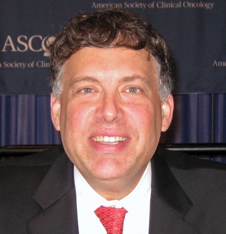
"Prospective identification of patients whose CML will progress despite treatment is not currently possible and the work we have been doing is with PP2A, a tumor suppressor gene," explained Claire M. Lucas, a specialist health care scientist in the department of hematology at the university. Ms. Lucas presented the findings at the annual European Hematology Congress.
PP2A is a serine/threonine phosphatase that down-regulates cell-signaling pathways that lead to tumor cell proliferation, differentiation, and survival. The protein is key target of BCR-ABL1 signaling, and inhibition of the latter via imatinib (Gleevec) is known to increase levels of PP2A, to kick start apoptosis, and to suppress further tumor growth.
Ms. Lucas was part of a team led by Dr. Richard E. Clark that investigated whether the expression of PP2A or its inhibitory proteins SET and CIP2A differed in patients with CML before diagnosis and at disease progression.
The study included 31 patients with CML who were diagnosed in a chronic phase and who were being treated with imatinib within 4 weeks of their presentation. Patients were stratified into three groups based on their clinical response to treatment after 12 months: complete cytogenic response (CCR; n = 14), no CCR (n = 11), and blast crisis (n = 6).
Patients’ peripheral blood was collected before and after treatment for analysis, and evaluated according to their eventual clinical outcome. PP2A protein expression and phosphorylation were found to be increased in cells that had been taken from patients who were seen in blast crisis, Ms. Lucas reported.
CIP2A protein levels at diagnosis were also predictive of patients’ eventual outcome and were found to play a key role in regulating PP2A in CML cells. Chronic-phase patients who had high CIP2A levels at diagnosis had a 100% probability of progressing to blast crisis. Furthermore, CIPA siRNA was found to restore PP2A function and to decrease BCR-ABL tyrosine kinase activity.
Taken together, these data show that CIP2A is not only biologically but also clinically important in CML, Ms. Lucas said.
This is the first time an association between CIP2A and patient outcome has been shown in any hematologic malignancy, the research team believes. CIP2A levels have previously been linked to the aggressiveness of breast and gastric cancers (Clin. Cancer Res. 2009;15:5092-100; Clin. Cancer Res. 2008;14:3722-8; J. Natl. Cancer Inst. 2009;101:793-805).
The study findings have recently been published online (Blood 2011 April 13 [doi:10.1182/blood-2010-08-304477]). Ms. Lucas had no financial disclosures.
LONDON – Measuring levels of a newly described protein at diagnosis could help identify patients that are going to fare worse than others with chronic myeloid leukemia, data from a small study suggest.
Cancerous inhibitor of protein phosphatase 2A (CIP2A) levels were found to be significantly higher in patients with chronic myeloid leukemia (CML) who later went into blast crisis, reported researchers from the University of Liverpool (England). CIP2A could, therefore, represent a novel drug target as well as a biomarker for the disease.
"Prospective identification of patients whose CML will progress despite treatment is not currently possible and the work we have been doing is with PP2A, a tumor suppressor gene," explained Claire M. Lucas, a specialist health care scientist in the department of hematology at the university. Ms. Lucas presented the findings at the annual European Hematology Congress.
PP2A is a serine/threonine phosphatase that down-regulates cell-signaling pathways that lead to tumor cell proliferation, differentiation, and survival. The protein is key target of BCR-ABL1 signaling, and inhibition of the latter via imatinib (Gleevec) is known to increase levels of PP2A, to kick start apoptosis, and to suppress further tumor growth.
Ms. Lucas was part of a team led by Dr. Richard E. Clark that investigated whether the expression of PP2A or its inhibitory proteins SET and CIP2A differed in patients with CML before diagnosis and at disease progression.
The study included 31 patients with CML who were diagnosed in a chronic phase and who were being treated with imatinib within 4 weeks of their presentation. Patients were stratified into three groups based on their clinical response to treatment after 12 months: complete cytogenic response (CCR; n = 14), no CCR (n = 11), and blast crisis (n = 6).
Patients’ peripheral blood was collected before and after treatment for analysis, and evaluated according to their eventual clinical outcome. PP2A protein expression and phosphorylation were found to be increased in cells that had been taken from patients who were seen in blast crisis, Ms. Lucas reported.
CIP2A protein levels at diagnosis were also predictive of patients’ eventual outcome and were found to play a key role in regulating PP2A in CML cells. Chronic-phase patients who had high CIP2A levels at diagnosis had a 100% probability of progressing to blast crisis. Furthermore, CIPA siRNA was found to restore PP2A function and to decrease BCR-ABL tyrosine kinase activity.
Taken together, these data show that CIP2A is not only biologically but also clinically important in CML, Ms. Lucas said.
This is the first time an association between CIP2A and patient outcome has been shown in any hematologic malignancy, the research team believes. CIP2A levels have previously been linked to the aggressiveness of breast and gastric cancers (Clin. Cancer Res. 2009;15:5092-100; Clin. Cancer Res. 2008;14:3722-8; J. Natl. Cancer Inst. 2009;101:793-805).
The study findings have recently been published online (Blood 2011 April 13 [doi:10.1182/blood-2010-08-304477]). Ms. Lucas had no financial disclosures.
FROM THE ANNUAL CONGRESS OF THE EUROPEAN HEMATOLOGY ASSOCIATION
Major Finding: At diagnosis, levels of CIP2A were significantly higher in patients with CML who progressed to blast crisis than in those who did not (P less than .0001).
Data Source: Study of 31 patients with CML treated with imatinib for 12 months.
Disclosures: Ms. Lucas had no financial disclosures.
Brentuximab Benefits Hodgkin's Patients Ineligible for Transplant
LONDON – The investigational agent brentuximab vedotin appears to have a beneficial effect in patients with refractory or relapsed Hodgkin’s lymphoma who are ineligible for or have refused to have an autologous stem cell transplant.
A retrospective analysis of two phase I studies performed with the drug used as a single agent have shown that almost one-third of these heavily pretreated patients are able to achieve an objective response to treatment.
Indeed, 6 of 20 patients (30%) aged a median of 32 years achieved an objective response (two complete and four partial remissions). The response can last longer than 6 months, with one patient achieving a durable remission for more than 2 years that later allowed for transplantation.
"These are very encouraging results in patients with [an] unmet need, and additional studies are ongoing," Dr. Ranjana Advani of Stanford University Medical Center in Palo Alto, Calif., said at the annual congress of the European Hematology Association.
"Patients who have primary refractory disease or fail to achieve a remission at relapse have a dismal outcome," Dr. Advani observed. She added that long-term survival prospects for such patients "were pretty bleak, with median overall survival in a small series as low as 4 months."
Brentuximab vedotin is a novel antibody-drug conjugate that comprises an anti-CD30 monoclonal antibody and a synthetic antimicrotubule agent, monomethyl auristatin E (MMAE). After binding to CD30 on the surface of T and B cells, the antibody-drug conjugate is internalized, the link between antibody and drug is severed, and MMAE is released – thus targeting malignant cells while, in theory, leaving normal cells unscathed.
MMAE is a potent antimicrotubule agent, and as with the taxanes, one of the expected side effects of the drug could be peripheral neuropathy. Although this was not seen in the small number of patients discussed by Dr. Advani, she said that peripheral neuropathy, mostly sensory, had been observed in about 15% of patients treated in the clinical trials program to date.
"Adverse events were seen in over 25% of patients, and these were not necessarily drug related; they could have been even disease related," Dr. Advani reported. Common side effects included fatigue (45%); nausea (40%); pyrexia (35%); decreased weight and diarrhea (30% each); vomiting, back pain, decreased appetite, anemia, and night sweats (25% each).
There were no deaths within 30 days of receiving the last dose of the novel agent.
The current findings add to data released separately from a pivotal phase II trial, recently updated and presented at this year’s American Society of Clinical Oncology meeting in Chicago. In that trial (J. Clin. Oncol. 2011 29[suppl.]: abstract 8031), brentuximab vedotin (SGN-35) induced objective responses in 75% of patients with relapsed or refractory Hodgkin’s disease; 34% achieved a durable complete remission, with two-thirds of patients remaining in complete remission.
Brentuximab has also recently been linked to durable remissions in patients with relapsed or refractory systemic anaplastic large-cell lymphoma (sALCL), an aggressive subtype of peripheral T-cell lymphoma.
The Food and Drug Administration has granted brentuximab vedotin orphan drug status for the treatment of Hodgkin’s lymphoma and sALCL.
Further studies are underway, and include the phase III AETHERA trial – which is comparing brentuximab vedotin to placebo in high-risk patients with Hodgkin’s lymphoma after autologous stem cell transplantation. Another phase I trial is also looking at the combination of brentuximab vedotin and the ABVD (doxorubicin, bleomycin, vinblastine, dacarbazine) regimen as de novo treatment in Hodgkin’s lymphoma.
Seattle Genetics and Millennium: the Takeda Oncology Co. funded the research. Dr. Advani disclosed acting as a principal investigator and receiving research support and advisory board fees from the company.
FROM THE ANNUAL CONGRESS OF THE EUROPEAN HEMATOLOGY ASSOCIATION
Major Finding: Six patients (30%) achieved an objective response (two complete and four partial remissions).
Data Source: Retrospective analysis of two phase I studies of brentuximab vedotin in 20 patients with relapsed or refractory Hodgkin’s lymphoma who were ineligible for or refused autologous stem cell transplantation.
Disclosures: Seattle Genetics and Millennium: the Takeda Oncology Co. funded the research. Dr. Advani disclosed acting as a principal investigator and receiving research support and advisory board fees from the company.
Treat HIV-Related Hodgkin's Like Non-HIV Disease
LONDON – HIV-infected patients with Hodgkin’s lymphoma can be treated more or less the same as any other Hodgkin’s patient, according to new data from the German HIV-related Lymphoma Study Group.
In a prospective, multicenter study of 108 patients with Hodgkin’s lymphoma (HL), all of whom were HIV positive, a risk-adapted treatment strategy was found to be feasible while patients were being treated with HAART (highly active antiretroviral therapy).
"I think the main message is that the prognosis [of patients with HIV-related HL] has dramatically improved with the [use of] HAART and with the stage- and risk-adapted treatment approach," study investigator Dr. Marcus Hentrich said in a June 11 interview at the annual congress of the European Hematology Association, where he presented the study.
"The results are approaching those we have obtained in the HIV-negative population," added Dr. Hentrich of Harlaching Hospital Munich. The findings show that "not every patient needs to be treated with full intensity" for six to eight courses of chemotherapy; rather, "we can distinguish [treatment] depending on the Hodgkin’s stage."
HL is one of the most common non–AIDS-defining cancers that often presents at an advanced stage. To date, there have been few prospective studies looking at how best to treat patients who are both HIV positive and have the hematologic malignancy; indeed, patients with HIV-related HL are often excluded from HL clinical trials. As a consequence, how best to manage such patients remains unknown (Adv. Hematol. 2011 [doi:10.1155/2011/402682]).
Combination therapy regimens have largely been used to treat HL in HIV-infected patients because of the generally late presentation of the disease, and controlling HIV infection via HAART has also been shown to improve the outcome of HL (Ann. Oncol. 2006;17:914-9).
The current study presented by Dr. Hentrich was conducted between March 2004 and October 2010. It included 100 male and 8 female patients with HL who were HIV positive, and its aim was to see whether a risk-adapted treatment strategy that was used in HIV-negative patients with HL could be applied to those infected with HIV.
Treatment for HL was determined by the stage of disease, with two to four cycles of ABVD (doxorubicin, bleomycin, vinblastine, dacarbazine) used with involved-field radiotherapy (30 Gy) in early-stage patients (that is, those with stage I-II HL and no additional risk factors).
In intermediate-stage patients (that is, those with stage I-II disease plus additional risk factors, such as large mediastinal tumor, extranodal involvement, and three or more lymph node regions involved), treatment consisted of four cycles of BEACOPP (bleomycin, etoposide, doxorubicin, cyclophosphamide, vincristine, procarbazine, and prednisone) plus the same radiotherapy regimen.
More advanced HL (stage III-IV) was treated with six to eight cycles of BEACOPP plus facultative radiotherapy (30 Gy initial bulk or rest).
In patients with very advanced HIV infection and a poor performance status, the BEACOPP regimen could be replaced by ABVD, and the dose of ABVD reduced according to individual circumstances.
The median age of recruited patients was 43.9 years (range, 27-70 years). The majority (65%) had advanced disease, with 14% identified as having intermediate-stage HL, and 21% with early-stage disease. Extranodal involvement was observed in about half of patients (54%), and almost two-thirds (65%) had B-symptoms (which includes systemic symptoms such as fever, night sweats, and weight loss; B-symptoms can occur in both HL and non-HL).
Most (77%) patients had received HAART, and the median time from HIV to HL diagnosis was 5.9 years (range, 0-26 years).
After 26 months’ follow-up, 96%, 100%, and 84% of patients with early-, intermediate-, and advanced-stage HL, respectively, were in complete remission.
Perhaps not surprisingly, some advanced-stage patients fared worse, with four (6%) toxic and one (1.4%) unknown cause of death, five (7%) cases of early progression, and one (1.4%) partial remission in this group. One (4%) patient in the early-stage group also died because of toxicity.
Dr. Hentrich pointed out that treatment relapses and failures mainly occurred in patients with advanced disease.
Grade 3/4 toxicity was common and tended to occur in more patients who were treated with BEACOPP than AVBD, but the differences were statistically significantly only in the early-stage patients. The major hematologic toxicity was severe neutropenia.
At 2-years, progression-free and overall survival were 91.7% and 90.2%, respectively, for the whole population, and did not differ greatly between early-, intermediate-, and advanced-stage disease. However, patients with advanced disease and advanced HIV infection were more likely to have a reduced overall survival, compared with the other two groups of patients.
"The next strategy is to focus the amount of intensity to special patients, so we want to incorporate early PET scans after two cycles [of chemotherapy] and then de-escalate therapy or even escalate therapy," Dr. Hentrich said.
Dr. Hentrich had no financial disclosures or conflicts of interest.
FROM THE ANNUAL CONGRESS OF THE EUROPEAN HEMATOLOGY ASSOCIATION
Major Finding: At 2-years, progression-free and overall survival rates were 91.7% and 90.2%, respectively, for the whole population.
Data Source: Prospective, multicenter study of 108 HIV-associated Hodgkin’s lymphoma patients who were treated with chemotherapy according to the stage of hematologic malignancy.
Disclosures: Dr. Hentrich had no financial disclosures or conflicts of interest.
Stopping Dasatinib and Nilotinib Explored in Chronic Myeloid Leukemia
LONDON – Stopping treatment with dasatinib and nilotinib might be safe in chronic myeloid leukemia patients who have achieved sustained and complete molecular responses, early data from a pilot study have shown.
The STOP 2GTKI study is a successor to the STop IMatinib (STIM) trial, which showed that stopping treatment with imatinib – the first-generation tyrosine kinase inhibitor (TKI) of BCR-ABL – could be feasible in patients who achieve long-lasting complete molecular responses (Lancet Oncol.2010;11:1029-35).
Although still preliminary, the new data suggest that it could be possible to do the same with these second-generation TKIs in patients who were previously intolerant or resistant to treatment with imatinib, Dr. Delphine Réa said at the annual Congress of the European Hematology Association.
"There are different kinds of reasons why we should aim at stopping tyrosine kinase inhibitors in chronic myeloid leukemia patients," said Dr. Réa of Hôpital Saint-Louis in Paris.
"The first one relates to patients. From the patients’ perspective it’s very important to have the hope of feeling cured," said Dr. Réa, a member of the French team that performed both the STIM and the STOP 2G-TKI studies.
Other reasons given include avoiding the adverse effects of prolonged therapy on quality of life, and reducing health care costs by steering clear of unnecessary long-term treatment for what becomes a chronic disease. It’s also important for clinicians to be able to show that a treatment really does cure a disease, she said, and the only way to do that is to stop the treatment.
Dr. Réa presented early data on 14 patients who had been enrolled in the STOP 2G-TKI pilot study until May 2011 and for whom there was at least 6 months’ follow-up. The median age of patients was 59 years.
"I think it’s very important to have 6 months’ follow-up at least because in the STop IMatinib trial, most of the relapses occurred within the first 6 months," Dr. Réa explained in an interview after her presentation.
"Here we didn’t have any relapses after 6 months," Dr. Réa added. "For the nine patients who didn’t relapse, the median follow-up is 10 months." The duration of time that these patients remained treatment free was between 6 months and 19 months.
For inclusion in the study, patients had to be aged 18 years or older with chronic or acute phase chronic myeloid leukemia at diagnosis, and be receiving ongoing dasatinib or nilotinib treatment after intolerance or resistance to imatinib therapy. TKI treatment had to have been administered for at least 3 years, and patients needed to have undetectable levels of BCR-ABL with at least 20,000 copies of ABL.
The primary end point was the achievement of a stable major molecular response (MMR) at 6 months, determined by monthly blood counts and real-time quantitative polymerase chain reaction for the first year of discontinuation, then every 2-3 months thereafter. Bone marrow smears and cytogenic and mutational analysis are undertaken if there is a rise in BCR-ABL above an internationally standardized (IS) ratio of 1%. Patients are re-treated with dasatinib or nilotinib if there is a loss of MMR of more than 0.1% IS.
Results showed that at 6 months’ follow-up, 71% of patients had a persistent MMR, and 64% remained treatment free. Five patients restarted TKI treatment at a median of 5 months, and four patients lost MMR after a median of 3 months. However, the patients who lost major molecular response retained their sensitivity to TKIs, and BCR-ABL was undetectable in all five patients who restarted therapy.
Asked what these results could mean for clinical practice, Dr. Réa noted that they are an early indicator that stopping treatment with second-generation TKIs will probably be an option for some patients who achieve good responses. Careful (at least monthly) follow-up of patients who cease treatment would be required, at least in the research setting.
"We don’t know, but we hope the relapse rate will be lower than we have seen with imatinib [discontinuation]," Dr. Réa said. She added, however, that there is an indication that the relapse rate could actually be higher than that seen in the STIM trial.
As yet, the optimal duration of treatment with a second-generation TKI before discontinuation can be attempted is unknown. "In the STop IMatinib trial, it seems to be between 4 and 5 years," Dr. Réa observed. "So I don’t think it will be reasonable to stop treatment before that period of time."
Further results from the trial can be expected later in the year, Dr. Réa noted. To date, 23 patients have been recruited and 8 more are to be included in the trial.
Dr. Réa disclosed serving on advisory boards of Bristol-Myers Squibb and Novartis.
FROM THE ANNUAL CONGRESS OF THE EUROPEAN HEMATOLOGY ASSOCIATION
Major Finding: At 6 months’ follow-up, 71% of patients achieved a stable MMR, and 64% remained treatment free.
Data Source: Preliminary data on 14 patients (median age, 59 years) with chronic myeloid leukemia participating in the ongoing STOP 2G-TKI pilot study.
Disclosures: Dr. Réa disclosed serving on advisory boards of Bristol-Myers Squibb and Novartis.
International Association Favors CT Screening of Heavy Smokers
AMSTERDAM – The International Association for the Study of Lung Cancer has issued a call for physicians to discuss lung cancer screening with patients who match the high-risk smoking history of the people enrolled in the landmark National Lung Screening Trial.
The National Lung Screening Trial (NLST) showed that an annual, low-dose CT chest scan can lead to significant reductions in lung cancer deaths and overall mortality in patients aged 55-74 years who smoked for at least 30 pack-years and, if former smokers, quit within the prior 15 years (New Engl. J. Med. 2011 [doi: 364:10.1056/NEJMoa1102873).
Based on these unprecedented findings, the International Association for the Study of Lung Cancer (IASLC)’s position-writing committee issued a call for physicians to discuss the data and its implications with such patients.
"It is appropriate for heavy smokers ages 55 to 74 to discuss relevant lung cancer screening information with their physicians to assist them in deciding whether to undergo spiral CT screening," said the statement, issued as IASLC started its world conference
Although some committee members, including the chairman, urged caution when routinely discussing screening with the target population before the cost effectiveness of this approach is proven, two of the Americans on the 10-member position-statement writing committee endorsed immediately offering screening to fully informed people who match the study’s screening profile.
"For patients with metastatic lung cancer the cure rate is essentially zero. Finding lung cancer early is the best way to deal with this disease, and that’s why this is such an extraordinary result," said Dr. Roy S. Herbst, a member of the task force and chief of medical oncology at Yale University in New Haven, Conn.
"Even with all the cautions, I think that in the United States at least you’ll see screening, especially since the NLST was largely sponsored by the National Cancer Institute. Assuming that the CMS [Centers for Medicare and Medicaid Services] and insurers will pick this up, I think [screening] is something we’re going to see. I think there will be great pressure in the United States for this to be covered, at a cost of about $300-$400 per scan.
"At Yale, we’ll start screening people who meet the enrollment criteria for the trial, as will several other U.S. centers. We’ll offer screening with all the caveats," including informing patients about the risks they will face from screening, their need to stop smoking, their need for ongoing screening, and the need to have a multidisciplinary team in place at the screening site to deal with all the possible consequences of screening, Dr. Herbst said.
"We know there is effectiveness from screening, but is there cost effectiveness? Is there value?" asked Dr. Richard Gralla, another member of the statement-writing committee and chief of hematology-oncology at North Shore University Medical Center and Long Island Jewish Medical Center in New Hyde Park, N.Y.
"My prediction is that screening will not only be shown to be cost effective, but it will be very cost effective. It will also be very expensive" to run annual screens on the millions of middle-aged smokers who meet the trial’s screening profile, he added. The NLST report estimated that 7 million Americans match the age and smoking history of the people enrolled in the trial.
By thrusting medicine into a new era of routine lung cancer screening, these developments will trigger creation of a new system of quality oversight for lung cancer screening that will likely follow the model of breast cancer screening.
"There is a laundry list of requirements that will need to be established by the institutions that want to do CT screening," said Dr. Denise R. Aberle, professor of radiology at the University of California, Los Angeles, and a collaborator on the NLST. "That will likely evolve into a form of accreditation to better guarantee quality assurance, as with breast cancer screening." Dr. Aberle also noted that the NLST researchers collected cost-effectiveness data, and that they will soon release a report on their analysis of those data.
Routine lung cancer screening will also place new responsibilities on the thoracic surgeons who follow up on suspicious lung lesions found though screening, most of which will not be cancers.
"For surgeons it will be a very large challenge to offer correct treatment to patients with very small cancers," said Dr. Jesper Pedersen, a thoracic surgeon at Copenhagen University Hospital. "We’re planning on writing guidelines for surgeons, because they will be at risk by operating on so many patients without lung cancer." The NLST results showed that 96% of suspicious lesions identified by CT screening were not cancers.
"There is potential for physical and psychiatric harm from cancer screening, but the results from many studies of breast cancer screening have shown that the benefits of screening outweigh its harms," said IASLC president David R. Gandara, professor of medicine and director of the thoracic oncology program at the University of California, Davis, in Sacramento.
"We’re in the early days of screening for lung cancer, and we must do everything to make sure that screening is done appropriately and that follow-up is appropriate. But our message to patients about screening is positive. We can’t overemphasize that," Dr. Gandara said.
Dr. Herbst said that he has been a consultant to Genentech, Agennix, Allos Therapeutics, Boehringer Ingelheim, and OSI Pharmaceuticals. He has been on the advisory boards of Amgen, Biothera, Genetics Squared (now Everist Genomics), MedTrust, N-of-One, SunDev, Targeted Molecular Diagnostics, and DiaTech. He has received research grants from Genentech, Amgen, Bristol-Myers Squibb, AstraZeneca, Novartis, OSI, Oncothyreon, Geron, Sanofi-Aventis, Pfizer, and ImClone.
Dr. Gralla and Dr. Aberle had no relevant disclosures. Dr. Pedersen said that he has been on the speakers bureau for Eli Lilly and Roche and received grant support from AstraZeneca. Dr. Gandara said that he has been a consultant to Amgen, AstraZeneca, Biodesix, Bristol-Myers Squibb/ImClone, GlaxoSmithKline, Genentech, Merck, Novartis, Sanofi-Aventis, and Response Genetics; he has received research support from Abbott, Bristol-Myers Squibb/ImClone, Genentech, Lilly, Merck, Novartis, and Pfizer.
FROM THE WORLD CONFERENCE ON LUNG CANCER
No Change in Statin Usage
A new study that links intensive-dose statin therapy to increased risk of developing diabetes mellitus is unlikely to convince HM or any other specialty to discontinue the treatment, a hospitalist focused on glycemic issues says.
A recent JAMA article reports that a meta-analysis of five clinical trials showed intensive statin regimen was associated with a 12% increased diabetes incidence over moderate-dose regimens (JAMA. 2011;305(24):2556-2564). The authors found that it would take 498 patients taking a statin to cause one extra case of diabetes. Conversely, only 155 people taking a statin would prevent one heart attack.
“The benefits of statins are just too well documented to ignore,” says Steven C. Smith, MD, FHM, medical director of hospitalist services at Healthcare Authority for Medical West in Bessemer, Ala. Quoting a cardiologist colleague, he adds, “statins are so beneficial, there is no way I wouldn’t use them because of a higher risk for diabetes.”
Dr. Smith, who leads the SHM-sponsored Glycemic Control Mentored Implementation program at his hospital, says the research is a formal link for what many hospitalists already realize: patients taking statins because of cardiovascular risk factors usually are at risk for diabetes. He adds that “modifiable risk factors”—including sedentary lifestyle, weight issues and diet—are prevalent in both patient groups. He refers to the JAMA research as “the last stone on the scale that tips the scale.”
“You have to weigh one versus the other,” Dr. Smith says. “The population of people on statins are already at risk for diabetes...you really have to address the risk factors that will solve both problems.”
First Responder
Every May, Mayo Clinic hospitalist Jason Persoff, MD, SFHM, sheds his doctor’s gear, grabs his camera and camcorder, and heads to the Midwest in search of ferocious weather for two weeks. This year, the Jacksonville, Fla.-based physician put his doctor’s gear back on sooner than he expected.
After 20 years of chasing storms, he found himself in what might have been considered a situation that was inevitable: helping people injured in a tornado. When a monstrous twister barreled through Joplin, Mo., last month, Dr. Persoff was less than a mile from its path. He and his “chase partner,” Robert Balogh, MD, an Oklahoma-based internist and former hospitalist, were able to rush in and assist in the aftermath.
One hospital serving the area, St. John’s Regional Medical Center, was destroyed, its roof ripped off by 200 mph winds.
Dr. Persoff checked in at the emergency room of the other one, Freeman Health System, and offered his help. He spent 10 hours there—first treating trauma patients.
“The initial trauma that came in was pretty fast and furious,” he says. “If somebody could be saved, and it wasn’t going to require an effort that would jeopardize resources, they did everything they could to save people.”
There were amputations, impalements, eviscerations. Some patients were covered in glass, he recalls. When the patients from St. John’s began to arrive at Freeman, Dr. Persoff treated them, doing admission orders on 24 patients.
Dr. Persoff plans to continue storm-chasing next year. But he says he’ll never forget the trauma nurse who was working as he arrived at Freeman and was still working as he left the hospital.
“That was one of the times where I was like, ‘Wow, this is really humbling,’ ” he says.
Check out photos and journal entries of Dr. Persoff’s storm-chasing adventures at http://stormdoctor.com/.
Gene Test Refines Prognosis For Lung Cancer
AMSTERDAM – A commercially available gene-expression test significantly improved discrimination between low- and high-risk stage I and IIa lung cancer patients in a pair of validation tests, leading investigators to propose routine use of the test to identify early-stage patients who should get adjuvant chemotherapy.
"The multigene assay can outperform conventional risk factors and staging, and may lead to personalized therapies for patients with early-stage nonsquamous non–small cell lung cancer," Dr. Johannes Kratz said at the World Conference on Lung Cancer.

Dr. Kratz conceded that no prospective, randomized study has yet tested whether identification of high-risk stage I patients singled out a subgroup that would definitely benefit from adjuvant chemotherapy. But the prognostic information that the genetic test already provides justifies its routine use in stage I and II patients, said Dr. Kratz, a surgeon who performed this study while at the University of California, San Francisco (UCSF), but who is now at Massachusetts General Hospital in Boston.
"I think [the test] is certainly ready for prognosis, to give patients information," he said in an interview. "We’ll start using it routinely for prognosis at UCSF. We believe the strength of the results show it’s ready for prime time. Whether it should also be used to guide treatment, especially for stage I patients, is up to each health care provider, but it opens an interesting possibility before anything is proven in a randomized, controlled trial. The hope is that by identifying high-risk patients, you’ll improve their survival by giving them adjuvant chemotherapy. And in some of the low-risk stage II patients, you can avoid some of the toxicities of adjuvant chemotherapy."
Although several different genetic tests for stage I lung cancer have been studied over the past decade, none have wound up as marketed tests. Dr. Kratz and his associates set out to develop a practical and commercially viable test. They worked in collaboration with Pinpoint Genomics, the company that has now begun marketing the test.
The test they developed uses polymerase chain reaction–based gene expression assays for 11 different genes, based on results from prior studies that identified genes critical to key causal pathways leading to lung cancer. "We took a truly blinded, one-shot approach" in putting together the genetic test panel, without any tinkering during the validation phase to boost the prognostic strength of the test, he explained at the conference sponsored by the International Association for the Study of Lung Cancer. They also focused on tests that use paraffin-embedded specimens.
"I don’t think a prospective validation study is needed" before routine prognostic use of the test begins, he said. The validation studies "were done retrospectively, but in a very controlled way that was equivalent to prospective validation. I think we have powerful evidence that these markers provide additional prognostic information. We’re not saying to abandon traditional staging, but this adds useful prognostic information."
The initial test development cohort consisted of 361 stage I, II, and III patients treated and followed at UCSF. Validation used two independent cohorts, 433 stage I patients treated by physicians from Kaiser Permanente of Northern California, and a second cohort of 1,006 patients with stage I, II, or II disease treated at hospitals affiliated with the China Clinical Trials Consortium. Median follow-up in the three cohorts ranged from just over 3 years to just short of 6 years. Five-year mortality was about 42% in each of the three cohorts. About 80% of the nonsquamous non–small cell lung cancer patients in the three cohorts had adenocarcinomas.
The genetic test used to discriminate among three risk levels in the UCSF cohort identified a low-risk group with a calculated 5-year survival of 78%, an intermediate group with a 5-year survival of 60%, and a high-risk group with a survival rate of 30%. Between-group differences were statistically significant (P = .00005). The U.S. and Chinese validation cohorts each led to identification of three very similar prognostic subgroups, "suggesting that the assay was based on principles of lung cancer biology that are fundamental to the disease and remain constant despite the diverse genetic backgrounds of the populations studied," Dr. Kratz said.
In a multivariate analysis that controlled for age, sex, tumor size, and smoking history, high-risk identification using the genetic test led to a near doubling of the mortality risk in the Kaiser cohort (hazard ratio = 1.93, P = .010) and a more than tripling of the mortality risk in the Chinese cohort, compared with the low-risk tertile (HR = 3.25, P less than .001).
On the basis of their findings, Dr. Kratz and his associates proposed a new variation on the conventional tumor size, lymph node status, metastases (TNM) staging system that they called TNMM; the second M stands for multigene assay.
Their revised system designates patients judged stage Ia or Ib by TNM who have a low-risk gene test result as a new class Ia. Patients who had been classified Ia or Ib by the old system who have intermediate- or high-risk gene test results form a new stage Ib, a stage that also includes old IIa patients by TNM who had a low-risk gene test outcome. Finally, the new stage IIa consists of patients scored as IIa by the TNM system who also score as intermediate or high risk on the genetic test.
To further assess the prognostic value of adding the gene test, the researchers ran receiver-operator curves for the standard and revised staging methods in each of the two validation cohorts, and found that adding the gene test led to statistically significant increases in the area under the curve for prognostic accuracy.
In commenting on the study, Dr. Giorgio V. Scagliotti, the designated discussant, said the prognostic factors in current use – tumor size, differentiation, vascular invasion, and surgical margins – are not enough. Additional prognostic factors are needed to identify the completely resected stage I patients who might benefit from adjuvant chemotherapy. "We also need to better identify the stage II patients who have a low risk of recurrence and will not benefit from adjuvant chemotherapy," he said.
Previously reported genetic tests for early-stage non–small cell lung cancer involved complicated microarray test methods and a need for fresh tissue. They lacked reproducibility and validation, and had other problems as well. "The new study avoided these pitfalls, but despite its advances, it remains a set of post hoc analyses that lack prospective, randomized testing. Additional study must prospectively establish the medical utility of the prognostic information before routine use begins. Do patients identified by the test as high risk get any benefit from systemic treatment? Is the genetic test significant in the context of tumor stage, patient age, and treatment with adjuvant chemotherapy? Adjuvant therapy has so far not been included in the analysis," said Dr. Scagliotti, professor of medicine at the University of Torino (Italy).
He said he has been a consultant to Eli Lilly, and has been on the speakers bureaus of AstraZeneca, Eli Lilly, and Roche. Dr. Kratz said that he has been a consultant to and has an equity interest in Pinpoint Genomics, the company that developed the genetic test used in the study.
AMSTERDAM – A commercially available gene-expression test significantly improved discrimination between low- and high-risk stage I and IIa lung cancer patients in a pair of validation tests, leading investigators to propose routine use of the test to identify early-stage patients who should get adjuvant chemotherapy.
"The multigene assay can outperform conventional risk factors and staging, and may lead to personalized therapies for patients with early-stage nonsquamous non–small cell lung cancer," Dr. Johannes Kratz said at the World Conference on Lung Cancer.
Dr. Kratz conceded that no prospective, randomized study has yet tested whether identification of high-risk stage I patients singled out a subgroup that would definitely benefit from adjuvant chemotherapy. But the prognostic information that the genetic test already provides justifies its routine use in stage I and II patients, said Dr. Kratz, a surgeon who performed this study while at the University of California, San Francisco (UCSF), but who is now at Massachusetts General Hospital in Boston.
"I think [the test] is certainly ready for prognosis, to give patients information," he said in an interview. "We’ll start using it routinely for prognosis at UCSF. We believe the strength of the results show it’s ready for prime time. Whether it should also be used to guide treatment, especially for stage I patients, is up to each health care provider, but it opens an interesting possibility before anything is proven in a randomized, controlled trial. The hope is that by identifying high-risk patients, you’ll improve their survival by giving them adjuvant chemotherapy. And in some of the low-risk stage II patients, you can avoid some of the toxicities of adjuvant chemotherapy."
Although several different genetic tests for stage I lung cancer have been studied over the past decade, none have wound up as marketed tests. Dr. Kratz and his associates set out to develop a practical and commercially viable test. They worked in collaboration with Pinpoint Genomics, the company that has now begun marketing the test.
The test they developed uses polymerase chain reaction–based gene expression assays for 11 different genes, based on results from prior studies that identified genes critical to key causal pathways leading to lung cancer. "We took a truly blinded, one-shot approach" in putting together the genetic test panel, without any tinkering during the validation phase to boost the prognostic strength of the test, he explained at the conference sponsored by the International Association for the Study of Lung Cancer. They also focused on tests that use paraffin-embedded specimens.
"I don’t think a prospective validation study is needed" before routine prognostic use of the test begins, he said. The validation studies "were done retrospectively, but in a very controlled way that was equivalent to prospective validation. I think we have powerful evidence that these markers provide additional prognostic information. We’re not saying to abandon traditional staging, but this adds useful prognostic information."
The initial test development cohort consisted of 361 stage I, II, and III patients treated and followed at UCSF. Validation used two independent cohorts, 433 stage I patients treated by physicians from Kaiser Permanente of Northern California, and a second cohort of 1,006 patients with stage I, II, or II disease treated at hospitals affiliated with the China Clinical Trials Consortium. Median follow-up in the three cohorts ranged from just over 3 years to just short of 6 years. Five-year mortality was about 42% in each of the three cohorts. About 80% of the nonsquamous non–small cell lung cancer patients in the three cohorts had adenocarcinomas.
The genetic test used to discriminate among three risk levels in the UCSF cohort identified a low-risk group with a calculated 5-year survival of 78%, an intermediate group with a 5-year survival of 60%, and a high-risk group with a survival rate of 30%. Between-group differences were statistically significant (P = .00005). The U.S. and Chinese validation cohorts each led to identification of three very similar prognostic subgroups, "suggesting that the assay was based on principles of lung cancer biology that are fundamental to the disease and remain constant despite the diverse genetic backgrounds of the populations studied," Dr. Kratz said.
In a multivariate analysis that controlled for age, sex, tumor size, and smoking history, high-risk identification using the genetic test led to a near doubling of the mortality risk in the Kaiser cohort (hazard ratio = 1.93, P = .010) and a more than tripling of the mortality risk in the Chinese cohort, compared with the low-risk tertile (HR = 3.25, P less than .001).
On the basis of their findings, Dr. Kratz and his associates proposed a new variation on the conventional tumor size, lymph node status, metastases (TNM) staging system that they called TNMM; the second M stands for multigene assay.
Their revised system designates patients judged stage Ia or Ib by TNM who have a low-risk gene test result as a new class Ia. Patients who had been classified Ia or Ib by the old system who have intermediate- or high-risk gene test results form a new stage Ib, a stage that also includes old IIa patients by TNM who had a low-risk gene test outcome. Finally, the new stage IIa consists of patients scored as IIa by the TNM system who also score as intermediate or high risk on the genetic test.
To further assess the prognostic value of adding the gene test, the researchers ran receiver-operator curves for the standard and revised staging methods in each of the two validation cohorts, and found that adding the gene test led to statistically significant increases in the area under the curve for prognostic accuracy.
In commenting on the study, Dr. Giorgio V. Scagliotti, the designated discussant, said the prognostic factors in current use – tumor size, differentiation, vascular invasion, and surgical margins – are not enough. Additional prognostic factors are needed to identify the completely resected stage I patients who might benefit from adjuvant chemotherapy. "We also need to better identify the stage II patients who have a low risk of recurrence and will not benefit from adjuvant chemotherapy," he said.
Previously reported genetic tests for early-stage non–small cell lung cancer involved complicated microarray test methods and a need for fresh tissue. They lacked reproducibility and validation, and had other problems as well. "The new study avoided these pitfalls, but despite its advances, it remains a set of post hoc analyses that lack prospective, randomized testing. Additional study must prospectively establish the medical utility of the prognostic information before routine use begins. Do patients identified by the test as high risk get any benefit from systemic treatment? Is the genetic test significant in the context of tumor stage, patient age, and treatment with adjuvant chemotherapy? Adjuvant therapy has so far not been included in the analysis," said Dr. Scagliotti, professor of medicine at the University of Torino (Italy).
He said he has been a consultant to Eli Lilly, and has been on the speakers bureaus of AstraZeneca, Eli Lilly, and Roche. Dr. Kratz said that he has been a consultant to and has an equity interest in Pinpoint Genomics, the company that developed the genetic test used in the study.
FROM THE WORLD CONFERENCE ON LUNG CANCER
Major Finding: Adding a commercially available genetic test to standard TNM staging significantly refined the prognosis of stage I and IIa patients. Patients identified as being at high risk for mortality by the genetic test had a statistically significant, 90% increased risk (P = .010) in a multivariate analysis in one validation cohort, and a threefold increased mortality risk (P less than .001) in the second validation cohort.
Data Source: Validation cohorts of 433 nonsquamous non–small cell lung cancer patients collected by Kaiser Permanente of Northern California and 1,006 similar lung cancer patients collected by the Chinese Clinical Trials Consortium, and a training cohort of 361 similar lung cancer patients collected at UCSF.
Disclosures: Dr. Kratz said that he has been a consultant to and has an equity interest in Pinpoint Genomics, the company that developed the genetic test used in the study.
Supporting Children's Grief within an Adult and Pediatric Palliative Care Program
Children are too often the forgotten mourners in the homes of dying patients. Children, even young children, and youth grieve and mourn the threatened and, then, actual loss of a dying parent, sibling, or other significant family member.1 At a time when the family resources and focus are pulled away and taxed, caregivers are tasked with the difficult job of sorting through their own emotions and a wealth of advice. Caregivers must decide how they will communicate with, include, and support the children/youth in their care.
Although evidence is incomplete and there is a clear need for further studies, links between unresolved childhood grief, or an inability to adequately process their grief, and subsequent psychiatric conditions such as depression and anxiety have been presented as far back as Freud.[2], [3], [4] and [5] In addition, prevalent feelings of responsibility and exclusion and poor communication are consistently identified by researchers interviewing bereaved children/youth about their own experience over the last couple of decades.[6], [7] and [8] Therefore, given the risk of negative psychological and social outcomes associated with children's grief and the struggles communicated by children themselves, it is critical to recognize the important and preventive role of supportive interventions, especially prior to the death of a significant family member.
Looking at caregivers' experiences, there is still a large divide between the advice given by many family and friends in this situation (see Table 1) and what has become accepted within the palliative and grief counseling fields as “best practice.”[6], [7] and [8] In addition, family members' access to professionals trained or knowledgeable in this area is growing but usually still limited.9 Many children/youth are left uninformed, unprepared, and cut off from their family's support.
Table 1. Myths and Realities about Speaking to Children about Grief and Dying. Adapted from MacPherson C.10
Professionals are not immune to subscribing to the myths listed in Table 1 and “are often inhibited by their anxieties about saying or doing the wrong thing and causing lasting emotional damage.”10 However, by communicating openly and honestly and including children/youth, informed care team members can offer many supportive interventions that a family can benefit from during the time leading up to and following the death. These interventions foster the best outcomes when they are offered early on in the palliative trajectory.[11] and [12]
Our Setting
The Temmy Latner Centre for Palliative Care (TLCPC) at Mount Sinai Hospital in Toronto is one of Canada's largest academic palliative care programs, incorporating a children's center, the Max and Beatrice Wolfe Children's Centre, which provides pediatric palliative care and children's grief programs (Dr. Jay Children's Grief Program). Our children's center supports children, youth, and their families when a family member is dying or has died. This support includes Canada's first Camp Erin, an overnight children's grief camp. Children are referred to these programs for grief counseling by our center's palliative care physicians, local palliative care units and hospices, and a wide variety of community agencies. Children's grief programs are very limited in our large urban setting, as is true in most communities across North America. We have four counselors devoted to child and youth grief support services.
What We Do
Action 1: Intake and Assessment
The center has an open referral policy, accepting referrals from any source, including self-referrals, regardless of the nature of the illness or cause of death.
An intake phone call, lasting between 15 and 60 minutes, is made to the family to assess their needs and to provide psychoeducation and relevant resources. Based on our belief that early intervention provides the most supportive opportunities, families in which the patient is dying are prioritized.
As a result of demand for services being greater than our resources, children and their families bereaved at the time of referral are provided with an initial psychoeducational visit and assessment and then placed on a waiting list if further counseling is deemed appropriate.
In either case, the initial assessment often reveals that what the family needs most is psychoeducation about child/youth grief, communication and development, and reassurance about the benefits of the things they are already doing. Caregivers are provided with educational materials, including a copy of the center's publication Living Dying: A Guide for Adults Supporting Grieving Children and Teenagers,11 a list of Web resources, books, and brochures written by us. All these resources are also available to professionals.
Action 2: Counseling
If further assessment and counseling are warranted, children are seen individually, with siblings and/or with their family depending on the needs and circumstances. The bulk of our counseling services are brief, typically lasting three or four sessions in total. However, more intensive counseling is available on a case-by-case basis, with progress and needs being assessed every four sessions. Counseling techniques including expressive arts, crafts, therapeutic play, and activities are used to support children/youth and families in the grieving process. Families are able to contact counselors when issues arise for them, which often occurs around anniversaries, holidays, other important events, and as children/youth develop and experience their grief in a new light.13
The center's model is resiliency-based, nonpathological, and family-centered. Caregivers are empowered as primary and ongoing sources of support and dominant role models for the grieving child and youth in their care. In addition to conversations with their counselor, families are offered monthly opportunities forcaregiver education and peer support and various therapeutic group activities for children/youth.
Action 3: Complex Cases
Referrals to secondary children's mental health services are facilitated for families with needs beyond the scope of our supportive grief services, including children with indicators of complicated grief who need more intensive counseling. For children with severe psychological distress, referrals are made to a pediatric psychiatrist with special interest in this area.
According to Rando,14 there are a number of forms of unresolved or complicated grief which can overlap, and each has components of denial or regression. These include feelings of grief and mourning being absent, an inhibition of some of the normal symptoms of grief, putting grief on hold for any reason, and when there is a dependent or ambivalent relationship with the deceased. Two common manifestations are extreme anger and extreme guilt.
Some types of death that place children/youth at risk for complicated grief include a sudden or unexpected death, a violent death, a death involving mutilation, the death of a child, and death as the result of a prolonged illness. Also included is complicated loss associated with social stigma such as imprisonment, suicide, AIDS, abortion, severe mental illness, serious family dysfunction, or addiction. However, the presence of these factors does not necessarily lead to complicated grief. Complicated loss is known to be mediated by personal, familial, and social factors that contribute to relative risk and resilience.
What We Say
Engaging a family early in the palliative journey allows greater opportunity to prepare children/youth and prevent possible negative outcomes.[6] and [15] Christ and Weisenfluh16 tell us that the greatest need for support is found during the weeks leading up to the death. A large component of the early intervention we offer is age-appropriate psychoeducation to help caregivers conceptualize how their child/youth may be experiencing and understanding what is happening. Caregivers who anticipate some of the thoughts, feelings, questions, and struggles that their child/youth might face are empowered and children benefit.
The 3 Cs
1. Can I catch it?
2. Did I cause it?
3. Who is going to take care of me?
Julie Stokes15 was able to summarize what children/youth think and worry about most when a family member is dying into three questions. We have coined these three questions the “Three Cs”: Catch, Cause, and Care.
Catch
The first “C” relates to the fear expressed by children/youth that they could catch the illness. Melanie, a 7-year-old, explained that she would have a brain tumor soon because her sister Sarah, who was dying of brain cancer at the time, “is my sister and we lived in the same room always.” If such concerns go unexplored and children are not given clear information, caregivers may see children/youth distance themselves from their ill family member, develop a fear that they and the rest of their family will get sick and die as well, and other implications.17
The weight of language in a child's understanding of illness, death, and dying cannot be emphasized enough.11 More often than not, indirect and generic language used by adults, such as referring to someone as being “sick” or “not doing well,” complicates a child's ability to differentiate between the common cold or flu and life-threatening diseases and illnesses. “My mom was sick and she died; therefore, all people that are sick die.” Many adults believe that their child is too young to understand what cancer means. While it is true that children may not be able to grasp the complex medical information about the illness, they are able to understand a great deal more than they are given credit for, and using the word “cancer” gives them a way to distinguish their dying family member's illness from others such as the common cold or flu.
Cause
The second “C” arises from the common thought in children/youth that they somehow caused or hastened the death and/or prevented the recovery of their family member. Cause is one of the five accepted subconcepts of the developmental understanding of death8 and among the last to be mastered due to its complexity and abstract components. As she sat under her pink bunk-bed, 5-year-old Tayah told her counselor that she had cancer when she was a little girl and then her mother got it after her. Eleven-year-old Joshua shared, “I just have to get to that cancer walk thing. If I can do that, then my mom will get her cure. That is what they said on TV you know, to walk for the cure!”
There are a number of ways that children/youth may try to own responsibility for the illness and death of their family member. They may believe that things they thought or did not think, did or did not do, felt or did not feel were directly related to the cause, progression, or death of the family member.18 These thoughts are especially strong for children in the magical thinking stage of development. In Piaget's theory of cognitive development, magical thinking dominates the preoperational stage (2–7 years old) and describes thinking disconnected from the laws of nature.19 Special attention should be given to assuring children in this age group that they are in no way responsible for the illness or death of their family member, as well as to avoid minimizing these fears and beliefs of responsibility.[6] and [17] This point will need to be readdressed often, with frequent reassurance that they are not responsible.
Care
The third “C” includes concerns about what will become of them and who will take care of them as well as the desire to help care for the dying person. While it is commonly thought that this fear is felt more significantly by children/youth who have a dying or deceased parent, siblings of dying or deceased children appear to be just as challenged. They will experience the same break in their belief that their family member will always be with them.20 Also, many children worry that their surviving parent will be incapacitated and unable to care for them after the death. Four-year-old Alex looked up at his mom one night and said, “You are dying in front of me. I'm scared. Who is going to turn out the light, I can't reach it, and who will cook for me, I can't turn on the stove?” This fear extends beyond who will take over the practical parental roles, to the worry that the child or youth will be orphaned.
Children will often connect their own experience to what they have seen or heard and fear the worst.[18] and [21] Orphans are ever present in children's literature and movies and are often depicted as abandoned to fend for themselves. For many, this fear is grounded in the reality that their current caregiver may not have been emotionally able to address issues of custody or guardianship or that they do not have anyone willing or able to care for the kids after their death. As Alice lay on her bedroom floor writing notes to her dying mother in the critical care unit, she shared, “My mommy and my daddy are dying … they is both going to die,” convinced that this was the truth. Alice's dad is healthy and has been for all of Alice's life.
“Three Cs” and Adolescents
Occasionally, adults have questioned the relativity of the “Three Cs” to our adolescent (ages 13–18) population. Our clinical work has led us to conclude that indeed the “Three Cs” are very real in the lives of the youth we work with. An example of this in relation to “Catch” is Sarah, who expressed distress over the familial traits of breast cancer and the likelihood that her fate will one day mirror that of her mother, aunt, and grandmother.
With regard to “Cause,” we hear youth talk about the relationship between the quality of their caregiving efforts and the death of their family member. Many youth express a feeling that if they had done a better job caregiving, their family member would not have died, especially for youth in primary caregiving roles and single-parent homes.14
When looking at “Care,” youth are impacted by their ability to think abstractly and to experience the loss of what is yet to come. Youth understand that they depend on their caregivers for much more than their practical needs. Joshua talked to his counselor about the loss he was feeling as he searched for employment without his father's help and network. He shared, “If my Dad was here, he would know someone; he would know what I should do.” Important to note is that age is not always the best predictor of cognitive capacity as we see school-aged children grapple with many of the thoughts more commonly expressed by our older population.
Whether or not a caregiver has already spoken with a child about a diagnosis and prognosis, it is helpful to get him or her to consider what the child may be thinking about. Using the foundation of the “Three Cs” will help caregivers understand the importance of open, honest, and concrete communication, as well as feel better equipped to provide their child/youth with support.
Teaching Parents How to Communicate with Their Children
It is difficult for a parent to witness the reaction of a child/youth to such difficult information. This challenge cannot be questioned. However, the benefit to children/youth of having their parents lead or participate in communications and psychoeducation about the cancer and terminal prognosis is clear.[6], [7], [8] and [12] Caregivers are able to clarify assumptions and misperceptions while modeling that it is permissible to talk about cancer and dying together.
Medical staff can facilitate opportunities for such family communications by including children in family meetings and discussions with medical staff and by explaining complex concepts to family members in a simple, concrete fashion. Caregivers often need professionals to welcome and encourage the children to be included. Staff can reassure parents that although they may fear that their children's inclusion will be harmful, on the contrary, inclusion is helpful.
Getting Started
1 Ask what the child/youth understands about the disease/illness.
2 Fill in any gaps in their understanding and explain the treatments that were given.
3 Explain cancer treatments such as chemotherapy, radiation, and surgery.
4 Ask the child/youth what he or she thinks is going to happen.
5 Explain in terms that can be understood that the family member will die.
In any conversation with children/youth about such complex and consequential material, follow their lead in regard to the amount of detail they want. Conceptualize it as an onion: You will want to provide the child/youth with clear and simple language describing basic information. Beyond that, peel off subsequent layers as the child/youth requests more information, always using clear and concrete language. The child/youth may be uncomfortable with the material and may attempt to change the topic. This is a healthy coping mechanism; the average child is quite good at knowing when he or she has reached the limit of their emotional attention span. Allow the child to take the conversation in a different direction, periodically checking to see if he or she is ready to come back to the topic at hand. The benefit of starting this course of conversation early is that it allows children/youth the chance to receive information in small, digestible pieces and the time to process and integrate the information to begin to make meaning of it.
Summary
“What about the kids?” is a dominant and consuming question for caregivers supporting children/youth around the dying and death of a family member. The concerns and fears encompassed in this question can overwhelm caregivers as they put vast amounts of energy into trying to protect children/youth from the suffering and pain that awaits them. Perhaps the hardest lesson these caregivers must learn is that they cannot protect their child/youth from the death any more than they can stop the death from happening. Instead, what is needed most from children/youth is to be included, prepared, and provided with a safe place for emotional expression. Children, as well as adults, will grieve in their own specific way, mediated by their developmental level, circumstances of the illness and death, and protective factors available to them. Providing comprehensive, whole-person care to palliative patients with children/youth in their care ought to include psychoeducation and support for all members of the family. Our communities' bereaved children/youth will be impacted by the death of their family members in countless ways throughout their lives. As professionals caring for people who are dying, we have a responsibility to mediate this impact to the best of our ability.
References1
1 J. Bowlby, Pathological mourning and childhood mourning, J Am Psychoanal Assoc 11 (1963), pp. 500–541. Full Text via CrossRef | View Record in Scopus | Cited By in Scopus (48)
2 L. Dowdney, Annotation: childhood bereavement following parental death, J Child Psychol Psychiat 41 (7) (2000), pp. 819–830. Full Text via CrossRef | View Record in Scopus | Cited By in Scopus (68)
3 K. Kirwin and V. Hamrin, Decreasing the risk of complicated bereavement and future psych disorders in children, J Child Adolesc Psychiat Nurs 18 (2) (2005), pp. 62–78. View Record in Scopus | Cited By in Scopus (10)
4 L. Saler and N. Skoinick, Childhood parental death and depression in adulthood: roles of surviving parent and family environment, Am J Orthopsychiatry 62 (4) (1992), pp. 504–516. Full Text via CrossRef | View Record in Scopus | Cited By in Scopus (35)
5 G. Mireault and L. Bond, Parental death in childhood: perceived vulnerability, and adult depression and anxiety, Am J Orthopsychiatry 62 (4) (1992), pp. 517–524. Full Text via CrossRef | View Record in Scopus | Cited By in Scopus (28)
6 G. Christ and A. Christ, Current approaches to helping children cope with a parent's terminal illness, CA Cancer J Clin 56 (2006), pp. 197–212. Full Text via CrossRef | View Record in Scopus | Cited By in Scopus (13)
7 P.R. Silverman, Never Too Young to Know: Death in Children's Lives, Oxford University Press, New York (2000).
8 J.W. Warden, Children and Grief: When a Parent Dies, Guilford Press, New York (1996).
9 L. Dowdney, R. Wilson, B. Maughan, M. Allerton, P. Schofield and D. Skuse, Psychological disturbance and service provision in parentally bereaved children: prospective case–control study, BMJ 319 (7206) (1999), pp. 354–357. View Record in Scopus | Cited By in Scopus (25)
10 C. MacPherson, Telling children their ill parent is dying: a study of the factors influencing the well parent, Mortality 10 (2) (2005), pp. 113–120.
11 C. Eaton-Russell, Living Dying: A Guide for Adults Supporting Grieving Children and Teenagers, Temmy Latner Centre for Palliative Care, Toronto (2007).
12 D. Black, Childhood bereavement: distress and long term sequelae can be lessened by early intervention, BMJ 312 (1996), p. 1496. View Record in Scopus | Cited By in Scopus (14)
13 S. Leighton, Bereavement therapy with adolescents—facilitating a process of spiritual growth, J Child Adolesc Psychiatr Nurs 21 (1) (2008), pp. 24–34. Full Text via CrossRef | View Record in Scopus | Cited By in Scopus (4)
14 T. Rando, Grief, Dying, and Death: Clinical Interventions for Caregivers, Research Press, Champaign, IL (1984).
15 J. Stokes, Anticipatory grief in families affected by HIV/AIDS, Prog Palliat Care 2 (1994), pp. 43–48.
16 C. Christ and S. Weisenfluh, Parent and child bereavement. In: D. Walsh, Editor, Palliative Medicine (1st ed.), Saunders, Philadelphia (2008).
17 L. Kroll, J. Barnes, A.L. Jones and A. Stein, Cancer in parents: telling children, BMJ 316 (1998), p. 880. View Record in Scopus | Cited By in Scopus (22)
18 J. Piaget, Piaget's theory. In: P. Mussen, Editor, Handbook of Child Psychology (4th ed.), John Wiley & Sons, New York (1970), pp. 703–732.
19 M. Van Riper, Death of a sibling: five sisters, five stories, Pediatr Nurs 23 (6) (1997), pp. 587–593.
20 F. Thompson and S. Payne, Bereaved children's questions to a doctor, Mortality 5 (1) (2000), pp. 74–96. Full Text via CrossRef
21 C.M. Burns, T.W. LeBlanc, A. Abernethy and D. Currow, Young caregivers in the end-of-life setting: a population-based profile of an emerging group, J Palliat Med 13 (10) (2010), pp. 1225–1235. Full Text via CrossRef | View Record in Scopus | Cited By in Scopus (0)