User login
Federal Grant Supports "eHospitalist" Pilot Program in Wisconsin
John Almquist, MD, FHM, director of hospitalist services for Ministry Health Care, a 15-hospital system serving rural Wisconsin, believes that an "e-hospitalist" pilot project now being tested at Ministry St. Mary's Hospital in Rhinelander, Wis., could be a boon for rural communities that have difficulty recruiting primary-care physicians (PCPs).
When the hospitals in those communities are unable to offer hospitalist coverage, it makes the setting less attractive to PCPs because they might have to follow their patients in the hospital day and night, he explains.
Ministry recruited and trained two nurse practitioners who will soon be deployed at a critical-access hospital in Eagle River, population 1,443, supported remotely by the eight-member HM group in Rhinelander for consultations, supervision, and multidisciplinary rounds. The training is bolstered by written order sets focused on 30 common medical conditions that lead to admissions to rural hospitals.
"The hospitalist in Rhinelander is also able to talk directly to the patient at the remote site," Dr. Almquist says.
The e-hospitalist program uses a telehealth network developed by Marshfield Clinic, a multispecialty physician group practice based in Marshfield, Wis. The clinic recently received a $1 million grant from the federal government to expand its 15-year-old telemedicine program. Part of the grant money is being used to expand the ehospitalist approach to new sites.
Visit our website for more information about hospitalists and telemedicine.
ITL: Physician Reviews of HM-Relevant Research
Clinical question: Does the addition of clopidogrel to aspirin reduce the risk of any type of recurrent stroke, or affect the risk of bleeding or death, in patients who recently suffered a lacunar stroke?
Background: There are no prior randomized, multicenter trials on secondary prevention of lacunar stroke; aspirin is the standard antiplatelet therapy in this setting.
Study design: Double-blind, randomized, multicenter trial.
Setting: Eighty-two clinical centers in North America, Latin America, and Spain.
Synopsis: Researchers enrolled 3,020 patients from 2003 to 2011; criteria included age >30 years old and symptomatic lacunar stroke (proven by MRI) in the preceding 180 days.
Results showed no significant difference between recurrent strokes (any type) in the aspirin-only group (2.7% per year) versus the aspirin-plus-clopidogrel group (2.5% per year). Major hemorrhage risk was much higher in the aspirin-plus-clopidogrel group (2.1% per year) versus aspirin-only group (1.1% per year). All-cause mortality also was much higher in the aspirin-plus-clopidogrel group (N=113) versus the aspirin-only group (N=77).
Bottom line: The addition of clopidogrel to aspirin for secondary prevention does not significantly reduce the risk of recurrent stroke, but it does significantly increase the risk of bleeding and death.
Citation: Benavente OR, Hart RG, McClure LA, et al. Effects of clopidogrel added to aspirin in patients with recent lacunar stroke. N Engl J Med. 2012;367:817-825.
For more physician reviews of recent HM-relevant literature, visit our website.
Woman with “Dull, Achy” Back Pain and Shortness of Breath
ANSWER
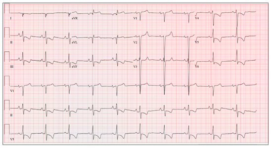
This ECG demonstrates normal sinus rhythm, right-axis deviation, evidence of a lateral MI, and inferolateral ST- and T-wave abnormalities.
Right-axis deviation is indicated by an R-wave axis between 90° and 180° and QS or QR complexes in lead I and/or aVL. While the most common cause of a right-axis deviation is right ventricular hypertrophy, it is also evident in a lateral MI. Evidence for the latter includes the presence of significant Q waves in leads I and aVL. Finally, inferolateral ST- and T-wave changes are evidenced by inverted T waves in leads II, III, aVF, and precordial leads V4 to V6.
ECG evidence of a lateral MI not present on a previous scan (eight months ago), in the presence of a normal troponin level, suggests a recent MI.
ANSWER
This ECG demonstrates normal sinus rhythm, right-axis deviation, evidence of a lateral MI, and inferolateral ST- and T-wave abnormalities.
Right-axis deviation is indicated by an R-wave axis between 90° and 180° and QS or QR complexes in lead I and/or aVL. While the most common cause of a right-axis deviation is right ventricular hypertrophy, it is also evident in a lateral MI. Evidence for the latter includes the presence of significant Q waves in leads I and aVL. Finally, inferolateral ST- and T-wave changes are evidenced by inverted T waves in leads II, III, aVF, and precordial leads V4 to V6.
ECG evidence of a lateral MI not present on a previous scan (eight months ago), in the presence of a normal troponin level, suggests a recent MI.

A 70-year-old woman has a 10-year history of a dilated nonischemic cardiomyopathy and New York Heart Association Class II heart failure. She presents with a one-week history of back pain and shortness of breath. She describes the pain as a “dull, achy” pressure, exacerbated by exertion and relieved with rest. She says the pain is localized in the back between her scapulas and does not radiate. She denies substernal chest pain, nausea, vomiting, or diaphoresis; the only associated symptom is dyspnea. Her most recent echocardiogram showed a dilated left ventricle, with a left ventricular ejection fraction of 29%, and a normal right ventricle, with mild hypertrophy and mildly reduced systolic function. She was also noted to have atherosclerotic changes in her ascending and descending thoracic aorta. Medical history is remarkable for diabetes, hypertension, chronic renal insufficiency, hyperlipidemia, and cataracts. Her current medications include aspirin, fer-rous sulfate, furosemide, hydralazine, glargine insulin, isosorbide dinitrate, lisinopril, metoprolol, and raloxifene. She is allergic to codeine, amiodarone, and radi-ographic contrast. Family history is positive for coronary artery disease, diabetes, and stroke. The patient is widowed, does not smoke, and does not consume alcohol. She is very active in her local quilting club. The review of systems is positive for increased weakness and diarrhea. She states that approximately two weeks ago, she experienced vague epigastric pain and diaphoresis; she did not seek medical attention, as it resolved. The physical exam reveals a thin, elderly woman in mild distress. Blood pressure is 139/82 mm Hg; pulse, 66 beats/min; respiratory rate, 21 breaths/¬min-1; and temperature, 35.9°C. Her weight is 108 lb. Pertinent physical findings include a grade II/VI diastolic murmur at the left lower sternal border, 2+ peripheral pulses with a bruit present in the right femoral artery, occasional late expiratory wheezes in both lung bases, vertebral tenderness at the T6-T7 level with no evidence of scoliosis or kyphosis, and no evidence of peripheral edema. She is intact from a neurologic standpoint. Significant laboratory data include a serum glucose level of 294 mg/dL; blood urea nitrogen (BUN), 68 mg/dL; creatinine, 1.75 mg/dL; glomerular filtration rate, 30 mL/min; B-type natriuretic peptide, 984 pg/mL; and serum troponin, 0.11 ng/mL. An ECG is obtained that reveals the following: a ventricular rate of 62 beats/min; PR interval, 160 ms; QRS duration, 94 ms; QT/QTc interval, 404/410 ms; P ax-is, 84°; R axis, 151°; and T axis, 253°. What is your interpretation of this ECG?
Topical Steroids: the Solution or the Cause?
ANSWER
The correct answer is all of the above (choice “d”). Prolonged injudicious use of topical steroids can cause a number of problems, including these; they are collectively termed iatrogenic since they are ultimately caused by prescribed medication. One of the more difficult aspects of this problem to deal with is the “addictive” state, in which withdrawal symptoms compel the patient to continue applying the offending steroid cream.
DISCUSSION
This is a relatively common scenario in dermatology offices. The misuse of topical steroids is well known, and something we strive to prevent—but with mixed results. It’s one of the reasons we’re stingy with refills of such medications, requiring the patient to be seen at least once a year. Unfortunately, this patient had been getting “refills” from friends in Mexico; patients often “borrow” steroid creams from household members or friends, or use products prescribed for one condition to treat others for which they were not intended.
The primary mode of action of topical steroids is vasoconstriction, a positive thing in terms of reduction of inflammation. The bad news is that continuous use of class 1 (the most powerful) steroids, such as clobetasol, can cause such profound and prolonged vasoconstriction that the skin effectively loses its blood supply and withers, sometimes down to adipose tissue. As one might suspect, this is more likely in already thin-skinned areas, including the antecubital area, face, neck, eyelids, and genitals, where the creation of striae is especially common.
Fairly early on in this process, before frank atrophy occurs, the condition being treated usually resolves. However, when the steroid is stopped, stinging and itching immediately return—which, of course, causes the patient to reapply the medication, perpetuating the vicious cycle.
The cycle is ultimately broken by gradual reduction in the frequency of application of successively weaker steroids. Usually, the skin gradually regenerates and returns to normal. In this case, the process will be lengthy and will almost certainly result in significant scarring.
Even injudicious application of weaker classes of steroids (eg, hydrocortisone 2.5% cream) to areas such as the face can result in a range of deleterious effects, including localized rosacea-like eruption or erythema. It has been reported that approximately 75% of cases of perioral dermatitis are either caused by or exacerbated by the application of topical steroids.
Topical application of even mid-strength steroids can also have systemic effects (eg, adrenal suppression, hyperglycemia) if applied over large areas. This is especially true when pediatric patients are involved.
Prevention of these iatrogenic effects lies in selecting the lowest strength steroid for the condition and area in question, then using them sparingly: no more than twice a day, and for no more than five days in a row, stopping for two consecutive days to allow the skin to regenerate. Even more caution should be exercised in treating children and when applying the product to intertriginous areas (skin-on-skin areas, such as the groin, in axillae, or under the breasts). Covering steroid-treated areas with anything—bandages, socks, even skin—effectively potentiates the positive and negative effects of steroids.
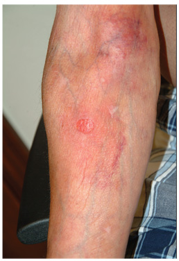
A 59-year-old man presents with skin changes on both antecubital areas. For more than a year, he has applied clobetasol 0.05% cream at least twice daily to the area ostensibly for treatment of long-standing eczema, which has affected not only the antecubital areas but also the patient’s legs. In addition to the eczema, he has a history of atopy, marked by seasonal allergies and asthma. He notes that his stress level has increased in the past several months, which he suspects has contributed to his itching. On examination, marked epidermal atrophy is seen in both antecubital areas, along with extensive purpura. Surface adnexal structures, such as hair, follicles, and skin lines, are sparse at best, but dermal and subdermal vasculature are readily visible. In the midst of the affected area on the right arm, a nickel-sized, full-thickness defect is noted. Beneath it, adipose tissue can be seen. Clearly, these changes are attributable to the effects of the clobetasol, which the patient is advised to stop. But he replies that when he does, the treated areas burn and itch even more, until he obtains relief by applying more clobetasol.

Cold weather and diarrhea: Don't forget yersiniosis
The genus Yersinia includes 11 species. Three species are generally associated with human disease; Y. enterocolitica, Y. pestis, and Y. pseudotuberculosis. Yersinia pestis is the causative agent of plague. Yersinia pseudotuberculosis can manifest with fever, abdominal pain, and scarlatiniform rash. Additional symptoms include diarrhea, sterile joint effusions, erythema nodosum, and septicemia; these symptoms can be indistinguishable from Kawasaki Disease. By report, almost 10 % of cases of Kawasaki Disease in Japan have serologic or bacteriologic evidence of Y. pseudotuberculosis infection [Redbook: 2012 Report of the Committee on Infectious Diseases, 795-7]. Y. enterocolitica is most often associated with yersiniosis.
Although Y. enterocolitica is not the most common cause of diarrheal illness in the United States, it is one of the nine pathogens that have been monitored by the Foodborne Diseases Active Surveillance Network (FoodNet) since 1996. In the United States, it is estimated that Y. enterocolitica causes slightly over 115,000 infections annually (Emerg. Infect. Dis. 2011;17:7-15). The disease is more common in cooler months. It is transmitted by consumption of contaminated food, especially raw or undercooked pork products.

Only a few outbreaks have been reported in the United States, and these were usually associated with consumption of pork, specifically chitterlings (pig intestines), a winter holiday dish prepared most frequently in black households in the South (MMWR 1990;39:819-20). Transmission to infants and young children is thought to occur from caretakers preparing chitterlings who have not adequately cleaned their hands prior to touching objects subsequently handled by the child.
The incubation period is usually 4-6 days (range, 1-14 days). The duration of diarrhea is variable and can persist up to 3 weeks. Organisms can be excreted an average of 6 weeks. Clinical manifestations vary by age. Younger children usually present with fever and diarrhea. Stools frequently contain blood and leucocytes. Vomiting is also reported in most series. In contrast, older children and adults often present with a pseudoappendicitis syndrome with right-sided abdominal pain and fever. Leukocytosis is often present. At surgery, mesenteric adenitis is observed, and the appendix generally is normal.
Bacteremia can occur and is usually associated with infection in children less than 1 year of age and in those with iron-overloaded states, including persons with sickle cell disease, beta-thalassemia, and those receiving deferoxamine therapy. While uncommon, focal manifestations including pharyngitis, osteomyelitis, pyomyositis, pneumonia, empyema, and meningitis may occur.

Diagnosis is confirmed by isolation of the organism from stool, blood, peritoneal fluid, lymph nodes, and throat cultures. Most laboratories do not routinely test for Yersinia in stool cultures. If Y. enterocolitica is suspected, you should notify the laboratory so the stool can be plated on appropriate media (CIN agar). Serologic tests to detect a rise in serum antibody titers to confirm infection are available in reference and research laboratories, but are not generally used for diagnosis. Cross reactivity with Brucella, Salmonella, Vibrio, and Rickettsia may lead to false positive titer results. Y. enterocolitica antibodies also have antigenic similarity with thyroid tissue. You may see persistent elevation of titers in patients with thyroid disease.
Benefit of antimicrobial therapy for isolated Y. enterocolitica gastrointestinal disease and Y. pseudotuberculosis has not been established. Therapy may decrease the duration of fecal shedding. Treatment is indicated for immunocompromised hosts and persons with septicemia and focal infections. Y. enterocolitica and Y. pseudotuberculosis are usually sensitive to trimethoprim-sulfamethoxazole, aminoglycosides, cefotaxime, fluoroquinolones (persons greater than 18 years of age or older), and tetracycline or doxycycline (for children at least 8 years of age and older).
So what is the actual incidence and when should the practitioner be concerned? Initial population based surveillance data for Y. enterocolitica infections in FoodNet sites between 1996 and 1999 reported an overall incidence of 0.9 cases per 100,000 population. The highest incidence was among black and Asian individuals and was 3.2 cases and 1.5 cases per 100,000 population, respectively. The incidence in Hispanics and whites was 0.6 and 0.4 cases per 100,000 respectively. Incidence increased with decreasing age in all racial/ethnic groups. Blacks infants had the highest incidence, 141.9 cases/100,000 population, and the highest incidence in infants was reported from Georgia (207 cases/100,000). Seasonal variation in incidence was noted only in black individuals with peak activity occurring in December (Clin. Infect. Dis. 2004;38[Suppl 3]:S181-9).

The most recent data from FoodNet (1996-2009) reveals an overall incidence of 0.5/100,000. There was a decline in incidence in all racial and ethnic groups. The highest incidence is still observed in black and Asians (0.9 and 0.7 per 100,000). The most dramatic decline occurred in black individuals (3.2 vs. 0.9 per 100,000). In 1998, an educational campaign was initiated in Georgia that targeted high-risk individuals and provided information on the safe handling and preparation of chitterlings. The state of Georgia reported the greatest decline to 0.4/100,000, which has almost eliminated the racial disparity reported in 2009. It is unclear if this campaign was the only reason for the decline in Georgia. The incidence in whites is 0.2/100,000. Since 2007, the incidence in Asian children less than 5 years of ages has been the highest amongst all racial and ethnic groups. Pork consumption is still assumed to be the major source. Seasonal variability persists amongst Black children less 5 years of age, implying that chitterlings may still be the source of infection for individuals in this group (Clin. Infect. Dis. 2012:54 [Suppl 5]:S385-S90).
In general, yersiniosis should be included in the differential of a febrile diarrheal illness, particularly during the cooler months and holiday season. It is prudent to determine if consumption and/or preparation of chitterlings or other pork products by the patient or caretakers has occurred. This will enable you to alert the laboratory so stool specimens can be cultured on the appropriate medium (CIN agar). Consumption of chitterlings is not limited to any specific racial or ethnic group. Individuals from rural and farming areas may also consume this product.
Dr. Word is a pediatric infectious disease specialist and director of the Houston Travel Medicine Clinic. She said she had no relevant financial disclosures. Write to Dr. Word at [email protected].
The genus Yersinia includes 11 species. Three species are generally associated with human disease; Y. enterocolitica, Y. pestis, and Y. pseudotuberculosis. Yersinia pestis is the causative agent of plague. Yersinia pseudotuberculosis can manifest with fever, abdominal pain, and scarlatiniform rash. Additional symptoms include diarrhea, sterile joint effusions, erythema nodosum, and septicemia; these symptoms can be indistinguishable from Kawasaki Disease. By report, almost 10 % of cases of Kawasaki Disease in Japan have serologic or bacteriologic evidence of Y. pseudotuberculosis infection [Redbook: 2012 Report of the Committee on Infectious Diseases, 795-7]. Y. enterocolitica is most often associated with yersiniosis.
Although Y. enterocolitica is not the most common cause of diarrheal illness in the United States, it is one of the nine pathogens that have been monitored by the Foodborne Diseases Active Surveillance Network (FoodNet) since 1996. In the United States, it is estimated that Y. enterocolitica causes slightly over 115,000 infections annually (Emerg. Infect. Dis. 2011;17:7-15). The disease is more common in cooler months. It is transmitted by consumption of contaminated food, especially raw or undercooked pork products.
Only a few outbreaks have been reported in the United States, and these were usually associated with consumption of pork, specifically chitterlings (pig intestines), a winter holiday dish prepared most frequently in black households in the South (MMWR 1990;39:819-20). Transmission to infants and young children is thought to occur from caretakers preparing chitterlings who have not adequately cleaned their hands prior to touching objects subsequently handled by the child.
The incubation period is usually 4-6 days (range, 1-14 days). The duration of diarrhea is variable and can persist up to 3 weeks. Organisms can be excreted an average of 6 weeks. Clinical manifestations vary by age. Younger children usually present with fever and diarrhea. Stools frequently contain blood and leucocytes. Vomiting is also reported in most series. In contrast, older children and adults often present with a pseudoappendicitis syndrome with right-sided abdominal pain and fever. Leukocytosis is often present. At surgery, mesenteric adenitis is observed, and the appendix generally is normal.
Bacteremia can occur and is usually associated with infection in children less than 1 year of age and in those with iron-overloaded states, including persons with sickle cell disease, beta-thalassemia, and those receiving deferoxamine therapy. While uncommon, focal manifestations including pharyngitis, osteomyelitis, pyomyositis, pneumonia, empyema, and meningitis may occur.
Diagnosis is confirmed by isolation of the organism from stool, blood, peritoneal fluid, lymph nodes, and throat cultures. Most laboratories do not routinely test for Yersinia in stool cultures. If Y. enterocolitica is suspected, you should notify the laboratory so the stool can be plated on appropriate media (CIN agar). Serologic tests to detect a rise in serum antibody titers to confirm infection are available in reference and research laboratories, but are not generally used for diagnosis. Cross reactivity with Brucella, Salmonella, Vibrio, and Rickettsia may lead to false positive titer results. Y. enterocolitica antibodies also have antigenic similarity with thyroid tissue. You may see persistent elevation of titers in patients with thyroid disease.
Benefit of antimicrobial therapy for isolated Y. enterocolitica gastrointestinal disease and Y. pseudotuberculosis has not been established. Therapy may decrease the duration of fecal shedding. Treatment is indicated for immunocompromised hosts and persons with septicemia and focal infections. Y. enterocolitica and Y. pseudotuberculosis are usually sensitive to trimethoprim-sulfamethoxazole, aminoglycosides, cefotaxime, fluoroquinolones (persons greater than 18 years of age or older), and tetracycline or doxycycline (for children at least 8 years of age and older).
So what is the actual incidence and when should the practitioner be concerned? Initial population based surveillance data for Y. enterocolitica infections in FoodNet sites between 1996 and 1999 reported an overall incidence of 0.9 cases per 100,000 population. The highest incidence was among black and Asian individuals and was 3.2 cases and 1.5 cases per 100,000 population, respectively. The incidence in Hispanics and whites was 0.6 and 0.4 cases per 100,000 respectively. Incidence increased with decreasing age in all racial/ethnic groups. Blacks infants had the highest incidence, 141.9 cases/100,000 population, and the highest incidence in infants was reported from Georgia (207 cases/100,000). Seasonal variation in incidence was noted only in black individuals with peak activity occurring in December (Clin. Infect. Dis. 2004;38[Suppl 3]:S181-9).
The most recent data from FoodNet (1996-2009) reveals an overall incidence of 0.5/100,000. There was a decline in incidence in all racial and ethnic groups. The highest incidence is still observed in black and Asians (0.9 and 0.7 per 100,000). The most dramatic decline occurred in black individuals (3.2 vs. 0.9 per 100,000). In 1998, an educational campaign was initiated in Georgia that targeted high-risk individuals and provided information on the safe handling and preparation of chitterlings. The state of Georgia reported the greatest decline to 0.4/100,000, which has almost eliminated the racial disparity reported in 2009. It is unclear if this campaign was the only reason for the decline in Georgia. The incidence in whites is 0.2/100,000. Since 2007, the incidence in Asian children less than 5 years of ages has been the highest amongst all racial and ethnic groups. Pork consumption is still assumed to be the major source. Seasonal variability persists amongst Black children less 5 years of age, implying that chitterlings may still be the source of infection for individuals in this group (Clin. Infect. Dis. 2012:54 [Suppl 5]:S385-S90).
In general, yersiniosis should be included in the differential of a febrile diarrheal illness, particularly during the cooler months and holiday season. It is prudent to determine if consumption and/or preparation of chitterlings or other pork products by the patient or caretakers has occurred. This will enable you to alert the laboratory so stool specimens can be cultured on the appropriate medium (CIN agar). Consumption of chitterlings is not limited to any specific racial or ethnic group. Individuals from rural and farming areas may also consume this product.
Dr. Word is a pediatric infectious disease specialist and director of the Houston Travel Medicine Clinic. She said she had no relevant financial disclosures. Write to Dr. Word at [email protected].

Is the Relational Approach to Diagnosis Possible or Desirable?
The American Family Therapy Academy recently issued a policy statement protesting the DSM-5, and asks the American Psychiatric Association to consider the importance of relational and family context to psychiatric diagnoses.
AFTA, a multidisciplinary group, does not support the current revision of the DSM, stating that it "continues a long history of ignoring research and excluding vital contributions of nonpsychiatric mental health disciplines." This statement refers to the substantial body of research concerning the role of relational factors in mental health and mental illness, and also refers to the large number of effective family treatments, including, but not limited to, family therapy.
The academy criticizes the DSM’s use of the biomedical model and its omission of the role of family and sociocultural contexts on well-being. AFTA states that the DSM "delegitimizes the focus on relationship, life stage, community, and access to power and resources." AFTA points out that the DSM fails to take into account culture, class and ‘destructive unjust social factors,’ such as poverty, hunger, homelessness, violence, racism, and other forms of oppression. AFTA considers these factors to be important in reaching a diagnosis that accurately describes patients.
Many psychiatrists, especially family, social, and cultural psychiatrists, agree with AFTA’s position. Several family researchers and family psychiatrists have been pushing for many years to get relational diagnoses included in the DSM-IV and the DSM-5 (J. Fam. Psychol. 2006;20:359-68), citing decades of excellent research into relational diagnoses. Their attempts are supported by nonmedical health care professionals who complain that they cannot get paid by insurance companies for treating families. However, putting any diagnosis in the DSM so the insurance companies get paid is a backward way of thinking. Any diagnostic system of American psychiatry should not be framed or influenced by financial organizations that want to ration health care.
Some psychiatrists who contributed to the DSM offer the disclaimer that "they do not mean this to be a bible." However, the DSM is frequently used "as a bible," for example, in the courts. More importantly, reductionist diagnostic descriptions in the DSM narrow the public’s and the professionals’ thinking about psychological difficulties, and, by extrapolation, limit the conceptualization of what types of interventions might be helpful.
For example, describing psychiatric illnesses as biological leads to the assumption that biological interventions are needed. If an illness is defined using a biopsychosocial explanation, however, this broader understanding leads to a wider array of possible treatments. A psychiatric diagnostic system should recognize all the factors that are known to contribute to psychological health and illness to be of most use in patient care.
There is also a strong argument for not including relational diagnoses in the DSM. The argument is this: Relational factors are process factors, rather than static factors. For example, expressed emotion (EE) is not a characteristic of a family but rather a description of family distress that arises as a result of living with a disease. It is a description of a family process. Providing psychoeducation to a distressed family substantially reduces the level of EE and the subsequent risk of patient relapse. EE is a measure of relational process. If EE is entered into the DSM, there is a danger of its being seen as a static entity.
A delicate balance exists between the utilitarian need for a system of diagnoses and the risk of overdefining people and their relationships as "pathological." It was not that long ago that we pathologized homosexuality and described the entity of the "schizophrenogenic mother."
Dr. Larry Freeman, a member of the Association of Family Psychiatrists, adds: "Be wary of a pressure beyond medical circles to utilize psychiatry as a force for social control. I do a great deal of workers’ [compensation], and so-called ‘preexisting conditions’ are commonly framed as the "cause" of a worker’s emotional response to injury, and therefore, [the worker’s] current psychiatric conditions are not accepted as a consequence of the original injury event.
"Be careful that we do not enable this distortion further in our efforts to include context and history."
How should we include patient contexts such as violence, abuse, trauma, poverty, injustice, or relational dysfunction? How do we acknowledge that these factors play a significant role in the lives of our patients? For children, this is especially important as treatment often focuses on changing or stabilizing their environment, and ensuring that there is adequate attachment and nurturance.
How do we ensure that these relationships and contexts are adequately defined so we can monitor the effectiveness (or not) of interventions? AFTA supports the creation of a work group that will focus on developing an alternative to the DSM for the conceptualization of emotional distress. David Elkins, Ph.D., is planning an international summit in 2013 with representatives from all therapist groups to discuss the feasibility of such a system.
Another way forward is to develop a diagnostic system that focuses on health. The Global Assessment of Functioning (GAF), describes with reasonable accuracy a person’s individual level of functioning on a scale of 1 to 100. The Global Assessment of Relational Functioning (GARF) describes the health of a relationship on a scale of 1-100. Using these scales, pathology and health coexist on a continuum, with anchors throughout the scale. These systems are currently crude instruments, but imagine how much better they could become if they were the focus of research, clinical trials, etc.
There will always be the need for individual diagnoses, where the melancholic continues to suffer despite having an excellent social and family context, and there will always be cases where we cannot decide if the patient is ill unto himself or if his illness is informed by the context of his life.
But consider the inverse, the person who is optimistic and functional in spite of the dire context of his life, people who hold beliefs, convictions, and so on that raise them above their life circumstances. (Think of visionaries like Gandhi or Mandela). In the same way, there are relationships that function well, despite the presence of adversity. How do we develop a system that aspires to "health" instead of pathology? The American health care system (or rather its illness care system) needs to morph into true health care with a focus on prevention on both an individual and relational front.
For additional information, see Relational Processes and DSM-V: Neuroscience, Assessment, Prevention, and Treatment (Washington: American Psychiatric Association Publishing, 2006).
Dr. Alison Heru is with the department of psychiatry at the University of Colorado at Denver, Aurora. She has been a member of the Association of Family Psychiatrists since 2002 and currently serves as the organization’s treasurer. In addition, she is the coauthor of two books on working with families and is the author of numerous articles on this topic.

Preventing VTE with Decision Support
Over 900,000 incident and recurrent venous thromboembolism (VTE) events occur in the United States each year, resulting in nearly 300,000 fatalities.[1] VTE, including deep vein thrombosis (DVT) and pulmonary embolism (PE), is among the most common causes of death in the United States, with more people dying annually from VTE than motor vehicle accidents and breast cancer.[2]
Accordingly, healthcare policy makers and regulators have placed greater emphasis on VTE prevention, including use of VTE prophylaxis measures in the Centers for Medicare and Medicaid Services (CMS) value‐based purchasing (pay for performance) program and the Joint Commission's adoption of a national hospital patient safety goal related to anticoagulation therapy.[3, 4] Beginning in 2008, VTE events following hip and knee procedures were included as 1 of 10 hospital‐acquired conditions for which CMS would not pay for associated additional costs of care.[5]
A typical 300‐bed hospital can expect roughly 150 cases of hospital‐acquired VTE annually.[6] Up to 75% of these cases will occur on the medicine service, where nearly every patient has 1 or more VTE risk factor.[7] Although effective preventive modalities exist, prophylaxis rates among medical patients have been noted to be <50%.[8, 9] While quality improvement interventions have been shown to be effective in improving compliance with VTE prophylaxis, there are few studies describing effectiveness of these interventions in electronic health record (EHR) environments.[10] As EHR implementation accelerates, it will be essential to define the strengths and limitations of various decision support approaches to optimally improve patient safety.
We sought to evaluate the effectiveness and safety of a computerized decision support application, which was designed as part of a quality improvement initiative to improve rates of VTE prophylaxis rates on the medicine services at 2 hospital sites.
METHODS
Setting
The initiative was conducted at Montefiore Medical Center, an academic medical center in the Bronx, New York. This article describes results from an effort to improve inpatient VTE prophylaxis rates as part of an overall medical center initiative to improve anticoagulation management beginning in 2007. The initiative was led by an interdisciplinary committee consisting of administrators, medical and surgical physicians, nursing staff, and information technology and performance improvement personnel.
Intervention
As part of the initial quality improvement project, the group analyzed factors associated with and rates of hospital‐acquired VTE. Among the findings was a predominance of hospital‐acquired VTE cases and suboptimal rates of VTE prophylaxis on medicine services. Accordingly, the medicine service, whose discharge volume was 36,500 in 2010, was the population of focus for the improvement effort. The analysis also demonstrated a 99% agreement rate between administratively coded VTE events and VTE diagnoses verified from chart review, validating the utility of institutional administrative data for ongoing study of VTE events. As the hospital sites had computerized physician order entry, the group sought to develop an electronic clinical decision support module. The primary objective of the quality improvement effort was to increase VTE prophylaxis rates and decrease VTE incidence among medicine patients.
A range of clinical decision support approaches was explored. Based on team review, key decision support design objectives were to:
- Minimize alert fatigue
Utilize existing clinical information system variables to:
- Avoid de novo physician data entry solely to support the application
- Automatically identify and exclude patients in whom pharmacologic VTE prophylaxis was contraindicated
- Utilize the 8th edition of the American College of Chest Physicians VTE guidelines[8] as a basis for recommendations (as the study was conducted prior to the 9th edition release)
The VTE decision support module was comprised of order sets with the following features:
- Patients were identified as on the medicine service based on admitting service designation.
- An order set was populated from this triggering mechanism offering pharmacologic VTE prophylaxis options, or alternately, options to document lack of a clinical indication for pharmacologic VTE prophylaxis, planned therapeutic anticoagulation, or contraindication to VTE prophylaxis.
- Alternate order sets were offered with mechanical VTE prophylaxis options if the physician indicated pharmacologic VTE prophylaxis was contraindicated or if the information system identified a clinical contraindication.
- If pharmacologic VTE prophylaxis was not prescribed, the rules logic was repeated every 5 days.
Analyses
The evaluation sought to assess the effectiveness and safety of the decision support module. VTE processes and outcomes for the 6‐month periods immediately before and after full scale decision support go‐live on September 9, 2009, were evaluated. This time window was chosen in relation to CMS' requirement that hospitals use present on admission codes for discharge diagnoses (including VTE) on October 1, 2007, and first implementation of a hospital‐acquired condition policy on October 1, 2008.[5] The 6‐month period prior to September 2009 was within the first calendar year where both CMS policies were in effect.
Effectiveness of the decision support module was measured by evaluating the proportion of medicine service discharges before and after module deployment who:
- Received any VTE prophylaxis modality
- Received a pharmacologic VTE prophylaxis modality
- Developed a hospital‐acquired VTE
Successful receipt of any VTE prophylaxis modality was defined as use of compression stockings, pneumatic compression devices, or pharmacologic VTE prophylaxis modalities, including therapeutic anticoagulation (eg, mechanical heart valve). Medications counting toward the definition of pharmacologic agents included unfractionated heparin, dalteparin, warfarin, fondaparinux, lepirudin, argatroban, or bivalirudin, which are all on formulary at the medical center. Heparin used as an intravenous flush or associated with dialysis was excluded. Hospital‐acquired VTE was defined by the numerator International Classification of Diseases, 9th Revision (ICD‐9) discharge diagnosis codes for DVT or PE events as specified in the Agency for Healthcare Research and Quality (AHRQ) postoperative PE or DVT Patient Safety Indicator 12, and where the codes were not present on admission.[11]
Patient discharges excluded from analyses were those with patient age <18 years, length of stay 1 day or less, VTE diagnosis present on admission, or patient with an inferior vena cava filter during the stay. For evaluation of pharmacologic VTE prophylaxis, patients were additionally excluded if they had a platelet count <50,000/L during their stay, were a neurosurgical patient, or had a discharge diagnosis that included gastrointestinal bleeding or coagulopathy.
The safety of the decision support application was measured by assessing the proportion of medicine service discharges before and after decision support deployment who developed bleeding or thrombocytopenia. Bleeding was defined as receipt of 1 or more packed red blood cell units following administration of an anticoagulant medication at a VTE prophylaxis dosage range. Exclusion criteria for bleeding evaluation were patients aged <18 years, with length of stay 1 day or less, VTE diagnosis present on admission, platelet count <50,000/L during the stay, were a neurosurgical patient, or had diagnoses of anemia, hematologic malignancy, or inferior vena cava filter during the stay, or diagnoses of gastrointestinal bleeding, hemorrhage, or hematoma on admission.
Thrombocytopenia was defined as a >50% decrease from the initial platelet count during the hospital stay, or a decrease from an admission platelet count of >100,000/L to <100,000/L during the hospital stay. Criteria for exclusion from these analyses were age <18 years, length of stay 1 day or less, diagnosis of VTE present on admission, and vena cava filter during the stay.
A medical center comparison group was defined to contrast the magnitude of change in study end points on medicine services where the intervention was deployed with the change on other services where decision support was not used, and to distinguish potential changes observed on medicine services from secular trends. The comparison group consisted of discharges from cardiology, cardiothoracic surgery, family medicine, general surgery, surgical subspecialty, oncology, psychiatry, and rehabilitation medicine services. Newborn, neurosurgery, obstetrics, and pediatrics service discharges were excluded from the comparison group because of their being at low risk for VTE or in a high‐risk group in whom pharmacologic VTE prophylaxis was frequently contraindicated. All parameters described above were evaluated in the comparison group using inclusion and exclusion criteria similar to the intervention group. Outcomes (hospital‐acquired VTE, bleeding, thrombocytopenia) were assessed similarly across index admissions and readmissions.
The significance of change in rates of prescribing, VTE incidence, and adverse event occurrence, were tested by comparing event proportions before and after decision support module implementation in both groups. As all variables were categorical, significance was assessed using 2‐sided Pearson 2 tests at an level of 0.05. Statistical analyses were performed using SPSS software (IBM, Armonk, NY). This project was reviewed by the Albert Einstein College of Medicine/Montefiore Medical Center institutional review board (protocol number 12‐02‐058X) and deemed exempt. Design of the decision support module and definition of the implementation and evaluation plan required approximately 1 year of monthly interdisciplinary team meetings and 200 hours of programmer development time.
RESULTS
Table 1 compares the effectiveness of the decision support module intervention in medicine intervention and in nonmedicine (nonintervention) services. Among medicine service patients, any VTE prophylaxis ordering increased from 61.9% to 82.1% (P < 0.001), and pharmacologic VTE prophylaxis increased from 59.0% to 74.5% (P < 0.001). Smaller but significant increases were observed on nonmedicine services. Hospital‐acquired VTE incidence on medicine services decreased significantly, from 0.65% to 0.42% (P = 0.008) and nonsignificantly on nonmedicine services.
| Medicine Service | Nonmedicine Services | |||||||
|---|---|---|---|---|---|---|---|---|
| Pre | Post | Pre | Post | |||||
| % (n) | % (n) | Relative Change | Significance | % (n) | % (n) | Relative Change | Significance | |
| ||||||||
| Any VTE prophylaxis | ||||||||
| Eligible | N = 15,254 | N = 15,065 | N/A | N/A | N = 8566 | N = 8162 | N/A | N/A |
| Received | 61.9 (9443) | 82.1 (12,372) | +32.7% | P < 0.001 | 70.5 (6040) | 73.6 (6010) | +4.4% | P < 0.001 |
| Pharmacologic VTE prophylaxis | ||||||||
| Eligible | N = 14,768 | N = 14,588 | N/A | N/A | N = 7883 | N = 7567 | N/A | N/A |
| Received | 59.0 (8712) | 74.5 (10,869) | +26.3% | P < 0.001 | 59.3 (4677) | 63.3 (4791) | +6.7% | P < 0.001 |
| Hospital‐acquired VTE incidence | ||||||||
| Susceptible | N = 15,254 | N = 15,065 | N/A | N/A | N = 8566 | N = 8162 | N/A | N/A |
| Developed | 0.65 (99) | 0.42 (64) | 34.5% | P = 0.008 | 0.82 (70) | 0.72 (59) | 11.5% | P = 0.486 |
Table 2 shows ordering patterns for major VTE prophylaxis modalities. Among eligible medicine service patients, rates of low molecular weight heparin prophylaxis increased from 13.0% to 23.7% (P < 0.001), and of unfractionated heparin prophylaxis from 35.1% to 40.7% (P < 0.001). On nonmedicine services, there was no significant change in low molecular weight heparin use, and unfractionated heparin use increased significantly from 37.2% to 40.9% (P < 0.001). Proportions of patients receiving mechanical prophylaxis or not receiving prophylaxis decreased significantly by 37.8% on medicine services and by 9.8% on nonmedicine services Table 3 shows the safety of the decision support module. Bleeding rates increased on medicine services from 2.9% to 4.0% (P < 0.001) and on nonmedicine services from 7.7% to 8.6% (P = 0.043). Nonsignificant changes in thrombocytopenia rates were observed on both services.
| Medicine Service | Nonmedicine Services | |||||||
|---|---|---|---|---|---|---|---|---|
| Pre % (n) | Post % (n) | Relative Change | Significance | Pre % (n) | Post % (n) | Relative Change | Significance | |
| ||||||||
| Eligible for pharmacologic VTE prophylaxis | N = 14,768 | N = 14,588 | N/A | N = 7883 | N = 7567 | N/A | ||
| Low molecular weight heparin | 13.0 (1922) | 23.7 (3463) | +82.4% | P < 0.001 | 15.3 (1206) | 15.9 (1204) | +4.0% | P = 0.294 |
| Unfractionated heparin | 35.1 (5181) | 40.7 (5936) | +16.0% | P < 0.001 | 37.2 (2932) | 40.9 (3093) | +9.9% | P < 0.001 |
| Warfarin | 10.8 (1594) | 10.0 (1461) | 7.2% | P = 0.029 | 6.8 (532) | 6.4 (483) | 5.4% | P = 0.359 |
| Other agent | 0.1 (15) | 0.1 (9) | 39.3% | P = 0.232 | 0.1 (7) | 0.2 (11) | +63.7 | P = 0.303 |
| Mechanical prophylaxis or did not receive | 41.0 (6056) | 25.5 (3719) | 37.8% | P < 0.001 | 40.7 (3206) | 36.7 (2776) | 9.8% | P < 0.001 |
| Medicine Service | Nonmedicine Services | |||||||
|---|---|---|---|---|---|---|---|---|
| Pre % (n) | Post % (n) | Relative Change | Significance | Pre % (n) | Post % (n) | Relative Change | Significance | |
| ||||||||
| Bleeding | ||||||||
| Susceptible | N = 13,614 | N = 13,445 | N/A | N = 7372 | N = 7061 | N/A | ||
| Developed | 2.9 (401) | 4.0 (534) | +34.8% | P < 0.001 | 7.7 (565) | 8.6 (606) | +12.0% | P = 0.043 |
| Thrombocytopenia | ||||||||
| Susceptible | N = 15,254 | N = 15,065 | N/A | N = 8566 | N = 8162 | N/A | ||
| Developed | 7.4 (1123) | 6.9 (1047) | 5.6% | P = 0.164 | 8.7 (749) | 8.8 (716) | +0.3% | P = 0.948 |
DISCUSSION
Following implementation of a computerized decision support application to improve VTE prophylaxis on 2 hospital medicine services, we observed a significant increase in the rate of overall and pharmacologic VTE prophylaxis use and a significant decrease in the incidence of hospital‐acquired VTE. Changes were of greater magnitude and significance on medicine services where the intervention was deployed.
Rates of any VTE prophylaxis and pharmacologic VTE prophylaxis ordering on medicine services increased significantly by 32.7% and 26.3%, respectively. These rates increased on nonmedicine comparison services by a more modest 4.4% for any VTE prophylaxis and 6.7% for pharmacologic VTE prophylaxis. Although the medicine service intervention was designed to be agnostic to the type of prophylactic heparin preparation, the intervention resulted in a significant 82.4% increase in low molecular weight heparin use and a significant 16.0% increase in unfractionated heparin use. With respect to outcomes, we observed a 34.5% decrease (P < 0.001) in hospital‐acquired VTE incidence on medicine services and a nonsignificant decrease on nonmedicine services.
In assessing intervention safety, increased usage of VTE prophylaxis was not accompanied by an increase in thrombocytopenia, but was associated with an increase in bleeding from 2.9% to 4.0% (P < 0.001) on medicine services and from 7.7% to 8.6% (P = 0.043) on non‐medicine services. As our intervention was a quality improvement project, we conducted a brief post hoc analysis to evaluate the increased bleeding rate on the medicine service following intervention. A random sample of 50 records of medicine patients who had received VTE prophylaxis and had a subsequent bleeding event was reviewed. Findings are summarized in Table 4. Prophylaxis was used appropriately in 100% of cases. Bleeding episodes were minor in that no case required more than 2 U of packed red blood cells. The most common clinical scenario was a patient with baseline anemia, typically with chronic kidney disease, who had a slight decrease in hematocrit of unclear etiology requiring 1 U of blood.
| Characteristic | % (N = 50) |
|---|---|
| |
| Prophylaxis indication | |
| Pharmacologic VTE prophylaxis indicateda | 100.0 |
| Clinical characteristic | |
| Anemia upon admission | 92.0 |
| Chronic kidney disease | 66.0 |
| Suspected bleeding source | |
| Unclear | 62.0 |
| Gastrointestinal | 18.0 |
| Catheter/external device site | 8.0 |
| Operative | 6.0 |
| Epistaxis | 4.0 |
| Gynecologic | 2.0 |
| Medication use | |
| Prophylactic agent associated with bleeding | |
| Unfractionated heparin | 66.0 |
| Dalteparin | 34.0 |
| On antiplatelet agent at time of bleedb | 52.0 |
| Transfusion outcome | |
| Required >2 packed red blood cell units | 0.0 |
Although the intervention occurred on medicine services, favorable albeit smaller changes were observed on nonmedicine services. We expected this favorable secular trend because of VTE prophylaxis awareness efforts across the organization as a whole. There was also ongoing focus on VTE prevention and outcomes by policymakers, regulatory agencies, and professional societies during the time period of study.[3, 4, 5, 12] Public reporting of CMS inpatient surgical VTE prophylaxis measures was required throughout the study period.[13] Changes observed on medicine services occurred during a period where there were no publicly reported measures of VTE prophylaxis for inpatient medicine services.
Our study had several limitations. We derived our eligibility criteria for VTE prophylaxis based on administrative data. To address this, we incorporated accepted standardized definitions,[11] used clinical data elements in our queries beyond ICD‐9 codes (eg, platelet count), and applied pertinent exclusion criteria (eg, length of stay 1 day or less). VTE events that were present on admission were excluded from analyses. However, as these community‐acquired VTE events may be caused by inadequate VTE prophylaxis during a prior hospitalization, the overall true incidence of hospital‐acquired VTE was likely underestimated.
With respect to the hospital‐acquired VTE outcome, we did not distinguish superficial from deep VTE. A consistent AHRQ definition of 13 ICD‐9 VTE codes was used to identify clinically significant VTE events for the periods before and after the intervention. Although the present on admission code identified VTE events that were hospital acquired, 1 new acute VTE ICD‐9 code was added in October 2009, allowing for more specific coding of acute, isolated, upper extremity VTE. Accordingly, our postintervention hospital‐acquired VTE rate may have slightly underestimated the true hospital‐acquired VTE incidence by omitting some coded acute, isolated, upper extremity VTE cases (if not coded using the prior Other VTE codes). In a study in a teaching hospital setting, isolated upper extremity VTE accounted for up to 21% of all symptomatic VTE events among adults.[14]
With respect to VTE prophylaxis, the study evaluated use in a dichotomous fashion but did not assess appropriateness, or adequacy of dosing of pharmacologic agents. We did not employ the intervention in a randomized fashion on the medicine service. As our project was a quality improvement intervention, we used a concurrent control group of nonmedicine service patients to assess potential secular trend bias.
With respect to the safety of the intervention, the record review we performed supported the appropriateness of prophylaxis use following the intervention, but was not designed to establish whether the increase in prophylaxis use was the proximate cause of bleeding events observed. Similarly, as specific testing for heparin‐induced thrombocytopenia was not used, the lack of significant change in thrombocytopenia rates before and after the intervention cannot directly establish the intervention's safety. Finally, our study also included only in‐hospital end points.
The rate of VTE prophylaxis use in hospitals has been noted to be disappointing.[15] Two large multinational studies found that VTE prophylaxis rates in at‐risk hospitalized medical patients in the United States were 48% and 52%.[9, 16] Amin and colleagues found the overall rate of VTE prophylaxis among 227 US hospitals to be 62%.[17] Accordingly, our intervention, which resulted in an 82% compliance rate on a large medical service and was associated with a significantly reduced VTE incidence, appears to be highly effective. Our results are likely more favorable in that beyond length of stay criteria, we did not exclude less acutely ill medical patients from analyses.
Michota summarized quality improvement studies for VTE prevention.[10] Among 9 studies attempting to improve VTE prophylaxis, 2 used electronic decision support as a primary strategy, and only 1, by Kucher et al., used a computerized approach on a medical service.[18] This study showed significant improvement in VTE prophylaxis and incidence in patients randomized to a provider computer program. The intervention was complex, requiring specification of 8 patient‐level risk factors via a customized database, and the physician to recommend specific prophylactic regimens accordingly. Our findings, using a more basic approach, similarly support the effectiveness of using automated decision support, which can be readily modified as evidence‐based guidelines evolve.
Overall adoption of information technology systems in US hospitals is low: only 7.6% of hospitals have a basic system, and 17% have computerized physician order entry.[19] As hospitals have been financially incentivized to adopt such systems, our relatively simple intervention may prove to be readily generalizable across varied vendor systems.[20] The intervention involved order sets triggered by automated logic, corollary information, and a hard stop to prompt VTE prophylaxis. Within the context of intensified emphasis on reducing harm in the inpatient setting and various pay for performance programs, our intervention is also of importance to payers.[3, 5] Using national data in year 2000, Zhan and Miller calculated the excess charges per case associated with VTE to be $21,709.[21]
In conclusion, a relatively simple automated clinical decision support application significantly improved rates of VTE prophylaxis and was associated with significantly lower hospital‐acquired VTE incidence in hospitalized medicine patients, with a reasonable safety profile.
Acknowledgments
The authors acknowledge the roles of Gillian Wendt and Maggie Feng in data acquisition.
Disclosure: The authors declare no conflict of interest related to the research, analyses, or preparation of this manuscript. M. J. Sinnett reports receiving payment for speaking on behalf of Amgen, which was not a funder of this study. All coauthors have seen and agree with the contents of the manuscript, and all coauthors fulfill the authorship criteria specified by the Journal of Hospital Medicine. Rohit Bhalla, MD, MPH, takes responsibility for the entire manuscript. This submission is not under review by any other publication. Development of the electronic decision support application was supported in part by funding under the 2008 Cardinal Health Foundation Patient Safety Grant Program.
- . The epidemiology of venous thromboembolism: implications for prevention and management. Paper presented at: Surgeon General's Workshop on Deep Vein Thrombosis; May 8, 2006; Bethesda, MD. Available at: http://www.surgeongeneral.gov/topics/deepvein/workshop/agenda.html. Accessed February 17,2012.
- American Public Health Association White Paper. Deep‐vein thrombosis: advancing awareness to protect patient lives. Public Health Leadership Conference on Deep‐Vein Thrombosis. Washington, D.C.; February 26,2003. Available at: http://www.apha.org/NR/rdonlyres/A209F84A‐7C0E‐4761–9ECF‐61D22E1E11F7/0/DVT_ White_Paper.pdf. Accessed February 27, 2012.
- Department of Health and Human Services. Centers for Medicare and Medicaid Services. Medicare program: hospital inpatient value based purchasing program. Fed Regist.2011;76(88):26490–26547.
- The Joint Commission. 2011 hospital national patient safety goals. Available at: http://www.jointcommission.org/assets/1/6/HAP_NPSG_6–10‐11.pdf. Accessed October 23,2011.
- US Department of Health and Human Services. Centers for Medicare and Medicaid Services. Medicare Learning Network. Hospital acquired conditions in acute inpatient prospective payment system (IPPS) hospitals. Available at: https://www.cms.gov/HospitalAcqCond/downloads/HACFactsheet.pdf. Accessed February 17,2012.
- , .Preventing Hospital‐Acquired Venous Thromboembolism: A Guide For Effective Quality Improvement. Society of Hospital Medicine. AHRQ Publication No. 08–0075.Rockville, MD:Agency for Healthcare Research and Quality;2008.
- . Prophylaxis for thromboembolism in hospitalized medical patients. N Engl J Med.2007;356:1438–1444.
- , , , et al. Prevention of venous thromboembolism: American College of Chest Physicians Evidence‐Based Clinical Practice Guidelines (8th Edition). Chest.2008;133;381S–453S.
- , , , et al. Venous thromboembolism risk and prophylaxis in the acute hospital care setting (ENDORSE study): a multinational cross‐sectional study. Lancet.2008;371(9610):387–394.
- . Bridging the gap between evidence and practice in venous thromboembolism prophylaxis: the quality improvement process. J Gen Intern Med.2007;22(12):1762–1770.
- Agency for Healthcare Research and Quality. PSI #12: Postoperative pulmonary embolism or deep vein thrombosis. Version 4.1.; 2009. Available at: http://www.qualityindicators.ahrq.gov/Downloads/Modules/PSI/V41/TechSpecs/PSI%2012%20Postoperative%20Pulmonary %20Embolism%20or%20Deep%20Vein%20Thrombosis.pdf. Accessed February 19,2012.
- , . The CMS ruling on venous thromboembolism after total knee or hip arthroplasty: weighing risks and benefits. JAMA.2009;301(10):1063–1065.
- US Department of Health and Human Services. Centers for Medicare and Medicaid Services. Hospital compare. Available at: http://www.hospitalcompare.hhs.gov. Accessed October 23,2011.
- , , , , , . Upper extremity deep venous thrombosis. Chest.2003;123;1953–1956.
- , . Thromboprophylaxis for adults in hospital: an intervention that would save many lives is still not being implemented. BMJ.2007;334:1017–1018.
- , , , et al. Venous thromboembolism prophylaxis in acutely ill hospitalized medical patients: findings from the International Medical Prevention Registry on Venous Thromboembolism. Chest.2007:132:936–945.
- , , , . Thromboprophylaxis rates in US medical centers: success or failure?J Thromb Haemost.2007;5:1610–1616.
- , , , et al. Electronic alerts to prevent venous thromboembolism among hospitalized patients. N Engl J Med.2005;352:969–977.
- , , , et al. Use of electronic health records in U.S. hospitals. N Engl J Med.2009;360:1628–1638.
- , . The “meaningful use” regulation for electronic health records. N Engl J Med.2010;363(6):501–504.
- , . Excess length of stay, charges, and mortality attributable to medical injuries during hospitalization. JAMA.2003;290(14):1868–1874.
Over 900,000 incident and recurrent venous thromboembolism (VTE) events occur in the United States each year, resulting in nearly 300,000 fatalities.[1] VTE, including deep vein thrombosis (DVT) and pulmonary embolism (PE), is among the most common causes of death in the United States, with more people dying annually from VTE than motor vehicle accidents and breast cancer.[2]
Accordingly, healthcare policy makers and regulators have placed greater emphasis on VTE prevention, including use of VTE prophylaxis measures in the Centers for Medicare and Medicaid Services (CMS) value‐based purchasing (pay for performance) program and the Joint Commission's adoption of a national hospital patient safety goal related to anticoagulation therapy.[3, 4] Beginning in 2008, VTE events following hip and knee procedures were included as 1 of 10 hospital‐acquired conditions for which CMS would not pay for associated additional costs of care.[5]
A typical 300‐bed hospital can expect roughly 150 cases of hospital‐acquired VTE annually.[6] Up to 75% of these cases will occur on the medicine service, where nearly every patient has 1 or more VTE risk factor.[7] Although effective preventive modalities exist, prophylaxis rates among medical patients have been noted to be <50%.[8, 9] While quality improvement interventions have been shown to be effective in improving compliance with VTE prophylaxis, there are few studies describing effectiveness of these interventions in electronic health record (EHR) environments.[10] As EHR implementation accelerates, it will be essential to define the strengths and limitations of various decision support approaches to optimally improve patient safety.
We sought to evaluate the effectiveness and safety of a computerized decision support application, which was designed as part of a quality improvement initiative to improve rates of VTE prophylaxis rates on the medicine services at 2 hospital sites.
METHODS
Setting
The initiative was conducted at Montefiore Medical Center, an academic medical center in the Bronx, New York. This article describes results from an effort to improve inpatient VTE prophylaxis rates as part of an overall medical center initiative to improve anticoagulation management beginning in 2007. The initiative was led by an interdisciplinary committee consisting of administrators, medical and surgical physicians, nursing staff, and information technology and performance improvement personnel.
Intervention
As part of the initial quality improvement project, the group analyzed factors associated with and rates of hospital‐acquired VTE. Among the findings was a predominance of hospital‐acquired VTE cases and suboptimal rates of VTE prophylaxis on medicine services. Accordingly, the medicine service, whose discharge volume was 36,500 in 2010, was the population of focus for the improvement effort. The analysis also demonstrated a 99% agreement rate between administratively coded VTE events and VTE diagnoses verified from chart review, validating the utility of institutional administrative data for ongoing study of VTE events. As the hospital sites had computerized physician order entry, the group sought to develop an electronic clinical decision support module. The primary objective of the quality improvement effort was to increase VTE prophylaxis rates and decrease VTE incidence among medicine patients.
A range of clinical decision support approaches was explored. Based on team review, key decision support design objectives were to:
- Minimize alert fatigue
Utilize existing clinical information system variables to:
- Avoid de novo physician data entry solely to support the application
- Automatically identify and exclude patients in whom pharmacologic VTE prophylaxis was contraindicated
- Utilize the 8th edition of the American College of Chest Physicians VTE guidelines[8] as a basis for recommendations (as the study was conducted prior to the 9th edition release)
The VTE decision support module was comprised of order sets with the following features:
- Patients were identified as on the medicine service based on admitting service designation.
- An order set was populated from this triggering mechanism offering pharmacologic VTE prophylaxis options, or alternately, options to document lack of a clinical indication for pharmacologic VTE prophylaxis, planned therapeutic anticoagulation, or contraindication to VTE prophylaxis.
- Alternate order sets were offered with mechanical VTE prophylaxis options if the physician indicated pharmacologic VTE prophylaxis was contraindicated or if the information system identified a clinical contraindication.
- If pharmacologic VTE prophylaxis was not prescribed, the rules logic was repeated every 5 days.
Analyses
The evaluation sought to assess the effectiveness and safety of the decision support module. VTE processes and outcomes for the 6‐month periods immediately before and after full scale decision support go‐live on September 9, 2009, were evaluated. This time window was chosen in relation to CMS' requirement that hospitals use present on admission codes for discharge diagnoses (including VTE) on October 1, 2007, and first implementation of a hospital‐acquired condition policy on October 1, 2008.[5] The 6‐month period prior to September 2009 was within the first calendar year where both CMS policies were in effect.
Effectiveness of the decision support module was measured by evaluating the proportion of medicine service discharges before and after module deployment who:
- Received any VTE prophylaxis modality
- Received a pharmacologic VTE prophylaxis modality
- Developed a hospital‐acquired VTE
Successful receipt of any VTE prophylaxis modality was defined as use of compression stockings, pneumatic compression devices, or pharmacologic VTE prophylaxis modalities, including therapeutic anticoagulation (eg, mechanical heart valve). Medications counting toward the definition of pharmacologic agents included unfractionated heparin, dalteparin, warfarin, fondaparinux, lepirudin, argatroban, or bivalirudin, which are all on formulary at the medical center. Heparin used as an intravenous flush or associated with dialysis was excluded. Hospital‐acquired VTE was defined by the numerator International Classification of Diseases, 9th Revision (ICD‐9) discharge diagnosis codes for DVT or PE events as specified in the Agency for Healthcare Research and Quality (AHRQ) postoperative PE or DVT Patient Safety Indicator 12, and where the codes were not present on admission.[11]
Patient discharges excluded from analyses were those with patient age <18 years, length of stay 1 day or less, VTE diagnosis present on admission, or patient with an inferior vena cava filter during the stay. For evaluation of pharmacologic VTE prophylaxis, patients were additionally excluded if they had a platelet count <50,000/L during their stay, were a neurosurgical patient, or had a discharge diagnosis that included gastrointestinal bleeding or coagulopathy.
The safety of the decision support application was measured by assessing the proportion of medicine service discharges before and after decision support deployment who developed bleeding or thrombocytopenia. Bleeding was defined as receipt of 1 or more packed red blood cell units following administration of an anticoagulant medication at a VTE prophylaxis dosage range. Exclusion criteria for bleeding evaluation were patients aged <18 years, with length of stay 1 day or less, VTE diagnosis present on admission, platelet count <50,000/L during the stay, were a neurosurgical patient, or had diagnoses of anemia, hematologic malignancy, or inferior vena cava filter during the stay, or diagnoses of gastrointestinal bleeding, hemorrhage, or hematoma on admission.
Thrombocytopenia was defined as a >50% decrease from the initial platelet count during the hospital stay, or a decrease from an admission platelet count of >100,000/L to <100,000/L during the hospital stay. Criteria for exclusion from these analyses were age <18 years, length of stay 1 day or less, diagnosis of VTE present on admission, and vena cava filter during the stay.
A medical center comparison group was defined to contrast the magnitude of change in study end points on medicine services where the intervention was deployed with the change on other services where decision support was not used, and to distinguish potential changes observed on medicine services from secular trends. The comparison group consisted of discharges from cardiology, cardiothoracic surgery, family medicine, general surgery, surgical subspecialty, oncology, psychiatry, and rehabilitation medicine services. Newborn, neurosurgery, obstetrics, and pediatrics service discharges were excluded from the comparison group because of their being at low risk for VTE or in a high‐risk group in whom pharmacologic VTE prophylaxis was frequently contraindicated. All parameters described above were evaluated in the comparison group using inclusion and exclusion criteria similar to the intervention group. Outcomes (hospital‐acquired VTE, bleeding, thrombocytopenia) were assessed similarly across index admissions and readmissions.
The significance of change in rates of prescribing, VTE incidence, and adverse event occurrence, were tested by comparing event proportions before and after decision support module implementation in both groups. As all variables were categorical, significance was assessed using 2‐sided Pearson 2 tests at an level of 0.05. Statistical analyses were performed using SPSS software (IBM, Armonk, NY). This project was reviewed by the Albert Einstein College of Medicine/Montefiore Medical Center institutional review board (protocol number 12‐02‐058X) and deemed exempt. Design of the decision support module and definition of the implementation and evaluation plan required approximately 1 year of monthly interdisciplinary team meetings and 200 hours of programmer development time.
RESULTS
Table 1 compares the effectiveness of the decision support module intervention in medicine intervention and in nonmedicine (nonintervention) services. Among medicine service patients, any VTE prophylaxis ordering increased from 61.9% to 82.1% (P < 0.001), and pharmacologic VTE prophylaxis increased from 59.0% to 74.5% (P < 0.001). Smaller but significant increases were observed on nonmedicine services. Hospital‐acquired VTE incidence on medicine services decreased significantly, from 0.65% to 0.42% (P = 0.008) and nonsignificantly on nonmedicine services.
| Medicine Service | Nonmedicine Services | |||||||
|---|---|---|---|---|---|---|---|---|
| Pre | Post | Pre | Post | |||||
| % (n) | % (n) | Relative Change | Significance | % (n) | % (n) | Relative Change | Significance | |
| ||||||||
| Any VTE prophylaxis | ||||||||
| Eligible | N = 15,254 | N = 15,065 | N/A | N/A | N = 8566 | N = 8162 | N/A | N/A |
| Received | 61.9 (9443) | 82.1 (12,372) | +32.7% | P < 0.001 | 70.5 (6040) | 73.6 (6010) | +4.4% | P < 0.001 |
| Pharmacologic VTE prophylaxis | ||||||||
| Eligible | N = 14,768 | N = 14,588 | N/A | N/A | N = 7883 | N = 7567 | N/A | N/A |
| Received | 59.0 (8712) | 74.5 (10,869) | +26.3% | P < 0.001 | 59.3 (4677) | 63.3 (4791) | +6.7% | P < 0.001 |
| Hospital‐acquired VTE incidence | ||||||||
| Susceptible | N = 15,254 | N = 15,065 | N/A | N/A | N = 8566 | N = 8162 | N/A | N/A |
| Developed | 0.65 (99) | 0.42 (64) | 34.5% | P = 0.008 | 0.82 (70) | 0.72 (59) | 11.5% | P = 0.486 |
Table 2 shows ordering patterns for major VTE prophylaxis modalities. Among eligible medicine service patients, rates of low molecular weight heparin prophylaxis increased from 13.0% to 23.7% (P < 0.001), and of unfractionated heparin prophylaxis from 35.1% to 40.7% (P < 0.001). On nonmedicine services, there was no significant change in low molecular weight heparin use, and unfractionated heparin use increased significantly from 37.2% to 40.9% (P < 0.001). Proportions of patients receiving mechanical prophylaxis or not receiving prophylaxis decreased significantly by 37.8% on medicine services and by 9.8% on nonmedicine services Table 3 shows the safety of the decision support module. Bleeding rates increased on medicine services from 2.9% to 4.0% (P < 0.001) and on nonmedicine services from 7.7% to 8.6% (P = 0.043). Nonsignificant changes in thrombocytopenia rates were observed on both services.
| Medicine Service | Nonmedicine Services | |||||||
|---|---|---|---|---|---|---|---|---|
| Pre % (n) | Post % (n) | Relative Change | Significance | Pre % (n) | Post % (n) | Relative Change | Significance | |
| ||||||||
| Eligible for pharmacologic VTE prophylaxis | N = 14,768 | N = 14,588 | N/A | N = 7883 | N = 7567 | N/A | ||
| Low molecular weight heparin | 13.0 (1922) | 23.7 (3463) | +82.4% | P < 0.001 | 15.3 (1206) | 15.9 (1204) | +4.0% | P = 0.294 |
| Unfractionated heparin | 35.1 (5181) | 40.7 (5936) | +16.0% | P < 0.001 | 37.2 (2932) | 40.9 (3093) | +9.9% | P < 0.001 |
| Warfarin | 10.8 (1594) | 10.0 (1461) | 7.2% | P = 0.029 | 6.8 (532) | 6.4 (483) | 5.4% | P = 0.359 |
| Other agent | 0.1 (15) | 0.1 (9) | 39.3% | P = 0.232 | 0.1 (7) | 0.2 (11) | +63.7 | P = 0.303 |
| Mechanical prophylaxis or did not receive | 41.0 (6056) | 25.5 (3719) | 37.8% | P < 0.001 | 40.7 (3206) | 36.7 (2776) | 9.8% | P < 0.001 |
| Medicine Service | Nonmedicine Services | |||||||
|---|---|---|---|---|---|---|---|---|
| Pre % (n) | Post % (n) | Relative Change | Significance | Pre % (n) | Post % (n) | Relative Change | Significance | |
| ||||||||
| Bleeding | ||||||||
| Susceptible | N = 13,614 | N = 13,445 | N/A | N = 7372 | N = 7061 | N/A | ||
| Developed | 2.9 (401) | 4.0 (534) | +34.8% | P < 0.001 | 7.7 (565) | 8.6 (606) | +12.0% | P = 0.043 |
| Thrombocytopenia | ||||||||
| Susceptible | N = 15,254 | N = 15,065 | N/A | N = 8566 | N = 8162 | N/A | ||
| Developed | 7.4 (1123) | 6.9 (1047) | 5.6% | P = 0.164 | 8.7 (749) | 8.8 (716) | +0.3% | P = 0.948 |
DISCUSSION
Following implementation of a computerized decision support application to improve VTE prophylaxis on 2 hospital medicine services, we observed a significant increase in the rate of overall and pharmacologic VTE prophylaxis use and a significant decrease in the incidence of hospital‐acquired VTE. Changes were of greater magnitude and significance on medicine services where the intervention was deployed.
Rates of any VTE prophylaxis and pharmacologic VTE prophylaxis ordering on medicine services increased significantly by 32.7% and 26.3%, respectively. These rates increased on nonmedicine comparison services by a more modest 4.4% for any VTE prophylaxis and 6.7% for pharmacologic VTE prophylaxis. Although the medicine service intervention was designed to be agnostic to the type of prophylactic heparin preparation, the intervention resulted in a significant 82.4% increase in low molecular weight heparin use and a significant 16.0% increase in unfractionated heparin use. With respect to outcomes, we observed a 34.5% decrease (P < 0.001) in hospital‐acquired VTE incidence on medicine services and a nonsignificant decrease on nonmedicine services.
In assessing intervention safety, increased usage of VTE prophylaxis was not accompanied by an increase in thrombocytopenia, but was associated with an increase in bleeding from 2.9% to 4.0% (P < 0.001) on medicine services and from 7.7% to 8.6% (P = 0.043) on non‐medicine services. As our intervention was a quality improvement project, we conducted a brief post hoc analysis to evaluate the increased bleeding rate on the medicine service following intervention. A random sample of 50 records of medicine patients who had received VTE prophylaxis and had a subsequent bleeding event was reviewed. Findings are summarized in Table 4. Prophylaxis was used appropriately in 100% of cases. Bleeding episodes were minor in that no case required more than 2 U of packed red blood cells. The most common clinical scenario was a patient with baseline anemia, typically with chronic kidney disease, who had a slight decrease in hematocrit of unclear etiology requiring 1 U of blood.
| Characteristic | % (N = 50) |
|---|---|
| |
| Prophylaxis indication | |
| Pharmacologic VTE prophylaxis indicateda | 100.0 |
| Clinical characteristic | |
| Anemia upon admission | 92.0 |
| Chronic kidney disease | 66.0 |
| Suspected bleeding source | |
| Unclear | 62.0 |
| Gastrointestinal | 18.0 |
| Catheter/external device site | 8.0 |
| Operative | 6.0 |
| Epistaxis | 4.0 |
| Gynecologic | 2.0 |
| Medication use | |
| Prophylactic agent associated with bleeding | |
| Unfractionated heparin | 66.0 |
| Dalteparin | 34.0 |
| On antiplatelet agent at time of bleedb | 52.0 |
| Transfusion outcome | |
| Required >2 packed red blood cell units | 0.0 |
Although the intervention occurred on medicine services, favorable albeit smaller changes were observed on nonmedicine services. We expected this favorable secular trend because of VTE prophylaxis awareness efforts across the organization as a whole. There was also ongoing focus on VTE prevention and outcomes by policymakers, regulatory agencies, and professional societies during the time period of study.[3, 4, 5, 12] Public reporting of CMS inpatient surgical VTE prophylaxis measures was required throughout the study period.[13] Changes observed on medicine services occurred during a period where there were no publicly reported measures of VTE prophylaxis for inpatient medicine services.
Our study had several limitations. We derived our eligibility criteria for VTE prophylaxis based on administrative data. To address this, we incorporated accepted standardized definitions,[11] used clinical data elements in our queries beyond ICD‐9 codes (eg, platelet count), and applied pertinent exclusion criteria (eg, length of stay 1 day or less). VTE events that were present on admission were excluded from analyses. However, as these community‐acquired VTE events may be caused by inadequate VTE prophylaxis during a prior hospitalization, the overall true incidence of hospital‐acquired VTE was likely underestimated.
With respect to the hospital‐acquired VTE outcome, we did not distinguish superficial from deep VTE. A consistent AHRQ definition of 13 ICD‐9 VTE codes was used to identify clinically significant VTE events for the periods before and after the intervention. Although the present on admission code identified VTE events that were hospital acquired, 1 new acute VTE ICD‐9 code was added in October 2009, allowing for more specific coding of acute, isolated, upper extremity VTE. Accordingly, our postintervention hospital‐acquired VTE rate may have slightly underestimated the true hospital‐acquired VTE incidence by omitting some coded acute, isolated, upper extremity VTE cases (if not coded using the prior Other VTE codes). In a study in a teaching hospital setting, isolated upper extremity VTE accounted for up to 21% of all symptomatic VTE events among adults.[14]
With respect to VTE prophylaxis, the study evaluated use in a dichotomous fashion but did not assess appropriateness, or adequacy of dosing of pharmacologic agents. We did not employ the intervention in a randomized fashion on the medicine service. As our project was a quality improvement intervention, we used a concurrent control group of nonmedicine service patients to assess potential secular trend bias.
With respect to the safety of the intervention, the record review we performed supported the appropriateness of prophylaxis use following the intervention, but was not designed to establish whether the increase in prophylaxis use was the proximate cause of bleeding events observed. Similarly, as specific testing for heparin‐induced thrombocytopenia was not used, the lack of significant change in thrombocytopenia rates before and after the intervention cannot directly establish the intervention's safety. Finally, our study also included only in‐hospital end points.
The rate of VTE prophylaxis use in hospitals has been noted to be disappointing.[15] Two large multinational studies found that VTE prophylaxis rates in at‐risk hospitalized medical patients in the United States were 48% and 52%.[9, 16] Amin and colleagues found the overall rate of VTE prophylaxis among 227 US hospitals to be 62%.[17] Accordingly, our intervention, which resulted in an 82% compliance rate on a large medical service and was associated with a significantly reduced VTE incidence, appears to be highly effective. Our results are likely more favorable in that beyond length of stay criteria, we did not exclude less acutely ill medical patients from analyses.
Michota summarized quality improvement studies for VTE prevention.[10] Among 9 studies attempting to improve VTE prophylaxis, 2 used electronic decision support as a primary strategy, and only 1, by Kucher et al., used a computerized approach on a medical service.[18] This study showed significant improvement in VTE prophylaxis and incidence in patients randomized to a provider computer program. The intervention was complex, requiring specification of 8 patient‐level risk factors via a customized database, and the physician to recommend specific prophylactic regimens accordingly. Our findings, using a more basic approach, similarly support the effectiveness of using automated decision support, which can be readily modified as evidence‐based guidelines evolve.
Overall adoption of information technology systems in US hospitals is low: only 7.6% of hospitals have a basic system, and 17% have computerized physician order entry.[19] As hospitals have been financially incentivized to adopt such systems, our relatively simple intervention may prove to be readily generalizable across varied vendor systems.[20] The intervention involved order sets triggered by automated logic, corollary information, and a hard stop to prompt VTE prophylaxis. Within the context of intensified emphasis on reducing harm in the inpatient setting and various pay for performance programs, our intervention is also of importance to payers.[3, 5] Using national data in year 2000, Zhan and Miller calculated the excess charges per case associated with VTE to be $21,709.[21]
In conclusion, a relatively simple automated clinical decision support application significantly improved rates of VTE prophylaxis and was associated with significantly lower hospital‐acquired VTE incidence in hospitalized medicine patients, with a reasonable safety profile.
Acknowledgments
The authors acknowledge the roles of Gillian Wendt and Maggie Feng in data acquisition.
Disclosure: The authors declare no conflict of interest related to the research, analyses, or preparation of this manuscript. M. J. Sinnett reports receiving payment for speaking on behalf of Amgen, which was not a funder of this study. All coauthors have seen and agree with the contents of the manuscript, and all coauthors fulfill the authorship criteria specified by the Journal of Hospital Medicine. Rohit Bhalla, MD, MPH, takes responsibility for the entire manuscript. This submission is not under review by any other publication. Development of the electronic decision support application was supported in part by funding under the 2008 Cardinal Health Foundation Patient Safety Grant Program.
- . The epidemiology of venous thromboembolism: implications for prevention and management. Paper presented at: Surgeon General's Workshop on Deep Vein Thrombosis; May 8, 2006; Bethesda, MD. Available at: http://www.surgeongeneral.gov/topics/deepvein/workshop/agenda.html. Accessed February 17,2012.
- American Public Health Association White Paper. Deep‐vein thrombosis: advancing awareness to protect patient lives. Public Health Leadership Conference on Deep‐Vein Thrombosis. Washington, D.C.; February 26,2003. Available at: http://www.apha.org/NR/rdonlyres/A209F84A‐7C0E‐4761–9ECF‐61D22E1E11F7/0/DVT_ White_Paper.pdf. Accessed February 27, 2012.
- Department of Health and Human Services. Centers for Medicare and Medicaid Services. Medicare program: hospital inpatient value based purchasing program. Fed Regist.2011;76(88):26490–26547.
- The Joint Commission. 2011 hospital national patient safety goals. Available at: http://www.jointcommission.org/assets/1/6/HAP_NPSG_6–10‐11.pdf. Accessed October 23,2011.
- US Department of Health and Human Services. Centers for Medicare and Medicaid Services. Medicare Learning Network. Hospital acquired conditions in acute inpatient prospective payment system (IPPS) hospitals. Available at: https://www.cms.gov/HospitalAcqCond/downloads/HACFactsheet.pdf. Accessed February 17,2012.
- , .Preventing Hospital‐Acquired Venous Thromboembolism: A Guide For Effective Quality Improvement. Society of Hospital Medicine. AHRQ Publication No. 08–0075.Rockville, MD:Agency for Healthcare Research and Quality;2008.
- . Prophylaxis for thromboembolism in hospitalized medical patients. N Engl J Med.2007;356:1438–1444.
- , , , et al. Prevention of venous thromboembolism: American College of Chest Physicians Evidence‐Based Clinical Practice Guidelines (8th Edition). Chest.2008;133;381S–453S.
- , , , et al. Venous thromboembolism risk and prophylaxis in the acute hospital care setting (ENDORSE study): a multinational cross‐sectional study. Lancet.2008;371(9610):387–394.
- . Bridging the gap between evidence and practice in venous thromboembolism prophylaxis: the quality improvement process. J Gen Intern Med.2007;22(12):1762–1770.
- Agency for Healthcare Research and Quality. PSI #12: Postoperative pulmonary embolism or deep vein thrombosis. Version 4.1.; 2009. Available at: http://www.qualityindicators.ahrq.gov/Downloads/Modules/PSI/V41/TechSpecs/PSI%2012%20Postoperative%20Pulmonary %20Embolism%20or%20Deep%20Vein%20Thrombosis.pdf. Accessed February 19,2012.
- , . The CMS ruling on venous thromboembolism after total knee or hip arthroplasty: weighing risks and benefits. JAMA.2009;301(10):1063–1065.
- US Department of Health and Human Services. Centers for Medicare and Medicaid Services. Hospital compare. Available at: http://www.hospitalcompare.hhs.gov. Accessed October 23,2011.
- , , , , , . Upper extremity deep venous thrombosis. Chest.2003;123;1953–1956.
- , . Thromboprophylaxis for adults in hospital: an intervention that would save many lives is still not being implemented. BMJ.2007;334:1017–1018.
- , , , et al. Venous thromboembolism prophylaxis in acutely ill hospitalized medical patients: findings from the International Medical Prevention Registry on Venous Thromboembolism. Chest.2007:132:936–945.
- , , , . Thromboprophylaxis rates in US medical centers: success or failure?J Thromb Haemost.2007;5:1610–1616.
- , , , et al. Electronic alerts to prevent venous thromboembolism among hospitalized patients. N Engl J Med.2005;352:969–977.
- , , , et al. Use of electronic health records in U.S. hospitals. N Engl J Med.2009;360:1628–1638.
- , . The “meaningful use” regulation for electronic health records. N Engl J Med.2010;363(6):501–504.
- , . Excess length of stay, charges, and mortality attributable to medical injuries during hospitalization. JAMA.2003;290(14):1868–1874.
Copyright © 2012 Society of Hospital Medicine
Steroids in Pneumonia
Community‐acquired pneumonia (CAP) is the most common lower respiratory tract infection in adults and a leading cause of infection‐related deaths in the United States.[1] According to a survey, pneumonia was the most common reason for hospital admissions through the emergency department in 2003.[2] CAP is associated with significant morbidity and mortality among those sick enough to require hospitalization. In a prospective study, hospital mortality rates ranged from 5% to 18% and length of stay from 9 to 23 days depending on patient location (intensive care unit [ICU] vs elsewhere) and severity of illness.[3]
Empirical evidence suggests that host inflammatory response contributes significantly to lung injury in pneumonia.[4] Studies have demonstrated reduction in the host inflammatory response as well as in mortality among animals with bacterial pneumonia when exposed to glucocorticoids.[5, 6] Furthermore, the efficacy of adjunctive steroid therapy in severe pneumonia caused by Pneumocystis jirovecii[7] and in pneumococcal meningitis[8, 9] is already established. However, due to equivocal, and at times conflicting, human clinical trial data on the impact of steroid therapy in CAP, the 2007 consensus guidelines (jointly published by the Infectious Diseases Society of America and American Thoracic Society) do not provide recommendations for or against use of steroids in CAP, except in the setting of hypotension secondary to adrenal insufficiency.[10]
In their meta‐analysis, Chen et al. analyzed data from 6 randomized clinical trials (RCTs) published between 1972 and 2007 (including 2 on pediatric patients) and concluded that adding steroids to current standard of care was not beneficial.[11] Earlier, Lamontagne et al.'s meta‐analysis included RCTs on hospitalized CAP patients as well as those on patients with acute lung injury (ALI) or acute respiratory distress syndrome (ARDS) from any cause.[12] They concluded that low‐dose corticosteroid therapy reduced all‐cause in‐hospital mortality in this mixed patient population (relative risk [RR]: 0.68 [95% confidence interval (CI): 0.49 to 0.96]). Recently, data from a number of additional RCTs have become available.[13, 14, 15, 16, 17] Therefore, an updated review of RCTs evaluating the role of adjunctive steroid therapy among adults hospitalized with CAP was warranted.
MATERIALS AND METHODS
We conducted this systematic review and meta‐analysis in accordance with the recommendations published in the Cochrane Handbook for Systematic Reviews of Interventions[18] and reported our findings according to the Preferred Reporting Items for Systematic Reviews and Meta‐analyses guidelines.[19] The overall quality of evidence was judged using the Grading of Recommendations Assessment, Development, and Evaluation (GRADE) framework.[20]
Data Sources and Search Strategies
A comprehensive search of several databases including PubMed, Ovid MEDLINE In‐Process & Other Non‐Indexed Citations, Ovid MEDLINE, Ovid EMBASE, Ovid Cochrane Database of Systematic Reviews, Ovid Cochrane Central Register of Controlled Trials, and Scopus was conducted. The time range for search started from each database's earliest inclusive dates up to July 2011. An experienced institutional librarian assisted with the design and conduct of our literature search. Controlled vocabulary, supplemented with keywords, was used to search for the topic: steroid therapy for community‐acquired pneumonia. We consulted expert colleagues to ensure the inclusion of all eligible reports and also checked the bibliographies of previously published systematic reviews.[12, 21]
Eligibility Criteria
Studies deemed eligible for inclusion were RCTs that met the following patients, intervention, control, outcomes (PICO) criteria: P, adults hospitalized with CAP; I, administration of systemic corticosteroids plus standard treatment; C, standard treatment without corticosteroids; O, primary outcome: hospital mortality; secondary outcomes, length of hospital stay, length of ICU stay and duration of mechanical ventilation. Under the P criterion we included RCTs that defined CAP as a lung infection (based on a reasonable combination of history, physical examination, imaging, and/or other investigative data, such as per the American Thoracic Society definition)[22] of presumed or proven bacterial etiology, in a patient who was not immunocompromised and had no exposure to a healthcare facility in the past 90 days.
Study Selection and Quality Assessment
Two reviewers independently performed study selection, data extraction, and quality assessment. Data were abstracted using standardized data collection instruments. Kappa statistic was calculated to assess the reviewers' level of agreement.
We perused full texts of all articles whose abstracts met selection criteria, performing an appraisal of their quality using the Cochrane risk‐of‐bias tool.[23] We also reviewed the baseline characteristics of patients in each study cohort.
Analysis
We estimated RR and weighted mean differences along with the respective 95% confidence intervals by pooling data using a random effects model.[24] Study heterogeneity was assessed using the I2 statistic, which estimates the percentage of variation that is not attributable to chance.[25] We performed a priori subgroup analyses based on the location (ICU vs non‐ICU) and mean age group of study participants (based on a cutoff of 50 years). A significant (P < 0.05) test of interaction would provide an explanation for any heterogeneity.[26] We also performed an a priori sensitivity analysis excluding any studies published before the year 2000 to exclude the impact of changing standards of care for inpatient management of CAP over time.
The original investigators were not contacted for purposes of obtaining raw data.
RESULTS
Eight RCTs, comprising 1119 subjects, were eventually chosen.[14]. Seven shortlisted studies were excluded due to methodological limitations, failure to fully meet PICO criteria, or gross insufficiency of descriptive data on subjects or methodology.[18, 31, 32, 33, 34, 35, 36] Figure 1 illustrates the study selection process.

Table 1 summarizes the baseline characteristics of patient populations from each study. Mean ages in 7 RCTs were between 60 years and 80 years. In Marik et al., the mean age of the intervention group was 31.7 years, whereas that of the control group was 40.6 years (P value not reported).[30] Three RCTs included ICU patients only,[17, 28, 30] whereas 4 only included general medical ward patients.[14, 15, 29, 31] Disease severity scores at admission were similar between the 2 groups in all RCTs except Sabry and Omar,[17] which was the only clinical trial to use a chest radiograph score. Only Sabry and Omar,[17] and Mikami et al.[29] excluded chronic obstructive pulmonary disease patients. Where possible, the serum C‐reactive protein (CRP) value on day one was subtracted from that on day eight to generate a one week delta CRP.
| Author, Year | Number of Patients | Gender: Males (% Age) | Age (y) | Steroids Used (Daily Dose and Duration) | COPD (% of Total) | Diabetes (% of Total) | Mean PaO2/FiO2 Ratio | Severity Score (Score: Mean) | Patients Already in ICU (% of Total) | One‐week Delta CRP (mg/dL) | ||||||||||
|---|---|---|---|---|---|---|---|---|---|---|---|---|---|---|---|---|---|---|---|---|
| Total | Steroid | Control | Steroid | Control | Steroid | Control | Steroid | Control | Steroid | Control | Steroid | Control | Steroid | Control | Steroid | Control | Steroid | Control | ||
| ||||||||||||||||||||
| McHardy 1972[31] | 126 | 40 | 86 | 45 | 50 | 62 | 59 | Prednisolone 20 mg, 7 d | 40 | 35 | Not reported | Not reported | Not reported | 0 | 0 | N/A | N/A | |||
| Marik 1993[30] | 30 | 14 | 16 | Not reported | 32 | 41 | Hydrocortisone 10 mg/kg 1 | Not reported | Not reported | 213 | 214 | APACHE II | 100 | 100 | N/A | N/A | ||||
| 11 | 14 | |||||||||||||||||||
| Confalonieri 2005[28] | 46 | 23 | 23 | 74 | 65 | 60 | 67 | Hydrocortisone 200 mg bolus, then 10 mg/h, 7 d | Not reported | Not reported | 141a | 178a | APACHE II | 100 | 100 | 37a | +5a | |||
| 17 | 18 | |||||||||||||||||||
| Mikami 2007[29] | 31 | 15 | 16 | 73.3 | 75 | 76 | 68 | Prednisolone 40 mg IV, 3 d | 0 | 0 | Not reported | PaO2 (FiO2 not reported) | PSI | 0 | 0 | N/A | N/A | |||
| 61 | 64 | 95 | 86 | |||||||||||||||||
| Snijders 2010[16] | 213 | 104 | 109 | 52.9 | 63.3 | 63 | 64 | Prednisolone 40 mg IV/PO, 7 d | 18 | 22 | 10 | 11 | Not reported | PSI class V (% of total) | 14.4 | 6.4 | N/A | N/A | ||
| 13 | 16 | |||||||||||||||||||
| Fernandez‐Serrano 2011[15] | 45 | 23 | 22 | 69.6 | 63.6 | 66 | 61 | Methylprednisone 200 mg IV; then 20 mg/6 h, 3 d; then 20 mg/12 h, 3 d; then 20 mg/d, 3 d | 17 | 9 | 9 | 18 | 200 | 257 | SAPS classes IV + V (% of total) | 0 | 0 | N/A | N/A | |
| 65 | 54 | |||||||||||||||||||
| Meijvis 2011[14] | 304 | 151 | 153 | 57 | 56 | 65 | 63 | Dexamethasone 5 mg/d, 4 d | 13 | 9 | 15 | 14 | Not reported | PSI class V (% of total) | 0 | 0 | N/A | N/A | ||
| 17 | 14 | |||||||||||||||||||
| Sabry 2011[17] | 80 | 40 | 40 | 30 | 28 | 62 | 63 | Hydrocortisone 200 mg IV, then 12.5 mg/h, 7 d | 0 | 0 | Not reported | 338a | 243a | Chest radiograph score | 100 | 100 | 38a | 23a | ||
| 1a | 3a | |||||||||||||||||||
The mean ICU length of stay was 12.7 days for the steroid group and 12.3 days for the control group. The mean hospital lengths of stay were 10.2 days and 13.6 days, respectively. Quality of the studies was moderate (see Supporting Information, Appendix I, in the online version of this article). Kappa score was >0.90.
Meta‐analysis
Figure 2 illustrates the results of our meta‐analyses. Although adjunctive steroid therapy had no effect on hospital mortality or ICU length of stay, it was associated with reduced hospital length of stay (RR: 1.21 days [95% CI: 2.12 to 0.29]). Of note, Mikami et al.[29] did not report mortality in their article, whereas in McHardy and Schonell,[31] using the factorial design, each of the 2 treatment groups were further subdivided into those patients who received 1 g of ampicillin and those who received 2 g of ampicillin (Figure 2A).
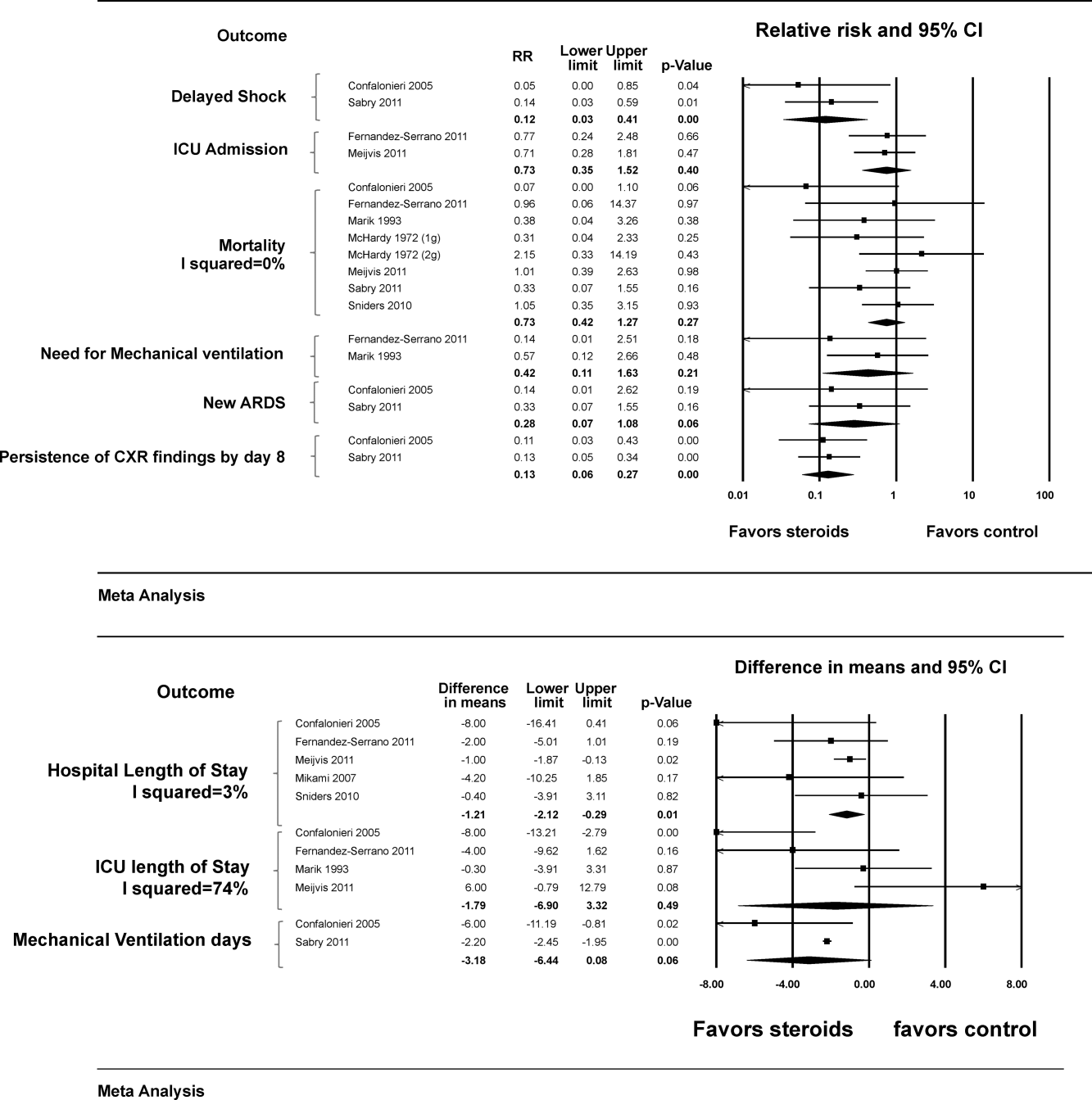
Analysis of other outcomes was limited by the fact that data were pooled from only a few studies. These included the need for and duration of mechanical ventilation, development of new ARDS and ICU admission rate, neither of which was associated with steroid therapy. However, steroid use was associated with lower incidence of delayed shock (ie, shock occurring after enrollment (RR: 0.12 [95% CI: 0.03 to 0.41]) and lower incidence of persistent chest x‐ray abnormalities at 1 week (RR: 0.13 [95% CI: 0.06 to 0.27]).
Subgroup and Sensitivity Analyses
Heterogeneity (I2 statistic) was <50% for all outcomes except ICU length of stay (74%). There were no significant interactions to suggest a subgroup effect based on older vs younger or ICU vs non‐ICU based patients (Table 2). In a priori sensitivity analysis that excluded McHardy and Schonell (published in 1972) and Marik et al. (published in 1993),[30] the results were not different from the main analysis.
| No. of Studies | Effect Size | LL | UL | P for Interaction (Difference Between Subgroups) | |
|---|---|---|---|---|---|
| |||||
| Mortality | |||||
| ICU | 3 | 0.27 | 0.08 | 0.83 | 0.06 |
| Non‐ICU | 4 | 0.96 | 0.45 | 2.05 | |
| Older | 7 | 0.75 | 0.40 | 1.38 | 0.56 |
| Young | 1 | 0.38 | 0.04 | 3.26 | |
| Need for mechanical ventilation | |||||
| ICU | 1 | 0.57 | 0.12 | 2.66 | 0.39 |
| Non‐ICU | 1 | 0.14 | 0.01 | 2.51 | |
| Older | 1 | 0.14 | 0.01 | 2.51 | 0.39 |
| Younger | 1 | 0.57 | 0.12 | 2.66 | |
| LOS | |||||
| ICU | 1 | 8.00 | 16.41 | 0.41 | 0.11 |
| Non‐ICU | 3 | 1.14 | 1.97 | 0.31 | |
| ICU LOS | |||||
| ICU | 2 | 3.91 | 11.44 | 3.62 | 0.45 |
| Non‐ICU | 2 | 0.81 | 8.98 | 10.60 | |
| Older | 3 | 2.26 | 9.95 | 5.43 | 0.65 |
| Younger | 1 | 0.30 | 3.91 | 3.31 | |
Quality of Evidence
Using the GRADE framework, the overall quality of evidence (confidence in the estimates) was judged to be moderate with the following main limitations: 1) methodological limitations among included studies (prognostic imbalance), 2) imprecision (small number of events and wide confidence intervals), and 3) inconsistency in the outcome ICU length of stay (as reflected by the I2 statistic).
Other Reported Outcomes
Four studies[15, 16, 29, 31] provided descriptive details of microbiologic data, whereas 1 study[16] provided analytical data on microbiology. In the latter, patients with Streptococcus pneumoniae infection (identified variably by sputum, pleural fluid, urine, or blood samples), had lower clinical cure rates in the steroid group at day 30 (P = 0.01) and higher numbers of late failures (defined as recurrence of signs and symptoms of pneumonia, P = 0.02).
Three[28, 30, 31] studies did not provide data on glycemic trends, whereas Fernandez‐Serrano et al., Mikami et al., and Snijders et al. reported that rates of hyperglycemia were not different across the 2 groups.[15, 16, 29] Meijvis et al.[14] reported more frequent hyperglycemia in the steroid group (44% vs 23%, P < 0.001) but no difference in the need for glucose‐lowering treatment (5% vs 3%, P = 0.57). Sabry and Omar[17] reported a higher incidence of hyperglycemia in the steroid group (no numerical data reported). Snijders et al.,[16] Meijvis et al.,[14] and Sabry and Omar[17] reported that the rates of super‐infection were not different between the 2 groups. No other adverse effects were consistently reported.
DISCUSSION
In this meta‐analysis of 8 RCTs, we found no significant association between steroid therapy and our primary outcome of interest (hospital mortality). However, length of hospital stay was shorter in the steroid group. These findings were not altered in various sensitivity and subgroup analyses. Although adverse effects of steroid therapy were not consistently reported, most of the RCTs reported that hyperglycemia was either no more common in the steroid group or did not require additional treatment.
Previous meta‐analyses have also concluded that adding corticosteroids to conventional therapy does not impact mortality among adults hospitalized with CAP.[12, 22] This may or may not be a consequence of inadequate statistical power. Although Lamontagne et al.[13] reported that low‐dose corticosteroid therapy (2 mg/kg/day or less of methylprednisolone or equivalent) was associated with reduced hospital mortality (RR: 0.68 [95% CI: 0.49 to 0.96]), this result was obtained by pooling data from 5 RCTs on adults hospitalized with CAP and 4 on adults with ALI/ARDS from any cause. In a subgroup analysis of RCTs conducted only on CAP patients, no impact on mortality was found. Of note, all RCTs involving CAP patients had used low‐dose steroids; the 3 RCTs using high‐dose steroids were carried out on ALI/ARDS patients.[36, 37, 38] Similarly, all RCTs in our meta‐analysis were also characterized by steroid doses under 2 mg/kg/day of methylprednisolone or equivalent.
Our study is the first to demonstrate decreased length of hospital stay in this patient population. Importantly, each of the 5 studies that reported this outcome (including 3 relatively recent RCTs) showed the same trend. However, it is not inconceivable that steroid use led to a quicker decline in cytokine levels resulting in an earlier resolution of fever and hence earlier discharge without a faster cure per se. The two studies whose data permitted calculation of delta CRP also demonstrated a faster CRP decline in the steroid group (Table 1).
Our analysis also suggested reduced incidence of delayed shock. However, these data were pooled from only 2 RCTs,[17, 28] and each of them used hydrocortisone, whose direct mineralocorticoid effect is an obvious confounder. Similarly, according to data pooled from 2 RCTs, steroid use was associated with fewer cases of persistent chest x‐ray abnormalities by day 8. Of note, although calculation of the I2 statistic was not possible because of too few studies, visual inspection of the forest plots suggested low levels of heterogeneity.
It is plausible that the impact of adjunctive steroids in CAP may vary based on the causative pathogen. This pathogen‐specific association has been observed in patients with bacterial meningitis, where most of the benefit is seemingly limited to pneumococcal meningitis.[9, 10] Unfortunately, as demonstrated by Snijders et al.,[16] establishing microbiologic etiology in CAP can be difficult, and most patients are treated empirically.
Our analysis showed no difference in duration of ventilation among patients who required ventilatory support on admission. However, only 2 studies reported this outcome.[17, 28] Second, in Confalonieri et al.,[28] the steroid group had a more severe baseline inflammatory response as illustrated by higher serum CRP levels (P = 0.04). Moreover, while mechanical ventilation was defined as either invasive or noninvasive ventilation, the steroid group had a higher number of patients who required noninvasive ventilation (P = 0.03), thus introducing selection bias. This study had additional areas of concern too, including a mortality of 0 among its 46 ICU patients, in contrast to established mortality rates of up to around 20%.[4] Unlike this study, Sabry and Omar[17] reported that none of their patients was on noninvasive ventilation. It may be pertinent to compare our findings with those of Steinberg et al.,[40] who studied patients with ARDS (pneumonia being the most common cause) who received methylprednisolone. This group had an early increase in ventilator‐free days, but that effect became less pronounced (though still significant) when the study end point was prolonged from 30 to 90 days.[41]
The 2 studies that were published before 2000 (McHardy and Schonell,[31] and Marik et al.[30]) were excluded in our a priori sensitivity analysis. A number of considerations led to this decision. First, standards of care for inpatient management of pneumoniaincluding pharmacologic therapies and ventilation strategieshave changed considerably over time. For instance, newer generation macrolides became available for clinical use in the early 1990s and meropenem in 1996.[41] Therefore, it would be hard to assume constancy of effect from that time period. Furthermore, the study by McHardy and Schonell[31] suffered from significant differences in the baseline characteristics of its 2 arms. There was incomplete randomization; patients with diabetes were excluded from only the steroid arm. Another issue with Marik et al.[30] was the considerably younger age of participants compared to other studies (Table 1).
Limitations
In spite of our relatively stringent selection criteria and a number of subgroup and sensitivity analyses, the overall quality of evidence was only moderate (Table 2). Key issues with the findings reported by Confalonieri et al.,[28] McHardy and Schonell,[31] and Marik et al.[30] were discussed earlier. Baseline severity of illness, patient comorbidities, and length of follow‐up were variable both within and across various studies. Another major limitation was that the intervention of interest (ie, steroid therapy) was not uniformly applied as the regimens varied considerably even though all regimens fit the designation of low‐dose steroids as previously noted (Table 1).
In conclusion, although evidence suggests that adjunctive steroid therapy is associated with reduced hospital length of stay, the data are not strong enough to recommend routine use of steroids among all adults hospitalized with CAP. However, considering that there was no increase in mortality or hospital length of stay with steroid use, it is reasonable to continue steroids if warranted for treatment of underlying comorbid conditions.
Due to the aforementioned limitations in RCTs published to date, we believe that additional studies that are more robustly designed and sufficiently powered to detect differences in key outcomes (including mortality) are warranted. Investigators should ensure appropriate randomization of groups, taking into account severity of illness, comorbid conditions and prior use of steroid therapy. Standardizing the intervention (including dose and duration of steroid therapy and time to first antibiotic dose) would be essential. Concurrent measurement of inflammatory markers such as delta CRP would be useful too. Finally, accurate measurement of all secondary outcomes of interest, including adverse effects and duration of both invasive and noninvasive mechanical ventilation, would be important to accurately study the benefit of steroids among the most likely beneficiaries: those patients who are the sickest.
Acknowledgments
The authors gratefully acknowledge the assistance of Dr. Jon Ebbert (Department of Medicine, Mayo Clinic, Rochester, MN) with proofreading the manuscript and providing thoughtful editorial suggestions.
Disclosures
The authors report no conflicts of interest.
- Centers for Disease Control and Prevention 2008. CDC/NCHS, National Vital Statistics System. Leading causes of Death. Available at: http://www.cdc.gov/nchs/nvss/mortality_tables.htm. Accessed August 14,2011.
- , . Reasons for being admitted to the hospital through the emergency department, 2003. HCUP Statistical Brief #2. February 2006. Agency for Healthcare Research and Quality, Rockville, MD. Available at: http://www.hcup‐us.ahrq.gov/reports/statbriefs/sb2.pdf. Accessed August 14,2011.
- , , , et al. Processes of care and outcomes for community‐acquired pneumonia. Am J Med. 2011;124(12):1175.e9–17.
- , , , , , . Cytokine kinetics and other host factors in response to pneumococcal pulmonary infection in mice. Infect Immun. 1998;66(3):912–922.
- , , , et al. Effects of glucocorticoids in ventilated piglets with severe pneumonia. Eur Respir J. 2008;32(4):1037–1046.
- , , , et al. Risk of death does not alter the efficacy of hydrocortisone therapy in a mouse E. coli pneumonia model: risk and corticosteroids in sepsis. Intensive Care Med. 2008;34(3):568–577.
- , , , . Adjunctive corticosteroids for Pneumocystis jiroveci pneumonia in patients with HIV‐infection. Cochrane Database Syst Rev. 2006;(3):CD006150.
- , . Dexamethasone in adults with bacterial meningitis. N Engl J Med. 2002;347(20):1549–1556.
- , , , . Steroids in adults with acute bacterial meningitis: a systematic review. Lancet Infect Dis. 2004;4(3):139–143.
- , , , et al. Infectious Diseases Society of America/American Thoracic Society consensus guidelines on the management of community‐acquired pneumonia in adults. Clin Infect Dis. 2007;44(suppl 2):S27–S72.
- , , , . Corticosteroids for pneumonia. Cochrane Database Syst Rev. 2011;(3):CD007720.
- , , , , , . Corticosteroid therapy for acute lung injury, acute respiratory distress syndrome, and severe pneumonia: a meta‐analysis of randomized controlled trials. J Crit Care. 2010;25(3):420–435.
- , , , et al. Dexamethasone and length of hospital stay in patients with community‐acquired pneumonia: a randomised, double‐blind, placebo‐controlled trial. Lancet. 2011;377(9782):2023–2030.
- , , , et al. Effect of corticosteroids on the clinical course of community‐acquired pneumonia: a randomized controlled trial. Crit Care. 2011;15(2):R96.
- , , , et al. Efficacy of corticosteroids in community‐acquired pneumonia: a randomized double‐blinded clinical trial. Am J Respir Crit Care Med. 2010;181:975–978.
- , . Corticosteroids and ICU course of community acquired pneumonia in Egyptian settings. Pharmacol Pharm. 2011;2(2):73–81.
- , , , , . Corticosteroid treatment in severe community‐acquired pneumonia: duration of treatment affects control of systemic inflammation and clinical improvement. Intensive Care Med. 2011;37(9):1553–1554.
- Higgins JPT, Green S, eds. Cochrane Handbook for Systematic Reviews of Interventions. West Sussex, UK:Wiley‐Blackwell;2008.
- , , , ;PRISMA Group. Preferred reporting items for systematic reviews and meta‐analyses: the PRISMA statement. J Clin Epidemiol. 2009;62:1006–1012.
- , , , et al. GRADE guidelines: 3. Rating the quality of evidence. J Clin Epidemiol. 2011;64(4):401–406.
- , , , , , . The role of corticosteroids in severe community‐acquired pneumonia: a systematic review. Crit Care. 2008;12(3):R76.
- , , , et al. Severe community‐acquired pneumonia: assessment of severity criteria. Am J Respir Crit Care Med. 1998;158(4):1102–1108.
- , .Assessing risk of bias in included studies. In: Higgins JPT, Green S eds. Cochrane Handbook for Systematic Reviews of Interventions. Chichester, UK:John Wiley 2009.
- , . Meta‐analysis in clinical trials. Control Clin Trials. 1986;7(3):177–188.
- , , , . Measuring inconsistency in meta‐analyses. BMJ. 2003;327(7414):557–560.
- , . Interaction revisited: the difference between two estimates. BMJ. 2003;326(7382):219.
- , , , et al. Hydrocortisone infusion for severe community‐acquired pneumonia: a preliminary randomized study. Am J Respir Crit Care Med. 2005;171(3):242–248.
- , , , , , . Efficacy of corticosteroids in the treatment of community‐acquired pneumonia requiring hospitalisation. Lung. 2007;185(5):249–255.
- , , , , , . Hydrocortisone and tumour necrosis factor in severe community acquired pneumonia. A randomised controlled study. Chest. 1993;104(2):389–392.
- , . Ampicillin dosage and use of prednisolone in treatment of pneumonia: co‐operative controlled trial. Br Med J. 1972;4:569–573.
- , , , et al. Impact of systemic corticosteroids on the clinical course and outcomes of patients with severe community‐acquired pneumonia: a cohort study. J Crit Care. 2011;26(2):193–200.
- , , , . Factors of importance for the long term prognosis after hospital treated pneumonia. Thorax. 1993;48(8):785–789.
- , , , . Assessment of mortality after long‐term follow‐up of patients with community‐acquired pneumonia. Clin Infect Dis. 2003;37(12):1617–1624.
- , , , , . Community‐acquired pneumonia on the intensive care unit: secondary analysis of 17,869 cases in the ICNARC Case Mix Programme Database. Crit Care. 2006;10(suppl 2):S1.
- , , , , , . Effects of systemic steroids in patients with severe community‐acquired pneumonia. Eur Respir J. 2007;30(5):951–956.
- , , , . Analysis of systemic corticosteroid usage and survival in patients requiring mechanical ventilation for severe community‐acquired pneumonia. J Infect Chemother. 2011;17(4):449–455.
- , , , et al. Effect of high‐dose prednisolone on lung fluid in patients with non‐cardiogenic lung edema [in German]. Wien Klin Wochenschr. 1987;99:245–249.
- , , , et al. Early steroid therapy for respiratory failure. Arch Surg. 1985;120:536–540.
- , , , et al. Efficacy and safety of corticosteroids for persistent acute respiratory distress syndrome. N Engl J Med. 2006;354(16):1671–1684.
- , , , et al. High‐dose corticosteroids in patients with the adult respiratory distress syndrome. N Engl J Med. 1987;317:1565–1570.
- . Antibiotics—past, present, and future. Med Clin North Am. 2006;90(6):1049–1076.
Community‐acquired pneumonia (CAP) is the most common lower respiratory tract infection in adults and a leading cause of infection‐related deaths in the United States.[1] According to a survey, pneumonia was the most common reason for hospital admissions through the emergency department in 2003.[2] CAP is associated with significant morbidity and mortality among those sick enough to require hospitalization. In a prospective study, hospital mortality rates ranged from 5% to 18% and length of stay from 9 to 23 days depending on patient location (intensive care unit [ICU] vs elsewhere) and severity of illness.[3]
Empirical evidence suggests that host inflammatory response contributes significantly to lung injury in pneumonia.[4] Studies have demonstrated reduction in the host inflammatory response as well as in mortality among animals with bacterial pneumonia when exposed to glucocorticoids.[5, 6] Furthermore, the efficacy of adjunctive steroid therapy in severe pneumonia caused by Pneumocystis jirovecii[7] and in pneumococcal meningitis[8, 9] is already established. However, due to equivocal, and at times conflicting, human clinical trial data on the impact of steroid therapy in CAP, the 2007 consensus guidelines (jointly published by the Infectious Diseases Society of America and American Thoracic Society) do not provide recommendations for or against use of steroids in CAP, except in the setting of hypotension secondary to adrenal insufficiency.[10]
In their meta‐analysis, Chen et al. analyzed data from 6 randomized clinical trials (RCTs) published between 1972 and 2007 (including 2 on pediatric patients) and concluded that adding steroids to current standard of care was not beneficial.[11] Earlier, Lamontagne et al.'s meta‐analysis included RCTs on hospitalized CAP patients as well as those on patients with acute lung injury (ALI) or acute respiratory distress syndrome (ARDS) from any cause.[12] They concluded that low‐dose corticosteroid therapy reduced all‐cause in‐hospital mortality in this mixed patient population (relative risk [RR]: 0.68 [95% confidence interval (CI): 0.49 to 0.96]). Recently, data from a number of additional RCTs have become available.[13, 14, 15, 16, 17] Therefore, an updated review of RCTs evaluating the role of adjunctive steroid therapy among adults hospitalized with CAP was warranted.
MATERIALS AND METHODS
We conducted this systematic review and meta‐analysis in accordance with the recommendations published in the Cochrane Handbook for Systematic Reviews of Interventions[18] and reported our findings according to the Preferred Reporting Items for Systematic Reviews and Meta‐analyses guidelines.[19] The overall quality of evidence was judged using the Grading of Recommendations Assessment, Development, and Evaluation (GRADE) framework.[20]
Data Sources and Search Strategies
A comprehensive search of several databases including PubMed, Ovid MEDLINE In‐Process & Other Non‐Indexed Citations, Ovid MEDLINE, Ovid EMBASE, Ovid Cochrane Database of Systematic Reviews, Ovid Cochrane Central Register of Controlled Trials, and Scopus was conducted. The time range for search started from each database's earliest inclusive dates up to July 2011. An experienced institutional librarian assisted with the design and conduct of our literature search. Controlled vocabulary, supplemented with keywords, was used to search for the topic: steroid therapy for community‐acquired pneumonia. We consulted expert colleagues to ensure the inclusion of all eligible reports and also checked the bibliographies of previously published systematic reviews.[12, 21]
Eligibility Criteria
Studies deemed eligible for inclusion were RCTs that met the following patients, intervention, control, outcomes (PICO) criteria: P, adults hospitalized with CAP; I, administration of systemic corticosteroids plus standard treatment; C, standard treatment without corticosteroids; O, primary outcome: hospital mortality; secondary outcomes, length of hospital stay, length of ICU stay and duration of mechanical ventilation. Under the P criterion we included RCTs that defined CAP as a lung infection (based on a reasonable combination of history, physical examination, imaging, and/or other investigative data, such as per the American Thoracic Society definition)[22] of presumed or proven bacterial etiology, in a patient who was not immunocompromised and had no exposure to a healthcare facility in the past 90 days.
Study Selection and Quality Assessment
Two reviewers independently performed study selection, data extraction, and quality assessment. Data were abstracted using standardized data collection instruments. Kappa statistic was calculated to assess the reviewers' level of agreement.
We perused full texts of all articles whose abstracts met selection criteria, performing an appraisal of their quality using the Cochrane risk‐of‐bias tool.[23] We also reviewed the baseline characteristics of patients in each study cohort.
Analysis
We estimated RR and weighted mean differences along with the respective 95% confidence intervals by pooling data using a random effects model.[24] Study heterogeneity was assessed using the I2 statistic, which estimates the percentage of variation that is not attributable to chance.[25] We performed a priori subgroup analyses based on the location (ICU vs non‐ICU) and mean age group of study participants (based on a cutoff of 50 years). A significant (P < 0.05) test of interaction would provide an explanation for any heterogeneity.[26] We also performed an a priori sensitivity analysis excluding any studies published before the year 2000 to exclude the impact of changing standards of care for inpatient management of CAP over time.
The original investigators were not contacted for purposes of obtaining raw data.
RESULTS
Eight RCTs, comprising 1119 subjects, were eventually chosen.[14]. Seven shortlisted studies were excluded due to methodological limitations, failure to fully meet PICO criteria, or gross insufficiency of descriptive data on subjects or methodology.[18, 31, 32, 33, 34, 35, 36] Figure 1 illustrates the study selection process.
Table 1 summarizes the baseline characteristics of patient populations from each study. Mean ages in 7 RCTs were between 60 years and 80 years. In Marik et al., the mean age of the intervention group was 31.7 years, whereas that of the control group was 40.6 years (P value not reported).[30] Three RCTs included ICU patients only,[17, 28, 30] whereas 4 only included general medical ward patients.[14, 15, 29, 31] Disease severity scores at admission were similar between the 2 groups in all RCTs except Sabry and Omar,[17] which was the only clinical trial to use a chest radiograph score. Only Sabry and Omar,[17] and Mikami et al.[29] excluded chronic obstructive pulmonary disease patients. Where possible, the serum C‐reactive protein (CRP) value on day one was subtracted from that on day eight to generate a one week delta CRP.
| Author, Year | Number of Patients | Gender: Males (% Age) | Age (y) | Steroids Used (Daily Dose and Duration) | COPD (% of Total) | Diabetes (% of Total) | Mean PaO2/FiO2 Ratio | Severity Score (Score: Mean) | Patients Already in ICU (% of Total) | One‐week Delta CRP (mg/dL) | ||||||||||
|---|---|---|---|---|---|---|---|---|---|---|---|---|---|---|---|---|---|---|---|---|
| Total | Steroid | Control | Steroid | Control | Steroid | Control | Steroid | Control | Steroid | Control | Steroid | Control | Steroid | Control | Steroid | Control | Steroid | Control | ||
| ||||||||||||||||||||
| McHardy 1972[31] | 126 | 40 | 86 | 45 | 50 | 62 | 59 | Prednisolone 20 mg, 7 d | 40 | 35 | Not reported | Not reported | Not reported | 0 | 0 | N/A | N/A | |||
| Marik 1993[30] | 30 | 14 | 16 | Not reported | 32 | 41 | Hydrocortisone 10 mg/kg 1 | Not reported | Not reported | 213 | 214 | APACHE II | 100 | 100 | N/A | N/A | ||||
| 11 | 14 | |||||||||||||||||||
| Confalonieri 2005[28] | 46 | 23 | 23 | 74 | 65 | 60 | 67 | Hydrocortisone 200 mg bolus, then 10 mg/h, 7 d | Not reported | Not reported | 141a | 178a | APACHE II | 100 | 100 | 37a | +5a | |||
| 17 | 18 | |||||||||||||||||||
| Mikami 2007[29] | 31 | 15 | 16 | 73.3 | 75 | 76 | 68 | Prednisolone 40 mg IV, 3 d | 0 | 0 | Not reported | PaO2 (FiO2 not reported) | PSI | 0 | 0 | N/A | N/A | |||
| 61 | 64 | 95 | 86 | |||||||||||||||||
| Snijders 2010[16] | 213 | 104 | 109 | 52.9 | 63.3 | 63 | 64 | Prednisolone 40 mg IV/PO, 7 d | 18 | 22 | 10 | 11 | Not reported | PSI class V (% of total) | 14.4 | 6.4 | N/A | N/A | ||
| 13 | 16 | |||||||||||||||||||
| Fernandez‐Serrano 2011[15] | 45 | 23 | 22 | 69.6 | 63.6 | 66 | 61 | Methylprednisone 200 mg IV; then 20 mg/6 h, 3 d; then 20 mg/12 h, 3 d; then 20 mg/d, 3 d | 17 | 9 | 9 | 18 | 200 | 257 | SAPS classes IV + V (% of total) | 0 | 0 | N/A | N/A | |
| 65 | 54 | |||||||||||||||||||
| Meijvis 2011[14] | 304 | 151 | 153 | 57 | 56 | 65 | 63 | Dexamethasone 5 mg/d, 4 d | 13 | 9 | 15 | 14 | Not reported | PSI class V (% of total) | 0 | 0 | N/A | N/A | ||
| 17 | 14 | |||||||||||||||||||
| Sabry 2011[17] | 80 | 40 | 40 | 30 | 28 | 62 | 63 | Hydrocortisone 200 mg IV, then 12.5 mg/h, 7 d | 0 | 0 | Not reported | 338a | 243a | Chest radiograph score | 100 | 100 | 38a | 23a | ||
| 1a | 3a | |||||||||||||||||||
The mean ICU length of stay was 12.7 days for the steroid group and 12.3 days for the control group. The mean hospital lengths of stay were 10.2 days and 13.6 days, respectively. Quality of the studies was moderate (see Supporting Information, Appendix I, in the online version of this article). Kappa score was >0.90.
Meta‐analysis
Figure 2 illustrates the results of our meta‐analyses. Although adjunctive steroid therapy had no effect on hospital mortality or ICU length of stay, it was associated with reduced hospital length of stay (RR: 1.21 days [95% CI: 2.12 to 0.29]). Of note, Mikami et al.[29] did not report mortality in their article, whereas in McHardy and Schonell,[31] using the factorial design, each of the 2 treatment groups were further subdivided into those patients who received 1 g of ampicillin and those who received 2 g of ampicillin (Figure 2A).
Analysis of other outcomes was limited by the fact that data were pooled from only a few studies. These included the need for and duration of mechanical ventilation, development of new ARDS and ICU admission rate, neither of which was associated with steroid therapy. However, steroid use was associated with lower incidence of delayed shock (ie, shock occurring after enrollment (RR: 0.12 [95% CI: 0.03 to 0.41]) and lower incidence of persistent chest x‐ray abnormalities at 1 week (RR: 0.13 [95% CI: 0.06 to 0.27]).
Subgroup and Sensitivity Analyses
Heterogeneity (I2 statistic) was <50% for all outcomes except ICU length of stay (74%). There were no significant interactions to suggest a subgroup effect based on older vs younger or ICU vs non‐ICU based patients (Table 2). In a priori sensitivity analysis that excluded McHardy and Schonell (published in 1972) and Marik et al. (published in 1993),[30] the results were not different from the main analysis.
| No. of Studies | Effect Size | LL | UL | P for Interaction (Difference Between Subgroups) | |
|---|---|---|---|---|---|
| |||||
| Mortality | |||||
| ICU | 3 | 0.27 | 0.08 | 0.83 | 0.06 |
| Non‐ICU | 4 | 0.96 | 0.45 | 2.05 | |
| Older | 7 | 0.75 | 0.40 | 1.38 | 0.56 |
| Young | 1 | 0.38 | 0.04 | 3.26 | |
| Need for mechanical ventilation | |||||
| ICU | 1 | 0.57 | 0.12 | 2.66 | 0.39 |
| Non‐ICU | 1 | 0.14 | 0.01 | 2.51 | |
| Older | 1 | 0.14 | 0.01 | 2.51 | 0.39 |
| Younger | 1 | 0.57 | 0.12 | 2.66 | |
| LOS | |||||
| ICU | 1 | 8.00 | 16.41 | 0.41 | 0.11 |
| Non‐ICU | 3 | 1.14 | 1.97 | 0.31 | |
| ICU LOS | |||||
| ICU | 2 | 3.91 | 11.44 | 3.62 | 0.45 |
| Non‐ICU | 2 | 0.81 | 8.98 | 10.60 | |
| Older | 3 | 2.26 | 9.95 | 5.43 | 0.65 |
| Younger | 1 | 0.30 | 3.91 | 3.31 | |
Quality of Evidence
Using the GRADE framework, the overall quality of evidence (confidence in the estimates) was judged to be moderate with the following main limitations: 1) methodological limitations among included studies (prognostic imbalance), 2) imprecision (small number of events and wide confidence intervals), and 3) inconsistency in the outcome ICU length of stay (as reflected by the I2 statistic).
Other Reported Outcomes
Four studies[15, 16, 29, 31] provided descriptive details of microbiologic data, whereas 1 study[16] provided analytical data on microbiology. In the latter, patients with Streptococcus pneumoniae infection (identified variably by sputum, pleural fluid, urine, or blood samples), had lower clinical cure rates in the steroid group at day 30 (P = 0.01) and higher numbers of late failures (defined as recurrence of signs and symptoms of pneumonia, P = 0.02).
Three[28, 30, 31] studies did not provide data on glycemic trends, whereas Fernandez‐Serrano et al., Mikami et al., and Snijders et al. reported that rates of hyperglycemia were not different across the 2 groups.[15, 16, 29] Meijvis et al.[14] reported more frequent hyperglycemia in the steroid group (44% vs 23%, P < 0.001) but no difference in the need for glucose‐lowering treatment (5% vs 3%, P = 0.57). Sabry and Omar[17] reported a higher incidence of hyperglycemia in the steroid group (no numerical data reported). Snijders et al.,[16] Meijvis et al.,[14] and Sabry and Omar[17] reported that the rates of super‐infection were not different between the 2 groups. No other adverse effects were consistently reported.
DISCUSSION
In this meta‐analysis of 8 RCTs, we found no significant association between steroid therapy and our primary outcome of interest (hospital mortality). However, length of hospital stay was shorter in the steroid group. These findings were not altered in various sensitivity and subgroup analyses. Although adverse effects of steroid therapy were not consistently reported, most of the RCTs reported that hyperglycemia was either no more common in the steroid group or did not require additional treatment.
Previous meta‐analyses have also concluded that adding corticosteroids to conventional therapy does not impact mortality among adults hospitalized with CAP.[12, 22] This may or may not be a consequence of inadequate statistical power. Although Lamontagne et al.[13] reported that low‐dose corticosteroid therapy (2 mg/kg/day or less of methylprednisolone or equivalent) was associated with reduced hospital mortality (RR: 0.68 [95% CI: 0.49 to 0.96]), this result was obtained by pooling data from 5 RCTs on adults hospitalized with CAP and 4 on adults with ALI/ARDS from any cause. In a subgroup analysis of RCTs conducted only on CAP patients, no impact on mortality was found. Of note, all RCTs involving CAP patients had used low‐dose steroids; the 3 RCTs using high‐dose steroids were carried out on ALI/ARDS patients.[36, 37, 38] Similarly, all RCTs in our meta‐analysis were also characterized by steroid doses under 2 mg/kg/day of methylprednisolone or equivalent.
Our study is the first to demonstrate decreased length of hospital stay in this patient population. Importantly, each of the 5 studies that reported this outcome (including 3 relatively recent RCTs) showed the same trend. However, it is not inconceivable that steroid use led to a quicker decline in cytokine levels resulting in an earlier resolution of fever and hence earlier discharge without a faster cure per se. The two studies whose data permitted calculation of delta CRP also demonstrated a faster CRP decline in the steroid group (Table 1).
Our analysis also suggested reduced incidence of delayed shock. However, these data were pooled from only 2 RCTs,[17, 28] and each of them used hydrocortisone, whose direct mineralocorticoid effect is an obvious confounder. Similarly, according to data pooled from 2 RCTs, steroid use was associated with fewer cases of persistent chest x‐ray abnormalities by day 8. Of note, although calculation of the I2 statistic was not possible because of too few studies, visual inspection of the forest plots suggested low levels of heterogeneity.
It is plausible that the impact of adjunctive steroids in CAP may vary based on the causative pathogen. This pathogen‐specific association has been observed in patients with bacterial meningitis, where most of the benefit is seemingly limited to pneumococcal meningitis.[9, 10] Unfortunately, as demonstrated by Snijders et al.,[16] establishing microbiologic etiology in CAP can be difficult, and most patients are treated empirically.
Our analysis showed no difference in duration of ventilation among patients who required ventilatory support on admission. However, only 2 studies reported this outcome.[17, 28] Second, in Confalonieri et al.,[28] the steroid group had a more severe baseline inflammatory response as illustrated by higher serum CRP levels (P = 0.04). Moreover, while mechanical ventilation was defined as either invasive or noninvasive ventilation, the steroid group had a higher number of patients who required noninvasive ventilation (P = 0.03), thus introducing selection bias. This study had additional areas of concern too, including a mortality of 0 among its 46 ICU patients, in contrast to established mortality rates of up to around 20%.[4] Unlike this study, Sabry and Omar[17] reported that none of their patients was on noninvasive ventilation. It may be pertinent to compare our findings with those of Steinberg et al.,[40] who studied patients with ARDS (pneumonia being the most common cause) who received methylprednisolone. This group had an early increase in ventilator‐free days, but that effect became less pronounced (though still significant) when the study end point was prolonged from 30 to 90 days.[41]
The 2 studies that were published before 2000 (McHardy and Schonell,[31] and Marik et al.[30]) were excluded in our a priori sensitivity analysis. A number of considerations led to this decision. First, standards of care for inpatient management of pneumoniaincluding pharmacologic therapies and ventilation strategieshave changed considerably over time. For instance, newer generation macrolides became available for clinical use in the early 1990s and meropenem in 1996.[41] Therefore, it would be hard to assume constancy of effect from that time period. Furthermore, the study by McHardy and Schonell[31] suffered from significant differences in the baseline characteristics of its 2 arms. There was incomplete randomization; patients with diabetes were excluded from only the steroid arm. Another issue with Marik et al.[30] was the considerably younger age of participants compared to other studies (Table 1).
Limitations
In spite of our relatively stringent selection criteria and a number of subgroup and sensitivity analyses, the overall quality of evidence was only moderate (Table 2). Key issues with the findings reported by Confalonieri et al.,[28] McHardy and Schonell,[31] and Marik et al.[30] were discussed earlier. Baseline severity of illness, patient comorbidities, and length of follow‐up were variable both within and across various studies. Another major limitation was that the intervention of interest (ie, steroid therapy) was not uniformly applied as the regimens varied considerably even though all regimens fit the designation of low‐dose steroids as previously noted (Table 1).
In conclusion, although evidence suggests that adjunctive steroid therapy is associated with reduced hospital length of stay, the data are not strong enough to recommend routine use of steroids among all adults hospitalized with CAP. However, considering that there was no increase in mortality or hospital length of stay with steroid use, it is reasonable to continue steroids if warranted for treatment of underlying comorbid conditions.
Due to the aforementioned limitations in RCTs published to date, we believe that additional studies that are more robustly designed and sufficiently powered to detect differences in key outcomes (including mortality) are warranted. Investigators should ensure appropriate randomization of groups, taking into account severity of illness, comorbid conditions and prior use of steroid therapy. Standardizing the intervention (including dose and duration of steroid therapy and time to first antibiotic dose) would be essential. Concurrent measurement of inflammatory markers such as delta CRP would be useful too. Finally, accurate measurement of all secondary outcomes of interest, including adverse effects and duration of both invasive and noninvasive mechanical ventilation, would be important to accurately study the benefit of steroids among the most likely beneficiaries: those patients who are the sickest.
Acknowledgments
The authors gratefully acknowledge the assistance of Dr. Jon Ebbert (Department of Medicine, Mayo Clinic, Rochester, MN) with proofreading the manuscript and providing thoughtful editorial suggestions.
Disclosures
The authors report no conflicts of interest.
- Centers for Disease Control and Prevention 2008. CDC/NCHS, National Vital Statistics System. Leading causes of Death. Available at: http://www.cdc.gov/nchs/nvss/mortality_tables.htm. Accessed August 14,2011.
- , . Reasons for being admitted to the hospital through the emergency department, 2003. HCUP Statistical Brief #2. February 2006. Agency for Healthcare Research and Quality, Rockville, MD. Available at: http://www.hcup‐us.ahrq.gov/reports/statbriefs/sb2.pdf. Accessed August 14,2011.
- , , , et al. Processes of care and outcomes for community‐acquired pneumonia. Am J Med. 2011;124(12):1175.e9–17.
- , , , , , . Cytokine kinetics and other host factors in response to pneumococcal pulmonary infection in mice. Infect Immun. 1998;66(3):912–922.
- , , , et al. Effects of glucocorticoids in ventilated piglets with severe pneumonia. Eur Respir J. 2008;32(4):1037–1046.
- , , , et al. Risk of death does not alter the efficacy of hydrocortisone therapy in a mouse E. coli pneumonia model: risk and corticosteroids in sepsis. Intensive Care Med. 2008;34(3):568–577.
- , , , . Adjunctive corticosteroids for Pneumocystis jiroveci pneumonia in patients with HIV‐infection. Cochrane Database Syst Rev. 2006;(3):CD006150.
- , . Dexamethasone in adults with bacterial meningitis. N Engl J Med. 2002;347(20):1549–1556.
- , , , . Steroids in adults with acute bacterial meningitis: a systematic review. Lancet Infect Dis. 2004;4(3):139–143.
- , , , et al. Infectious Diseases Society of America/American Thoracic Society consensus guidelines on the management of community‐acquired pneumonia in adults. Clin Infect Dis. 2007;44(suppl 2):S27–S72.
- , , , . Corticosteroids for pneumonia. Cochrane Database Syst Rev. 2011;(3):CD007720.
- , , , , , . Corticosteroid therapy for acute lung injury, acute respiratory distress syndrome, and severe pneumonia: a meta‐analysis of randomized controlled trials. J Crit Care. 2010;25(3):420–435.
- , , , et al. Dexamethasone and length of hospital stay in patients with community‐acquired pneumonia: a randomised, double‐blind, placebo‐controlled trial. Lancet. 2011;377(9782):2023–2030.
- , , , et al. Effect of corticosteroids on the clinical course of community‐acquired pneumonia: a randomized controlled trial. Crit Care. 2011;15(2):R96.
- , , , et al. Efficacy of corticosteroids in community‐acquired pneumonia: a randomized double‐blinded clinical trial. Am J Respir Crit Care Med. 2010;181:975–978.
- , . Corticosteroids and ICU course of community acquired pneumonia in Egyptian settings. Pharmacol Pharm. 2011;2(2):73–81.
- , , , , . Corticosteroid treatment in severe community‐acquired pneumonia: duration of treatment affects control of systemic inflammation and clinical improvement. Intensive Care Med. 2011;37(9):1553–1554.
- Higgins JPT, Green S, eds. Cochrane Handbook for Systematic Reviews of Interventions. West Sussex, UK:Wiley‐Blackwell;2008.
- , , , ;PRISMA Group. Preferred reporting items for systematic reviews and meta‐analyses: the PRISMA statement. J Clin Epidemiol. 2009;62:1006–1012.
- , , , et al. GRADE guidelines: 3. Rating the quality of evidence. J Clin Epidemiol. 2011;64(4):401–406.
- , , , , , . The role of corticosteroids in severe community‐acquired pneumonia: a systematic review. Crit Care. 2008;12(3):R76.
- , , , et al. Severe community‐acquired pneumonia: assessment of severity criteria. Am J Respir Crit Care Med. 1998;158(4):1102–1108.
- , .Assessing risk of bias in included studies. In: Higgins JPT, Green S eds. Cochrane Handbook for Systematic Reviews of Interventions. Chichester, UK:John Wiley 2009.
- , . Meta‐analysis in clinical trials. Control Clin Trials. 1986;7(3):177–188.
- , , , . Measuring inconsistency in meta‐analyses. BMJ. 2003;327(7414):557–560.
- , . Interaction revisited: the difference between two estimates. BMJ. 2003;326(7382):219.
- , , , et al. Hydrocortisone infusion for severe community‐acquired pneumonia: a preliminary randomized study. Am J Respir Crit Care Med. 2005;171(3):242–248.
- , , , , , . Efficacy of corticosteroids in the treatment of community‐acquired pneumonia requiring hospitalisation. Lung. 2007;185(5):249–255.
- , , , , , . Hydrocortisone and tumour necrosis factor in severe community acquired pneumonia. A randomised controlled study. Chest. 1993;104(2):389–392.
- , . Ampicillin dosage and use of prednisolone in treatment of pneumonia: co‐operative controlled trial. Br Med J. 1972;4:569–573.
- , , , et al. Impact of systemic corticosteroids on the clinical course and outcomes of patients with severe community‐acquired pneumonia: a cohort study. J Crit Care. 2011;26(2):193–200.
- , , , . Factors of importance for the long term prognosis after hospital treated pneumonia. Thorax. 1993;48(8):785–789.
- , , , . Assessment of mortality after long‐term follow‐up of patients with community‐acquired pneumonia. Clin Infect Dis. 2003;37(12):1617–1624.
- , , , , . Community‐acquired pneumonia on the intensive care unit: secondary analysis of 17,869 cases in the ICNARC Case Mix Programme Database. Crit Care. 2006;10(suppl 2):S1.
- , , , , , . Effects of systemic steroids in patients with severe community‐acquired pneumonia. Eur Respir J. 2007;30(5):951–956.
- , , , . Analysis of systemic corticosteroid usage and survival in patients requiring mechanical ventilation for severe community‐acquired pneumonia. J Infect Chemother. 2011;17(4):449–455.
- , , , et al. Effect of high‐dose prednisolone on lung fluid in patients with non‐cardiogenic lung edema [in German]. Wien Klin Wochenschr. 1987;99:245–249.
- , , , et al. Early steroid therapy for respiratory failure. Arch Surg. 1985;120:536–540.
- , , , et al. Efficacy and safety of corticosteroids for persistent acute respiratory distress syndrome. N Engl J Med. 2006;354(16):1671–1684.
- , , , et al. High‐dose corticosteroids in patients with the adult respiratory distress syndrome. N Engl J Med. 1987;317:1565–1570.
- . Antibiotics—past, present, and future. Med Clin North Am. 2006;90(6):1049–1076.
Copyright © 2012 Society of Hospital Medicine
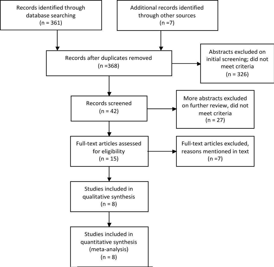
Moving Beyond Readmission Penalties
Containing the rise of healthcare costs has taken on a new sense of urgency in the wake of the recent economic recession and continued growth in the cost of healthcare. Accordingly, many stakeholders seek solutions to improve value (reducing costs while improving care)[1]; hospital readmissions, which are common and costly,[2] have emerged as a key target. The Centers for Medicare and Medicaid Services (CMS) have instituted several programs intended to reduce readmissions, including funding for community‐based, care‐transition programs; penalties for hospitals with elevated risk‐adjusted readmission rates for selected diagnoses; pioneer Accountable Care Organizations (ACOs) with incentives to reduce global costs of care; and Hospital Engagement Networks (HENs) through the Partnership for Patients.[3] A primary aim of these initiatives is to enhance the quality of care transitions as patients are discharged from the hospital.
Though the recent focus on hospital readmissions has appropriately drawn attention to transitions in care, some have expressed concerns. Among these are questions about: 1) the extent to which readmissions truly reflect the quality of hospital care[4]; 2) the preventability of readmissions[5]; 3) limitations in risk‐adjustment techniques[6]; and 4) best practices for preventing readmissions.[7] We believe these concerns stem in part from deficiencies in the state of the science of transitional care, and that future efforts in this area will be hindered without a clear vision of an ideal transition in care. We propose the key components of an ideal transition in care and discuss the implications of this concept as it pertains to hospital readmissions.
THE IDEAL TRANSITION IN CARE
We propose the key components of an ideal transition in care in Figure 1 and Table 1. Figure 1 represents 10 domains described more fully below as structural supports of the bridge patients must cross from one care environment to another during a care transition. This figure highlights key domains and suggests that lack of a domain makes the bridge weaker and more prone to gaps in care and poor outcomes. It also implies that the more components are missing, the less safe is the bridge or transition. Those domains that mainly take place prior to discharge are placed closer to the hospital side of the bridge, those that mainly take place after discharge are placed closer to the community side of the bridge, while those that take place both prior to and after discharge are in the middle. Table 1 provides descriptions of the key content for each of these domains, as well as guidance about which personnel might be involved and where in the transition process that domain should be implemented. We support these domains with supporting evidence where available.
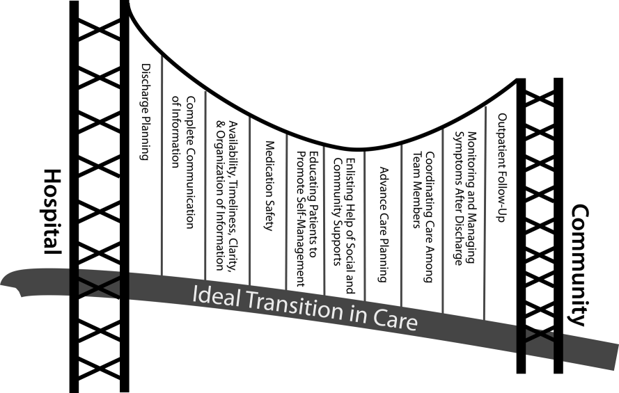
| Domain | Who | When | References |
|---|---|---|---|
| |||
| Discharge planning | |||
| Use a multidisciplinary team to create a discharge plan | Discharging clinician | Predischarge | 911 |
| Collaborate with PCP regarding discharge and follow‐up plan | Care managers/discharge planners | ||
| Arrange follow‐up appointments prior to discharge | Nurses | ||
| Make timely appointments for follow‐up care | |||
| Make appointments that take patient and caregiver's schedules and transportation needs into account | |||
| Complete communication of information | |||
| Includes: | Discharging clinician | Time of discharge | 1214 |
| Patient's full name | |||
| Age | |||
| Dates of admission and discharge | |||
| Names of responsible hospital physicians | |||
| Name of physician preparing discharge summary | |||
| Name of PCP | |||
| Main diagnosis | |||
| Other relevant diagnoses, procedures, and complications | |||
| Relevant findings at admission | |||
| Treatment and response for each active problem | |||
| Results of procedures and abnormal laboratory test results | |||
| Recommendations of any subspecialty consultants | |||
| Patient's functional status at discharge | |||
| Discharge medications | |||
| Follow‐up appointments made and those to be made | |||
| Tests to be ordered and pending tests to be followed‐up | |||
| Counseling provided to patient and caregiver, when applicable | |||
| Contingency planning | |||
| Code status | |||
| Availability, timeliness, clarity, and organization of information | |||
| Timely communication with postdischarge providers verbally (preferred) or by fax/e‐mail | Discharging clinician | Time of discharge | 1214 |
| Timely completion of discharge summary and reliable transmission to postdischarge providers | |||
| Availability of information in medical record | |||
| Use of a structured template with subheadings in discharge communication | |||
| Medication safety | |||
| Take an accurate preadmission medication history | Clinicians | Admission | 1521 |
| Reconcile preadmission medications with all ordered medications at all transfers in care, including discharge | Pharmacists | Throughout hospitalization | |
| Communicate discharge medications to all outpatient providers, including all changes and rationale for those changes | Nurses | Time of discharge | |
| Educating patients, promoting self‐management | |||
| Focus discharge counseling on major diagnoses, medication changes, dates of follow‐up appointments, self‐care instructions, warning signs and symptoms, and who to contact for problems | Clinicians | Daily | 911, 2228, 30 |
| Include caregivers as appropriate | Nurses | Time of discharge | |
| Ensure staff members provide consistent messages | Care managers/discharge planners | Postdischarge | |
| Provide simply written patient‐centered materials with instructions | Transition coaches | ||
| Use teach‐back methods to confirm understanding | |||
| Encourage questions | |||
| Continue teaching during postdischarge follow‐up | |||
| Use transition coaches in high‐risk patients: focus on medication management, keeping a personal medical record, follow‐up appointments, and knowledge of red flags | |||
| Enlisting help of social and community supports | |||
| Assess needs and appropriately arrange for home services | Clinicians | Predischarge and postdischarge | 29, 30 |
| Enlist help of caregivers | Nurses | ||
| Enlist help of community supports | Care managers | ||
| Home health staff | |||
| Advanced care planning | |||
| Establish healthcare proxy | Clinicians | Predischarge and postdischarge | 31, 32 |
| Discuss goals of care | Palliative care staff | ||
| Palliative care consultation (if appropriate) | Social workers | ||
| Enlist hospice services (if appropriate) | Nurses | ||
| Hospice workers | |||
| Coordinating care among team members | |||
| Share medical records | Clinicians | Predischarge and postdischarge | 33 |
| Communicate involving all team members | Nurses | ||
| Optimize continuity of providers and formal handoffs of care | Office personnel | ||
| IT staff | |||
| Monitoring and managing symptoms after discharge | |||
| Monitor for: | Clinicians | Postdischarge | 1113, 28, 3436 |
| Worsening disease control | Nurses | ||
| Medication side effects, discrepancies, nonadherence | Pharmacists | ||
| Therapeutic drug monitoring | Care managers | ||
| Inability to manage conditions at home | Visiting nurses and other home health staff | ||
| Via: | |||
| Postdischarge phone calls | |||
| Home visits | |||
| Postdischarge clinic visits | |||
| Patient hotline | |||
| Availability of inpatient providers after discharge | |||
| Follow‐up with outpatient providers | |||
| Within an appropriate time frame (eg, 7 d or sooner for high‐risk patients) | Clinicians | Postdischarge | 3740 |
| With appropriate providers (eg, most related to reasons for hospitalization, who manage least stable conditions, and/or PCP) | Nurses Pharmacists | ||
| Utilize multidisciplinary teams as appropriate | Care managers | ||
| Ensure appropriate progress along plan of care and safe transition | Office personnel | ||
| Other clinical staff as appropriate | |||
Our concept of an ideal transition in care began with work by Naylor, who described several important components of a safe transition in care, including complete communication of information, patient education, enlisting the help of social and community supports, ensuring continuity of care, and coordinating care among team members.[8] It is supplemented by the Transitions of Care Consensus Policy Statement proposed by representatives from hospital medicine, primary care, and emergency medicine, which emphasized aspects of timeliness and content of communication between providers.[9] Our present articulation of these key components includes 10 organizing domains.
The Discharge Planning domain highlights the important principle of planning ahead for hospital discharge while the patient is still being treated in the hospital, a paradigm espoused by Project RED[10] and other successful care transitions interventions.[11, 12] Collaborating with the outpatient provider and taking the patient and caregiver's preferences for appointment scheduling into account can help ensure optimal outpatient follow‐up.
Complete Communication of Information refers to the content that should be included in discharge summaries and other means of information transfer from hospital to postdischarge care. The specific content areas are based on the Society of Hospital Medicine and Society of General Internal Medicine Continuity of Care Task Force systematic review and recommendations,[13] which takes into account information requested by primary care physicians after discharge.
Availability, Timeliness, Clarity, and Organization of that information is as important as the content because postdischarge providers must be able to access and quickly understand the information they have been provided before assuming care of the patient.[14, 15]
The Medication Safety domain is of central importance because medications are responsible for most postdischarge adverse events.[16] Taking an accurate medication history,[17] reconciling changes throughout the hospitalization,[18] and communicating the reconciled medication regimen to patients and providers across transitions of care can reduce medication errors and improve patient safety.[19, 20, 21, 22]
The Patient Education and Promotion of Self‐Management domain involves teaching patients and their caregivers about the main hospital diagnoses and instructions for self‐care, including medication changes, appointments, and whom to contact if issues arise. Confirming comprehension of instructions through assessments of acute (delirium) and chronic (dementia) cognitive impairments[23, 24, 25, 26] and teach‐back from the patient (or caregiver) is an important aspect of such counseling, as is providing patients and caregivers with educational materials that are appropriate for their level of health literacy and preferred language.[14] High‐risk patients may benefit from patient coaching to improve their self‐management skills.[12] These recommendations are based on years of health literacy research,[27, 28, 29] and such elements are generally included in effective interventions (including Project RED,[10] Naylor and colleagues' Transitional Care Model,[11] and Coleman and colleagues' Care Transitions Intervention[12]).
Enlisting the help of Social and Community Supports is an important adjunct to medical care and is the rationale for the recent increase in CMS funding for community‐based, care‐transition programs. These programs are crucial for assisting patients with household activities, meals, and other necessities during the period of recovery, though they should be distinguished from care management or care coordination interventions, which have not been found to be helpful in preventing readmissions unless high touch in nature.[30, 31]
The Advanced Care Planning domain may begin in the hospital or outpatient setting, and involves establishing goals of care and healthcare proxies, as well as engaging with palliative care or hospice services if appropriate. Emerging evidence supports the intuitive conclusion that this approach prevents readmissions, particularly in patients who do not benefit from hospital readmission.[32, 33]
Attention to the Coordinating Care Among Team Members domain is needed to synchronize efforts across settings and providers. Clearly, many healthcare professionals as well as other involved parties can be involved in helping a single patient during transitions in care. It is vital that they coordinate information, assessments, and plans as a team.[34]
We recognize the domain of Monitoring and Managing Symptoms After Discharge as increasingly crucial as reflected in our growing understanding of the reasons for readmission, especially among patients with fragile conditions such as heart failure, chronic lung disease, gastrointestinal disorders, dementia,[23, 24, 25, 26] and vascular disease.[35] Monitoring for new or worsening symptoms; medication side effects, discrepancies, or nonadherence; and other self‐management challenges will allow problems to be detected and addressed early, before they result in unplanned healthcare utilization. It is noteworthy that successful interventions in this regard rely on in‐home evaluation[13, 14, 29] by nurses rather than telemonitoring, which in isolation has not been effective to date.[36, 37]
Finally, optimal Outpatient Follow‐Up with appropriate postdischarge providers is crucial for providing ideal transitions. These appointments need to be prompt[38, 39] (eg, within 7 days if not sooner for high‐risk patients) and with providers who have a longitudinal relationship to the patient, as prior work has shown increased readmissions when the provider is unfamiliar with the patient.[40] The advantages and disadvantages of hospitalist‐run postdischarge clinics as one way to increase access and expedite follow‐up are currently being explored. Although the optimal content of a postdischarge visit has not been defined, logical tasks to be completed are myriad and imply the need for checklists, adequate time, and a multidisciplinary team of providers.[41]
IMPLICATIONS OF THE IDEAL TRANSITION IN CARE
Our conceptualization of an ideal transition in care provides insight for hospital and healthcare system leadership, policymakers, researchers, clinicians, and educators seeking to improve transitions of care and reduce hospital readmissions. In the sections below, we briefly review commonly cited concerns about the recent focus on readmissions as a quality measure, illustrate how the Ideal Transition in Care addresses these concerns, and propose fruitful areas for future work.
How Does the Framework Address the Extent to Which Readmissions Reflect Hospital Quality?
One of the chief problems with readmissionrates as a hospital quality measure is that many of the factors that influence readmission may not currently be under the hospital's control. The healthcare environment to which a patient is being discharged (and was admitted from in the first place) is an important determinant of readmission.[42] In this context, it is noteworthy that successful interventions to reduce readmission are generally those that focus on outpatient follow‐up, while inpatient‐only interventions have had less success.[7] This is reflected in our framework above, informed by the literature, highlighting the importance of coordination between inpatient and outpatient providers and the importance of postdischarge care, including monitoring and managing symptoms after discharge, prompt follow‐up appointments, the continuation of patient self‐management activities, monitoring for drug‐related problems after discharge, and the effective utilization of community supports. Accountable care organizations, once established, would be responsible for several components of this environment, including the provision of prompt and effective follow‐up care.
The implication of the framework is that if a hospital does not have control over most of the factors that influence its readmission rate, it should see financial incentives to reduce readmission rates as an opportunity to invest in relationships with the outpatient environment from which their patients are admitted and to which they are discharged. One can envision hospitals growing ever‐closer relationships with their network of primary care physician groups, community agencies, and home health services, rehabilitation facilities, and nursing homes through coordinated discharge planning, medication management, patient education, shared electronic medical records, structured handoffs in care, and systems of intensive outpatient monitoring. Our proposed framework, in other words, emphasizes that hospitals cannot reduce their readmission rates by focusing on aspects of care within their walls. They must forge new and stronger relationships with their communities if they are to be successful.
How Does the Framework Help Us Understand Which Readmissions Are Preventable?
Public reporting and financial penalties are currently tied to all‐cause readmission, but preventable readmissions are a more appealing outcome to target. In one study, the ranking of hospitals by all‐cause readmission rate had very little correlation with the ranking by preventable readmission rate.[5] However, researchers have struggled to establish standardized, valid, and reliable measures for determining what proportion of readmissions are in fact preventable, with estimates ranging from 5% to 79% in the published literature.[43]
The difficulty of accurately determining preventability stems from an inadequate understanding of the roles that patient comorbidities, transitional processes of care, individual patient behaviors, and social and environmental determinants of health play in the complex process of hospital recidivism. Our proposed elements of an ideal transition in care provide a structure to frame this discussion and suggest future research opportunities to allow a more accurate and reliable understanding of the spectrum of preventability. Care system leadership can use the framework to rigorously evaluate their readmissions and determine the extent to which the transitions process approached the ideal. For example, if a readmission occurs despite care processes that addressed most of the domains with high fidelity, it becomes much less likely that the readmission was preventable. It should be noted that the converse is not always true: When a transition falls well short of the ideal, it does not always imply that provision of a more ideal transition would necessarily have prevented the readmission, but it does make it more likely.
For educators, the framework may provide insights for trainees into the complexity of the transitions process and vulnerability of patients during this time, highlighting preventable aspects of readmissions that are within the grasp of the discharging clinician or team. It highlights the importance of medication reconciliation, synchronous communication, and predischarge teaching, which are measurable and teachable skills for non‐physician providers, housestaff, and medical students. It also may allow for more structured feedback, for example, on the quality of discharge summaries produced by trainees.
How Could the Framework Improve Risk Adjustment for Between‐Hospital Comparisons?
Under the Patient Protection and Affordable Care Act (PPACA), hospitals will be compared to one another using risk‐standardized readmission rates as a way to penalize poorly performing hospitals. However, risk‐adjustment models have only modest ability to predict hospital readmission.[6] Moreover, current approaches predominantly adjust for patients' medical comorbidities (which are easily measurable), but they do not adequately take into account the growing literature on other factors that influence readmission rates, including a patient's health literacy, visual or cognitive impairment, functional status, language barriers, and community‐level factors such as social supports.[44, 45]
The Ideal Transition of Care provides a comprehensive framework of hospital discharge quality that provides additional process measures on which hospitals could be compared rather than focusing solely on (inadequately) risk‐adjusted readmission rates. Indeed, most other quality and safety measures (such as the National Quality Forum's Safe Practices[46] and The Joint Commission's National Patient Safety Goals),[47] emphasize process over outcome, in part because of issues of fairness. Process measures are less subject to differences in patient populations and also change the focus from simply reducing readmissions to improving transitional care more broadly. These process measures should be based on our framework and should attempt to capture as many dimensions of an optimal care transition as possible.
Possible examples of process measures include: the accuracy of medication reconciliation at admission and discharge; provision of prompt outpatient follow‐up; provision of adequate systems to monitor and manage symptoms after discharge; advanced care planning in appropriate patients; and the quality of discharge education, incorporating measurements of the patient's understanding and ability to self‐manage their illness. At least some of these could be used now as part of a performance measurement set that highlights opportunities for immediate system change and can serve as performance milestones.
The framework could also be used to validate risk‐adjustment techniques. After accounting for patient factors, the remaining variability in outcomes should be accounted for by processes of care that are in the transitions framework. Once these processes are accurately measured, one can determine if indeed the remaining variability is due to transitions processes, or rather unaccounted factors that are not being measured and that hospitals may have little control over. Such work can lead to iterative refinement of patient risk‐adjustment models.
What Does the Framework Imply About Best Practices for Reducing Readmission Rates?
Despite the limitations of readmission rates as a quality measure noted above, hospitals presently face potentially large financial penalties for readmissions and are allocating resources to readmission reduction efforts. However, hospitals currently may not have enough guidance to know what actions to take to reduce readmissions, and thus could be spending money inefficiently and reducing the value proposition of focusing on readmissions.
A recent systematic review of interventions hospitals could employ to reduce readmissions identified several positive studies, but also many negative studies, and there were significant barriers to understanding what works to reduce readmissions.[7] For example, most of the interventions described in both positive and negative studies were multifaceted, and the authors were unable to identify which components of the intervention were most effective. Also, while several studies have identified risk factors for readmission,[6, 48, 49] very few studies have identified which subgroups of patients benefit most from specific interventions. Few of the studies described key contextual factors that may have led to successful or failed implementation, or the fidelity with which the intervention was implemented.[50, 51, 52]
Few if any of the studies were guided by a concept of the ideal transition in care.[10] Such a framework will better guide development of multifaceted interventions and provide an improved means for interpreting the results. Clearly, rigorously conducted, multicenter studies of readmission prevention interventions are needed to move the field forward. These studies should: 1) correlate implementation of specific intervention components with reductions in readmission rates to better understand the most effective components; 2) be adequately powered to show effect modification, ie, which patients benefit most from these interventions; and 3) rigorously measure environmental context and intervention fidelity, and employ mixed methods to better understand predictors of implementation success and failure.
Our framework can be used in the design and evaluation of such interventions. For example, interventions could be designed that incorporate as many of the domains of an ideal transition as possible, in particular those that span the inpatient and outpatient settings. Processes of care metrics can be developed that measure the extent to which each domain is delivered, analogous to the way the Joint Commission might aggregate individual scores on the 10 items in Acute Myocardial Infarction Core Measure Set[53] to provide a composite of the quality of care provided to patients with this diagnosis. These can be used to correlate certain intervention components with success in reducing readmissions and also in measuring intervention fidelity.
NEXT STEPS
For hospital and healthcare system leaders, who need to take action now to avoid financial penalties, we recommend starting with proven, high‐touch interventions such as Project RED and the Care Transitions Intervention, which are durable, cost‐effective, robustly address multiple domains of the Ideal Transition in Care, and have been implemented at numerous sites.[54, 55] Each hospital or group will need to decide on a bundle of interventions and customize them based on local workflow, resources, and culture.
Risk‐stratification, to match the intensity of the intervention to the risk of readmission of the patient, will undoubtedly be a key component for the efficient use of resources. We anticipate future research will allow risk stratification to be a robust part of any implementation plan. However, as noted above, current risk prediction models are imperfect,[6] and more work is needed to determine which patients benefit most from which interventions. Few if any studies have described interventions tailored to risk for this reason.
Based on our ideal transition in care, our collective experience, and published evidence,[7, 10, 11, 12] potential elements to start with include: early discharge planning; medication reconciliation[56]; patient/caregiver education using health literacy principles, cognitive assessments, and teach‐back to confirm understanding; synchronous communication (eg, by phone) between inpatient and postdischarge providers; follow‐up phone calls to patients within 72 hours of discharge; 24/7 availability of a responsible inpatient provider to address questions and problems (both from the patient/caregiver and from postdischarge providers); and prompt appointments for patients discharged home. High‐risk patients will likely require additional interventions, including in‐home assessments, disease‐monitoring programs, and/or patient coaching. Lastly, patients with certain conditions prone to readmission (such as heart failure and chronic obstructive pulmonary disease) may benefit from disease‐specific programs, including patient education, outpatient disease management, and monitoring protocols.
It is likely that the most effective interventions are those that come from combined, coordinated interventions shared between inpatient and outpatient settings, and are intensive in nature. We expect that the more domains in the framework that are addressed, the safer and more seamless transitions in care will be, with improvement in patient outcomes. To the extent that fragmentation of care has been a barrier to the implementation of these types of interventions in the past, ACOs, perhaps with imbedded Patient‐Centered Medical Homes, may be in the best position to take advantage of newly aligned financial incentives to design comprehensive transitional care. Indeed, we anticipate that Figure 1 may provide substrate for a discussion of postdischarge care and division of responsibilities between inpatient and outpatient care teams at the time of transition, so effort is not duplicated and multiple domains are addressed.
Other barriers to implementation of ideal transitions in care will continue to be an issue for most healthcare systems. Financial constraints that have been a barrier up until now will be partially overcome by penalties for high readmission rates and by ACOs, bundled payments, and alternative care contracts (ie, global payments), but the extent to which each institution feels rewarded for investing in transitional interventions will vary greatly. Healthcare leadership that sees the value of improving transitions in care will be critical to overcoming this barrier. Competing demands (such as lowering hospital length of stay and carrying out other patient care responsibilities),[57] lack of coordination and diffusion of responsibility among various clinical personnel, and lack of standards are other barriers[58] that will require clear prioritization from leadership, policy changes, team‐based care, provider education and feedback, and adequate allocation of personnel resources. In short, process redesign using continuous quality improvement efforts and effective tools will be required to maximize the possibility of success.
CONCLUSIONS
Readmissions are costly and undesirable. Intuition suggests they are a marker of poor care and that hospitals should be capable of reducing them, thereby improving care and decreasing costs. In a potential future world of ACOs based on global payments, financial incentives would be aligned for each system to reduce readmissions below their current baseline, therefore obviating the need for external financial rewards and penalties. In the meantime, financial penalties do exist, and controversy exists over their fairness and likelihood of driving appropriate behavior. To address these controversies and promote better transitional care, we call for the development and use of multifaceted, collaborative transitions interventions that span settings, risk‐adjustment models that allow for fairer comparisons among hospitals, better and more widespread measurement of processes of transitional care, a better understanding of what interventions are most effective and in whom, and better guidance in how to implement these interventions. Our conceptualization of an ideal transition of care serves as a guide and provides a common vocabulary for these efforts. Such research is likely to produce the knowledge needed for healthcare systems to improve transitions in care, reduce readmissions, and reduce costs.
Disclosure
Funding for Dr Vasilevskis has been provided by the National Institutes of Health (K23AG040157) and the VA Tennessee Valley Geriatric Research, Education and Clinical Center (GRECC). The content is solely the responsibility of the authors and does not necessarily represent the official views of the National Institute on Aging, the National Institutes of Health, or the US Department of Veterans Affairs.
- , . Redefining Health Care: Creating Value‐Based Competition on Results. Boston, MA:Harvard Business School Press;2006.
- , , . Rehospitalizations among patients in the Medicare fee‐for‐service program. N Engl J Med. 2009;360(14):1418–1428.
- Patient Protection and Affordable Care Act (PPACA). Public Law 111–148; 2010. Available at: http://www.gpo.gov/fdsys/pkg/PLAW‐111publ148/pdf/PLAW‐111publ148.pdf. Accessed on June 4, 2012.
- , . Hospital readmission as an accountability measure. JAMA. 2011;305(5):504–505.
- , , , et al. Incidence of potentially avoidable urgent readmissions and their relation to all‐cause urgent readmissions. Can Med Assoc J. 2011;183(14):E1067–E1072.
- , , , et al. Risk prediction models for hospital readmission: a systematic review. JAMA. 2011;306(15):1688–1698.
- , , , , . Interventions to reduce 30‐day rehospitalization: a systematic review. Ann Intern Med. 2011;155(8):520–528.
- . A decade of transitional care research with vulnerable elders. J Cardiovasc Nurs. 2000;14(3):1–14.
- , , , et al;for the American College of Physicians; Society of General Internal Medicine; Society of Hospital Medicine; American Geriatrics Society; American College of Emergency Physicians; Society of Academic Emergency Medicine. Transitions of Care Consensus Policy Statement American College of Physicians–Society of General Internal Medicine–Society of Hospital Medicine–American Geriatrics Society–American College of Emergency Physicians–Society of Academic Emergency Medicine. J Gen Intern Med. 2009;24(8):971–976.
- , , , et al. A reengineered hospital discharge program to decrease rehospitalization: a randomized trial. Ann Intern Med. 2009;150(3):178–187.
- , , , et al. Comprehensive discharge planning and home follow‐up of hospitalized elders: a randomized clinical trial. JAMA. 1999;281(7):613–620.
- , , , . The care transitions intervention: results of a randomized controlled trial. Arch Intern Med. 2006;166(17):1822–1828.
- , , , , , . Deficits in communication and information transfer between hospital‐based and primary care physicians: implications for patient safety and continuity of care. JAMA. 2007;297(8):831–841.
- , , , . Promoting effective transitions of care at hospital discharge: a review of key issues for hospitalists. J Hosp Med. 2007;2(5):314–323.
- , , , , , . Discharge documentation of patients discharged to subacute facilities: a three‐year quality improvement process across an integrated health care system. Jt Comm J Qual Patient Saf. 2010;36(6):243–251.
- , , , , . The incidence and severity of adverse events affecting patients after discharge from the hospital. Ann Intern Med. 2003;138(3):161–167.
- , , , , , . Frequency, type and clinical importance of medication history errors at admission to hospital: a systematic review. Can Med Assoc J. 2005;173(5):510–515.
- , , , et al. Classifying and predicting errors of inpatient medication reconciliation. J Gen Intern Med. 2008;23(9):1414–1422.
- , , et al;for the PILL‐CVD (Pharmacist Intervention for Low Literacy in Cardiovascular Disease) Study Group. Effect of a pharmacist intervention on clinically important medication errors after hospital discharge: a randomized trial. Ann Intern Med. 2012;157(1):1–10.
- , , , . Hospital‐based medication reconciliation practices: a systematic review. Arch Intern Med. 2012;172(14):1057–1069.
- , , , et al. Effect of an electronic medication reconciliation application and process redesign on potential adverse drug events: a cluster‐randomized trial. Arch Intern Med. 2009;169(8):771–780.
- , , , et al. Role of pharmacist counseling in preventing adverse drug events after hospitalization. Arch Intern Med. 2006;166(5):565–571.
- , , , , . Insufficient help for activity of daily living disabilities and risk of all–cause hospitalization. J Am Geriatr Soc. 2012;60(5):927–933.
- , , , et al. Transitions in care for older adults with and without dementia. J Am Geriatr Soc. 2012;60(5):813–820.
- , , , , . Association of incident dementia with hospitalizations. JAMA. 2012;307(2):165–172.
- , , , et al. Potentially avoidable hospitalizations of dually eligible Medicare and Medicaid beneficiaries from nursing facility and home– and community–based services waiver programs. J Am Geriatr Soc. 2012;60(5):821–829.
- , . Teaching about health literacy and clear communication. J Gen Intern Med. 2006;21(8):888–890.
- , , , et al. Health literacy and outcomes among patients with heart failure. JAMA. 2011;305(16):1695–1701.
- , , , , . Patient experiences of transitioning from hospital to home: an ethnographic quality improvement project. J Hosp Med. 2012;7(5):382–387.
- , , , . Effects of care coordination on hospitalization, quality of care, and health care expenditures among Medicare beneficiaries: 15 randomized trials. JAMA. 2009;301(6):603–618.
- , , , , . How changes in Washington University's Medicare coordinated care demonstration pilot ultimately achieved savings. Health Aff (Millwood). 2012;31(6):1216–1226.
- , , , et al. Quality of care and rehospitalization rate in the last stage of disease in brain tumor patients assisted at home: a cost effectiveness study. J Palliat Med. 2012;15(2):225–227.
- , , , , . Inpatient palliative care consults and the probability of hospital readmission. Perm J. 2011;15(2):48–51.
- , , , et al. TeamSTEPPS™: team strategies and tools to enhance performance and patient safety. In: Henriksen K, Battles JB, Keyes MA, Grady ML, ed. Advances in Patient Safety: New Directions and Alternative Approaches. Vol 3: Performance and Tools. Rockville, MD:Agency for Healthcare Research and Quality; August2008.
- , , , et al. Factors contributing to all‐cause 30‐day readmissions: a structured case series across 18 hospitals. Med Care. 2012;50(7):599–605.
- , , , et al. Telemonitoring in patients with heart failure [erratum, N Engl J Med. 2011;364(5):490]. N Engl J Med. 2010;363(24):2301–2309.
- , , , et al. A randomized controlled trial of telemonitoring in older adults with multiple health issues to prevent hospitalizations and emergency department visits. Arch Intern Med. 2012;172(10):773–779.
- , , , et al. Relationship between early physician follow‐up and 30‐day readmission among Medicare beneficiaries hospitalized for heart failure. JAMA. 2010;303(17):1716–1722.
- , , . Post‐hospitalization transitions: examining the effects of timing of primary care provider follow‐up. J Hosp Med. 2010;5(7):392–397.
- , , . Does increased access to primary care reduce hospital readmissions? Veterans Affairs Cooperative Study Group on Primary Care and Hospital Readmission. N Engl J Med. 1996;334(22):1441–1447.
- . The Post‐Hospital Follow‐Up Visit: A Physician Checklist to Reduce Readmissions. California Healthcare Foundation; October 2010. Available at: http://www.chcf.org/publications/2010/10/the‐post‐hospital‐follow‐up‐visit‐a‐physician‐checklist. Accessed on January 10, 2012.
- , , . Thirty‐day readmission rates for Medicare beneficiaries by race and site of care. JAMA. 2011;305(7):675–681.
- , , , , . Proportion of hospital readmissions deemed avoidable: a systematic review. Can Med Assoc J. 2011;183(7):E391–E402.
- , , , et al. Postdischarge environmental and socioeconomic factors and the likelihood of early hospital readmission among community‐dwelling Medicare beneficiaries. Gerontologist. 2008;48(4):495–504.
- , , , , . Low health literacy and health outcomes: an updated systematic review. Ann Intern Med. 2011;155(2):97–107.
- National Quality Forum. Safe Practices for Better Healthcare—2010 Update: A Consensus Report. Washington, DC;2010.
- Joint Commission on Accreditation of Healthcare Organizations. Accreditation Program: Hospital 2010 National Patient Safety Goals (NPSGs). 2010. Available at: http://www.jointcommission.org/PatientSafety/NationalPatientSafetyGoals/. Accessed on March 20, 2012.
- , , , et al. Hospital readmission in general medicine patients: a prediction model. J Gen Intern Med. 2010;25(3):211–219.
- , , , et al. Derivation and validation of an index to predict early death or unplanned readmission after discharge from hospital to the community. Can Med Assoc J. 2010;182(6):551–557.
- , . Evaluating service delivery interventions to enhance patient safety. BMJ. 2008;337:a2764.
- , , . Assessing the Evidence for Context‐Sensitive Effectiveness and Safety of Patient Safety Practices: Developing Criteria. Rockville, MD:Agency for Healthcare Research and Quality; December2010.
- , , , et al. Advancing the science of patient safety. Ann Intern Med. 2011;154(10):693–696.
- The Joint Commission. Acute Myocardial Infarction Core Measure Set. Available at: http://www.jointcommission.org/assets/1/6/Acute%20Myocardial%20Infarction.pdf. Accessed August 20,2012.
- , , , , , . The care transitions intervention: translating from efficacy to effectiveness. Arch Intern Med. 2011;171(14):1232–1237.
- Project RED toolkit, AHRQ Innovations Exchange. Available at:http://www.innovations.ahrq.gov/content.aspx?id=2180. Accessed on July 2, 2012.
- , , , et al. A comprehensive pharmacist intervention to reduce morbidity in patients 80 years or older: a randomized controlled trial. Arch Intern Med. 2009;169(9):894–900.
- , . Thirty‐day readmissions—truth and consequences. N Engl J Med. 2012;366(15):1366–1369.
- , , , , . “Out of sight, out of mind”: housestaff perceptions of quality‐limiting factors in discharge care at teaching hospitals. J Hosp Med. 2012;7(5):376–381.
Containing the rise of healthcare costs has taken on a new sense of urgency in the wake of the recent economic recession and continued growth in the cost of healthcare. Accordingly, many stakeholders seek solutions to improve value (reducing costs while improving care)[1]; hospital readmissions, which are common and costly,[2] have emerged as a key target. The Centers for Medicare and Medicaid Services (CMS) have instituted several programs intended to reduce readmissions, including funding for community‐based, care‐transition programs; penalties for hospitals with elevated risk‐adjusted readmission rates for selected diagnoses; pioneer Accountable Care Organizations (ACOs) with incentives to reduce global costs of care; and Hospital Engagement Networks (HENs) through the Partnership for Patients.[3] A primary aim of these initiatives is to enhance the quality of care transitions as patients are discharged from the hospital.
Though the recent focus on hospital readmissions has appropriately drawn attention to transitions in care, some have expressed concerns. Among these are questions about: 1) the extent to which readmissions truly reflect the quality of hospital care[4]; 2) the preventability of readmissions[5]; 3) limitations in risk‐adjustment techniques[6]; and 4) best practices for preventing readmissions.[7] We believe these concerns stem in part from deficiencies in the state of the science of transitional care, and that future efforts in this area will be hindered without a clear vision of an ideal transition in care. We propose the key components of an ideal transition in care and discuss the implications of this concept as it pertains to hospital readmissions.
THE IDEAL TRANSITION IN CARE
We propose the key components of an ideal transition in care in Figure 1 and Table 1. Figure 1 represents 10 domains described more fully below as structural supports of the bridge patients must cross from one care environment to another during a care transition. This figure highlights key domains and suggests that lack of a domain makes the bridge weaker and more prone to gaps in care and poor outcomes. It also implies that the more components are missing, the less safe is the bridge or transition. Those domains that mainly take place prior to discharge are placed closer to the hospital side of the bridge, those that mainly take place after discharge are placed closer to the community side of the bridge, while those that take place both prior to and after discharge are in the middle. Table 1 provides descriptions of the key content for each of these domains, as well as guidance about which personnel might be involved and where in the transition process that domain should be implemented. We support these domains with supporting evidence where available.
| Domain | Who | When | References |
|---|---|---|---|
| |||
| Discharge planning | |||
| Use a multidisciplinary team to create a discharge plan | Discharging clinician | Predischarge | 911 |
| Collaborate with PCP regarding discharge and follow‐up plan | Care managers/discharge planners | ||
| Arrange follow‐up appointments prior to discharge | Nurses | ||
| Make timely appointments for follow‐up care | |||
| Make appointments that take patient and caregiver's schedules and transportation needs into account | |||
| Complete communication of information | |||
| Includes: | Discharging clinician | Time of discharge | 1214 |
| Patient's full name | |||
| Age | |||
| Dates of admission and discharge | |||
| Names of responsible hospital physicians | |||
| Name of physician preparing discharge summary | |||
| Name of PCP | |||
| Main diagnosis | |||
| Other relevant diagnoses, procedures, and complications | |||
| Relevant findings at admission | |||
| Treatment and response for each active problem | |||
| Results of procedures and abnormal laboratory test results | |||
| Recommendations of any subspecialty consultants | |||
| Patient's functional status at discharge | |||
| Discharge medications | |||
| Follow‐up appointments made and those to be made | |||
| Tests to be ordered and pending tests to be followed‐up | |||
| Counseling provided to patient and caregiver, when applicable | |||
| Contingency planning | |||
| Code status | |||
| Availability, timeliness, clarity, and organization of information | |||
| Timely communication with postdischarge providers verbally (preferred) or by fax/e‐mail | Discharging clinician | Time of discharge | 1214 |
| Timely completion of discharge summary and reliable transmission to postdischarge providers | |||
| Availability of information in medical record | |||
| Use of a structured template with subheadings in discharge communication | |||
| Medication safety | |||
| Take an accurate preadmission medication history | Clinicians | Admission | 1521 |
| Reconcile preadmission medications with all ordered medications at all transfers in care, including discharge | Pharmacists | Throughout hospitalization | |
| Communicate discharge medications to all outpatient providers, including all changes and rationale for those changes | Nurses | Time of discharge | |
| Educating patients, promoting self‐management | |||
| Focus discharge counseling on major diagnoses, medication changes, dates of follow‐up appointments, self‐care instructions, warning signs and symptoms, and who to contact for problems | Clinicians | Daily | 911, 2228, 30 |
| Include caregivers as appropriate | Nurses | Time of discharge | |
| Ensure staff members provide consistent messages | Care managers/discharge planners | Postdischarge | |
| Provide simply written patient‐centered materials with instructions | Transition coaches | ||
| Use teach‐back methods to confirm understanding | |||
| Encourage questions | |||
| Continue teaching during postdischarge follow‐up | |||
| Use transition coaches in high‐risk patients: focus on medication management, keeping a personal medical record, follow‐up appointments, and knowledge of red flags | |||
| Enlisting help of social and community supports | |||
| Assess needs and appropriately arrange for home services | Clinicians | Predischarge and postdischarge | 29, 30 |
| Enlist help of caregivers | Nurses | ||
| Enlist help of community supports | Care managers | ||
| Home health staff | |||
| Advanced care planning | |||
| Establish healthcare proxy | Clinicians | Predischarge and postdischarge | 31, 32 |
| Discuss goals of care | Palliative care staff | ||
| Palliative care consultation (if appropriate) | Social workers | ||
| Enlist hospice services (if appropriate) | Nurses | ||
| Hospice workers | |||
| Coordinating care among team members | |||
| Share medical records | Clinicians | Predischarge and postdischarge | 33 |
| Communicate involving all team members | Nurses | ||
| Optimize continuity of providers and formal handoffs of care | Office personnel | ||
| IT staff | |||
| Monitoring and managing symptoms after discharge | |||
| Monitor for: | Clinicians | Postdischarge | 1113, 28, 3436 |
| Worsening disease control | Nurses | ||
| Medication side effects, discrepancies, nonadherence | Pharmacists | ||
| Therapeutic drug monitoring | Care managers | ||
| Inability to manage conditions at home | Visiting nurses and other home health staff | ||
| Via: | |||
| Postdischarge phone calls | |||
| Home visits | |||
| Postdischarge clinic visits | |||
| Patient hotline | |||
| Availability of inpatient providers after discharge | |||
| Follow‐up with outpatient providers | |||
| Within an appropriate time frame (eg, 7 d or sooner for high‐risk patients) | Clinicians | Postdischarge | 3740 |
| With appropriate providers (eg, most related to reasons for hospitalization, who manage least stable conditions, and/or PCP) | Nurses Pharmacists | ||
| Utilize multidisciplinary teams as appropriate | Care managers | ||
| Ensure appropriate progress along plan of care and safe transition | Office personnel | ||
| Other clinical staff as appropriate | |||
Our concept of an ideal transition in care began with work by Naylor, who described several important components of a safe transition in care, including complete communication of information, patient education, enlisting the help of social and community supports, ensuring continuity of care, and coordinating care among team members.[8] It is supplemented by the Transitions of Care Consensus Policy Statement proposed by representatives from hospital medicine, primary care, and emergency medicine, which emphasized aspects of timeliness and content of communication between providers.[9] Our present articulation of these key components includes 10 organizing domains.
The Discharge Planning domain highlights the important principle of planning ahead for hospital discharge while the patient is still being treated in the hospital, a paradigm espoused by Project RED[10] and other successful care transitions interventions.[11, 12] Collaborating with the outpatient provider and taking the patient and caregiver's preferences for appointment scheduling into account can help ensure optimal outpatient follow‐up.
Complete Communication of Information refers to the content that should be included in discharge summaries and other means of information transfer from hospital to postdischarge care. The specific content areas are based on the Society of Hospital Medicine and Society of General Internal Medicine Continuity of Care Task Force systematic review and recommendations,[13] which takes into account information requested by primary care physicians after discharge.
Availability, Timeliness, Clarity, and Organization of that information is as important as the content because postdischarge providers must be able to access and quickly understand the information they have been provided before assuming care of the patient.[14, 15]
The Medication Safety domain is of central importance because medications are responsible for most postdischarge adverse events.[16] Taking an accurate medication history,[17] reconciling changes throughout the hospitalization,[18] and communicating the reconciled medication regimen to patients and providers across transitions of care can reduce medication errors and improve patient safety.[19, 20, 21, 22]
The Patient Education and Promotion of Self‐Management domain involves teaching patients and their caregivers about the main hospital diagnoses and instructions for self‐care, including medication changes, appointments, and whom to contact if issues arise. Confirming comprehension of instructions through assessments of acute (delirium) and chronic (dementia) cognitive impairments[23, 24, 25, 26] and teach‐back from the patient (or caregiver) is an important aspect of such counseling, as is providing patients and caregivers with educational materials that are appropriate for their level of health literacy and preferred language.[14] High‐risk patients may benefit from patient coaching to improve their self‐management skills.[12] These recommendations are based on years of health literacy research,[27, 28, 29] and such elements are generally included in effective interventions (including Project RED,[10] Naylor and colleagues' Transitional Care Model,[11] and Coleman and colleagues' Care Transitions Intervention[12]).
Enlisting the help of Social and Community Supports is an important adjunct to medical care and is the rationale for the recent increase in CMS funding for community‐based, care‐transition programs. These programs are crucial for assisting patients with household activities, meals, and other necessities during the period of recovery, though they should be distinguished from care management or care coordination interventions, which have not been found to be helpful in preventing readmissions unless high touch in nature.[30, 31]
The Advanced Care Planning domain may begin in the hospital or outpatient setting, and involves establishing goals of care and healthcare proxies, as well as engaging with palliative care or hospice services if appropriate. Emerging evidence supports the intuitive conclusion that this approach prevents readmissions, particularly in patients who do not benefit from hospital readmission.[32, 33]
Attention to the Coordinating Care Among Team Members domain is needed to synchronize efforts across settings and providers. Clearly, many healthcare professionals as well as other involved parties can be involved in helping a single patient during transitions in care. It is vital that they coordinate information, assessments, and plans as a team.[34]
We recognize the domain of Monitoring and Managing Symptoms After Discharge as increasingly crucial as reflected in our growing understanding of the reasons for readmission, especially among patients with fragile conditions such as heart failure, chronic lung disease, gastrointestinal disorders, dementia,[23, 24, 25, 26] and vascular disease.[35] Monitoring for new or worsening symptoms; medication side effects, discrepancies, or nonadherence; and other self‐management challenges will allow problems to be detected and addressed early, before they result in unplanned healthcare utilization. It is noteworthy that successful interventions in this regard rely on in‐home evaluation[13, 14, 29] by nurses rather than telemonitoring, which in isolation has not been effective to date.[36, 37]
Finally, optimal Outpatient Follow‐Up with appropriate postdischarge providers is crucial for providing ideal transitions. These appointments need to be prompt[38, 39] (eg, within 7 days if not sooner for high‐risk patients) and with providers who have a longitudinal relationship to the patient, as prior work has shown increased readmissions when the provider is unfamiliar with the patient.[40] The advantages and disadvantages of hospitalist‐run postdischarge clinics as one way to increase access and expedite follow‐up are currently being explored. Although the optimal content of a postdischarge visit has not been defined, logical tasks to be completed are myriad and imply the need for checklists, adequate time, and a multidisciplinary team of providers.[41]
IMPLICATIONS OF THE IDEAL TRANSITION IN CARE
Our conceptualization of an ideal transition in care provides insight for hospital and healthcare system leadership, policymakers, researchers, clinicians, and educators seeking to improve transitions of care and reduce hospital readmissions. In the sections below, we briefly review commonly cited concerns about the recent focus on readmissions as a quality measure, illustrate how the Ideal Transition in Care addresses these concerns, and propose fruitful areas for future work.
How Does the Framework Address the Extent to Which Readmissions Reflect Hospital Quality?
One of the chief problems with readmissionrates as a hospital quality measure is that many of the factors that influence readmission may not currently be under the hospital's control. The healthcare environment to which a patient is being discharged (and was admitted from in the first place) is an important determinant of readmission.[42] In this context, it is noteworthy that successful interventions to reduce readmission are generally those that focus on outpatient follow‐up, while inpatient‐only interventions have had less success.[7] This is reflected in our framework above, informed by the literature, highlighting the importance of coordination between inpatient and outpatient providers and the importance of postdischarge care, including monitoring and managing symptoms after discharge, prompt follow‐up appointments, the continuation of patient self‐management activities, monitoring for drug‐related problems after discharge, and the effective utilization of community supports. Accountable care organizations, once established, would be responsible for several components of this environment, including the provision of prompt and effective follow‐up care.
The implication of the framework is that if a hospital does not have control over most of the factors that influence its readmission rate, it should see financial incentives to reduce readmission rates as an opportunity to invest in relationships with the outpatient environment from which their patients are admitted and to which they are discharged. One can envision hospitals growing ever‐closer relationships with their network of primary care physician groups, community agencies, and home health services, rehabilitation facilities, and nursing homes through coordinated discharge planning, medication management, patient education, shared electronic medical records, structured handoffs in care, and systems of intensive outpatient monitoring. Our proposed framework, in other words, emphasizes that hospitals cannot reduce their readmission rates by focusing on aspects of care within their walls. They must forge new and stronger relationships with their communities if they are to be successful.
How Does the Framework Help Us Understand Which Readmissions Are Preventable?
Public reporting and financial penalties are currently tied to all‐cause readmission, but preventable readmissions are a more appealing outcome to target. In one study, the ranking of hospitals by all‐cause readmission rate had very little correlation with the ranking by preventable readmission rate.[5] However, researchers have struggled to establish standardized, valid, and reliable measures for determining what proportion of readmissions are in fact preventable, with estimates ranging from 5% to 79% in the published literature.[43]
The difficulty of accurately determining preventability stems from an inadequate understanding of the roles that patient comorbidities, transitional processes of care, individual patient behaviors, and social and environmental determinants of health play in the complex process of hospital recidivism. Our proposed elements of an ideal transition in care provide a structure to frame this discussion and suggest future research opportunities to allow a more accurate and reliable understanding of the spectrum of preventability. Care system leadership can use the framework to rigorously evaluate their readmissions and determine the extent to which the transitions process approached the ideal. For example, if a readmission occurs despite care processes that addressed most of the domains with high fidelity, it becomes much less likely that the readmission was preventable. It should be noted that the converse is not always true: When a transition falls well short of the ideal, it does not always imply that provision of a more ideal transition would necessarily have prevented the readmission, but it does make it more likely.
For educators, the framework may provide insights for trainees into the complexity of the transitions process and vulnerability of patients during this time, highlighting preventable aspects of readmissions that are within the grasp of the discharging clinician or team. It highlights the importance of medication reconciliation, synchronous communication, and predischarge teaching, which are measurable and teachable skills for non‐physician providers, housestaff, and medical students. It also may allow for more structured feedback, for example, on the quality of discharge summaries produced by trainees.
How Could the Framework Improve Risk Adjustment for Between‐Hospital Comparisons?
Under the Patient Protection and Affordable Care Act (PPACA), hospitals will be compared to one another using risk‐standardized readmission rates as a way to penalize poorly performing hospitals. However, risk‐adjustment models have only modest ability to predict hospital readmission.[6] Moreover, current approaches predominantly adjust for patients' medical comorbidities (which are easily measurable), but they do not adequately take into account the growing literature on other factors that influence readmission rates, including a patient's health literacy, visual or cognitive impairment, functional status, language barriers, and community‐level factors such as social supports.[44, 45]
The Ideal Transition of Care provides a comprehensive framework of hospital discharge quality that provides additional process measures on which hospitals could be compared rather than focusing solely on (inadequately) risk‐adjusted readmission rates. Indeed, most other quality and safety measures (such as the National Quality Forum's Safe Practices[46] and The Joint Commission's National Patient Safety Goals),[47] emphasize process over outcome, in part because of issues of fairness. Process measures are less subject to differences in patient populations and also change the focus from simply reducing readmissions to improving transitional care more broadly. These process measures should be based on our framework and should attempt to capture as many dimensions of an optimal care transition as possible.
Possible examples of process measures include: the accuracy of medication reconciliation at admission and discharge; provision of prompt outpatient follow‐up; provision of adequate systems to monitor and manage symptoms after discharge; advanced care planning in appropriate patients; and the quality of discharge education, incorporating measurements of the patient's understanding and ability to self‐manage their illness. At least some of these could be used now as part of a performance measurement set that highlights opportunities for immediate system change and can serve as performance milestones.
The framework could also be used to validate risk‐adjustment techniques. After accounting for patient factors, the remaining variability in outcomes should be accounted for by processes of care that are in the transitions framework. Once these processes are accurately measured, one can determine if indeed the remaining variability is due to transitions processes, or rather unaccounted factors that are not being measured and that hospitals may have little control over. Such work can lead to iterative refinement of patient risk‐adjustment models.
What Does the Framework Imply About Best Practices for Reducing Readmission Rates?
Despite the limitations of readmission rates as a quality measure noted above, hospitals presently face potentially large financial penalties for readmissions and are allocating resources to readmission reduction efforts. However, hospitals currently may not have enough guidance to know what actions to take to reduce readmissions, and thus could be spending money inefficiently and reducing the value proposition of focusing on readmissions.
A recent systematic review of interventions hospitals could employ to reduce readmissions identified several positive studies, but also many negative studies, and there were significant barriers to understanding what works to reduce readmissions.[7] For example, most of the interventions described in both positive and negative studies were multifaceted, and the authors were unable to identify which components of the intervention were most effective. Also, while several studies have identified risk factors for readmission,[6, 48, 49] very few studies have identified which subgroups of patients benefit most from specific interventions. Few of the studies described key contextual factors that may have led to successful or failed implementation, or the fidelity with which the intervention was implemented.[50, 51, 52]
Few if any of the studies were guided by a concept of the ideal transition in care.[10] Such a framework will better guide development of multifaceted interventions and provide an improved means for interpreting the results. Clearly, rigorously conducted, multicenter studies of readmission prevention interventions are needed to move the field forward. These studies should: 1) correlate implementation of specific intervention components with reductions in readmission rates to better understand the most effective components; 2) be adequately powered to show effect modification, ie, which patients benefit most from these interventions; and 3) rigorously measure environmental context and intervention fidelity, and employ mixed methods to better understand predictors of implementation success and failure.
Our framework can be used in the design and evaluation of such interventions. For example, interventions could be designed that incorporate as many of the domains of an ideal transition as possible, in particular those that span the inpatient and outpatient settings. Processes of care metrics can be developed that measure the extent to which each domain is delivered, analogous to the way the Joint Commission might aggregate individual scores on the 10 items in Acute Myocardial Infarction Core Measure Set[53] to provide a composite of the quality of care provided to patients with this diagnosis. These can be used to correlate certain intervention components with success in reducing readmissions and also in measuring intervention fidelity.
NEXT STEPS
For hospital and healthcare system leaders, who need to take action now to avoid financial penalties, we recommend starting with proven, high‐touch interventions such as Project RED and the Care Transitions Intervention, which are durable, cost‐effective, robustly address multiple domains of the Ideal Transition in Care, and have been implemented at numerous sites.[54, 55] Each hospital or group will need to decide on a bundle of interventions and customize them based on local workflow, resources, and culture.
Risk‐stratification, to match the intensity of the intervention to the risk of readmission of the patient, will undoubtedly be a key component for the efficient use of resources. We anticipate future research will allow risk stratification to be a robust part of any implementation plan. However, as noted above, current risk prediction models are imperfect,[6] and more work is needed to determine which patients benefit most from which interventions. Few if any studies have described interventions tailored to risk for this reason.
Based on our ideal transition in care, our collective experience, and published evidence,[7, 10, 11, 12] potential elements to start with include: early discharge planning; medication reconciliation[56]; patient/caregiver education using health literacy principles, cognitive assessments, and teach‐back to confirm understanding; synchronous communication (eg, by phone) between inpatient and postdischarge providers; follow‐up phone calls to patients within 72 hours of discharge; 24/7 availability of a responsible inpatient provider to address questions and problems (both from the patient/caregiver and from postdischarge providers); and prompt appointments for patients discharged home. High‐risk patients will likely require additional interventions, including in‐home assessments, disease‐monitoring programs, and/or patient coaching. Lastly, patients with certain conditions prone to readmission (such as heart failure and chronic obstructive pulmonary disease) may benefit from disease‐specific programs, including patient education, outpatient disease management, and monitoring protocols.
It is likely that the most effective interventions are those that come from combined, coordinated interventions shared between inpatient and outpatient settings, and are intensive in nature. We expect that the more domains in the framework that are addressed, the safer and more seamless transitions in care will be, with improvement in patient outcomes. To the extent that fragmentation of care has been a barrier to the implementation of these types of interventions in the past, ACOs, perhaps with imbedded Patient‐Centered Medical Homes, may be in the best position to take advantage of newly aligned financial incentives to design comprehensive transitional care. Indeed, we anticipate that Figure 1 may provide substrate for a discussion of postdischarge care and division of responsibilities between inpatient and outpatient care teams at the time of transition, so effort is not duplicated and multiple domains are addressed.
Other barriers to implementation of ideal transitions in care will continue to be an issue for most healthcare systems. Financial constraints that have been a barrier up until now will be partially overcome by penalties for high readmission rates and by ACOs, bundled payments, and alternative care contracts (ie, global payments), but the extent to which each institution feels rewarded for investing in transitional interventions will vary greatly. Healthcare leadership that sees the value of improving transitions in care will be critical to overcoming this barrier. Competing demands (such as lowering hospital length of stay and carrying out other patient care responsibilities),[57] lack of coordination and diffusion of responsibility among various clinical personnel, and lack of standards are other barriers[58] that will require clear prioritization from leadership, policy changes, team‐based care, provider education and feedback, and adequate allocation of personnel resources. In short, process redesign using continuous quality improvement efforts and effective tools will be required to maximize the possibility of success.
CONCLUSIONS
Readmissions are costly and undesirable. Intuition suggests they are a marker of poor care and that hospitals should be capable of reducing them, thereby improving care and decreasing costs. In a potential future world of ACOs based on global payments, financial incentives would be aligned for each system to reduce readmissions below their current baseline, therefore obviating the need for external financial rewards and penalties. In the meantime, financial penalties do exist, and controversy exists over their fairness and likelihood of driving appropriate behavior. To address these controversies and promote better transitional care, we call for the development and use of multifaceted, collaborative transitions interventions that span settings, risk‐adjustment models that allow for fairer comparisons among hospitals, better and more widespread measurement of processes of transitional care, a better understanding of what interventions are most effective and in whom, and better guidance in how to implement these interventions. Our conceptualization of an ideal transition of care serves as a guide and provides a common vocabulary for these efforts. Such research is likely to produce the knowledge needed for healthcare systems to improve transitions in care, reduce readmissions, and reduce costs.
Disclosure
Funding for Dr Vasilevskis has been provided by the National Institutes of Health (K23AG040157) and the VA Tennessee Valley Geriatric Research, Education and Clinical Center (GRECC). The content is solely the responsibility of the authors and does not necessarily represent the official views of the National Institute on Aging, the National Institutes of Health, or the US Department of Veterans Affairs.
- , . Redefining Health Care: Creating Value‐Based Competition on Results. Boston, MA:Harvard Business School Press;2006.
- , , . Rehospitalizations among patients in the Medicare fee‐for‐service program. N Engl J Med. 2009;360(14):1418–1428.
- Patient Protection and Affordable Care Act (PPACA). Public Law 111–148; 2010. Available at: http://www.gpo.gov/fdsys/pkg/PLAW‐111publ148/pdf/PLAW‐111publ148.pdf. Accessed on June 4, 2012.
- , . Hospital readmission as an accountability measure. JAMA. 2011;305(5):504–505.
- , , , et al. Incidence of potentially avoidable urgent readmissions and their relation to all‐cause urgent readmissions. Can Med Assoc J. 2011;183(14):E1067–E1072.
- , , , et al. Risk prediction models for hospital readmission: a systematic review. JAMA. 2011;306(15):1688–1698.
- , , , , . Interventions to reduce 30‐day rehospitalization: a systematic review. Ann Intern Med. 2011;155(8):520–528.
- . A decade of transitional care research with vulnerable elders. J Cardiovasc Nurs. 2000;14(3):1–14.
- , , , et al;for the American College of Physicians; Society of General Internal Medicine; Society of Hospital Medicine; American Geriatrics Society; American College of Emergency Physicians; Society of Academic Emergency Medicine. Transitions of Care Consensus Policy Statement American College of Physicians–Society of General Internal Medicine–Society of Hospital Medicine–American Geriatrics Society–American College of Emergency Physicians–Society of Academic Emergency Medicine. J Gen Intern Med. 2009;24(8):971–976.
- , , , et al. A reengineered hospital discharge program to decrease rehospitalization: a randomized trial. Ann Intern Med. 2009;150(3):178–187.
- , , , et al. Comprehensive discharge planning and home follow‐up of hospitalized elders: a randomized clinical trial. JAMA. 1999;281(7):613–620.
- , , , . The care transitions intervention: results of a randomized controlled trial. Arch Intern Med. 2006;166(17):1822–1828.
- , , , , , . Deficits in communication and information transfer between hospital‐based and primary care physicians: implications for patient safety and continuity of care. JAMA. 2007;297(8):831–841.
- , , , . Promoting effective transitions of care at hospital discharge: a review of key issues for hospitalists. J Hosp Med. 2007;2(5):314–323.
- , , , , , . Discharge documentation of patients discharged to subacute facilities: a three‐year quality improvement process across an integrated health care system. Jt Comm J Qual Patient Saf. 2010;36(6):243–251.
- , , , , . The incidence and severity of adverse events affecting patients after discharge from the hospital. Ann Intern Med. 2003;138(3):161–167.
- , , , , , . Frequency, type and clinical importance of medication history errors at admission to hospital: a systematic review. Can Med Assoc J. 2005;173(5):510–515.
- , , , et al. Classifying and predicting errors of inpatient medication reconciliation. J Gen Intern Med. 2008;23(9):1414–1422.
- , , et al;for the PILL‐CVD (Pharmacist Intervention for Low Literacy in Cardiovascular Disease) Study Group. Effect of a pharmacist intervention on clinically important medication errors after hospital discharge: a randomized trial. Ann Intern Med. 2012;157(1):1–10.
- , , , . Hospital‐based medication reconciliation practices: a systematic review. Arch Intern Med. 2012;172(14):1057–1069.
- , , , et al. Effect of an electronic medication reconciliation application and process redesign on potential adverse drug events: a cluster‐randomized trial. Arch Intern Med. 2009;169(8):771–780.
- , , , et al. Role of pharmacist counseling in preventing adverse drug events after hospitalization. Arch Intern Med. 2006;166(5):565–571.
- , , , , . Insufficient help for activity of daily living disabilities and risk of all–cause hospitalization. J Am Geriatr Soc. 2012;60(5):927–933.
- , , , et al. Transitions in care for older adults with and without dementia. J Am Geriatr Soc. 2012;60(5):813–820.
- , , , , . Association of incident dementia with hospitalizations. JAMA. 2012;307(2):165–172.
- , , , et al. Potentially avoidable hospitalizations of dually eligible Medicare and Medicaid beneficiaries from nursing facility and home– and community–based services waiver programs. J Am Geriatr Soc. 2012;60(5):821–829.
- , . Teaching about health literacy and clear communication. J Gen Intern Med. 2006;21(8):888–890.
- , , , et al. Health literacy and outcomes among patients with heart failure. JAMA. 2011;305(16):1695–1701.
- , , , , . Patient experiences of transitioning from hospital to home: an ethnographic quality improvement project. J Hosp Med. 2012;7(5):382–387.
- , , , . Effects of care coordination on hospitalization, quality of care, and health care expenditures among Medicare beneficiaries: 15 randomized trials. JAMA. 2009;301(6):603–618.
- , , , , . How changes in Washington University's Medicare coordinated care demonstration pilot ultimately achieved savings. Health Aff (Millwood). 2012;31(6):1216–1226.
- , , , et al. Quality of care and rehospitalization rate in the last stage of disease in brain tumor patients assisted at home: a cost effectiveness study. J Palliat Med. 2012;15(2):225–227.
- , , , , . Inpatient palliative care consults and the probability of hospital readmission. Perm J. 2011;15(2):48–51.
- , , , et al. TeamSTEPPS™: team strategies and tools to enhance performance and patient safety. In: Henriksen K, Battles JB, Keyes MA, Grady ML, ed. Advances in Patient Safety: New Directions and Alternative Approaches. Vol 3: Performance and Tools. Rockville, MD:Agency for Healthcare Research and Quality; August2008.
- , , , et al. Factors contributing to all‐cause 30‐day readmissions: a structured case series across 18 hospitals. Med Care. 2012;50(7):599–605.
- , , , et al. Telemonitoring in patients with heart failure [erratum, N Engl J Med. 2011;364(5):490]. N Engl J Med. 2010;363(24):2301–2309.
- , , , et al. A randomized controlled trial of telemonitoring in older adults with multiple health issues to prevent hospitalizations and emergency department visits. Arch Intern Med. 2012;172(10):773–779.
- , , , et al. Relationship between early physician follow‐up and 30‐day readmission among Medicare beneficiaries hospitalized for heart failure. JAMA. 2010;303(17):1716–1722.
- , , . Post‐hospitalization transitions: examining the effects of timing of primary care provider follow‐up. J Hosp Med. 2010;5(7):392–397.
- , , . Does increased access to primary care reduce hospital readmissions? Veterans Affairs Cooperative Study Group on Primary Care and Hospital Readmission. N Engl J Med. 1996;334(22):1441–1447.
- . The Post‐Hospital Follow‐Up Visit: A Physician Checklist to Reduce Readmissions. California Healthcare Foundation; October 2010. Available at: http://www.chcf.org/publications/2010/10/the‐post‐hospital‐follow‐up‐visit‐a‐physician‐checklist. Accessed on January 10, 2012.
- , , . Thirty‐day readmission rates for Medicare beneficiaries by race and site of care. JAMA. 2011;305(7):675–681.
- , , , , . Proportion of hospital readmissions deemed avoidable: a systematic review. Can Med Assoc J. 2011;183(7):E391–E402.
- , , , et al. Postdischarge environmental and socioeconomic factors and the likelihood of early hospital readmission among community‐dwelling Medicare beneficiaries. Gerontologist. 2008;48(4):495–504.
- , , , , . Low health literacy and health outcomes: an updated systematic review. Ann Intern Med. 2011;155(2):97–107.
- National Quality Forum. Safe Practices for Better Healthcare—2010 Update: A Consensus Report. Washington, DC;2010.
- Joint Commission on Accreditation of Healthcare Organizations. Accreditation Program: Hospital 2010 National Patient Safety Goals (NPSGs). 2010. Available at: http://www.jointcommission.org/PatientSafety/NationalPatientSafetyGoals/. Accessed on March 20, 2012.
- , , , et al. Hospital readmission in general medicine patients: a prediction model. J Gen Intern Med. 2010;25(3):211–219.
- , , , et al. Derivation and validation of an index to predict early death or unplanned readmission after discharge from hospital to the community. Can Med Assoc J. 2010;182(6):551–557.
- , . Evaluating service delivery interventions to enhance patient safety. BMJ. 2008;337:a2764.
- , , . Assessing the Evidence for Context‐Sensitive Effectiveness and Safety of Patient Safety Practices: Developing Criteria. Rockville, MD:Agency for Healthcare Research and Quality; December2010.
- , , , et al. Advancing the science of patient safety. Ann Intern Med. 2011;154(10):693–696.
- The Joint Commission. Acute Myocardial Infarction Core Measure Set. Available at: http://www.jointcommission.org/assets/1/6/Acute%20Myocardial%20Infarction.pdf. Accessed August 20,2012.
- , , , , , . The care transitions intervention: translating from efficacy to effectiveness. Arch Intern Med. 2011;171(14):1232–1237.
- Project RED toolkit, AHRQ Innovations Exchange. Available at:http://www.innovations.ahrq.gov/content.aspx?id=2180. Accessed on July 2, 2012.
- , , , et al. A comprehensive pharmacist intervention to reduce morbidity in patients 80 years or older: a randomized controlled trial. Arch Intern Med. 2009;169(9):894–900.
- , . Thirty‐day readmissions—truth and consequences. N Engl J Med. 2012;366(15):1366–1369.
- , , , , . “Out of sight, out of mind”: housestaff perceptions of quality‐limiting factors in discharge care at teaching hospitals. J Hosp Med. 2012;7(5):376–381.

Patients with Aspiration Pneumonia
Pneumonia is a common clinical syndrome with well‐described epidemiology and microbiology. Aspiration pneumonia comprises 5% to 15% of patients with pneumonia,[1] but is less well‐characterized despite being a major syndrome of pneumonia in the elderly.[2, 3] Difficulties in studying aspiration pneumonia include the lack of a sensitive and specific marker for aspiration, the overlap between aspiration pneumonia and other forms of pneumonia, and the lack of differentiation between aspiration pneumonia and aspiration pneumonitis by many clinicians.[4, 5, 6] Aspiration pneumonia, which develops after the aspiration of oropharyngeal contents, differs from aspiration pneumonitis, wherein inhalation of gastric contents causes inflammation without the subsequent development of bacterial infection.[7, 8]
A number of validated mortality prediction models exist for community‐acquired pneumonia (CAP), using a variety of clinical predictors. One clinical prediction rule endorsed by the British Thoracic Society is the CURB‐65, which assigns a score for Confusion, Uremia >19 mg/dL, Respiratory rate >= 30 breaths/min, Blood Pressure < 90 mmHg systolic or < 60 mmHg diastolic, and age 65). We favor eCURB, a version of the CURB‐65 model that uses continuously weighted variables to more accurately predict mortality, validated in CAP populations.[9] Most studies validating pneumonia severity scoring systems excluded aspiration pneumonia from their study population.[10, 11, 12] Severity scoring systems for CAP may not accurately predict disease severity patients with aspiration pneumonia.
The aims of our study were to: (1) identify a population of patients with aspiration pneumonia; (2) compare characteristics and outcomes in patients with community‐acquired aspiration pneumonia to those with CAP; and (3) study the performance of eCURB and CURB‐65 in predicting mortality for patients with community‐acquired aspiration pneumonia.
PATIENTS AND METHODS
Study Design and Setting
The study was performed at LDS Hospital, a university‐affiliated community teaching hospital in Salt Lake City, Utah, with 520 beds. In retrospective analysis of data from the electronic medical records, we identified all patients older than 18 years who were evaluated in the emergency department at LDS Hospital or admitted patients from other sources (direct admission, transfer from another hospital) from 1996 to 2006 with International Statistical Classification of Disease and Health Related Problems, 9th Revision (ICD‐9) codes specific for aspiration pneumonia and pneumonitis (507.x). The treating physicians were mostly hospitalists and intensivists. Two physicians (M.L. and N.D.) manually reviewed the electronic medical records, including the emergency room physician's notes, the admission histories and physicals, the discharge summaries, and radiographic reports of the patients identified in the query. Consensus regarding the diagnosis of aspiration pneumonia was achieved in all patients reviewed using criteria listed in Table 1. This study was approved by the LDS Hospital institutional review board, and permission was granted to use the Utah Population Database for determining mortality (#1008505), with a waiver of informed consent. For the contemporaneous group of CAP patients, we used a previously described population identified using ICD‐9 codes 481.x to 487.x, captured from the same hospital during the same period.[13]
| Inclusion Criteria | Exclusion Criteria |
|---|---|
| |
| 1. Age 18 years | 1. Absence of radiographic evidence of pneumonia within 48 hours after evaluation |
| 2. Either admitted to hospital or evaluated in emergency department | 2. Previous episode of aspiration pneumonia within 12 months |
| 3. 507.x code as primary diagnosis | 3. Initial admission date >48 hours before transfer to LDS Hospital |
| 4. 507.x code as secondary diagnosis with a primary diagnosis of pneumonia, respiratory failure, or septicemia | 4. AIDS 5. Receipt of antiretroviral therapy 6. History of solid organ transplant |
| 5. Treating physician indicated a diagnosis of aspiration pneumonia in the history and physical and/or discharge summary | 7. Hematologic malignancy 8. Witnessed isolated aspiration event within 24 hours prior to evaluation 9. Drug overdose, cardiopulmonary arrest, or seizure prior to hospital admission 10. Laryngoscopic or bronchoscopic evidence of food material in airway |
Inclusion and Exclusion Criteria
Inclusion and exclusion criteria are listed in Table 1. To exclude patients with recurrent pneumonia, we included only the first episode of pneumonia in a given 12‐month period. LDS Hospital frequently receives patients transferred from surrounding emergency departments and intensive care units. We excluded patients who were transferred >48 hours from their initial emergency department admission and therefore were late in their disease course. Exclusion criteria 8 to 10 were used to exclude patients with clinical presentations more consistent with aspiration pneumonitis. We also excluded immunocompromised patients (criteria 4 to 7).
Healthcare‐associated aspiration pneumonia was defined as receipt of chronic hemodialysis, residence in a nursing facility, or hospitalization within any Intermountain Healthcare‐affiliated hospital within the past 90 days.[14] The remaining patients were defined as community‐acquired aspiration pneumonia.
Measurements
The first vital signs, orientation status, and first 12 hours of routine laboratory results were extracted from the electronic medical records and used to calculate predicted mortality by eCURB and CURB‐65. We determined 30‐day mortality from the merger of the electronic medical records with vital status information from the Utah Population Database.[15] The first measured SpO2 and FiO2 were used to estimate the PaO2/FiO2 ratio, using the Severinghaus calculation[16] if no arterial blood gas was available. Presence of American Thoracic Society/Infectious Diseases Society of America (IDSA/ATS) 2007 minor criteria for severe community‐acquired pneumonia (SCAP)[17] were obtained from baseline patient characteristics (Table 2). A Charlson comorbidity index was calculated from ICD‐9 codes using published methodology.[18, 19] Presence of an abnormal swallow was defined as dysphagia or aspiration on modified barium swallow study, fiberoptic endoscopic evaluation, or clinical determination by a speech language pathologist during the index hospitalization. We also looked for causative pathogens, defined by a positive pneumococcus or legionella urinary antigen, or a positive culture from blood, bronchoalveolar lavage, pleural fluid, or tracheal aspirate, collected within 24 hours of admission. Antibiotics administered within the first 24 hours of admission were classified into 4 broad groups based on local physician prescribing patterns. Clindamycin and metronidazole were considered anaerobic‐specific antibiotics. Vancomycin or linezolid were considered methicillin‐resistant Staphylococcus aureus (MRSA) antibiotics. Broad‐spectrum antibiotics included any of the following: carbapenems, aztreonam, piperacillin/tazobactam, ticarcillin/clavulanate, cefepime, and ceftazidime. Macrolides, respiratory fluoroquinolones, and third‐generation cephalosporins were considered standard‐care antibiotics.
| Respiratory rate 30 breaths/minute |
| PaO2/FiO2 250 |
| Multilobar infiltrates |
| Confusion/disorientation |
| Uremia (blood urea nitrogen 20 mg/dL) |
| Leukopenia (white blood cell count <4000 cells/mm3) |
| Thrombocytopenia (platelet count <100,000 cells/mm3) |
| Hypothermia (core temperature 36C) |
| Hypotension requiring aggressive fluid resuscitation |
Statistical Analysis
We compared baseline patient characteristics and clinical outcomes using the Fisher exact test to compare proportions of categorical variables, and Mann‐Whitney U test or Student t test to compare central tendencies of continuous variables, as dictated by the normality of the data. Receiver operating characteristic curves calculated the ability of eCURB and CURB‐65 to predict 30‐day mortality prediction in patients with community‐acquired aspiration pneumonia and CAP, as well as the ability of IDSA/ATS minor criteria for SCAP to predict admission to the intensive care unit (ICU). We performed multivariate logistic regression to predict 30‐day mortality in patients with community‐acquired aspiration pneumonia and CAP, using stepwise backward elimination. Confounders were included if they were significant at a 0.05 level or if they altered the coefficient of the main variable by more than 10%. For logistic models, we evaluated goodness of fit with the Hosmer‐Lemeshow technique; comparisons of area under the curve (AUC) among models were made using the technique of DeLong.[20] Two‐tailed P values of 0.05 were considered statistically significant. Stata version 12 (StataCorp, College Station, TX) was used for all analyses.
RESULTS
Our initial query identified 1165 patients. Physician review of the medical records resulted in 628 patients, 118 of whom were classified as healthcare‐associated aspiration pneumonia (Figure 1, Table 3). Of all aspiration pneumonia patients, 80% were seen in the emergency department, 12.5% were directly admitted from the community, and 7.5% were transferred from another healthcare facility. Almost all patients seen in the emergency department (99.0%) were admitted to the hospital, with median length of hospitalization 6.7 days among survivors.

| Aspiration Pneumonia (N = 628) | Community‐Acquired Aspiration Pneumonia (N = 510) | Healthcare Associated Aspiration Pneumonia (N=118) | P Value | |
|---|---|---|---|---|
| ||||
| Age (range), y | 77 (6585) | 77 (6485) | 80 (6786) | 0.42 |
| Female, % | 49.8 | 50.2 | 48.3 | 0.76 |
| 30‐day mortality, % | 21.0% | 19.0% | 29.7% | 0.02 |
| CURB‐65 score | 2 (13) | 2 (13) | 2 (13) | 0.0012 |
| Confusion | 13.9% | 12.7% | 18.6% | 0.10 |
| Blood urea nitrogen (mg/dL) | 22 (1634) | 21 (1532) | 30 (2047) | <0.0001 |
| Respiratory rate (breaths/min) | 20 (1826) | 20 (1824) | 21 (1828) | 0.30 |
| Systolic blood pressure (mm Hg) | 128 (110149) | 129 (110150) | 127 (105146) | 0.28 |
| eCURB 30‐day mortality estimate (median, %) | 5.6 (2.414.2) | 5.2 (2.212.4) | 8.9 (4.222.5) | <0.0001 |
| eCURB 30‐day mortality estimate (mean, %) | 10.6 12.2 | 9.7 11.5 | 14.614.1 | <0.0001 |
| Hospital admission (of ED visits), % | 99.0 | 98.8 | 100 | 0.59 |
| Hospital LOS, d | 6.7 (4.111.1) | 6.5 (4.011.0) | 7.8 (5.412.3) | 0.05 |
| ICU admission, % | 37.9 | 37.1 | 41.5 | 0.21 |
| ICU LOS, d | 3.5 (1.98.8) | 3.1 (1.87.6) | 5.6 (3.810.8) | 0.02 |
| Mean ventilator‐free days (of ICU patients, out of 30 days) | 25.28.3 | 25.97.7 | 22.710.0 | 0.01 |
| Receipt of mechanical ventilation, % | 18.6 | 17.2 | 24.6 | 0.09 |
| Duration of ventilation, d | 2.8 (0.96.5) | 3.1 (1.06.6) | 1.9 (0.86.3) | 0.05 |
| Receipt of vasopressor, % | 1.8 | 1.4 | 3.4 | 0.13 |
| Charlson comorbidity index | 4 (26) | 3 (26) | 4 (36) | 0.0024 |
| Cerebrovascular disease, % | 33.9 | 32.4 | 40.7 | 0.11 |
| Chronic pulmonary disease, % | 51.0 | 51.8 | 47.5 | 0.42 |
| Congestive heart failure, % | 52.4 | 50.0 | 62.7 | 0.01 |
| Connective tissue disease, % | 8.4 | 8.8 | 6.8 | 0.58 |
| Dementia, % | 14.2 | 12.0 | 23.7 | 0.0019 |
| Hemiplegia/paraplegia | 9.4 | 8.0 | 15.2 | 0.02 |
| Myocardial infarction, % | 21.0 | 17.8 | 29.7 | 0.02 |
| Peripheral vascular disease, % | 17.7 | 16.3 | 23.7 | 0.06 |
| Peptic ulcer disease, % | 18.8 | 19.2 | 16.9 | 0.70 |
| Diabetes without complications, % | 10.7 | 9.2 | 16.9 | 0.02 |
| Diabetes with complications, % | 31.5 | 30.4 | 36.4 | 0.23 |
| Mild liver disease, % | 8.6 | 8.0 | 11.0 | 0.28 |
| Moderate or severe liver disease, % | 1.8 | 1.6 | 2.5 | 0.44 |
| Malignant solid tumor, % | 16.6 | 17.3 | 13.6 | 0.41 |
| Metastatic cancer, % | 5.4 | 5.7 | 4.2 | 0.66 |
| Renal disease, % | 14.7 | 4.2 | 18.6 | 0.19 |
| 3 or more minor SCAP criteria, %* | 24.7 | 23.1 | 31.4 | 0.08 |
| PaO2/FiO2 ratio | 221 (161280) | 226 (169280) | 181 (133245) | 0.0004 |
| Multilobar disease, % | 46.3 | 43.2 | 53.9 | 0.11 |
| Presence of an effusion, % | 23.1 | 19.7 | 31.9 | 0.03 |
| Swallow impairment (of tested survivors), % | 34.1 | 34.1 | 34.1 | 0.22 |
| Presence of a DNR/DNI order, % | 26.4 | 23.7 | 38.1 | 0.0024 |
| Mortality of patients with DNR/DNI order, % | 39.1 | 38.8 | 40.0 | 1.00 |
| Receipt of broad‐spectrum antibiotic, % | 35.4 | 32.5 | 47.5 | 0.0028 |
| Receipt of MRSA antibiotic, % | 7.5 | 5.7 | 15.3 | 0.0014 |
| Receipt of anaerobe antibiotic, % | 28.7 | 27.6 | 33.1 | 0.26 |
Observed mortality was 21.0%. eCURB significantly underestimated mortality in this group, predicting a mortality rate of 10.6%. When classifying patients by the 2007 IDSA/ATS guidelines, 24.7% of the patients had 3 or more minor criteria for SCAP.[17] The PaO2/FiO2 ratio was obtained in 99.7% of patients. The median PaO2/FiO2 ratio observed in this population was 221 mm Hg (equivalent to 260 mm Hg at sea level barometric pressure, adjusted for our altitude of 1400 meters), near the threshold sea level definition (250 mm Hg) for SCAP.[13, 17] Admission to the ICU was common, as were admission orders for do not resuscitate (DNR) or do not intubate (DNI). Patients with healthcare‐associated aspiration pneumonia had a higher comorbidity index and had a higher mortality rate than patients with community‐acquired aspiration pneumonia, although we found no significant difference in the rate of hospital or ICU admission or the receipt of critical care therapies. Inpatient assessment of dysphagia and aspiration was conducted in 177 patients. Abnormal swallow was noted in 96% of those tested.
We found several differences between patients with community‐acquired aspiration pneumonia and 2584 patients with CAP identified during the same time period[13] (Table 4). Patients with community‐acquired aspiration pneumonia were older, more likely to have multilobar disease or effusion on imaging, and had greater disease severity. They also had a higher frequency of ICU and hospital admission, IDSA/ATS minor criteria for SCAP, and higher Charlson comorbidity indices. Patients with community‐acquired aspiration pneumonia were more likely to receive mechanical ventilation than CAP patients, although there was no difference in 30‐day mortality among intubated patients or a difference in ventilator‐free days.
| Community‐Acquired Aspiration Pneumonia (N = 510) | Community‐ Acquired Pneumonia (N = 2584) | P Value | |
|---|---|---|---|
| |||
| Age (range), y | 77 (6485) | 59 (4176) | <0.0001 |
| Female, % | 50.2 | 49.5 | 0.81 |
| 30‐day mortality, % | 19.0 | 4.2 | <0.0001 |
| CURB‐65 score | 2 (13) | 1 (02) | <0.0001 |
| Confusion, % | 12.7 | 5.1 | <0.0001 |
| Blood urea nitrogen | 21 (1532) | 16 (1124) | <0.0001 |
| Respiratory rate | 20 (1824) | 20 (1824) | <0.0001 |
| Systolic blood pressure | 129 (110150) | 130 (112146) | 0.67 |
| eCURB 30‐day mortality estimate, median, % | 5.2 (2.212.4) | 1.7 (0.94.3) | <0.0001 |
| eCURB 30‐day mortality estimate, mean, % | 9.7 11.5 | 4.4 7.5 | <0.0001 |
| AUC of eCURB versus mortality | 0.71 (0.660.75) | 0.86 (0.830.90) | <0.0001 |
| Excluding DNR/DNI patients | 0.69 (0.650.74) | 0.87 (0.830.90) | 0.0001 |
| AUC of CURB‐65 versus mortality | 0.66 (0.620.69) | 0.81 (0.780.85) | <0.0001 |
| Excluding DNR/DNI patients | 0.65 (0.600.70) | 0.81 (0.760.85) | 0.0003 |
| Hospital admission (of ED visits), % | 98.8 | 57.8 | <0.0001 |
| Hospital LOS, d | 6.5 (4.011.0) | 3.3 (2.25.2) | <0.0001 |
| ICU admission, % | 37.1 | 14.2 | <0.0001 |
| ICU LOS, d | 3.1 (1.87.6) | 2.5 (1.17.7) | 0.01 |
| Mean ventilator‐free days (of ICU patients, out of 30 days) | 25.9 7.7 | 25 9 | 0.75 |
| Receipt of mechanical ventilation, % | 17.2 | 7.8 | <0.0001 |
| Duration of ventilation, d | 3.1 (1.06.6) | 3.5 (1.57.2) | 0.09 |
| Receipt of vasopressor, % | 1.4 | 3.3 | 0.02 |
| Charlson comorbidity index | 3 (26) | 1 (03) | <0.0001 |
| Cerebrovascular disease, % | 32.4 | 10.0 | <0.0001 |
| Chronic pulmonary disease, % | 51.8 | 42.5 | <0.0001 |
| Congestive heart failure, % | 50.0 | 22.1 | <0.0001 |
| Connective tissue disease, % | 8.8 | 5.6 | 0.0084 |
| Dementia, % | 12.0 | 2.8 | <0.0001 |
| Hemiplegia/paraplegia, % | 8.0 | 2.7 | <0.0001 |
| Myocardial infarction, % | 17.8 | 10.8 | <0.0001 |
| Peripheral vascular disease, % | 16.3 | 7.4 | <0.0001 |
| Peptic ulcer disease, % | 19.2 | 7.6 | <0.0001 |
| Diabetes without complications, % | 9.2 | 24.7 | <0.0001 |
| Diabetes with complications, % | 30.4 | 5.1 | <0.0001 |
| Mild liver disease, % | 8.0 | 6.2 | 0.14 |
| Moderate or severe liver disease, % | 1.6 | 0.8 | 0.13 |
| Malignant solid tumor, % | 17. | 8.9 | <0.0001 |
| Metastatic cancer, % | 5.7 | 1.3 | <0.0001 |
| Renal disease, % | 4.2 | 5.6 | <0.0001 |
| 3 or more minor SCAP criteria, %* | 24.7 | 19.1 | 0.01 |
| PaO2/FiO2 ratio | 226 (169280) | 260 (148338) | 0.0004 |
| Multilobar disease, % | 43.2 | 37.2 | 0.0012 |
| Presence of an effusion, % | 19.7 | 18.3 | <0.0001 |
| Presence of a DNR/DNI order, % | 23.7 | 9.7 | <0.0001 |
| Mortality of patients with DNR/DNI order, % | 38.8 | 12.4 | <0.0001 |
| Receipt of broad‐spectrum antibiotic, % | 32.5 | 8.4 | <0.0001 |
| Receipt of MRSA antibiotic, % | 5.7 | 2.2 | <0.0001 |
| Receipt of anaerobe antibiotic, % | 27.6 | 3.1 | <0.0001 |
Thirty‐day mortality for patients with community‐acquired aspiration pneumonia was significantly higher than in CAP patients. Patients with community‐acquired aspiration pneumonia also had higher eCURB and CURB‐65 scores. However, eCURB was a poor predictor of 30‐day mortality, with an AUC of 0.71, compared to 0.86 calculated for the CAP population (Figure 2). CURB‐65 performed similarly: AUC was 0.66 vs 0.81. The presence of a DNR/DNI order was twice as prevalent in the community‐acquired aspiration pneumonia population vs the CAP population; those patients with a DNR/DNI order were 3 times as likely to die. Excluding patients with a DNR/DNI order did not improve performance of eCURB or CURB‐65 (Table 4). The presence of IDSA/ATS minor criteria for SCAP was not predictive of triage to the ICU in the group of patients with community‐acquired aspiration pneumonia (AUC: 0.51), compared with CAP patients (AUC: 0.88, P < 0.01 for comparison, Figure 3). This finding persisted in the subset of patients without a DNR/DNI order (AUC: 0.52 in community‐acquired aspiration pneumonia vs 0.88 in CAP, P < 0.01).


Our regression model of mortality incorporated gender, presence of effusion or multilobar pneumonia, presence of a DNR/DNI order, and all the components of the CURB‐65, IDSA/ATS minor criteria for SCAP, and Charlson comorbidity index. The regression model demonstrated that even after adjustment for age, comorbidities, disease severity, and presence of a DNR/DNI order, the presence of aspiration pneumonia was associated with higher mortality than CAP (odds ratio [OR]: 3.46, P < 0.001, Table 5). In this model, systolic blood pressure did not predict mortality, and diabetes with complications was associated with decreased mortality.
| Odds Ratio | P Value | |
|---|---|---|
| ||
| Presence of aspiration pneumonia | 3.46 (2.115.67) | <0.001 |
| Age, y | 1.03 (1.011.04) | <0.001 |
| Confusion | 3.14 (1.955.05) | <0.001 |
| Blood urea nitrogen, mg/dL | 1.03 (1.021.04) | <0.001 |
| Respiratory rate, breaths/minute | 1.03 (1.001.06) | 0.04 |
| PaO2/FiO2 ratio, per 1 mm Hg | 0.99 (0.991.00) | <0.001 |
| Moderate or severe liver disease | 9.21 (3.1626.86) | <0.001 |
| Paraplegia/hemiplegia | 2.43 (1.135.27) | 0.02 |
| Diabetes with complications | 0.42 (0.200.87) | 0.02 |
| Leukocytosis | 4.47 (2.278.82) | <0.001 |
| DNR/DNI | 1.75 (1.112.75) | 0.02 |
Microbiological Findings
Blood cultures were performed at admission in 67.4% of aspiration‐pneumonia patients, and a tracheal aspirate in half (50.7%) of intubated patients with aspiration pneumonia. Organisms were recovered in 90 patients (14.3%), although 41 of those patients had tracheal aspirates of organisms commonly thought to be nonpathogenic (nonpneumococcal alpha‐hemolytic streptococcus, nonhemolytic streptococcus, diphtheroids, micrococci, coagulase negative staphylococccus). Tracheal aspirate was the most common method of recovering an organism (7.8% of patients), followed by blood culture (4.3%). Bronchoalveolar lavage, urinary antigen, and pleural fluid culture were less common (1.3%, 1.1%, 0.3%, respectively). The microbiologic results were grouped into: Staphylococcus aureus, Streptococcus pneumoniae, enteric bacilli, Haemophilus species, Neisseria species, Moraxella catarrhalis, and Pseudomonas aeruginosa (Figure 4). Comparing healthcare‐associated with community‐acquired aspiration pneumonia, healthcare‐associated patients were more likely to have a confirmed infection with MRSA (4.2% vs 1.4%, P = 0.06) and enteric bacteria (5.1% vs 1.6%, P = 0.03). There were no other statistically significant differences in microbiologic recovery between the 2 groups. Antibiotics targeting anaerobic pathogens were administered in 28.7% of patients with aspiration pneumonia, with no correlation to the presence of healthcare‐associated risks. Healthcare‐associated patients were more likely to receive broad‐spectrum antibiotics (47.5% vs 32.5%, P < 0.01) and MRSA coverage (15.3% vs 5.7%, P < 0.01) than patients with community‐acquired aspiration pneumonia.
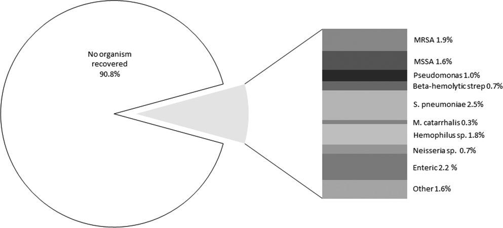
DISCUSSION
Our study identifies a larger cohort of patients with aspiration pneumonia than previous studies.[21, 22, 23, 24, 25] Patients with community‐acquired aspiration pneumonia are older and more likely to die than CAP patients. They are more likely to be admitted to the hospital or ICU. Thirty‐day mortality in this patient population was significantly underestimated by CURB‐65 and eCURB, models developed and validated in CAP populations.[9, 26] This finding supports a prior study.[27] It appears that a traditional prognostic model assessing mortality risk in the CAP patient does not apply to the aspiration‐pneumonia patient. One reason for eCURB and CURB‐65s poor utility in community‐acquired aspiration pneumonia may be their reliance on objective clinical features rather than comorbidities, which may influence mortality to a greater degree in aspiration pneumonia.
This study has several limitations. There is no gold standard for the definition of aspiration pneumonia, and it is difficult to distinguish aspiration pneumonia from typical pneumonia. It is plausible that older patients with greater comorbidities are being designated as aspiration pneumonia. If this is the case, then aspiration pneumonia merely represents the end of the pneumonia spectrum with highest mortality risk, and it is no surprise that these patients fare poorly.
It appears that the hospitalist or emergency department physician implicitly appreciates that aspiration pneumonia has a higher mortality risk than predicted by traditional severity assessment. With such high mortality and morbidity, a patient presenting to the emergency room with aspiration pneumonia is almost always admitted to the hospital. Further work in this area should investigate other factors to improve prognostic modeling in patients with aspiration pneumonia, although the utility of such a model may be limited to determining ICU admission. Our data indicate that IDSA/ATS minor criteria for SCAP are not useful in predicting admission to the ICU in patients with aspiration pneumonia.
In this study, a DNR/DNI order was twice as common in the community‐acquired aspiration pneumonia population than the CAP population. However, patients with community‐acquired aspiration pneumonia and a DNR/DNI order were more than 3 times more likely to die than patients with CAP and a DNR/DNI order. Our regression model suggested that the presence of a DNR/DNI order was an independent predictor of mortality (OR: 1.75, P < 0.001). Although a DNR/DNI order may correlate with the withholding or withdrawal of medical therapy, it is also a surrogate for increased age or comorbidities.[28] In our study, however, the increased prevalence of DNR/DNI orders did not explain the poor mortality prediction of the eCURB or CURB‐65, as exclusion of those patients did not significantly alter the AUCs in either the aspiration group or the CAP group.
Controversy exists regarding treatment of aspiration pneumonia. Historically, some have advocated for treatment of aspiration pneumonia with a regimen designed to cover anaerobic bacteria.[29] This recommendation was based on early microbiologic studies that obtained the samples late in the course of illness, or other studies where the sample was obtained transtracheally, where oropharyngeal flora may contaminate the sample.[30, 31, 32] Our clinically obtained microbiologic recovery of organisms was similar to the flora recovered in more recent CAP studies, in respect to both the incidence of pathogen recovery and the relative frequencies of recovered organisms.[33, 34] Our data do not support inferences regarding the prevalence of anaerobic infections, as the recovery of anaerobic organisms was limited to blood and pleural fluid cultures in this study, rather than techniques used in research settings that might have greater yield. As expected, patients with healthcare‐associated risk factors trended toward increased incidence of MRSA. Given the similarity of the organisms recovered to those recovered in CAP,[35] this study supports IDSA/ATS recommendations that antibiotic therapy in aspiration pneumonia be similar to that of higher‐risk CAP, with the addition of vancomycin or linezolid for MRSA coverage in patients with risk factors for healthcare‐associated pneumonia.[17]
Our study is limited by its single‐center retrospective design. However, beginning in 1995, the LDS Hospital emergency department initiated a standardized pneumonia therapy protocol and deployed electronic medical records, which prospectively recorded a wide array of clinical, therapeutic, and biometric data. Most data elements used in this analysis were routinely charted for clinical purposes in real time. Although the eCURB, CURB‐65, and some comorbidities could be extracted electronically for each patient, it was not possible to calculate the pneumonia severity index score due to our inability to rigorously identify the necessary comorbid illness elements. Other comorbidities, not present in our model, may have been identified by the physician who makes a diagnosis of aspiration pneumonia. Our identification of swallow impairment is also methodologically limited. The decision to obtain a swallow study was clinical, usually occurring upon convalescence. Therefore, it is not possible to distinguish between antecedent oropharyngeal dysfunction and post‐critical illness dysfunction.
Our definition of aspiration pneumonia required the treating physician to diagnose and code the patient as having aspiration pneumonia, followed by excluding patients more likely to have aspiration pneumonitis. Although we relied on ICD‐9 codes to initially identify aspiration pneumonia, all patients in our database were confirmed by physician chart review. Our incidence of community‐acquired aspiration pneumonia is congruent with other studies using different methodologies.[1, 36, 37] Unfortunately, there is no standard and widely accepted definition for separating aspiration pneumonia from usual CAP. A younger and healthier patient who has developed pneumonia subsequent to aspiration may be more likely to be diagnosed with CAP, resulting in selection bias for older patients with greater comorbidities.
CONCLUSION
Patients diagnosed with aspiration pneumonia are older, have more comorbid conditions, and demonstrate greater disease severity and higher 30‐day mortality than CAP patients. Mortality prediction using CURB‐65 and eCURB in this population was poor, possibly due to a greater effect of comorbidities on mortality. The pneumonia severity index, which incorporates patient comorbidities, might perform better than the eCURB or CURB‐65, and should be studied in aspiration pneumonia populations where comorbid illness information is prospectively collected. Further areas of study include creating an improved mortality prediction model for aspiration pneumonia that incorporates comorbid conditions, DNR/DNI status, and disease severity.
Acknowledgments
The authors acknowledge Al Jephson for database support, Yao Li for statistical analysis, and Anita Austin for help reviewing the medical records. Dr. Lanspa had full access to all of the data in the study and takes responsibility for the integrity of the data and the accuracy of the data analysis.
Disclosure
Preliminary versions of this work were presented as posters at the American Thoracic Society Meeting, Denver, Colorado, May 17, 2011. This study was supported by grants from the Intermountain Research and Medical Foundation. Dr. Brown is supported by a career development award from National Institute of General Medical Sciences (K23GM094465). Dr. Dean served on an advisory board for Merck, has been a paid consultant for Cerexa, and has received an investigator‐initiated competitive grant from Pfizer for development of an electronic pneumonia decision support tool. All other authors report no relevant financial disclosures.
- , , , et al. Severe community‐acquired pneumonia. Epidemiology and prognostic factors. Am Rev Respir Dis. 1991;144(2):312–318.
- , , . Risk factors for pneumonia in the elderly. Am J Med. 1994;96(4):313–320.
- , . Aspiration pneumonia and dysphagia in the elderly. Chest. 2003;124(1):328–336.
- , , . Pneumonia versus aspiration pneumonitis in nursing home residents: diagnosis and management. J Am Geriatr Soc. 2003;51(1):17–23.
- . Aspiration pneumonia: mixing apples with oranges and tangerines. Crit Care Med. 2004;32(5):1236; author reply 1236–1237.
- , , , , . Epidemiology and impact of aspiration pneumonia in patients undergoing surgery in Maryland, 1999–2000. Crit Care Med. 2003;31(7):1930–1937.
- . Aspiration syndromes: aspiration pneumonia and pneumonitis. Hosp Pract (Minneap). 2010;38(1):35–42.
- . Aspiration pneumonitis and aspiration pneumonia. N Engl J Med. 2001;344(9):665–671.
- , , , et al. CURB‐65 pneumonia severity assessment adapted for electronic decision support. Chest. 2011;140(1):156–163.
- , , , et al. Defining community acquired pneumonia severity on presentation to hospital: an international derivation and validation study. Thorax. 2003;58(5):377–382.
- , , , et al. Comparison of a disease‐specific and a generic severity of illness measure for patients with community‐acquired pneumonia. J Gen Intern Med. 1995;10(7):359–368.
- , , , et al. Development and validation of a clinical prediction rule for severe community‐acquired pneumonia. Am J Respir Crit Care Med. 2006;174(11):1249–1256.
- , , , . Validation of the Infectious Disease Society of America/American Thoracic Society 2007 guidelines for severe community‐acquired pneumonia. Crit Care Med. 2009;37(12):3010–3016.
- Guidelines for the management of adults with hospital‐acquired, ventilator‐associated, and healthcare‐associated pneumonia. Am J Respir Crit Care Med. 2005;171(4):388–416.
- .The Utah genealogical database: a resource for genetic epidemiology. In: Cairns JL, Skolnick M, eds. Banbury Report No 4; Cancer Incidence in Defined Populations. New York, NY:Cold Spring Harbor Laboratory;1980:285–297.
- . Simple, accurate equations for human blood O2 dissociation computations. J Appl Physiol. 1979;46(3):599–602.
- , , , et al. Infectious Diseases Society of America/American Thoracic Society consensus guidelines on the management of community‐acquired pneumonia in adults. Clin Infect Dis. 2007;44(suppl 2):S27–S72.
- , , , . Validation of a combined comorbidity index. J Clin Epidemiol. 1994;47(11):1245–1251.
- , , . Adapting a clinical comorbidity index for use with ICD‐9‐CM administrative databases. J Clin Epidemiol. 1992;45(6):613–619.
- , , . Comparing the areas under two or more correlated receiver operating characteristic curves: a nonparametric approach. Biometrics. 1988;44(3):837–845.
- , , . Prandial aspiration and pneumonia in an elderly population followed over 3 years. Dysphagia. 1996;11(2):104–109.
- , , , . Predictors of aspiration pneumonia in nursing home residents. Dysphagia. 2002;17(4):298–307.
- , , , , , . Aspiration pneumonia: dental and oral risk factors in an older veteran population. J Am Geriatr Soc. 2001;49(5):557–563.
- , , , et al. Microbiology of severe aspiration pneumonia in institutionalized elderly. Am J Respir Crit Care Med. 2003;167(12):1650–1654.
- , , . Aspiration pneumonia in stroke. Arch Phys Med Rehabil. 1993;74(9):973–976.
- , , , et al. Validation of a predictive rule for the management of community‐acquired pneumonia. Eur Respir J. 2006;27(1):151–157.
- , , , , . Pneumonia Severity Index (PSI), CURB‐65, and mortality in hospitalized elderly patients with aspiration pneumonia [in German]. Zeitschrift fur Gerontologie und Geriatrie. 2011;44(4):229–234.
- , , , . Do‐not‐resuscitate orders for critically ill patients in the hospital. How are they used and what is their impact?JAMA. 1986;256(2):233–237.
- , , , , . The Sanford Guide to Antimicrobial Therapy: 2010.40th ed.Sperryville, VA:Antimicrobial Therapy, Inc.;2009.
- , , . The bacteriology of aspiration pneumonia. Am J Med. 1974;56(2):202–207.
- , , . Bacteriologic flora of aspiration‐induced pulmonary infections. Arch Intern Med. 1975;135(5):711–714.
- , . Bacteriology of aspiration pneumonia. A prospective study of community‐ and hospital‐acquired cases. Ann Intern Med. 1974;81(3):329–331.
- , . The role of anaerobes in patients with ventilator‐associated pneumonia and aspiration pneumonia: a prospective study. Chest. 1999;115(1):178–183.
- , , , et al. Is penicillin G an adequate initial treatment for aspiration pneumonia? A prospective evaluation using a protected specimen brush and quantitative cultures. Intensive Care Med. 1993;19(5):279–284.
- , , , , . Community‐acquired pneumonia in the elderly. Semin Respir Infect. 1999;14(2):173–183.
- , , . Nursing home‐acquired pneumonia. A case‐control study. J Am Geriatr Soc. 1986;34(10):697–702.
- , , , , . Severe community‐acquired pneumonia. Etiology, epidemiology, and prognosis factors. French Study Group for Community‐Acquired Pneumonia in the Intensive Care Unit. Chest. 1994;105(5):1487–1495.
Pneumonia is a common clinical syndrome with well‐described epidemiology and microbiology. Aspiration pneumonia comprises 5% to 15% of patients with pneumonia,[1] but is less well‐characterized despite being a major syndrome of pneumonia in the elderly.[2, 3] Difficulties in studying aspiration pneumonia include the lack of a sensitive and specific marker for aspiration, the overlap between aspiration pneumonia and other forms of pneumonia, and the lack of differentiation between aspiration pneumonia and aspiration pneumonitis by many clinicians.[4, 5, 6] Aspiration pneumonia, which develops after the aspiration of oropharyngeal contents, differs from aspiration pneumonitis, wherein inhalation of gastric contents causes inflammation without the subsequent development of bacterial infection.[7, 8]
A number of validated mortality prediction models exist for community‐acquired pneumonia (CAP), using a variety of clinical predictors. One clinical prediction rule endorsed by the British Thoracic Society is the CURB‐65, which assigns a score for Confusion, Uremia >19 mg/dL, Respiratory rate >= 30 breaths/min, Blood Pressure < 90 mmHg systolic or < 60 mmHg diastolic, and age 65). We favor eCURB, a version of the CURB‐65 model that uses continuously weighted variables to more accurately predict mortality, validated in CAP populations.[9] Most studies validating pneumonia severity scoring systems excluded aspiration pneumonia from their study population.[10, 11, 12] Severity scoring systems for CAP may not accurately predict disease severity patients with aspiration pneumonia.
The aims of our study were to: (1) identify a population of patients with aspiration pneumonia; (2) compare characteristics and outcomes in patients with community‐acquired aspiration pneumonia to those with CAP; and (3) study the performance of eCURB and CURB‐65 in predicting mortality for patients with community‐acquired aspiration pneumonia.
PATIENTS AND METHODS
Study Design and Setting
The study was performed at LDS Hospital, a university‐affiliated community teaching hospital in Salt Lake City, Utah, with 520 beds. In retrospective analysis of data from the electronic medical records, we identified all patients older than 18 years who were evaluated in the emergency department at LDS Hospital or admitted patients from other sources (direct admission, transfer from another hospital) from 1996 to 2006 with International Statistical Classification of Disease and Health Related Problems, 9th Revision (ICD‐9) codes specific for aspiration pneumonia and pneumonitis (507.x). The treating physicians were mostly hospitalists and intensivists. Two physicians (M.L. and N.D.) manually reviewed the electronic medical records, including the emergency room physician's notes, the admission histories and physicals, the discharge summaries, and radiographic reports of the patients identified in the query. Consensus regarding the diagnosis of aspiration pneumonia was achieved in all patients reviewed using criteria listed in Table 1. This study was approved by the LDS Hospital institutional review board, and permission was granted to use the Utah Population Database for determining mortality (#1008505), with a waiver of informed consent. For the contemporaneous group of CAP patients, we used a previously described population identified using ICD‐9 codes 481.x to 487.x, captured from the same hospital during the same period.[13]
| Inclusion Criteria | Exclusion Criteria |
|---|---|
| |
| 1. Age 18 years | 1. Absence of radiographic evidence of pneumonia within 48 hours after evaluation |
| 2. Either admitted to hospital or evaluated in emergency department | 2. Previous episode of aspiration pneumonia within 12 months |
| 3. 507.x code as primary diagnosis | 3. Initial admission date >48 hours before transfer to LDS Hospital |
| 4. 507.x code as secondary diagnosis with a primary diagnosis of pneumonia, respiratory failure, or septicemia | 4. AIDS 5. Receipt of antiretroviral therapy 6. History of solid organ transplant |
| 5. Treating physician indicated a diagnosis of aspiration pneumonia in the history and physical and/or discharge summary | 7. Hematologic malignancy 8. Witnessed isolated aspiration event within 24 hours prior to evaluation 9. Drug overdose, cardiopulmonary arrest, or seizure prior to hospital admission 10. Laryngoscopic or bronchoscopic evidence of food material in airway |
Inclusion and Exclusion Criteria
Inclusion and exclusion criteria are listed in Table 1. To exclude patients with recurrent pneumonia, we included only the first episode of pneumonia in a given 12‐month period. LDS Hospital frequently receives patients transferred from surrounding emergency departments and intensive care units. We excluded patients who were transferred >48 hours from their initial emergency department admission and therefore were late in their disease course. Exclusion criteria 8 to 10 were used to exclude patients with clinical presentations more consistent with aspiration pneumonitis. We also excluded immunocompromised patients (criteria 4 to 7).
Healthcare‐associated aspiration pneumonia was defined as receipt of chronic hemodialysis, residence in a nursing facility, or hospitalization within any Intermountain Healthcare‐affiliated hospital within the past 90 days.[14] The remaining patients were defined as community‐acquired aspiration pneumonia.
Measurements
The first vital signs, orientation status, and first 12 hours of routine laboratory results were extracted from the electronic medical records and used to calculate predicted mortality by eCURB and CURB‐65. We determined 30‐day mortality from the merger of the electronic medical records with vital status information from the Utah Population Database.[15] The first measured SpO2 and FiO2 were used to estimate the PaO2/FiO2 ratio, using the Severinghaus calculation[16] if no arterial blood gas was available. Presence of American Thoracic Society/Infectious Diseases Society of America (IDSA/ATS) 2007 minor criteria for severe community‐acquired pneumonia (SCAP)[17] were obtained from baseline patient characteristics (Table 2). A Charlson comorbidity index was calculated from ICD‐9 codes using published methodology.[18, 19] Presence of an abnormal swallow was defined as dysphagia or aspiration on modified barium swallow study, fiberoptic endoscopic evaluation, or clinical determination by a speech language pathologist during the index hospitalization. We also looked for causative pathogens, defined by a positive pneumococcus or legionella urinary antigen, or a positive culture from blood, bronchoalveolar lavage, pleural fluid, or tracheal aspirate, collected within 24 hours of admission. Antibiotics administered within the first 24 hours of admission were classified into 4 broad groups based on local physician prescribing patterns. Clindamycin and metronidazole were considered anaerobic‐specific antibiotics. Vancomycin or linezolid were considered methicillin‐resistant Staphylococcus aureus (MRSA) antibiotics. Broad‐spectrum antibiotics included any of the following: carbapenems, aztreonam, piperacillin/tazobactam, ticarcillin/clavulanate, cefepime, and ceftazidime. Macrolides, respiratory fluoroquinolones, and third‐generation cephalosporins were considered standard‐care antibiotics.
| Respiratory rate 30 breaths/minute |
| PaO2/FiO2 250 |
| Multilobar infiltrates |
| Confusion/disorientation |
| Uremia (blood urea nitrogen 20 mg/dL) |
| Leukopenia (white blood cell count <4000 cells/mm3) |
| Thrombocytopenia (platelet count <100,000 cells/mm3) |
| Hypothermia (core temperature 36C) |
| Hypotension requiring aggressive fluid resuscitation |
Statistical Analysis
We compared baseline patient characteristics and clinical outcomes using the Fisher exact test to compare proportions of categorical variables, and Mann‐Whitney U test or Student t test to compare central tendencies of continuous variables, as dictated by the normality of the data. Receiver operating characteristic curves calculated the ability of eCURB and CURB‐65 to predict 30‐day mortality prediction in patients with community‐acquired aspiration pneumonia and CAP, as well as the ability of IDSA/ATS minor criteria for SCAP to predict admission to the intensive care unit (ICU). We performed multivariate logistic regression to predict 30‐day mortality in patients with community‐acquired aspiration pneumonia and CAP, using stepwise backward elimination. Confounders were included if they were significant at a 0.05 level or if they altered the coefficient of the main variable by more than 10%. For logistic models, we evaluated goodness of fit with the Hosmer‐Lemeshow technique; comparisons of area under the curve (AUC) among models were made using the technique of DeLong.[20] Two‐tailed P values of 0.05 were considered statistically significant. Stata version 12 (StataCorp, College Station, TX) was used for all analyses.
RESULTS
Our initial query identified 1165 patients. Physician review of the medical records resulted in 628 patients, 118 of whom were classified as healthcare‐associated aspiration pneumonia (Figure 1, Table 3). Of all aspiration pneumonia patients, 80% were seen in the emergency department, 12.5% were directly admitted from the community, and 7.5% were transferred from another healthcare facility. Almost all patients seen in the emergency department (99.0%) were admitted to the hospital, with median length of hospitalization 6.7 days among survivors.
| Aspiration Pneumonia (N = 628) | Community‐Acquired Aspiration Pneumonia (N = 510) | Healthcare Associated Aspiration Pneumonia (N=118) | P Value | |
|---|---|---|---|---|
| ||||
| Age (range), y | 77 (6585) | 77 (6485) | 80 (6786) | 0.42 |
| Female, % | 49.8 | 50.2 | 48.3 | 0.76 |
| 30‐day mortality, % | 21.0% | 19.0% | 29.7% | 0.02 |
| CURB‐65 score | 2 (13) | 2 (13) | 2 (13) | 0.0012 |
| Confusion | 13.9% | 12.7% | 18.6% | 0.10 |
| Blood urea nitrogen (mg/dL) | 22 (1634) | 21 (1532) | 30 (2047) | <0.0001 |
| Respiratory rate (breaths/min) | 20 (1826) | 20 (1824) | 21 (1828) | 0.30 |
| Systolic blood pressure (mm Hg) | 128 (110149) | 129 (110150) | 127 (105146) | 0.28 |
| eCURB 30‐day mortality estimate (median, %) | 5.6 (2.414.2) | 5.2 (2.212.4) | 8.9 (4.222.5) | <0.0001 |
| eCURB 30‐day mortality estimate (mean, %) | 10.6 12.2 | 9.7 11.5 | 14.614.1 | <0.0001 |
| Hospital admission (of ED visits), % | 99.0 | 98.8 | 100 | 0.59 |
| Hospital LOS, d | 6.7 (4.111.1) | 6.5 (4.011.0) | 7.8 (5.412.3) | 0.05 |
| ICU admission, % | 37.9 | 37.1 | 41.5 | 0.21 |
| ICU LOS, d | 3.5 (1.98.8) | 3.1 (1.87.6) | 5.6 (3.810.8) | 0.02 |
| Mean ventilator‐free days (of ICU patients, out of 30 days) | 25.28.3 | 25.97.7 | 22.710.0 | 0.01 |
| Receipt of mechanical ventilation, % | 18.6 | 17.2 | 24.6 | 0.09 |
| Duration of ventilation, d | 2.8 (0.96.5) | 3.1 (1.06.6) | 1.9 (0.86.3) | 0.05 |
| Receipt of vasopressor, % | 1.8 | 1.4 | 3.4 | 0.13 |
| Charlson comorbidity index | 4 (26) | 3 (26) | 4 (36) | 0.0024 |
| Cerebrovascular disease, % | 33.9 | 32.4 | 40.7 | 0.11 |
| Chronic pulmonary disease, % | 51.0 | 51.8 | 47.5 | 0.42 |
| Congestive heart failure, % | 52.4 | 50.0 | 62.7 | 0.01 |
| Connective tissue disease, % | 8.4 | 8.8 | 6.8 | 0.58 |
| Dementia, % | 14.2 | 12.0 | 23.7 | 0.0019 |
| Hemiplegia/paraplegia | 9.4 | 8.0 | 15.2 | 0.02 |
| Myocardial infarction, % | 21.0 | 17.8 | 29.7 | 0.02 |
| Peripheral vascular disease, % | 17.7 | 16.3 | 23.7 | 0.06 |
| Peptic ulcer disease, % | 18.8 | 19.2 | 16.9 | 0.70 |
| Diabetes without complications, % | 10.7 | 9.2 | 16.9 | 0.02 |
| Diabetes with complications, % | 31.5 | 30.4 | 36.4 | 0.23 |
| Mild liver disease, % | 8.6 | 8.0 | 11.0 | 0.28 |
| Moderate or severe liver disease, % | 1.8 | 1.6 | 2.5 | 0.44 |
| Malignant solid tumor, % | 16.6 | 17.3 | 13.6 | 0.41 |
| Metastatic cancer, % | 5.4 | 5.7 | 4.2 | 0.66 |
| Renal disease, % | 14.7 | 4.2 | 18.6 | 0.19 |
| 3 or more minor SCAP criteria, %* | 24.7 | 23.1 | 31.4 | 0.08 |
| PaO2/FiO2 ratio | 221 (161280) | 226 (169280) | 181 (133245) | 0.0004 |
| Multilobar disease, % | 46.3 | 43.2 | 53.9 | 0.11 |
| Presence of an effusion, % | 23.1 | 19.7 | 31.9 | 0.03 |
| Swallow impairment (of tested survivors), % | 34.1 | 34.1 | 34.1 | 0.22 |
| Presence of a DNR/DNI order, % | 26.4 | 23.7 | 38.1 | 0.0024 |
| Mortality of patients with DNR/DNI order, % | 39.1 | 38.8 | 40.0 | 1.00 |
| Receipt of broad‐spectrum antibiotic, % | 35.4 | 32.5 | 47.5 | 0.0028 |
| Receipt of MRSA antibiotic, % | 7.5 | 5.7 | 15.3 | 0.0014 |
| Receipt of anaerobe antibiotic, % | 28.7 | 27.6 | 33.1 | 0.26 |
Observed mortality was 21.0%. eCURB significantly underestimated mortality in this group, predicting a mortality rate of 10.6%. When classifying patients by the 2007 IDSA/ATS guidelines, 24.7% of the patients had 3 or more minor criteria for SCAP.[17] The PaO2/FiO2 ratio was obtained in 99.7% of patients. The median PaO2/FiO2 ratio observed in this population was 221 mm Hg (equivalent to 260 mm Hg at sea level barometric pressure, adjusted for our altitude of 1400 meters), near the threshold sea level definition (250 mm Hg) for SCAP.[13, 17] Admission to the ICU was common, as were admission orders for do not resuscitate (DNR) or do not intubate (DNI). Patients with healthcare‐associated aspiration pneumonia had a higher comorbidity index and had a higher mortality rate than patients with community‐acquired aspiration pneumonia, although we found no significant difference in the rate of hospital or ICU admission or the receipt of critical care therapies. Inpatient assessment of dysphagia and aspiration was conducted in 177 patients. Abnormal swallow was noted in 96% of those tested.
We found several differences between patients with community‐acquired aspiration pneumonia and 2584 patients with CAP identified during the same time period[13] (Table 4). Patients with community‐acquired aspiration pneumonia were older, more likely to have multilobar disease or effusion on imaging, and had greater disease severity. They also had a higher frequency of ICU and hospital admission, IDSA/ATS minor criteria for SCAP, and higher Charlson comorbidity indices. Patients with community‐acquired aspiration pneumonia were more likely to receive mechanical ventilation than CAP patients, although there was no difference in 30‐day mortality among intubated patients or a difference in ventilator‐free days.
| Community‐Acquired Aspiration Pneumonia (N = 510) | Community‐ Acquired Pneumonia (N = 2584) | P Value | |
|---|---|---|---|
| |||
| Age (range), y | 77 (6485) | 59 (4176) | <0.0001 |
| Female, % | 50.2 | 49.5 | 0.81 |
| 30‐day mortality, % | 19.0 | 4.2 | <0.0001 |
| CURB‐65 score | 2 (13) | 1 (02) | <0.0001 |
| Confusion, % | 12.7 | 5.1 | <0.0001 |
| Blood urea nitrogen | 21 (1532) | 16 (1124) | <0.0001 |
| Respiratory rate | 20 (1824) | 20 (1824) | <0.0001 |
| Systolic blood pressure | 129 (110150) | 130 (112146) | 0.67 |
| eCURB 30‐day mortality estimate, median, % | 5.2 (2.212.4) | 1.7 (0.94.3) | <0.0001 |
| eCURB 30‐day mortality estimate, mean, % | 9.7 11.5 | 4.4 7.5 | <0.0001 |
| AUC of eCURB versus mortality | 0.71 (0.660.75) | 0.86 (0.830.90) | <0.0001 |
| Excluding DNR/DNI patients | 0.69 (0.650.74) | 0.87 (0.830.90) | 0.0001 |
| AUC of CURB‐65 versus mortality | 0.66 (0.620.69) | 0.81 (0.780.85) | <0.0001 |
| Excluding DNR/DNI patients | 0.65 (0.600.70) | 0.81 (0.760.85) | 0.0003 |
| Hospital admission (of ED visits), % | 98.8 | 57.8 | <0.0001 |
| Hospital LOS, d | 6.5 (4.011.0) | 3.3 (2.25.2) | <0.0001 |
| ICU admission, % | 37.1 | 14.2 | <0.0001 |
| ICU LOS, d | 3.1 (1.87.6) | 2.5 (1.17.7) | 0.01 |
| Mean ventilator‐free days (of ICU patients, out of 30 days) | 25.9 7.7 | 25 9 | 0.75 |
| Receipt of mechanical ventilation, % | 17.2 | 7.8 | <0.0001 |
| Duration of ventilation, d | 3.1 (1.06.6) | 3.5 (1.57.2) | 0.09 |
| Receipt of vasopressor, % | 1.4 | 3.3 | 0.02 |
| Charlson comorbidity index | 3 (26) | 1 (03) | <0.0001 |
| Cerebrovascular disease, % | 32.4 | 10.0 | <0.0001 |
| Chronic pulmonary disease, % | 51.8 | 42.5 | <0.0001 |
| Congestive heart failure, % | 50.0 | 22.1 | <0.0001 |
| Connective tissue disease, % | 8.8 | 5.6 | 0.0084 |
| Dementia, % | 12.0 | 2.8 | <0.0001 |
| Hemiplegia/paraplegia, % | 8.0 | 2.7 | <0.0001 |
| Myocardial infarction, % | 17.8 | 10.8 | <0.0001 |
| Peripheral vascular disease, % | 16.3 | 7.4 | <0.0001 |
| Peptic ulcer disease, % | 19.2 | 7.6 | <0.0001 |
| Diabetes without complications, % | 9.2 | 24.7 | <0.0001 |
| Diabetes with complications, % | 30.4 | 5.1 | <0.0001 |
| Mild liver disease, % | 8.0 | 6.2 | 0.14 |
| Moderate or severe liver disease, % | 1.6 | 0.8 | 0.13 |
| Malignant solid tumor, % | 17. | 8.9 | <0.0001 |
| Metastatic cancer, % | 5.7 | 1.3 | <0.0001 |
| Renal disease, % | 4.2 | 5.6 | <0.0001 |
| 3 or more minor SCAP criteria, %* | 24.7 | 19.1 | 0.01 |
| PaO2/FiO2 ratio | 226 (169280) | 260 (148338) | 0.0004 |
| Multilobar disease, % | 43.2 | 37.2 | 0.0012 |
| Presence of an effusion, % | 19.7 | 18.3 | <0.0001 |
| Presence of a DNR/DNI order, % | 23.7 | 9.7 | <0.0001 |
| Mortality of patients with DNR/DNI order, % | 38.8 | 12.4 | <0.0001 |
| Receipt of broad‐spectrum antibiotic, % | 32.5 | 8.4 | <0.0001 |
| Receipt of MRSA antibiotic, % | 5.7 | 2.2 | <0.0001 |
| Receipt of anaerobe antibiotic, % | 27.6 | 3.1 | <0.0001 |
Thirty‐day mortality for patients with community‐acquired aspiration pneumonia was significantly higher than in CAP patients. Patients with community‐acquired aspiration pneumonia also had higher eCURB and CURB‐65 scores. However, eCURB was a poor predictor of 30‐day mortality, with an AUC of 0.71, compared to 0.86 calculated for the CAP population (Figure 2). CURB‐65 performed similarly: AUC was 0.66 vs 0.81. The presence of a DNR/DNI order was twice as prevalent in the community‐acquired aspiration pneumonia population vs the CAP population; those patients with a DNR/DNI order were 3 times as likely to die. Excluding patients with a DNR/DNI order did not improve performance of eCURB or CURB‐65 (Table 4). The presence of IDSA/ATS minor criteria for SCAP was not predictive of triage to the ICU in the group of patients with community‐acquired aspiration pneumonia (AUC: 0.51), compared with CAP patients (AUC: 0.88, P < 0.01 for comparison, Figure 3). This finding persisted in the subset of patients without a DNR/DNI order (AUC: 0.52 in community‐acquired aspiration pneumonia vs 0.88 in CAP, P < 0.01).
Our regression model of mortality incorporated gender, presence of effusion or multilobar pneumonia, presence of a DNR/DNI order, and all the components of the CURB‐65, IDSA/ATS minor criteria for SCAP, and Charlson comorbidity index. The regression model demonstrated that even after adjustment for age, comorbidities, disease severity, and presence of a DNR/DNI order, the presence of aspiration pneumonia was associated with higher mortality than CAP (odds ratio [OR]: 3.46, P < 0.001, Table 5). In this model, systolic blood pressure did not predict mortality, and diabetes with complications was associated with decreased mortality.
| Odds Ratio | P Value | |
|---|---|---|
| ||
| Presence of aspiration pneumonia | 3.46 (2.115.67) | <0.001 |
| Age, y | 1.03 (1.011.04) | <0.001 |
| Confusion | 3.14 (1.955.05) | <0.001 |
| Blood urea nitrogen, mg/dL | 1.03 (1.021.04) | <0.001 |
| Respiratory rate, breaths/minute | 1.03 (1.001.06) | 0.04 |
| PaO2/FiO2 ratio, per 1 mm Hg | 0.99 (0.991.00) | <0.001 |
| Moderate or severe liver disease | 9.21 (3.1626.86) | <0.001 |
| Paraplegia/hemiplegia | 2.43 (1.135.27) | 0.02 |
| Diabetes with complications | 0.42 (0.200.87) | 0.02 |
| Leukocytosis | 4.47 (2.278.82) | <0.001 |
| DNR/DNI | 1.75 (1.112.75) | 0.02 |
Microbiological Findings
Blood cultures were performed at admission in 67.4% of aspiration‐pneumonia patients, and a tracheal aspirate in half (50.7%) of intubated patients with aspiration pneumonia. Organisms were recovered in 90 patients (14.3%), although 41 of those patients had tracheal aspirates of organisms commonly thought to be nonpathogenic (nonpneumococcal alpha‐hemolytic streptococcus, nonhemolytic streptococcus, diphtheroids, micrococci, coagulase negative staphylococccus). Tracheal aspirate was the most common method of recovering an organism (7.8% of patients), followed by blood culture (4.3%). Bronchoalveolar lavage, urinary antigen, and pleural fluid culture were less common (1.3%, 1.1%, 0.3%, respectively). The microbiologic results were grouped into: Staphylococcus aureus, Streptococcus pneumoniae, enteric bacilli, Haemophilus species, Neisseria species, Moraxella catarrhalis, and Pseudomonas aeruginosa (Figure 4). Comparing healthcare‐associated with community‐acquired aspiration pneumonia, healthcare‐associated patients were more likely to have a confirmed infection with MRSA (4.2% vs 1.4%, P = 0.06) and enteric bacteria (5.1% vs 1.6%, P = 0.03). There were no other statistically significant differences in microbiologic recovery between the 2 groups. Antibiotics targeting anaerobic pathogens were administered in 28.7% of patients with aspiration pneumonia, with no correlation to the presence of healthcare‐associated risks. Healthcare‐associated patients were more likely to receive broad‐spectrum antibiotics (47.5% vs 32.5%, P < 0.01) and MRSA coverage (15.3% vs 5.7%, P < 0.01) than patients with community‐acquired aspiration pneumonia.
DISCUSSION
Our study identifies a larger cohort of patients with aspiration pneumonia than previous studies.[21, 22, 23, 24, 25] Patients with community‐acquired aspiration pneumonia are older and more likely to die than CAP patients. They are more likely to be admitted to the hospital or ICU. Thirty‐day mortality in this patient population was significantly underestimated by CURB‐65 and eCURB, models developed and validated in CAP populations.[9, 26] This finding supports a prior study.[27] It appears that a traditional prognostic model assessing mortality risk in the CAP patient does not apply to the aspiration‐pneumonia patient. One reason for eCURB and CURB‐65s poor utility in community‐acquired aspiration pneumonia may be their reliance on objective clinical features rather than comorbidities, which may influence mortality to a greater degree in aspiration pneumonia.
This study has several limitations. There is no gold standard for the definition of aspiration pneumonia, and it is difficult to distinguish aspiration pneumonia from typical pneumonia. It is plausible that older patients with greater comorbidities are being designated as aspiration pneumonia. If this is the case, then aspiration pneumonia merely represents the end of the pneumonia spectrum with highest mortality risk, and it is no surprise that these patients fare poorly.
It appears that the hospitalist or emergency department physician implicitly appreciates that aspiration pneumonia has a higher mortality risk than predicted by traditional severity assessment. With such high mortality and morbidity, a patient presenting to the emergency room with aspiration pneumonia is almost always admitted to the hospital. Further work in this area should investigate other factors to improve prognostic modeling in patients with aspiration pneumonia, although the utility of such a model may be limited to determining ICU admission. Our data indicate that IDSA/ATS minor criteria for SCAP are not useful in predicting admission to the ICU in patients with aspiration pneumonia.
In this study, a DNR/DNI order was twice as common in the community‐acquired aspiration pneumonia population than the CAP population. However, patients with community‐acquired aspiration pneumonia and a DNR/DNI order were more than 3 times more likely to die than patients with CAP and a DNR/DNI order. Our regression model suggested that the presence of a DNR/DNI order was an independent predictor of mortality (OR: 1.75, P < 0.001). Although a DNR/DNI order may correlate with the withholding or withdrawal of medical therapy, it is also a surrogate for increased age or comorbidities.[28] In our study, however, the increased prevalence of DNR/DNI orders did not explain the poor mortality prediction of the eCURB or CURB‐65, as exclusion of those patients did not significantly alter the AUCs in either the aspiration group or the CAP group.
Controversy exists regarding treatment of aspiration pneumonia. Historically, some have advocated for treatment of aspiration pneumonia with a regimen designed to cover anaerobic bacteria.[29] This recommendation was based on early microbiologic studies that obtained the samples late in the course of illness, or other studies where the sample was obtained transtracheally, where oropharyngeal flora may contaminate the sample.[30, 31, 32] Our clinically obtained microbiologic recovery of organisms was similar to the flora recovered in more recent CAP studies, in respect to both the incidence of pathogen recovery and the relative frequencies of recovered organisms.[33, 34] Our data do not support inferences regarding the prevalence of anaerobic infections, as the recovery of anaerobic organisms was limited to blood and pleural fluid cultures in this study, rather than techniques used in research settings that might have greater yield. As expected, patients with healthcare‐associated risk factors trended toward increased incidence of MRSA. Given the similarity of the organisms recovered to those recovered in CAP,[35] this study supports IDSA/ATS recommendations that antibiotic therapy in aspiration pneumonia be similar to that of higher‐risk CAP, with the addition of vancomycin or linezolid for MRSA coverage in patients with risk factors for healthcare‐associated pneumonia.[17]
Our study is limited by its single‐center retrospective design. However, beginning in 1995, the LDS Hospital emergency department initiated a standardized pneumonia therapy protocol and deployed electronic medical records, which prospectively recorded a wide array of clinical, therapeutic, and biometric data. Most data elements used in this analysis were routinely charted for clinical purposes in real time. Although the eCURB, CURB‐65, and some comorbidities could be extracted electronically for each patient, it was not possible to calculate the pneumonia severity index score due to our inability to rigorously identify the necessary comorbid illness elements. Other comorbidities, not present in our model, may have been identified by the physician who makes a diagnosis of aspiration pneumonia. Our identification of swallow impairment is also methodologically limited. The decision to obtain a swallow study was clinical, usually occurring upon convalescence. Therefore, it is not possible to distinguish between antecedent oropharyngeal dysfunction and post‐critical illness dysfunction.
Our definition of aspiration pneumonia required the treating physician to diagnose and code the patient as having aspiration pneumonia, followed by excluding patients more likely to have aspiration pneumonitis. Although we relied on ICD‐9 codes to initially identify aspiration pneumonia, all patients in our database were confirmed by physician chart review. Our incidence of community‐acquired aspiration pneumonia is congruent with other studies using different methodologies.[1, 36, 37] Unfortunately, there is no standard and widely accepted definition for separating aspiration pneumonia from usual CAP. A younger and healthier patient who has developed pneumonia subsequent to aspiration may be more likely to be diagnosed with CAP, resulting in selection bias for older patients with greater comorbidities.
CONCLUSION
Patients diagnosed with aspiration pneumonia are older, have more comorbid conditions, and demonstrate greater disease severity and higher 30‐day mortality than CAP patients. Mortality prediction using CURB‐65 and eCURB in this population was poor, possibly due to a greater effect of comorbidities on mortality. The pneumonia severity index, which incorporates patient comorbidities, might perform better than the eCURB or CURB‐65, and should be studied in aspiration pneumonia populations where comorbid illness information is prospectively collected. Further areas of study include creating an improved mortality prediction model for aspiration pneumonia that incorporates comorbid conditions, DNR/DNI status, and disease severity.
Acknowledgments
The authors acknowledge Al Jephson for database support, Yao Li for statistical analysis, and Anita Austin for help reviewing the medical records. Dr. Lanspa had full access to all of the data in the study and takes responsibility for the integrity of the data and the accuracy of the data analysis.
Disclosure
Preliminary versions of this work were presented as posters at the American Thoracic Society Meeting, Denver, Colorado, May 17, 2011. This study was supported by grants from the Intermountain Research and Medical Foundation. Dr. Brown is supported by a career development award from National Institute of General Medical Sciences (K23GM094465). Dr. Dean served on an advisory board for Merck, has been a paid consultant for Cerexa, and has received an investigator‐initiated competitive grant from Pfizer for development of an electronic pneumonia decision support tool. All other authors report no relevant financial disclosures.
- , , , et al. Severe community‐acquired pneumonia. Epidemiology and prognostic factors. Am Rev Respir Dis. 1991;144(2):312–318.
- , , . Risk factors for pneumonia in the elderly. Am J Med. 1994;96(4):313–320.
- , . Aspiration pneumonia and dysphagia in the elderly. Chest. 2003;124(1):328–336.
- , , . Pneumonia versus aspiration pneumonitis in nursing home residents: diagnosis and management. J Am Geriatr Soc. 2003;51(1):17–23.
- . Aspiration pneumonia: mixing apples with oranges and tangerines. Crit Care Med. 2004;32(5):1236; author reply 1236–1237.
- , , , , . Epidemiology and impact of aspiration pneumonia in patients undergoing surgery in Maryland, 1999–2000. Crit Care Med. 2003;31(7):1930–1937.
- . Aspiration syndromes: aspiration pneumonia and pneumonitis. Hosp Pract (Minneap). 2010;38(1):35–42.
- . Aspiration pneumonitis and aspiration pneumonia. N Engl J Med. 2001;344(9):665–671.
- , , , et al. CURB‐65 pneumonia severity assessment adapted for electronic decision support. Chest. 2011;140(1):156–163.
- , , , et al. Defining community acquired pneumonia severity on presentation to hospital: an international derivation and validation study. Thorax. 2003;58(5):377–382.
- , , , et al. Comparison of a disease‐specific and a generic severity of illness measure for patients with community‐acquired pneumonia. J Gen Intern Med. 1995;10(7):359–368.
- , , , et al. Development and validation of a clinical prediction rule for severe community‐acquired pneumonia. Am J Respir Crit Care Med. 2006;174(11):1249–1256.
- , , , . Validation of the Infectious Disease Society of America/American Thoracic Society 2007 guidelines for severe community‐acquired pneumonia. Crit Care Med. 2009;37(12):3010–3016.
- Guidelines for the management of adults with hospital‐acquired, ventilator‐associated, and healthcare‐associated pneumonia. Am J Respir Crit Care Med. 2005;171(4):388–416.
- .The Utah genealogical database: a resource for genetic epidemiology. In: Cairns JL, Skolnick M, eds. Banbury Report No 4; Cancer Incidence in Defined Populations. New York, NY:Cold Spring Harbor Laboratory;1980:285–297.
- . Simple, accurate equations for human blood O2 dissociation computations. J Appl Physiol. 1979;46(3):599–602.
- , , , et al. Infectious Diseases Society of America/American Thoracic Society consensus guidelines on the management of community‐acquired pneumonia in adults. Clin Infect Dis. 2007;44(suppl 2):S27–S72.
- , , , . Validation of a combined comorbidity index. J Clin Epidemiol. 1994;47(11):1245–1251.
- , , . Adapting a clinical comorbidity index for use with ICD‐9‐CM administrative databases. J Clin Epidemiol. 1992;45(6):613–619.
- , , . Comparing the areas under two or more correlated receiver operating characteristic curves: a nonparametric approach. Biometrics. 1988;44(3):837–845.
- , , . Prandial aspiration and pneumonia in an elderly population followed over 3 years. Dysphagia. 1996;11(2):104–109.
- , , , . Predictors of aspiration pneumonia in nursing home residents. Dysphagia. 2002;17(4):298–307.
- , , , , , . Aspiration pneumonia: dental and oral risk factors in an older veteran population. J Am Geriatr Soc. 2001;49(5):557–563.
- , , , et al. Microbiology of severe aspiration pneumonia in institutionalized elderly. Am J Respir Crit Care Med. 2003;167(12):1650–1654.
- , , . Aspiration pneumonia in stroke. Arch Phys Med Rehabil. 1993;74(9):973–976.
- , , , et al. Validation of a predictive rule for the management of community‐acquired pneumonia. Eur Respir J. 2006;27(1):151–157.
- , , , , . Pneumonia Severity Index (PSI), CURB‐65, and mortality in hospitalized elderly patients with aspiration pneumonia [in German]. Zeitschrift fur Gerontologie und Geriatrie. 2011;44(4):229–234.
- , , , . Do‐not‐resuscitate orders for critically ill patients in the hospital. How are they used and what is their impact?JAMA. 1986;256(2):233–237.
- , , , , . The Sanford Guide to Antimicrobial Therapy: 2010.40th ed.Sperryville, VA:Antimicrobial Therapy, Inc.;2009.
- , , . The bacteriology of aspiration pneumonia. Am J Med. 1974;56(2):202–207.
- , , . Bacteriologic flora of aspiration‐induced pulmonary infections. Arch Intern Med. 1975;135(5):711–714.
- , . Bacteriology of aspiration pneumonia. A prospective study of community‐ and hospital‐acquired cases. Ann Intern Med. 1974;81(3):329–331.
- , . The role of anaerobes in patients with ventilator‐associated pneumonia and aspiration pneumonia: a prospective study. Chest. 1999;115(1):178–183.
- , , , et al. Is penicillin G an adequate initial treatment for aspiration pneumonia? A prospective evaluation using a protected specimen brush and quantitative cultures. Intensive Care Med. 1993;19(5):279–284.
- , , , , . Community‐acquired pneumonia in the elderly. Semin Respir Infect. 1999;14(2):173–183.
- , , . Nursing home‐acquired pneumonia. A case‐control study. J Am Geriatr Soc. 1986;34(10):697–702.
- , , , , . Severe community‐acquired pneumonia. Etiology, epidemiology, and prognosis factors. French Study Group for Community‐Acquired Pneumonia in the Intensive Care Unit. Chest. 1994;105(5):1487–1495.
Copyright © 2012 Society of Hospital Medicine