User login
CCSs more likely to claim social security support

Photo courtesy of
Huntsman Cancer Institute
A new study indicates that childhood cancer survivors (CCSs) are more likely than individuals without a cancer history to enroll on federal programs that provide disability benefits.
CCSs diagnosed between 1970 and 1986 were about 2 to 5 times as likely as control subjects to utilize such a program.
“The long-term impact of cancer can affect other issues besides health outcomes,” said study author Anne Kirchhoff, PhD, of the Huntsman Cancer Institute at the University of Utah.
“We need to do a better job of helping people function throughout their lives, not just when they’re finishing their cancer therapy.”
Dr Kirchhoff and her colleagues conducted this research and detailed the results in the Journal of the National Cancer Institute.
The researchers looked at health insurance surveys completed in 2011 and 2012 by a random sample of 698 CCSs who were diagnosed between the ages of 0 and 20 years. Today, they range in age from 20s to early 60s.
The patients are part of a National Cancer Institute initiative, called the Childhood Cancer Survivor Study, which has followed more than 14,000 children and adolescents since 1994 who were diagnosed with cancer and survived at least 5 years after diagnosis. A comparison group of 210 siblings without cancer also responded to the survey and were used as controls.
Dr Kirchhoff and her colleagues looked at current or former enrollment on 2 federal disability programs:
- Supplemental security income (SSI), which is for people with limited income who have no prior work history
- Social security disability insurance (SSDI), which pays disability benefits to adults ages 18 years and older who have worked and paid social security taxes.
In all, 13.5% of CCSs reported being enrolled on SSI in the past or present, and 10% of survivors reported being enrolled on SSDI at some point. This was substantially higher than for the comparison group, in which 2.6% of patients reported SSI enrollment and 5.4% reported SSDI enrollment.
In addition, CCSs reported current enrollment in SSI more frequently than the US population, at rates of 7.3% and 2.5%, respectively.
Dr Kirchoff and her colleagues also identified survivor socio-demographic and treatment characteristics that were associated with a higher rate of enrollment in federal support programs.
“Survivors that were younger at diagnosis, age 4 or under, were about 7 times more likely to be on SSI than we see with survivors that were diagnosed in their adolescence,” she said.
SSI enrollment was more likely for female CCSs and for survivors with a history of cranial radiation treatment as well.
Dr Kirchhoff noted that, over the years, research on CCSs has caused hospitals to rethink how to better care for cancer survivors.
“There’s really a growing strategy to support survivors in the long-term,” she said. “For example, here at Huntsman Cancer Institute, we have a pediatric cancer late-effects clinic, which helps manage issues that might come up with childhood cancer survivors in the long term, including health-management support, health-behavior support, and access to providers to help them with other issues.” ![]()
Team images immune response

Photo by Aaron Logan
Researchers say they have devised an approach that allows for real-time imaging of the immune system’s response to tumors, without the need for blood draws or biopsies.
The method harnesses PET to identify areas of immune cell activity associated with inflammation or tumor development.
The researchers believe the approach offers a potential breakthrough in diagnostics and the ability to monitor the efficacy of cancer therapies.
The team described their method in PNAS.
Study author Hidde Ploegh, PhD, of the Whitehead Institute for Biomedical Research in Cambridge, Massachusetts, said that every experimental immunologist wants to monitor an ongoing immune response, but options are limited.
“One can look at blood, but blood is a vehicle of transport for immune cells and is not where immune responses occur,” he said. “Surgical biopsies are invasive and non-random, so, for example, a fine-needle aspirate of a tumor could miss a significant feature of that condition.”
In search of a better monitoring approach, Dr Ploegh and his colleagues leveraged two research tools that have become staples in his lab in recent years.
The first exploits single-domain antibodies known as VHHs, derived from the heavy chain-only antibodies made by the immune systems of animals in the camelid family. Dr Ploegh’s lab immunizes alpacas—his camelid of choice—to generate VHHs specific to immune cells of interest.
The second tool, sortagging, labels the VHHs in a site-specific fashion so that researchers can track the VHHs and their targets in a living animal.
Knowing that the tissue around tumors often contains immune cells such as neutrophils and macrophages, Dr Ploegh and his colleagues hypothesized that appropriately labeled VHHs might allow them to pinpoint tumor locations by finding the tumor-associated immune cells.
Dr Ploegh noted that VHHs’ extremely small size—approximately one-tenth that of conventional antibodies—are likely responsible for their superior tissue penetration and, thus, makes them particularly well suited for such use.
So the researchers generated VHHs that recognize mouse immune cells, then labeled these VHHs with radioisotopes and injected them into tumor-bearing mice. Subsequent PET imaging detected the location of immune cells around the tumor quickly and accurately.
“We were able to image tumors as small as 1 mm in size and within just a few days of their starting to grow,” said Mohammad Rashidian, PhD, a researcher in Dr Ploegh’s lab.
“We’re very excited about this because it’s a powerful approach to pick up inflammation in and around the tumor.”
Drs Rashidian and Ploegh believe that, with further refinement, the method could be used to monitor response to—and perhaps modify—cancer immunotherapy.
“To succeed with immunotherapy, we need more information about the tumor microenvironment,” Dr Rashidian said. “With this method, you could perhaps start immunotherapy and then, a few weeks later, image with VHHs to figure out progress and success of treatment.”
“PET imaging should allow a much more comprehensive look at the entire tumor in its environment,” Dr Ploegh added. “Then we can ask, ‘Did the tumor grow? Did immune cells invade? What has happened to the tumor?’ And to be able to see this without going in invasively is a significant achievement.” ![]()
PET scans could prevent unnecessary RT in HL
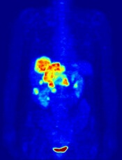
Image by Jens Langner
Performing PET scans immediately after chemotherapy may reveal which Hodgkin lymphoma (HL) patients need radiotherapy (RT).
A study published in NEJM showed similar rates of progression-free survival in HL patients who werePET-negative after chemotherapy, whether they received subsequent RT or not.
However, the investigators said longer follow-up is needed to determine if eliminating RT in PET-negative patients will lead to fewer late effects and improved overall survival.
The 602 patients who agreed to take part in this trial, known as RAPID, had a PET scan performed after chemotherapy. Patients who tested positive received RT.
Those who tested negative were divided into 2 groups. One group of 211 patients received no further treatment, and the other group of 209 patients had the standard RT.
At a median of 60 months of follow-up, the proportion of patients who were alive and disease-free was 94.6% in the RT group and 90.8% in the group that hadn’t received further treatment.
Eight patients in the RT group progressed, and 8 died (3 with disease progression, 1 of whom died from HL). Five of the deaths occurred in patients who did not ultimately receive RT.
In the untreated group, 20 patients progressed, and 4 patients died (2 with disease progression and none from HL).
“This research is an important step forward,” said study author John Radford, of The University of Manchester and The Christie NHS Foundation Trust in the UK.
“The results of RAPID show that, in early stage Hodgkin lymphoma, radiotherapy after initial chemotherapy marginally reduces the recurrence rate, but this is bought at the expense of exposing to radiation all patients with negative PET findings, most of whom are already cured.” ![]()
Concerns Regarding Long‐term Opioid Use
Overall rates of opioid use and chronic use for noncancer pain have increased markedly in the last 1 to 2 decades.[1, 2] Recognition of such rapidly increasing use has prompted a flurry of investigations examining the impact of what has been referred to as the opioid epidemic.[1, 3, 4] Patients receiving chronic opioid analgesics have previously been demonstrated to consume disproportionate shares of healthcare resources, including significantly more emergency room visits and days in the hospital.[5] In this issue of the Journal of Hospital Medicine, Liang and Turner[6] further demonstrate the impressive scope of healthcare resources consumed by this patient population, and extend these findings by examining the relationship between opioid dose and subsequent hospitalization in a large national cohort of middle‐aged health maintenance organization enrollees with noncancer pain. This is the first study to investigate the relationship in a general cohort of patients.
Perhaps the most striking finding of the study was an all‐cause hospitalization rate of 1120 per 10,000 person‐years among their cohort of opioid users. Considering that 5 to 8 million Americans use long‐term opioids,[7] this translates to about 500,000 to 900,000 admissions per year. The degree to which opioids themselves contribute to such hospitalizations (attributable risk) is uncertain, and it is likely that some of this risk can be explained by the idea that opioids are a marker for comorbidity, and that the conditions prompting opioid use independently increase risk of hospitalization. Studies examining more homogeneous patient populations could serve to shed light on this question. The issue of attributable risk notwithstanding, it is clear that this is a patient population that should have the attention of physicians, hospital administrators, and policy makers.
The main finding of their study is that the total opioid dose in any given 6‐month interval was more strongly associated with subsequent all‐cause hospitalization than the daily dose. This suggests that cumulative exposure is important, and possibly more important than the strength of any given prescription, at least when it comes to the outcome of hospitalization. That is not to say that the daily dose is unimportant, and the authors appropriately caution against such an interpretation. Daily dose matters, and to conclude otherwise would be incorrect for several reasons. First, among patients receiving high total doses of opioids, higher daily doses did seem to confer additional risk. Second, hospitalization is only 1 measure of risk, and multiple prior studies and a recent systematic review have concluded that higher opioid doses are strongly associated with adverse events, including overdose, abuse, addiction, motor vehicle accidents, and myocardial infarction.[8] Last, their finding that total dose more strongly predicts hospitalization than daily dose may reflect confounding by indication and underlying patient characteristics not captured in their analysis. Patients receiving a daily dose of 100 mg or more, but with a total dose of 1830 mg over 6 months, would necessarily have received opioids for a relatively brief period of time (20 days). The indications forand patients receivingsuch short‐course, high‐dose therapy are likely to be vastly different from those for longer‐course, high‐dose therapy, in ways that could be meaningfully associated with hospitalization risk. Nonetheless, their results suggest that cumulative exposure is important as an additional metric by which to predict possible adverse consequences of opioid use.
That cumulative exposure and percent of time on opioids are associated with increased risk of subsequent hospitalization casts further doubt on the already questionable risk‐to‐benefit ratio of long‐term use of opioids for noncancer pain. A recent systematic review of the effectiveness and risks of long‐term opioid therapy for chronic pain found existing evidence insufficient to determine effectiveness for chronic pain and function, owing to lack of a single study evaluating long‐term outcomes in patients on opioid therapy versus no opioid therapy, and found evidence for a dose‐dependent risk for serious harms.[8] The authors conclude that the lack of scientific evidence on effectiveness of long‐term opioid therapy for chronic pain is in striking contrast to its widespread use in this setting. Studies examining the effect of long‐term opioid therapy on pain and function, and defining patient subgroups that may benefit from such therapy, are imperative and long overdue.
In the absence of data showing benefit, and in the face of a growing body of evidence demonstrating harm, we are obligated to reevaluate opioid prescribing for chronic noncancer pain. Until studies have evaluated the impact of opioid use on long‐term outcomes, physicians are missing a key piece of the risk‐benefit calculation, and prescribing must be done judiciously. Curbing the opioid epidemic will require initiatives of epidemic proportions, involving the entire spectrum of healthcare, from the primary care setting to the emergency department (where up to 25% of patients with chronic pain receive their opioids[7]), from researchers to policy makers, and ultimately from patient expectations to physician decision making.
Disclosures
Dr. Herzig was funded by grant number K23AG042459 from the National Institute on Aging. The funding organization had no involvement in any aspect of the study, including design, conduct, and reporting of the study. The author reports no conflicts of interest.
- Vital signs: overdoses of prescription opioid pain relievers—United States, 1999—2008. MMWR Morb Mortal Wkly Rep. 2011;60:1487–1492.
- , , , et al. Trends in long‐term opioid therapy for chronic non‐cancer pain. Pharmacoepidemiol Drug Saf. 2009;18:1166–1175.
- CDC grand rounds: prescription drug overdoses—a U.S. epidemic. MMWR Morb Mortal Wkly Rep. 2012;61:10–13.
- . A flood of opioids, a rising tide of deaths. N Engl J Med. 2010;363:1981–1985.
- , , , , , . Co‐morbidity and utilization of medical services by pain patients receiving opioid medications: data from an insurance claims database. Pain. 2009;144:20–27.
- , . National cohort study of opioid analgesic dose and risk of future hospitalization. J Hosp Med. 2015;10:000–000.
- , , , et al. National institutes of health pathways to prevention workshop: the role of opioids in the treatment of chronic pain. Ann Intern Med. 2015;162:295–300.
- , , , et al. The effectiveness and risks of long‐term opioid therapy for chronic pain: a systematic review for a national institutes of health pathways to prevention workshop. Ann Intern Med. 2015;162:276–286.
Overall rates of opioid use and chronic use for noncancer pain have increased markedly in the last 1 to 2 decades.[1, 2] Recognition of such rapidly increasing use has prompted a flurry of investigations examining the impact of what has been referred to as the opioid epidemic.[1, 3, 4] Patients receiving chronic opioid analgesics have previously been demonstrated to consume disproportionate shares of healthcare resources, including significantly more emergency room visits and days in the hospital.[5] In this issue of the Journal of Hospital Medicine, Liang and Turner[6] further demonstrate the impressive scope of healthcare resources consumed by this patient population, and extend these findings by examining the relationship between opioid dose and subsequent hospitalization in a large national cohort of middle‐aged health maintenance organization enrollees with noncancer pain. This is the first study to investigate the relationship in a general cohort of patients.
Perhaps the most striking finding of the study was an all‐cause hospitalization rate of 1120 per 10,000 person‐years among their cohort of opioid users. Considering that 5 to 8 million Americans use long‐term opioids,[7] this translates to about 500,000 to 900,000 admissions per year. The degree to which opioids themselves contribute to such hospitalizations (attributable risk) is uncertain, and it is likely that some of this risk can be explained by the idea that opioids are a marker for comorbidity, and that the conditions prompting opioid use independently increase risk of hospitalization. Studies examining more homogeneous patient populations could serve to shed light on this question. The issue of attributable risk notwithstanding, it is clear that this is a patient population that should have the attention of physicians, hospital administrators, and policy makers.
The main finding of their study is that the total opioid dose in any given 6‐month interval was more strongly associated with subsequent all‐cause hospitalization than the daily dose. This suggests that cumulative exposure is important, and possibly more important than the strength of any given prescription, at least when it comes to the outcome of hospitalization. That is not to say that the daily dose is unimportant, and the authors appropriately caution against such an interpretation. Daily dose matters, and to conclude otherwise would be incorrect for several reasons. First, among patients receiving high total doses of opioids, higher daily doses did seem to confer additional risk. Second, hospitalization is only 1 measure of risk, and multiple prior studies and a recent systematic review have concluded that higher opioid doses are strongly associated with adverse events, including overdose, abuse, addiction, motor vehicle accidents, and myocardial infarction.[8] Last, their finding that total dose more strongly predicts hospitalization than daily dose may reflect confounding by indication and underlying patient characteristics not captured in their analysis. Patients receiving a daily dose of 100 mg or more, but with a total dose of 1830 mg over 6 months, would necessarily have received opioids for a relatively brief period of time (20 days). The indications forand patients receivingsuch short‐course, high‐dose therapy are likely to be vastly different from those for longer‐course, high‐dose therapy, in ways that could be meaningfully associated with hospitalization risk. Nonetheless, their results suggest that cumulative exposure is important as an additional metric by which to predict possible adverse consequences of opioid use.
That cumulative exposure and percent of time on opioids are associated with increased risk of subsequent hospitalization casts further doubt on the already questionable risk‐to‐benefit ratio of long‐term use of opioids for noncancer pain. A recent systematic review of the effectiveness and risks of long‐term opioid therapy for chronic pain found existing evidence insufficient to determine effectiveness for chronic pain and function, owing to lack of a single study evaluating long‐term outcomes in patients on opioid therapy versus no opioid therapy, and found evidence for a dose‐dependent risk for serious harms.[8] The authors conclude that the lack of scientific evidence on effectiveness of long‐term opioid therapy for chronic pain is in striking contrast to its widespread use in this setting. Studies examining the effect of long‐term opioid therapy on pain and function, and defining patient subgroups that may benefit from such therapy, are imperative and long overdue.
In the absence of data showing benefit, and in the face of a growing body of evidence demonstrating harm, we are obligated to reevaluate opioid prescribing for chronic noncancer pain. Until studies have evaluated the impact of opioid use on long‐term outcomes, physicians are missing a key piece of the risk‐benefit calculation, and prescribing must be done judiciously. Curbing the opioid epidemic will require initiatives of epidemic proportions, involving the entire spectrum of healthcare, from the primary care setting to the emergency department (where up to 25% of patients with chronic pain receive their opioids[7]), from researchers to policy makers, and ultimately from patient expectations to physician decision making.
Disclosures
Dr. Herzig was funded by grant number K23AG042459 from the National Institute on Aging. The funding organization had no involvement in any aspect of the study, including design, conduct, and reporting of the study. The author reports no conflicts of interest.
- Vital signs: overdoses of prescription opioid pain relievers—United States, 1999—2008. MMWR Morb Mortal Wkly Rep. 2011;60:1487–1492.
- , , , et al. Trends in long‐term opioid therapy for chronic non‐cancer pain. Pharmacoepidemiol Drug Saf. 2009;18:1166–1175.
- CDC grand rounds: prescription drug overdoses—a U.S. epidemic. MMWR Morb Mortal Wkly Rep. 2012;61:10–13.
- . A flood of opioids, a rising tide of deaths. N Engl J Med. 2010;363:1981–1985.
- , , , , , . Co‐morbidity and utilization of medical services by pain patients receiving opioid medications: data from an insurance claims database. Pain. 2009;144:20–27.
- , . National cohort study of opioid analgesic dose and risk of future hospitalization. J Hosp Med. 2015;10:000–000.
- , , , et al. National institutes of health pathways to prevention workshop: the role of opioids in the treatment of chronic pain. Ann Intern Med. 2015;162:295–300.
- , , , et al. The effectiveness and risks of long‐term opioid therapy for chronic pain: a systematic review for a national institutes of health pathways to prevention workshop. Ann Intern Med. 2015;162:276–286.
Teens who tan indoors do it often
More than one-third of teens who undergo indoor tanning do so frequently, according to data from a survey of 1,850 public high school students from 27 schools in New Jersey.
The data were part of the 2012 New Jersey Youth Tobacco Survey, taken before New Jersey banned indoor tanning for individuals younger than 17 years of age in 2013. A total of 146 respondents reported indoor tanning within the past year, and approximately 38% reported 10 or more tanning sessions, wrote Elliot J. Coups, Ph.D., of Rutgers Cancer Institute of New Jersey, New Brunswick, and colleagues.
Overall, “frequent indoor tanning was significantly more common among indoor tanners who were current smokers, tanned to improve their mood, had used social media related to tanning salons, or indicated it would be very hard to stop indoor tanning,” the researchers noted, adding that social media might be an opportunity for interventions to reduce indoor tanning in this population.
Find the study in the Journal of the American Academy of Dermatology (doi:10.1016/j.jaad.2015.01.035).
Short but simple theme guides AK treatment recommendations
An expert panel of seven dermatologists has generated recommendations to aid clinicians in prescribing topical treatments for actinic keratoses. Dr. Eggert Stockfleth and his colleagues acknowledged that patient compliance with topical AK therapy remains a challenge.
Their recommendations included the need to develop topical treatments with shorter duration but equal effectiveness, to keep regimens as simple as possible, and to manage patient expectations.“Our experts believe that real-world efficacy could be vastly improved by shorter treatments coupled with effective patient management,” they noted.Read the full article in the International Journal of Dermatology (doi:10.1111/ijd.12840).
Finally, an end to the SGR game of chicken
Well, in a remarkable moment in what is likely the most dysfunctional American government on record, all sides agreed to repeal the sustainable growth rate formula and change the way Medicare payments are done.
For the foreseeable future, this eliminates the annual game of chicken that political parties play at our expense.

For more than 10 years now, the cuts have come up, the medical journals decry them, doctors threaten to stop seeing Medicare patients, and eventually the politicians reach an agreement to stop the cuts that had grown to a high of 27.4% in 2012 ... until the next year. The classic game of political kick the can.
The dysfunctional waltz had been going on for so long that only the medical press and doctors seemed to care.
So, after years of wrapping duct tape around a leaking pipe, they called a plumber and replaced the pipe. The cost is far higher than it would have been if they’d actually addressed the problem when it started in 2003.
I’m sure the new law will have its own issues, but it stops – for now – the nightmare scenario where the cuts are allowed to go through. That would have left many of us in the difficult situation of turning away new Medicare patients. None of us wants to do that.
It’s nice to have seen the government function in the way it’s supposed to – with negotiations and compromise – rather than more threats, vitriolic posturing, and finger-pointing that have been the recent norm. Members of Congress, regrettably, don’t have their health care affected by their decisions. Regardless of age, the majority aren’t on Medicare. They’re all covered through the Federal Employees Health Benefits Program, which uses private plans. So it doesn’t directly affect them if a doctor’s office is shuttered or sick constituents can’t find someone to help them. They figure by the next election cycle they’ll be able to distract voters with some other topic.
I’m glad that reason and concern for the health care of ordinary citizens prevailed. It’s easy to play chicken when someone else’s life and livelihood are involved, and not yours. I hope this leads to greater cooperation and willingness to work together between the two sides in the future.
But I’m not optimistic.
Dr. Block has a solo neurology practice in Scottsdale, Ariz.
Well, in a remarkable moment in what is likely the most dysfunctional American government on record, all sides agreed to repeal the sustainable growth rate formula and change the way Medicare payments are done.
For the foreseeable future, this eliminates the annual game of chicken that political parties play at our expense.
For more than 10 years now, the cuts have come up, the medical journals decry them, doctors threaten to stop seeing Medicare patients, and eventually the politicians reach an agreement to stop the cuts that had grown to a high of 27.4% in 2012 ... until the next year. The classic game of political kick the can.
The dysfunctional waltz had been going on for so long that only the medical press and doctors seemed to care.
So, after years of wrapping duct tape around a leaking pipe, they called a plumber and replaced the pipe. The cost is far higher than it would have been if they’d actually addressed the problem when it started in 2003.
I’m sure the new law will have its own issues, but it stops – for now – the nightmare scenario where the cuts are allowed to go through. That would have left many of us in the difficult situation of turning away new Medicare patients. None of us wants to do that.
It’s nice to have seen the government function in the way it’s supposed to – with negotiations and compromise – rather than more threats, vitriolic posturing, and finger-pointing that have been the recent norm. Members of Congress, regrettably, don’t have their health care affected by their decisions. Regardless of age, the majority aren’t on Medicare. They’re all covered through the Federal Employees Health Benefits Program, which uses private plans. So it doesn’t directly affect them if a doctor’s office is shuttered or sick constituents can’t find someone to help them. They figure by the next election cycle they’ll be able to distract voters with some other topic.
I’m glad that reason and concern for the health care of ordinary citizens prevailed. It’s easy to play chicken when someone else’s life and livelihood are involved, and not yours. I hope this leads to greater cooperation and willingness to work together between the two sides in the future.
But I’m not optimistic.
Dr. Block has a solo neurology practice in Scottsdale, Ariz.

Bath salts – the new designer high
“Bath salts” is a dangerous illicit drug that teens may be using. It hit the U.S. scene in 2010 after being a big hit in Europe. It’s called “bath salts” because it resembled Epsom salt, but shares no chemical properties with it.
Because it was called bath salts, it initially evaded the Food and Drug Administration, and was sold at gas stations and clubs marked “not for human consumption,” which earned it its name as the “legal high.” Bath salts are creatively packaged in containers that are colorful and attractive, and have aliases such as “plant food,” “Ivory Wave,” “Purple Wave,” “Bloom,” “Cloud Nine,” or “Vanilla Sky,” according to the National Institute on Drug Abuse.

Shortly after the drug’s debut, emergency departments were seeing several hundreds of people with amphetamine-like intoxication and “naked rages.” In 2012, when it became known that these substances were being use as illicit drugs, they were made illegal, but the problem now is there are many derivatives of this drug and their use is becoming more widespread.
Mephedrone is one of the possible active ingredients, and it is derived from the synthetic cathinone, similar to cathinone found naturally in the khat plant (Catha edulis). It stimulates the adrenergic receptors to release dopamine and norepinephrine, and block their reuptake, which makes them act like a stimulant on the nervous system. The result is agitation, hyperalertness, hypertension, tachycardia, diaphoresis, and elevated temperatures to 106<scaps>˚ </scaps>F. The term “naked rage” came into use because users would become overheated and angry, and would rip off their clothes as they raged.
Other synthetic canthinones being used as drugs of abuse include butylone, dimethylcathinone, ethcathinone, ethylone, 3- and 4-fluoromethcathinone, mephedrone, methedrone, methylenedioxypyrovalerone (MDPV), methylone, and pyrovalerone (J. Med. Toxicol. 2012;8:33-42).
MDPV raises dopamine levels in the brain as does cocaine, but is at least 10 times as potent, according to the National Institute on Drug Abuse.
Bath salts can be administered by snorting, ingesting, smoking, or injecting. Necrotizing fasciitis has been associated with its use. Most patients present with elevated temperatures, hypertension, palpitations, and delirium. Hydration and sedation are usually required, and patients usually need to be restrained to prevent them from hurting themselves further. Psychiatric symptoms may include paranoia, hallucinations, and panic attacks. Several deaths have been reported in association with its use.
In the time since this drug was made illegal, several other designer drugs have presented, which are just modifications of the mephedrone. “Flakka” and “jewelry cleaner” are examples of the modified synthetic cathinone. They all cause amphetamine-like reactions and the risk of overdose is great.
The cost to manufacture the drug is about $8 per unit, but its retail price is $20-$40. Though it takes only 3-5 mg to be effective, packages are sold with 500 mg, making the risk for overdose even greater. It usually takes about 1.5 hours to reach peak rush and the entire experience can last 6-8 hours. There is little difference between the euphoric level and intoxication level, which is what makes the drug so dangerous.
Synthetic drugs are a great danger to all age groups, including toddlers, because of the misleading packaging. But they are especially dangerous because the ingredients vary, and overdose occurs quickly.
It is imperative that parents and teens are made aware of the dangers associated with these drugs and their street names; www.streetdrugs.org is a website designed to educate teachers, parents and students about these drugs and their dangers.
Dr. Pearce is a pediatrician in Frankfort, Ill. E-mail her at [email protected].
“Bath salts” is a dangerous illicit drug that teens may be using. It hit the U.S. scene in 2010 after being a big hit in Europe. It’s called “bath salts” because it resembled Epsom salt, but shares no chemical properties with it.
Because it was called bath salts, it initially evaded the Food and Drug Administration, and was sold at gas stations and clubs marked “not for human consumption,” which earned it its name as the “legal high.” Bath salts are creatively packaged in containers that are colorful and attractive, and have aliases such as “plant food,” “Ivory Wave,” “Purple Wave,” “Bloom,” “Cloud Nine,” or “Vanilla Sky,” according to the National Institute on Drug Abuse.
Shortly after the drug’s debut, emergency departments were seeing several hundreds of people with amphetamine-like intoxication and “naked rages.” In 2012, when it became known that these substances were being use as illicit drugs, they were made illegal, but the problem now is there are many derivatives of this drug and their use is becoming more widespread.
Mephedrone is one of the possible active ingredients, and it is derived from the synthetic cathinone, similar to cathinone found naturally in the khat plant (Catha edulis). It stimulates the adrenergic receptors to release dopamine and norepinephrine, and block their reuptake, which makes them act like a stimulant on the nervous system. The result is agitation, hyperalertness, hypertension, tachycardia, diaphoresis, and elevated temperatures to 106<scaps>˚ </scaps>F. The term “naked rage” came into use because users would become overheated and angry, and would rip off their clothes as they raged.
Other synthetic canthinones being used as drugs of abuse include butylone, dimethylcathinone, ethcathinone, ethylone, 3- and 4-fluoromethcathinone, mephedrone, methedrone, methylenedioxypyrovalerone (MDPV), methylone, and pyrovalerone (J. Med. Toxicol. 2012;8:33-42).
MDPV raises dopamine levels in the brain as does cocaine, but is at least 10 times as potent, according to the National Institute on Drug Abuse.
Bath salts can be administered by snorting, ingesting, smoking, or injecting. Necrotizing fasciitis has been associated with its use. Most patients present with elevated temperatures, hypertension, palpitations, and delirium. Hydration and sedation are usually required, and patients usually need to be restrained to prevent them from hurting themselves further. Psychiatric symptoms may include paranoia, hallucinations, and panic attacks. Several deaths have been reported in association with its use.
In the time since this drug was made illegal, several other designer drugs have presented, which are just modifications of the mephedrone. “Flakka” and “jewelry cleaner” are examples of the modified synthetic cathinone. They all cause amphetamine-like reactions and the risk of overdose is great.
The cost to manufacture the drug is about $8 per unit, but its retail price is $20-$40. Though it takes only 3-5 mg to be effective, packages are sold with 500 mg, making the risk for overdose even greater. It usually takes about 1.5 hours to reach peak rush and the entire experience can last 6-8 hours. There is little difference between the euphoric level and intoxication level, which is what makes the drug so dangerous.
Synthetic drugs are a great danger to all age groups, including toddlers, because of the misleading packaging. But they are especially dangerous because the ingredients vary, and overdose occurs quickly.
It is imperative that parents and teens are made aware of the dangers associated with these drugs and their street names; www.streetdrugs.org is a website designed to educate teachers, parents and students about these drugs and their dangers.
Dr. Pearce is a pediatrician in Frankfort, Ill. E-mail her at [email protected].

May 2015 Quiz 2
ANSWER: C
Critique
Sulfasalazine can cause reversible azoospermia and infertility. About 80% of patients taking sulfasalazine have semen abnormalities and 72% have oligospermia. Only one case of azoospermia has been reported in patients taking pure mesalamines. The sperm abnormalities are thought to be caused by the sulfapyridine moiety in the sulfasalazine molecule. Therefore, a switch from sulfasalazine to Asacol® or any other pure mesalamine is indicated.
Sulfasalazine inhibits dihydrofolate reductase and can cause folate deficiency. As a result, it should always be given along with oral folic acid supplementation. There is no clear evidence that 6-mercaptopurine affects male fertility. Holding both medications in a patient with UC would put him at increased risk of recurrence and should not be recommended. Withdrawal of an immunomodulatory agent such as 6-mercaptopurine or azathioprine in patients who are in remission can lead to rapid relapse in up to one-third of patients in 1 year and two-thirds within 5 years. Since 6-mercaptopurine is maintaining remission, there is no need for a switch to infliximab. An in vitro fertilization specialist might be required if there is no conception despite sulfasalazine withdrawal.
References
1. Nielsen O.H., Munck L.K. Drug insight: aminosalicylates for the treatment of IBD. Nat. Clin. Pract. Gastroenterol. Hepatol. 2007;4:160-70.
2. Cassinotti A., Actis G.C., Duca P., et al. Maintenance treatment with azathioprine in ulcerative colitis: outcome and predictive factors after drug withdrawal. Am. J. Gastroenterol. 2009;104:2760-7.
May 2015 Quiz 1
ANSWER: C
Critique
A recent Olmstead County, Minn., study was undertaken to understand the epidemiology of community-acquired C. difficile infections. In the cohort of 385 proven C. difficile infections, 41% were community acquired. Compared to cases acquired in the hospital, community C. difficile infections were observed in younger (B) females (A) who had lower comorbidity scores and fewer chronic illnesses (D). Also, these outpatient cases were less likely to have recent antibiotic exposure (E) and fortunately had less severe clinical courses. A large population-based study and several others have confirmed that the use of gastric acid-suppressive agents such as PPIs (C) increases the risk of C. difficile-associated disease (adjusted risk ratio 2.9, 95% CI: 2.4-3.4).
References
1. Khanna S., Pardi D.S., Aronson S.L., et al. The epidemiology of community-acquired Clostridium difficile infection: a population-based study. Am. J. Gastroenterol. 2012;107:89-95.
2. Dial S., Delaney J.A., Barkun A.N., Suissa S. Use of gastric acid-suppressive agents and the risk of community-acquired Clostridium difficile-associated disease. JAMA 2005;294:2989.