User login
Startup Pharmacy Takes Mail-Order to Next Level, Could Solve Medication Management Issue for Millions
It only takes one idea to help change the face of medicine and, recently, Forbes posted an article outlining a small startup pharmacy that could change the way we get our medication. Manchester, N.H.-based Pill Pack takes the whole mail-order pharmacy to a new level.
Medication management can be a major issue for seniors and their caregivers. Seniors are at risk for such problems as overmedication and drug interactions, if medications are not properly managed.
An UpToDate article says that a survey for adults aged 57-85 shows:
- At least one prescription medication was used by 81%;
- Five or more prescription medications were used by 29% of the overall survey population and by 36% of people aged 75 to 85 years; and
- 46% of prescription users also took at least one over-the-counter medication.
Hospitalists see it every day; the readmit because prescribed medication isn’t taken correctly. Possibly, Pill Pack might be one step in the right direction. TH
Lisa Courtney is director of operations at Baptist Health Systems in Birmingham, Ala., and a member of Team Hospitalist.
Stent-retriever thrombectomy reduces poststroke disability
For patients with proximal large-vessel anterior stroke, neurovascular thrombectomy with a stent retriever plus medical therapy in the REVASCAT trial reduced the severity of poststroke disability and raised the rate of functional independence, compared with medical therapy alone, according to a report in the New England Journal of Medicine.
To assess the efficacy and safety of thrombectomy with a stent retriever, investigators performed a prospective, open-label, phase III clinical trial involving 206 adults up to 85 years of age treated at four designated comprehensive stroke centers in Catalonia, Spain.
All the participants in the Randomized Trial of Revascularization With Solitaire FR Device Versus Best Medical Therapy in the Treatment of Acute Stroke Due to Anterior Circulation Large Vessel Occlusion Presenting Within Eight Hours of Symptom Onset (REVASCAT) had either not responded to intravenous alteplase administered within 4.5 hours of symptom onset or had contraindications to alteplase therapy. They were randomly assigned in equal numbers to undergo endovascular treatment with a stent retriever or medical therapy alone, said Dr. Tudor G. Jovin, director of the Stroke Institute, University of Pittsburgh Medical Center, and his associates.
The trial was halted early when the first interim analysis showed “lack of equipoise” between the two study groups, and because emerging results from three other studies demonstrated the superior efficacy of thrombectomy. The primary efficacy outcome measure – severity of disability at 90 days, as measured by expert assessors blinded to treatment assignment – significantly favored thrombectomy over medical therapy. The proportion of patients who achieved functional independence by day 90 on the modified Rankin scale also demonstrated the clear superiority of thrombectomy (43.7%) over medical therapy (28.2%).
Only 6.5 patients would need to be treated with thrombectomy to prevent 1 case of functional dependency or death. In addition, “thrombectomy was associated with a shift toward better outcomes across the entire spectrum of disability,” Dr. Jovin and his associates said (N. Engl. J. Med. 2015 [doi:10.1056/NEJMoa1503780]).
Regarding safety, the rate of death at 90 days did not differ significantly between patients who underwent thrombectomy (18.4%) and control subjects (15.5%). Rates of intracranial hemorrhage were the same, 1.9%, in both groups, and rates of other serious adverse events also were similar.
These findings are consistent with those of several other recently reported clinical trials and show that “in patients with acute stroke caused by a proximal large-vessel occlusion and an absence of a large infarct on baseline imaging, mechanical thrombectomy with [a] stent retriever was safe and led to improved clinical outcomes, as compared with medical therapy alone,” the investigators said.
REVASCAT was funded by an unrestricted grant from Covidien, maker of the stent retriever, and by grants from several Spanish research institutes. Dr. Jovin reported ties to Covidien, Silk Road Medical, Air Liquide, Medtronic, and Stryker Neurovascular.
FROM THE NEW ENGLAND JOURNAL OF MEDICINE
Key clinical point: Thrombectomy with a stent retriever reduced poststroke disability in patients with occlusion of the proximal anterior circulation.
Major finding: The proportion of patients who achieved functional independence by day 90 showed the clear superiority of thrombectomy (43.7%) over medical therapy (28.2%); only 6.5 patients would need to be treated with thrombectomy to prevent 1 case of functional dependency or death.
Data source: REVASCAT, a prospective, open-label, randomized phase III trial involving 206 adults treated during a 4-year period at four stroke centers in Spain.
Disclosures: REVASCAT was funded by an unrestricted grant from Covidien, maker of the stent retriever, and by grants from several Spanish research institutes. Dr. Jovin reported ties to Covidien, Silk Road Medical, Air Liquide, Medtronic, and Stryker Neurovascular.
Study suggests need for change in matching criteria
![]()
Photo by Chad McNeeley
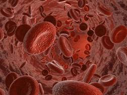
A donor’s CD8 cell count can affect the outcome of hematopoietic stem cell transplant (HSCT) in older patients, according to a study published in the Journal of Clinical Oncology.
Older patients who received stem cells from unrelated donors with higher CD8 counts had a significantly lower risk of relapse and higher rate of
survival than patients who received grafts with lower CD8 counts, even if those grafts were from matched sibling donors.
This suggests that screening for donor T-cell characteristics could optimize donor selection and ultimately lead to more successful transplants in an older population, study investigators said.
“Developing better tools to identify ideal donors is an exciting prospect and fundamental to improving transplant outcomes,” said Ran Reshef, MD, of the Abramson Cancer Center at the University of Pennsylvania in Philadelphia.
“There may be suitable donors out there who are overlooked because they are considered a poorer match by today’s donor selection algorithms. Refining the screening method could greatly increase the chances of finding the most appropriate donor, one that will induce the most potent graft-vs-tumor response.”
For this study, Dr Reshef and his colleagues retrospectively evaluated associations between a graft’s T-cell dose and outcomes in 200 patients undergoing HSCT to treat acute myeloid leukemia, myelodysplastic syndrome, non-Hodgkin lymphoma, and other hematologic disorders.
The investigators looked specifically at CD4 and CD8 counts and found the number of CD8 cells in the graft had an impact on survival. They also found that high CD8 cell counts were much more common among young donors.
The 4-year overall survival rates were 59% for patients who received grafts from younger (<50 years), unrelated donors with high CD8 counts; 18% for those who received grafts from younger, unrelated donors with low CD8 counts; and 33% for patients who received grafts from older (>50 years), HLA-matched sibling donors.
In multivariable analysis, CD8 count was an independent predictor of relapse (adjusted hazard ratio [aHR]=0.43; P=0.009), relapse-free survival (aHR=0.50; P=0.006), and overall survival (aHR=0.57; P=0.04).
The investigators also assessed the effect of CD8 count on survival using a cutoff of 0.72 x 10⁸ CD8 cells/kg. They found that patients with CD8 doses above this cutoff had significantly better relapse-free survival (P=0.005) and overall survival (P=0.007) rates than patients with CD8 doses below this cutoff.
These results suggest it would be better to use a younger, unrelated donor graft with a high CD8 cell count rather than an older sibling donor, the investigators said. However, this strategy should be tested in a prospective trial.
“This is a method deserving additional investigation,” Dr Reshef said, “which could refine the standardized matching system used by registries, such as Be the Match and others, and ultimately optimize the donor pool for older patients undergoing these transplants.” ![]()
![]()
Photo by Chad McNeeley
A donor’s CD8 cell count can affect the outcome of hematopoietic stem cell transplant (HSCT) in older patients, according to a study published in the Journal of Clinical Oncology.
Older patients who received stem cells from unrelated donors with higher CD8 counts had a significantly lower risk of relapse and higher rate of
survival than patients who received grafts with lower CD8 counts, even if those grafts were from matched sibling donors.
This suggests that screening for donor T-cell characteristics could optimize donor selection and ultimately lead to more successful transplants in an older population, study investigators said.
“Developing better tools to identify ideal donors is an exciting prospect and fundamental to improving transplant outcomes,” said Ran Reshef, MD, of the Abramson Cancer Center at the University of Pennsylvania in Philadelphia.
“There may be suitable donors out there who are overlooked because they are considered a poorer match by today’s donor selection algorithms. Refining the screening method could greatly increase the chances of finding the most appropriate donor, one that will induce the most potent graft-vs-tumor response.”
For this study, Dr Reshef and his colleagues retrospectively evaluated associations between a graft’s T-cell dose and outcomes in 200 patients undergoing HSCT to treat acute myeloid leukemia, myelodysplastic syndrome, non-Hodgkin lymphoma, and other hematologic disorders.
The investigators looked specifically at CD4 and CD8 counts and found the number of CD8 cells in the graft had an impact on survival. They also found that high CD8 cell counts were much more common among young donors.
The 4-year overall survival rates were 59% for patients who received grafts from younger (<50 years), unrelated donors with high CD8 counts; 18% for those who received grafts from younger, unrelated donors with low CD8 counts; and 33% for patients who received grafts from older (>50 years), HLA-matched sibling donors.
In multivariable analysis, CD8 count was an independent predictor of relapse (adjusted hazard ratio [aHR]=0.43; P=0.009), relapse-free survival (aHR=0.50; P=0.006), and overall survival (aHR=0.57; P=0.04).
The investigators also assessed the effect of CD8 count on survival using a cutoff of 0.72 x 10⁸ CD8 cells/kg. They found that patients with CD8 doses above this cutoff had significantly better relapse-free survival (P=0.005) and overall survival (P=0.007) rates than patients with CD8 doses below this cutoff.
These results suggest it would be better to use a younger, unrelated donor graft with a high CD8 cell count rather than an older sibling donor, the investigators said. However, this strategy should be tested in a prospective trial.
“This is a method deserving additional investigation,” Dr Reshef said, “which could refine the standardized matching system used by registries, such as Be the Match and others, and ultimately optimize the donor pool for older patients undergoing these transplants.” ![]()
Follow-up PET/CT has ‘clinical value’ in NHL
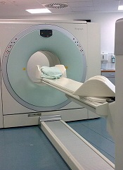
BALTIMORE—Post-treatment PET/CT scans may be necessary for some patients with non-Hodgkin lymphoma (NHL), according to a presentation at the 2015 SNMMI Annual Meeting.
“[T]he role of post-treatment PET/CT has been controversial,” said Mehdi Taghipour, MD, of Johns Hopkins University in Baltimore, Maryland.
“Our study proves that 39% of follow-up PET/CT scans added clinical value, which represents a significant improvement in NHL patient care.”
Dr Taghipour presented the details of this study at the meeting as abstract 599.
He and his colleagues had examined 560 PET/CT scans from 204 patients. Imaging was performed 6 months or more after the patient completed primary therapy.
The researchers assessed the value of follow-up PET/CT by conducting statistical analyses to determine changes in patient management and evaluated the accuracy of these scans compared to either histopathology or an additional 6-month follow-up.
Results showed that the sensitivity of PET/CT for detecting relapsed NHL was 95.1%, and the specificity was 90.5%. Positive and negative predictive values were 84.5% and 97.1%, respectively.
The overall accuracy of follow-up PET/CT with the common imaging agent fluorodeoxyglucose (FDG) was 92.1%.
Follow-up PET/CT led to changes in patient management in 17% of cases, and new treatments were initiated after 15.7% of scans. More than 69% of scans were performed without prior clinical suspicion of recurrence, and 30.7% of scans were ordered because of suspected disease.
More than 22% of follow-up scans showed suspected disease when there was no clinical suspicion for disease recurrence, and the presence of disease was ruled out in 17.4% of scans where the treating physician had suspected recurrence prior to the scan. ![]()
A better FLT3 inhibitor for AML?
© ASCO/Scott Morgan
CHICAGO—A dual inhibitor of FLT3 and Axl may produce more durable responses than other FLT3 inhibitors and improve survival in patients with FLT3-positive, relapsed or refractory acute myeloid leukemia (AML), according to a speaker at the 2015 ASCO Annual Meeting.
The FLT3/Axl inhibitor, ASP2215, has not been compared against other FLT3 inhibitors directly, and the data presented were from a phase 1/2 study.
However, the speaker said ASP2215 provided “potent and sustained” inhibition of FLT3 and produced an overall response rate (ORR) of 52% among FLT3-positive patients.
The median duration of response for these patients was 18 weeks, and their median overall survival was about 27 weeks.
Mark J. Levis, MD, PhD, of the Sidney Kimmel Comprehensive Cancer Center at Johns Hopkins in Baltimore, Maryland, presented these results at ASCO as abstract 7003.*
“We’ve been studying FLT3 inhibitors for a number of years now,” Dr Levis began, “and we think they show significant clinical promise, [but] they also have problems.”
He noted that some of these drugs haven’t been particularly safe or well-tolerated. They can cause gastrointestinal toxicity, hand-foot syndrome, QT prolongation, and myelosuppression.
However, the most intriguing problem with FLT3 inhibitors, according to Dr Levis, is the emergence of resistance-conferring point mutations observed in studies of some of the newer drugs, such as sorafenib and quizartinib.
“So in that context, here we have ASP2215,” Dr Levis said. “This is a type 1 FLT3 tyrosine kinase inhibitor, and, as such, it has activity against not only wild-type and ITD-mutated FLT3 but also against those resistance-conferring point mutations typically found in the activation loop at the so-called gatekeeper residue (F691L).”
With this in mind, he and his colleagues conducted a phase 1/2 trial of ASP2215. The study was sponsored by Astellas Pharma Global Development, Inc., the company developing ASP2215.
The trial was open to patients with relapsed or refractory AML, irrespective of their FLT3 mutation status. The researchers’ goal was to identify a safe, tolerable dose of ASP2215 that fully inhibited FLT3.
The team used a standard 3+3 design, with dose levels ranging from 20 mg to 450 mg once daily. They expanded every cohort until they reached a dose-limiting toxicity.
In all, the trial enrolled 198 patients, 24 in the dose-escalation cohorts and 174 in the dose-expansion cohorts. The patients’ median age was 62 (range, 21-90), and 53.1% were male.
Nearly 66% of patients had FLT3 mutations, 29.4% were FLT3-negative, and 5.2% had unknown FLT3 status. About 35% of patients had received 1 prior line of therapy, 26.3% had 2, 33.5% had 3 or more, and 5.7% had an unknown number of prior therapies.
Safety results
In the 194 patients who were evaluable for safety, treatment-emergent adverse events included diarrhea (13.4%), fatigue (12.4%), AST increase (11.3%), ALT increase (9.3%), thrombocytopenia (7.7%), anemia (7.2%), peripheral edema (7.2%), constipation (6.7%), nausea (6.7%), dizziness (6.2%), vomiting (5.7%), and dysgeusia (5.2%).
Serious adverse events included febrile neutropenia (27.3%), sepsis (11.9%), disease progression (10.3%), pneumonia (8.8%), hypotension (5.7%), and respiratory failure (5.7%).
“The kinds of side effects we saw were typical for a relapsed/refractory AML population,” Dr Levis said. “Nothing really stood out. Any trial of relapsed/refractory AML is going to have febrile neutropenia and sepsis, and those were our dominant, serious adverse events. There was no real safety signal here that was unique to the drug, we felt.”
The researchers said the maximum-tolerated dose of ASP2215 was 300 mg, as 2 patients who received the 450 mg dose experienced dose-limiting toxicities. One was grade 3 diarrhea, and the other was grade 3 AST elevation.
Response and survival
Among the 127 patients who were FLT3-positive, the ORR was 52% (n=66). The complete response (CR) rate was 6.3% (n=8). The composite CR rate, which includes CRs with incomplete hematologic recovery (CRi) and incomplete platelet recovery (CRp), was 40.9% (n=52). And the partial response (PR) rate was 11% (n=14).
“As we scale up the dose, the PRs shift on over to CRis, and the dominant response is, in fact, a complete response with incomplete count recovery,” Dr Levis said. “The categories where we had the largest number of responses were the 120 mg and 200 mg categories. We didn’t really have enough patients in the 300 mg category to comment on it. ”
For the FLT3-positive patients, the median duration of response was 126 days.
“The duration of response really stood out here,” Dr Levis said. “It’s over 4 months. That is something we really didn’t see with the other drugs, and I suspect that is a reflection of the suppression of the outgrowth of these resistance mutations.”
Unfortunately, FLT3-wild-type patients did not fare as well. The ORR among these patients was 8.8%. None of the patients achieved a CR, 3 had a composite CR (5.3%), and 2 had a PR (3.5%).
Among the FLT3-positive patients, the median overall survival was about 27 weeks. It was 128 days in the 20 mg dose cohort (n=13), 105.5 days in the 40 mg cohort (n=8), 201 days in the 80 mg cohort (n=12), 199 days in the 120 mg cohort (n=40), and 161 days in the 200 mg cohort (n=45). Dr Levis did not present survival data for the 300 mg or 450 mg cohorts, which included 7 and 2 patients, respectively.
“[Relapsed/refractory AML] is a population that has a median survival of about 3 months with conventional therapy, at least by historical publications,” Dr Levis noted. “If you look at survival in this trial, patients treated at the FLT3-inhibitory doses [had a] greater than 6-month median survival.”
He added that studies of ASP2215 in combination with other agents are ongoing in patients with newly diagnosed AML. And phase 3 trials of ASP2215 at the 120 mg dose, with the option of scaling up to 200 mg, are planned. ![]()
*Information in the abstract differs from that presented at the meeting.
Lymphocyte recovery linked to outcome of HSCT

Photo courtesy of VCU
Massey Cancer Center
Results of a retrospective study suggest lymphocyte recovery is associated with outcomes after allogeneic hematopoietic stem cell transplant (HSCT).
Researchers found that, after transplant, lymphocyte recovery occurred in 1 of 3 general patterns.
And these patterns were associated with the rate of survival, relapse, and graft-vs-host disease (GVHD), as well as the need for further donor immune cell infusions to treat the patients’ disease.
Amir Toor, MD, of VCU Massey Cancer Center in Richmond, Virginia, and his colleagues reported these findings in Biology of Blood & Marrow Transplantation.
The team had examined lymphocyte recovery and clinical outcome data from a phase 2 trial (NCT00709592) of 41 patients who received an HSCT from a related or unrelated donor.
As part of the trial protocol, the patients underwent low-dose radiation therapy and received 1 of 2 different doses of anti-thymocyte globulin as GVHD prophylaxis.
The researchers found that, after transplant, lymphocyte recovery followed 1 of 3 general patterns that correlated with patient outcomes.
“We began considering lymphocyte reconstitution following stem cell transplantation as similar to population growth models,” Dr Toor explained. “So we graphed the lymphocyte counts of our patients at various times following their transplant as a logistic function and observed distinct patterns that correlated with clinical outcomes.”
Patients in group A experienced fast, early lymphoid expansion, culminating in a high absolute lymphoid count (ALC) within 2 months of HSCT. Group B experienced a slower, but steady, lymphoid expansion that peaked much later than group A with a lower ALC. Group C experienced very poor lymphocyte recovery that demonstrated an early, but brief, lymphoid expansion with a very low ALC.
Group B had the best survival rate—86%—compared to 67% in group A and 30% in group C. Relapse rates between groups A and B were similar, at 33% and 29%, respectively, while group C experienced a 90% relapse rate.
GVHD occurred in 67% of patients in group A, 43% in group B, and 10% in group C. And adoptive immunotherapy with donor cell infusions was required for 13% of patients in group A, 21% in group B, and 70% in group C.
“Our goal is to use this data to develop models that can predict complications from stem cell transplantation,” Dr Toor said. “Then, we may be able to intervene at key points in time with appropriate clinical treatments that will make the most positive impact on patients’ outcomes.” ![]()
Hospital Medicine in 2015
This year, we celebrate the 10th anniversary of this esteemed publication, and it is indeed an occasion for celebration. For those of us who were there at the creation of the hospitalist field, the establishment of a vibrant academic journal was a dream, one whose fulfillment was central to the legitimization of our field as a full‐fledged specialty. After a decade and 83 issues, the Journal of Hospital Medicine is a formidable source of information, cohesion, and pride.
The anniversary comes at a particularly interesting time for hospitals and hospitalists. Our field's lifeblood has been in trailblazing and continuous reinvention. We were the first physician specialty that embraced the mantra of systems thinking, as captured in our famous metaphor that we care for two sick patients: the person and the system. We were the first field that proudly, and without a hint of shame, allied ourselves with hospital leaders, believing that we were mutually dependent on one another, and that our ability to make change happen and stick was better if we were working with our institutions' leaders. In creating our professional society (and this journal), we took unusual pains to be inclusiveof academic and community‐based hospitalists, or hospitalists entering the field from a variety of backgrounds, of hospitalists caring for adults and kids, and of nonphysician providers.
Our efforts have paid off. Leaders as prominent as Don Berwick have observed that hospitalists have become the essential army of improvers in hospitals and healthcare systems. Hospitalists have made immense contributions at their own institutions, and are increasingly assuming leadership roles both locally and nationally. It is not a coincidence that Medicare's top physician (Patrick Conway) and the Surgeon General (Vivek Murthy) are both hospitalists. Although there have been a few bumps along the way, hospitalists are generally satisfied with their careers, respected by their colleagues, accepted by their patients, and pleased to be members of the fastest growing specialty in the history of modern medicine.
All of this should leave us all feeling warm, proud and more than a little nervous. We are now a mature medical specialty, no longer upstarts, and the natural inclination, in a changing world, will be to hunker down and protect what we have. Of course, some of that is reasonable and appropriate (for example, to fight for our fair share of a bundled payment pie),[1] but some of it will be wrong, even self‐defeating. The world of healthcare is changing fast, and our ability to stay relevant and indispensable will depend on our ability to evolve to meet new conditions and needs.
Let us consider some of the major trends playing out in healthcare. The biggest is the brisk and unmistakable shift from volume to value.[2] This is a trend we have been on top of, because this really has been our field's raison d'tre: improving value in the hospital by cutting costs and length of stay while improving (or at least keeping neutral) quality and safety.[3] However, a world under intense value pressure will work hard to move patients from hospital to less expensive postacute settings, and will insist on seamless handoffs between the hospital and such settings. Thoughtful hospital medicine groups are thinking hard about this trend, and many are placing colleagues in skilled nursing facilities, or at the very least tightening their connections to the postacute facilities in their healthcare ecosystem. We no longer have the luxury of confining our talents and energies to those things that take place within the 4 walls of the hospital.
Another trend is the digitization of healthcare, a trend turbocharged by $30 billion in federal incentive payments distributed between 2009 and 2014.[4] Here too, hospitalists have emerged as leaders in information technology (IT) implementations, and a disproportionate number of chief medical information officers and other IT leaders seem to be hospitalists. Splendid. But it is also up to us to help figure out how to use IT tools effectively. The notes have morphed into bloated, copy‐and‐pasteridden monstrosities: let us figure out what a good note should look like in the digital era, and then implement educational and system changes to create a new standard. We no longer go to radiology because we do not need to to see our films; let us think about what the loss of the collegial exchange with our radiology colleagues has cost, and then set out to develop new systems to reimagine it. Right now, big data are mostly hype and unrequited promise. Who better than hospitalists to dive in and start making sense of the data to predict risks or help point to better treatments?
Another trend is population health. Although I do not foresee a return to the Marcus Welby model of a kindly physician following the patient everywhere, I can imagine certain patients (mostly those with several social and clinical comorbidities and at least 3 admissions per year) who might be well served by a back‐to‐the‐future system in which a primary care provider follows them into the hospital, perhaps comanaging the patients with the on‐service hospitalist. David Meltzer, at the University of Chicago, is currently studying such a model, and I look forward to seeing his results.[5] Rather than rejecting such experiments as violating the usual hospitalist structure, we must embrace them, at least until the evidence is in.
In the end, the field of hospital medicine emerged and thrived because of the promise, and later the evidence, that our presence led to better quality, safety, patient experience, education, and efficiency. This mandate must remain our mantra, even if it means that we have to evolve our model in keeping with a changing healthcare landscape. The minute we stop evolving is the minute our field starts planting the seeds of its own destruction.
Disclosure
Dr. Wachter reports that he is a member of the board of directors of IPC Healthcare.
- . Bundled payment. Hospitals see the advantages but face big challenges, too. Hospitals 367:292–295.
- , . The emerging role of “hospitalists” in the American health care system. N Engl J Med. 1995;335:514–517.
- . The Digital Doctor: Hope, Hype, and Harm at the Dawn of Medicine's Computer Age. New York, NY: McGraw‐Hill; 2015.
- . Comprehensive care physicians: an emerging specialty for chronic care. Fierce Healthcare website. Available at: http://www.fiercehealthcare.com/story/comprehensivists‐close‐chronic‐care‐communication‐gaps/2011‐05‐02. Published May 2, 2011. Last accessed May 29, 2015.
This year, we celebrate the 10th anniversary of this esteemed publication, and it is indeed an occasion for celebration. For those of us who were there at the creation of the hospitalist field, the establishment of a vibrant academic journal was a dream, one whose fulfillment was central to the legitimization of our field as a full‐fledged specialty. After a decade and 83 issues, the Journal of Hospital Medicine is a formidable source of information, cohesion, and pride.
The anniversary comes at a particularly interesting time for hospitals and hospitalists. Our field's lifeblood has been in trailblazing and continuous reinvention. We were the first physician specialty that embraced the mantra of systems thinking, as captured in our famous metaphor that we care for two sick patients: the person and the system. We were the first field that proudly, and without a hint of shame, allied ourselves with hospital leaders, believing that we were mutually dependent on one another, and that our ability to make change happen and stick was better if we were working with our institutions' leaders. In creating our professional society (and this journal), we took unusual pains to be inclusiveof academic and community‐based hospitalists, or hospitalists entering the field from a variety of backgrounds, of hospitalists caring for adults and kids, and of nonphysician providers.
Our efforts have paid off. Leaders as prominent as Don Berwick have observed that hospitalists have become the essential army of improvers in hospitals and healthcare systems. Hospitalists have made immense contributions at their own institutions, and are increasingly assuming leadership roles both locally and nationally. It is not a coincidence that Medicare's top physician (Patrick Conway) and the Surgeon General (Vivek Murthy) are both hospitalists. Although there have been a few bumps along the way, hospitalists are generally satisfied with their careers, respected by their colleagues, accepted by their patients, and pleased to be members of the fastest growing specialty in the history of modern medicine.
All of this should leave us all feeling warm, proud and more than a little nervous. We are now a mature medical specialty, no longer upstarts, and the natural inclination, in a changing world, will be to hunker down and protect what we have. Of course, some of that is reasonable and appropriate (for example, to fight for our fair share of a bundled payment pie),[1] but some of it will be wrong, even self‐defeating. The world of healthcare is changing fast, and our ability to stay relevant and indispensable will depend on our ability to evolve to meet new conditions and needs.
Let us consider some of the major trends playing out in healthcare. The biggest is the brisk and unmistakable shift from volume to value.[2] This is a trend we have been on top of, because this really has been our field's raison d'tre: improving value in the hospital by cutting costs and length of stay while improving (or at least keeping neutral) quality and safety.[3] However, a world under intense value pressure will work hard to move patients from hospital to less expensive postacute settings, and will insist on seamless handoffs between the hospital and such settings. Thoughtful hospital medicine groups are thinking hard about this trend, and many are placing colleagues in skilled nursing facilities, or at the very least tightening their connections to the postacute facilities in their healthcare ecosystem. We no longer have the luxury of confining our talents and energies to those things that take place within the 4 walls of the hospital.
Another trend is the digitization of healthcare, a trend turbocharged by $30 billion in federal incentive payments distributed between 2009 and 2014.[4] Here too, hospitalists have emerged as leaders in information technology (IT) implementations, and a disproportionate number of chief medical information officers and other IT leaders seem to be hospitalists. Splendid. But it is also up to us to help figure out how to use IT tools effectively. The notes have morphed into bloated, copy‐and‐pasteridden monstrosities: let us figure out what a good note should look like in the digital era, and then implement educational and system changes to create a new standard. We no longer go to radiology because we do not need to to see our films; let us think about what the loss of the collegial exchange with our radiology colleagues has cost, and then set out to develop new systems to reimagine it. Right now, big data are mostly hype and unrequited promise. Who better than hospitalists to dive in and start making sense of the data to predict risks or help point to better treatments?
Another trend is population health. Although I do not foresee a return to the Marcus Welby model of a kindly physician following the patient everywhere, I can imagine certain patients (mostly those with several social and clinical comorbidities and at least 3 admissions per year) who might be well served by a back‐to‐the‐future system in which a primary care provider follows them into the hospital, perhaps comanaging the patients with the on‐service hospitalist. David Meltzer, at the University of Chicago, is currently studying such a model, and I look forward to seeing his results.[5] Rather than rejecting such experiments as violating the usual hospitalist structure, we must embrace them, at least until the evidence is in.
In the end, the field of hospital medicine emerged and thrived because of the promise, and later the evidence, that our presence led to better quality, safety, patient experience, education, and efficiency. This mandate must remain our mantra, even if it means that we have to evolve our model in keeping with a changing healthcare landscape. The minute we stop evolving is the minute our field starts planting the seeds of its own destruction.
Disclosure
Dr. Wachter reports that he is a member of the board of directors of IPC Healthcare.
- . Bundled payment. Hospitals see the advantages but face big challenges, too. Hospitals 367:292–295.
- , . The emerging role of “hospitalists” in the American health care system. N Engl J Med. 1995;335:514–517.
- . The Digital Doctor: Hope, Hype, and Harm at the Dawn of Medicine's Computer Age. New York, NY: McGraw‐Hill; 2015.
- . Comprehensive care physicians: an emerging specialty for chronic care. Fierce Healthcare website. Available at: http://www.fiercehealthcare.com/story/comprehensivists‐close‐chronic‐care‐communication‐gaps/2011‐05‐02. Published May 2, 2011. Last accessed May 29, 2015.
LISTEN NOW: Yale hospitalists' brush with cancer leads to healthcare cost awareness training program
ROBERT FOGERTY, MD, MPH, a hospitalist and assistant professor of medicine at Yale University, talks about how his own bout with cancer as a college senior heading to medical school helped influence his I-CARE education initiative, which introduces cost awareness into internal medicine residency programs.
ROBERT FOGERTY, MD, MPH, a hospitalist and assistant professor of medicine at Yale University, talks about how his own bout with cancer as a college senior heading to medical school helped influence his I-CARE education initiative, which introduces cost awareness into internal medicine residency programs.
ROBERT FOGERTY, MD, MPH, a hospitalist and assistant professor of medicine at Yale University, talks about how his own bout with cancer as a college senior heading to medical school helped influence his I-CARE education initiative, which introduces cost awareness into internal medicine residency programs.
‘Modest’ uptake of novel anticoagulants in real-world practice
The novel oral anticoagulants dabigatran and rivaroxaban have had only “modest” uptake into real-world clinical practice as thromboembolism prevention in high-risk patients with atrial fibrillation, according to a report published online June 9 in Circulation: Cardiovascular Outcomes and Quality.
These drugs have proved to be at least as effective as warfarin at prophylaxis in this patient population, appear to be safer than warfarin regarding the risk of intracranial hemorrhage, and are more practical because they don’t require routine monitoring and have more predictable treatment effects. Yet little is known about their adoption into real-world practice, said Dr. Priyesh A. Patel of Duke Clinical Research Institute in Durham, N.C., and his associates.
The investigators examined the issue by analyzing hospital records in the database of the Get With The Guidelines–Stroke initiative, a project aimed at improving overall stroke care. They focused on 61,655 patients with AF who were hospitalized for ischemic stroke or transient ischemic attack (TIA) and discharged on dabigatran, rivaroxaban, or warfarin during a 2-year period after FDA approval of the new agents. Dabigatran was prescribed to 9.6% of these patients, rivaroxaban to 1.5%, and warfarin to 88.9%.
“Our study shows modest early adoption rates for novel oral anticoagulant therapy” of 16%-17%. Yet this rate falls in the upper range of estimated uptakes in studies that describe early utilization patterns of drugs – including the initially modest adoption of warfarin for this indication, Dr. Patel and his associates said (Circ. Cardiovasc. Qual. Outcomes 2015 June 9 [doi:10.1161/circoutcomes.114.000907]).
The relative expense of the newer agents likely played a role in hindering diffusion into clinical practice because patients who lacked health insurance or were covered by Medicare/Medicaid were more likely to receive warfarin. In addition, clinicians may hesitate to prescribe dabigatran or rivaroxaban because real-world patients differ markedly from those included in clinical trials of these drugs. In this study, for example, participants were older, more likely to be female, less likely to be ambulatory, and had higher scores on measures of risk such as CHADS2 and CHA2DS2-VASc scores, compared with clinical study subjects, they said.
This study also uncovered one particularly worrying fact: 1.4% of patients discharged on dabigatran or rivaroxaban had prosthetic heart valves and 31% had known coronary artery disease, which can be contraindications to using these agents. “Further education on the risks and benefits of novel oral anticoagulation therapy may be needed to increase familiarity with these drugs and prevent risk-treatment mismatches or adverse events,” Dr. Patel and his associates added.
This study was funded by grants from the American Heart Association and the National Institutes of Health. Dr. Patel reported having no financial disclosures; his associates reported ties to numerous industry sources.
FROM CIRCULATION: CARDIOVASCULAR QUALITY AND OUTCOMES
Key clinical point: Clinicians have only modestly adopted novel oral anticoagulants into real-world clinical practice.
Major finding: Early adoption rates for novel oral anticoagulant therapy were deemed “modest” at 16%-17% of cases.
Data source: Analysis of 61,655 hospital records in the Get With The Guidelines–Stroke database concerning AF patients hospitalized for ischemic stroke or TIA in 2010-2012 who were discharged on warfarin, dabigatran, or rivaroxaban.
Disclosures: This study was funded by grants from the American Heart Association and the National Institutes of Health. Dr. Patel reported having no financial disclosures; his associates reported ties to numerous industry sources.
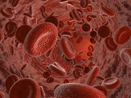
LISTEN NOW: UCSF's Christopher Moriates, MD, discusses waste-reduction efforts in hospitals
CHRISTOPHER MORIATES, MD, assistant clinical professor in the Division of Hospital Medicine at the University of California, San Francisco, talks about the change in focus and priorities needed for medicine to make progress in waste-reduction efforts.
CHRISTOPHER MORIATES, MD, assistant clinical professor in the Division of Hospital Medicine at the University of California, San Francisco, talks about the change in focus and priorities needed for medicine to make progress in waste-reduction efforts.
CHRISTOPHER MORIATES, MD, assistant clinical professor in the Division of Hospital Medicine at the University of California, San Francisco, talks about the change in focus and priorities needed for medicine to make progress in waste-reduction efforts.