User login
Researchers Report Opioid Modulation with the Combination of Buprenorphine and Samidorphan Improves Symptoms in Patients with Depression
NEW YORK (Reuters Health) - Opioid modulation with the combination of buprenorphine and samidorphan (ALKS 5461) improves symptoms in patients whose depression has not responded adequately to antidepressant treatment, researchers report.
"If the findings are confirmed in phase 3 studies, ALKS 5461 could be used as an augmenting agent in patients not responding to standard antidepressant therapies as an alternative to the atypical antipsychotic agents currently approved for the treatment of this population," Dr. Maurizio Fava, from Massachusetts General Hospital and Harvard Medical School, Boston, told Reuters Health by email.
Buprenorphine is a mu- and kappa-opioid partial agonist, and samidorphan blocks the mu agonist effects of buprenorphine associated with its abuse and addictive potential. A growing body of evidence implicates dysregulation of the endogenous mu- and kappa-opioid system in mood disorders.
Dr. Fava and colleagues at 31 sites in the U.S. used a sequential parallel comparison design to investigate the efficacy of buprenorphine/samidorphan (2 mg/2 mg or 8 mg/8 mg) in 142 patients with major depression inadequately responsive to antidepressant therapy.
At the end of four weeks of treatment, patients in the ALKS 5461 2 mg/2 mg group showed significantly greater improvements in Hamilton Depression Rating Scale (HAM-D), Montgomery-Asberg Depression Rating Scale (MADRS), and Clinical Global Impression severity scores, compared with patients in the placebo group. The ALKS 5461 8 mg/8 mg group showed smaller, nonsignificant improvements.
"The overall effect sizes for the 2/2 dosage were 0.50 for HAM-D and 0.54 for MADRS," the researchers noted. "The result compares favorably with results from a meta-analysis of 14 studies with atypical antipsychotics as adjunctive therapy for major depression, with reported effect sizes of 0.35 to 0.48 for individual drugs."
Treatment response rates according to HAM-D and MADRS (at least 50% reduction in scores) were highest with ALKS 5461 2 mg/2 mg treatment group, according to the February 12 onlinereport in the American Journal of Psychiatry.
Two patients (1.6%) in the placebo group and 17 patients (19.3%) in the ALKS 5461 groups discontinued because of treatment-emergent adverse events, but there was no evidence of opioid withdrawal in any patient.
"When depressed patients do not respond to standard monoamine-based therapies for depression, consider the use of an augmenting agent that modulates other systems, such as the opioid one," Dr. Fava concluded.
Dr. Jeffrey F. Scherrer, from Saint Louis University School of Medicine, St. Louis, Missouri, recently examined the association between opioid use and increased depression rates. He told Reuters Health by email, "Our analysis of opioid use and depression did not include buprenorphine among the opioid exposure variable. Therefore, it is difficult to extrapolate our results to a trial of buprenorphine/samidorphan and major depression."
"As the authors noted, the clinical trial was of short duration and the risks of depression that we have observed appears to be greatest among those patients remaining on opioids for more than 90 days," he explained. "Additional work is currently being done to determine if some opioid medications have a greater depressogenic effect than others, which further limits direct comparison of our findings to the current study."
"I will say that it is unlikely for oxycodone, codeine, and hydrocodone (which together account for more than 90% of prescribed opioids) would help depressed patients and be more likely to worsen their depression with chronic treatment," Dr. Scherrer concluded.
Alkermes sponsored the trial, employed seven coauthors, and had various relationships with the other four coauthors.
Team identifies potential target for aggressive AML

Photo by Rhoda Baer
Research published in The Journal of Clinical Investigation has revealed a potential therapeutic target for an aggressive form of acute myeloid leukemia (AML).
Investigators studied AML characterized by overexpression of the gene meningioma-1 (MN1), which is not a druggable target.
The team found that MN1 overexpression induces aggressive AML that is dependent on a gene expression program controlled by 2 histone methyltransferases.
And 1 of these histone methyltransferases can be targeted by drugs currently in clinical development.
To make these discoveries, the investigators forced expression of MN1 in mice, which induced AML, and looked for changes in other genes. They found that MN1 overexpression prompted the activation of genes already linked to AML development—HoxA9 and Meis1.
HoxA9 and Meis1 are key targets of the histone methyltransferases Mll1 and Dot1l. It turned out that Mll1 and Dotl1 are essential for creating the environment MN1 needs to cause AML.
“In mice, we put MN1 in first, leading to AML,” explained study author Kathrin Bernt, MD, of the University of Colorado Anschutz Medical Campus.
“Then, we knocked out these chromatin-regulating molecules, Mll1 or Dot1l. When we did that, the leukemia collapsed.”
The investigators also studied samples from AML patients and found that samples with overexpression of MN1 and HOXA9 were sensitive to the DOT1L inhibitor EPZ004777. The drug induced dose-dependent decreases in cell growth and the fraction of cycling cells, as well as an increase in apoptosis.
Anticancer agents targeting DOT1L are already in clinical trials. One such inhibitor, EPZ-5676, is being tested in a phase 1 trial of pediatric patients with aggressive leukemias (NCT02141828).
“The existing trial targets patients with rearrangements in the gene MLL1,” Dr Bernt noted. “Our study shows another subset of patients that may benefit from this or other therapies aimed at DOT1L inhibition—namely, patients with MN1 overexpression.”
Dr Bernt added, however, that the investigators must still determine the cutoff of MN1 overexpression at which AML is susceptible to DOT1L inhibition.
“Overexpression exists along a spectrum,” she explained. “At what degree of MN1 overexpression does it become clinically significant?” ![]()
EC approves drug for iron-deficiency anemia in IBD

The European Commission (EC) has granted marketing authorization for ferric maltol (Feraccru) to treat iron-deficiency anemia in adults with inflammatory bowel disease (IBD).
The product can now be marketed for this indication in the 28 member countries of the European Union, as well as Iceland, Liechtenstein, and Norway.
The company developing the drug, Shield Therapeutics, said it will begin a roll-out of commercialization in the coming months.
Feraccru contains iron in a stable ferric state as a complex with a trimaltol ligand—ferric maltol. It is formulated as 30 mg of ferric iron in a hard gelatin capsule.
The complex is designed to provide iron for uptake across the intestinal wall and transfer to the iron transport and storage proteins—transferrin and ferritin, respectively. Feraccru dissociates on uptake from the gastrointestinal tract, and ferric maltol itself does not appear to enter the systemic circulation.
Phase 3 trials
The EC’s approval of ferric maltol is based on results of 2 phase 3 studies—AEGIS 1 and AEGIS 2. The results of these studies were published in Inflammatory Bowel Diseases in March 2015.
Together, the trials included 128 adult patients with IBD (ulcerative colitis and Crohn’s disease), iron-deficiency anemia, and recorded intolerance of ferrous sulphate. They were randomized to receive either 30 mg of ferric maltol twice a day or a matched placebo capsule for 12 weeks.
The primary efficacy endpoint was the change in hemoglobin (Hb) levels from baseline to week 12. The mean improvement in Hb in the ferric maltol group compared to the placebo group was 2.25 g/dL (P<0.0001).
The absolute mean Hb concentration improved from 11.00 g/dL at baseline to 13.20 g/dL at week 12 in the ferric maltol group. In the placebo group, the mean Hb values were similar at baseline and week 12—11.10 g/dL and 11.20 g/dL, respectively.
The incidence of treatment-emergent adverse events (AEs) was 58% in the ferric maltol group and 72% in the placebo group. However, not all of these events were considered treatment-related.
AEs that were considered treatment-related occurred in 25% of ferric maltol-treated patients and 11.7% of placebo-treated patients. The most common treatment-related AEs in the ferric maltol group were abdominal pain, constipation, and flatulence—each occurring in 6.7% of patients.
“The phase 3 clinical studies clearly demonstrated Feraccru’s effectiveness,” said Andreas Stallmach, MD, of University Clinic Jena in Germany.
“[T]his pan-European marketing authorization gives treating physicians like myself the opportunity to fulfill an important and currently unmet need for patients who are unable to tolerate other oral products, as Feraccru could provide an oral alternative to intravenous iron infusion.’’ ![]()
Vaccine candidate fails to prevent malaria in humans
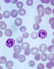
Plasmodium vivax
Image by Mae Melvin
Results of a phase 1 trial suggest a vaccine candidate does not prevent Plasmodium vivax malaria.
The vaccine, VMP001/AS01B, did not prevent malaria in any of the 30 subjects tested, although it did significantly delay parasitemia in 59% of subjects.
Lieutenant Colonel Jason W. Bennett, of the Walter Reed Army Institute
of Research (WRAIR) in Silver Spring, Maryland, and his colleagues reported these results in PLOS Neglected Tropical Diseases.
The vaccine antigen VMP001 is an Escherichia coli-produced, synthetic, chimeric, recombinant protein that incorporates the 3 major domains of circumsporozoite protein but is distinct from the native molecule. The VMP001 antigen was adjuvanted with AS01B, a proprietary liposome-based adjuvant system from GlaxoSmithKline.
For this phase 1 study, the investigators tested VMP001/AS01B in 30 healthy volunteers. The subjects received 3 intramuscular injections of VMP001 at 15 μg (n=10), 30 μg (n=10), or 60 μg (n=10), respectively, all formulated in 500 μL of AS01B at each immunization.
Fourteen days after the third immunization, the subjects were exposed to mosquitoes carrying P vivax. Six non-vaccinated control subjects were exposed to the mosquitoes as well.
The investigators said VMP001/AS01B was well tolerated and generated robust immune responses. However, it did not protect the subjects from malaria infection.
Still, the vaccine induced a “small but significant” delay in time to parasitemia in 59% of vaccinated subjects, when compared to controls.
Colonel Robert Paris, director of the US Military Malaria Research Program at WRAIR, believes the investigators can use the results of this study to design a better vaccine for P vivax malaria.
“Findings from the analysis of the immune response of vaccinated subjects have given us clues to improve vaccine candidates,” he said. “[S]tudies are now underway at WRAIR to develop next-generation vivax vaccines.” ![]()
No Change in Duration of Mechanical Ventilation with Acetazolamide for COPD Patients with Metabolic Alkalosis
Clinical question: Does acetazolamide decrease the duration of mechanical ventilation for patients with chronic obstructive pulmonary disease and metabolic alkalosis?
Bottom line: Although acetazolamide effectively decreased the levels of serum bicarbonate and the number of days with metabolic alkalosis in critically ill patients with chronic obstructive pulmonary disease (COPD) on mechanical ventilation, it did not produce a statistically significant difference in the duration of mechanical ventilation. However, this lack of statistical significance may have been due to an underpowered study. (LOE = 1b-)
Reference: Faisy C, Meziani F, Planquette B, et al, for the DIABOLO Investigators. Effect of acetazolamide vs placebo on duration of invasive mechanical ventilation among patients with chronic obstructive pulmonary disease. JAMA 2016;315(5):480-488.
Study design: Randomized controlled trial (double-blinded)
Funding source: Industry + govt
Allocation: Concealed
Setting: Inpatient (ICU only)
Synopsis:
Acetazolamide is used as a respiratory stimulant for patients with COPD and metabolic alkalosis because of its ability to reduce serum bicarbonate, which may lead to a rise in minute ventilation. However, its clinical benefit has not been demonstrated previously in controlled trials.
Using concealed allocation, these investigators randomized patients with COPD who required invasive mechanical ventilation to receive either acetazolamide 500 mg intravenously twice daily (1000 mg if loop diuretics were also prescribed) or matching placebo up to 28 days. Only those patients found to have pure or mixed metabolic alkalosis received the treatment. The intention-to-treat population consisted of 380 patients and baseline characteristics were similar in both groups. The use of acetazolamide led to decreased levels of serum bicarbonate and fewer days with metabolic alkalosis but did not improve respiratory parameters such as respiratory rate, tidal volume, or minute ventilation.
For the primary outcome of duration of mechanical ventilation, the magnitude of the difference between the 2 groups was large, suggesting a clinically important effect, but the difference did not reach statistical significance (between-group difference -16 hours with acetazolamide; 95% CI -36.5 to 4.0 hours). The lack of statistical significance may be due to an underpowered study because of fewer-than-expected patients who received the treatment (more than 20% of patients in each group did not receive the assigned treatment because of lack of metabolic alkalosis or temporary contraindications) and a shorter-than-anticipated duration of mechanical ventilation in the placebo group.
Dr. Kulkarni is an assistant professor of hospital medicine at Northwestern University in Chicago.
Tamsulosin Effective as Expulsion Therapy for Distal Ureteric Stones
Clinical question: Is tamsulosin effective in the management of distal ureteric stones?
Bottom line: Tamsulosin promotes stone passage of ureteric stones that are 5 mm to 10 mm. You would need to treat 5 patients with tamsulosin to cause the expulsion of one such stone. Stones smaller than 5 mm have a high rate of spontaneous passage without any intervention. (LOE = 1b-)
Reference: Furyk JS, Chu K, Banks C et al. Distal ureteric stones and tamsulosin: a double-blind, placebo-controlled, randomized, multicenter trial. Ann Emerg Med 2016;67(1):86-95.
Study design: Randomized controlled trial (double-blinded)
Funding source: Foundation
Allocation: Concealed
Setting: Outpatient (primary care)
Synopsis: These authors recruited adult patients who presented to the emergency department with symptoms and imaging consistent with distal ureteric stones. Patients with fever, hypotension, stones larger than 10 mm, or kidney disease were excluded. Using concealed allocation, the investigators randomized the patients to receive either tamsulosin 0.4 mg daily or matching placebo for 28 days or until stone passage. The 2 groups had similar baseline characteristics and analysis was by intention to treat.
The primary outcome was stone expulsion as confirmed by computed tomography (CT) and time to stone expulsion was defined by self-reported passage of stone or 48-hour pain-free period. Compliance to the study medications was poor in both groups, and almost one-fifth of the patients did not have follow-up imaging. Of the approximately 80% of patients in each group who underwent follow-up CT, there was no difference in the percentage of patients with passed stones (87% in the tamsulosin group vs 82% in the placebo group; P = .22). In the subset of patients with larger stones (5 mm -10 mm), the tamsulosin group had a significantly higher rate of stone passage than the placebo group (83% vs 61%; P = .03). There were no significant differences detected in time to stone passage, pain, analgesia requirements, need for urological intervention, or adverse events.
Dr. Kulkarni is an assistant professor of hospital medicine at Northwestern University in Chicago.
Leading scope manufacturer to pay hefty settlement for alleged kickbacks, bribery
Embattled scope maker Olympus Corp., already under federal investigation for its role in superbug outbreaks, has agreed to pay $646 million to resolve criminal and civil probes into illegal kickbacks and bribes to doctors and hospitals.
Federal prosecutors said Tuesday that the company’s settlement is the largest ever for violations of the U.S. anti-kickback law. A portion of the company’s payout, $22.8 million, will resolve foreign bribery allegations in Latin America.
U.S. investigators said the company’s “greed-fueled kickback scheme” from 2006 to 2011 used grants, consulting deals, trips to Japan, lavish gifts, and free equipment to induce influential doctors to order more Olympus devices at prominent hospitals and keep out competitors.
After a key doctor for a Midwest hospital system took a week-long trip to Japan and received a company grant, an Olympus vice president wrote an internal email in 2006 that said, “We have received all of the orders expected and have kept (a competitor) completely out of the (Midwestern hospital) system. Hooray!”
The federal complaint against Olympus mentioned similar scenarios at a prominent California institution and a New York medical center, but the exact names were redacted.
The violations produced about $600 million in sales and $230 million in gross profits for Olympus, federal officials said.
Federal prosecutors credited the help of a former compliance officer at Olympus, John Slowik, who filed a whistle-blower case against the company under seal in 2010.
Olympus agreed to a corporate-integrity agreement and the appointment of an independent monitor.
“Olympus leadership acknowledges the company’s responsibility for the past conduct, which does not represent the values of Olympus or its employees,” Nacho Abia, chief executive of the Olympus Corp. of the Americas unit in Center Valley, Pa., said in a statement. “Olympus is committed to complying with all laws and regulations and to adhering to our own rigorous code of conduct.”
Olympus has come under fire for failing to alert U.S. regulators and hospitals sooner about the risks of infection from its duodenoscopes, a gastrointestinal scope that has proven difficult to clean of dangerous bacteria because of its intricate design.
A Senate investigation released in January identified 19 scope-related outbreaks at U.S. medical centers during 2012-2015 that sickened nearly 200 patients with drug-resistant infections.
Olympus announced in January it would recall its duodenoscopes nationwide and make repairs to improve patient safety. The company controls 85% of the U.S. market for gastrointestinal scopes.
Last year, the Tokyo company disclosed it had received a subpoena from federal prosecutors in March 2015 seeking “information relating to duodenoscopes that Olympus manufactures and sells.”
This article was originally published by Kaiser Health News, a national health policy news service that is part of the nonpartisan Henry J. Kaiser Family Foundation.
Legacy Society members sustain research
AGA Legacy Society members are showing their gratitude for what funding and research has brought to our specialty by giving back.
Legacy Society members are the most generous individual donors to the AGA Research Foundation. Members of the AGA Legacy Society provide tax-deductible gifts to the AGA Research Foundation of $25,000 or more (payable over 5 years) or $50,000 or more in a planned gift, such as a bequest.
The AGA Research Foundation’s mission is to raise funds to support young researchers in gastroenterology and hepatology. Richard M. Peek, Jr., M.D., AGAF, Legacy Society member said, “I have a huge appreciation for the AGA Research Foundation, because they were the first foundation that really took a chance on my research. Today, I am proud to be a donor myself because I know it is making a difference [for] yet another young investigator.”
Donors who make gifts at the Legacy Society level before DDW will receive an invitation to the annual Benefactors’ Dinner at The Don Room and Terrace at El Cortez in San Diego. Individuals interested in learning more about Legacy Society membership may contact Stacey Hinton Tuneski, Senior Director of Development at [email protected] or via phone (301) 222-4005.
A celebration of research support
Beginning with a memorable gathering at the United States Library of Congress in 2007, the Benefactors’ Dinner has welcomed members of the AGA Legacy Society and other AGA dignitaries to special locations nationwide. The Don Room and Terrace at El Cortez will be the location of the 2016 AGA Research Foundation Benefactors’ Dinner during DDW in San Diego. Guests will enjoy a wonderful evening in the historic setting that has hosted dignitaries since 1927. Members of the AGA Legacy Society will be among the distinguished honorees at the annual event.
My day advocating for GI on Capitol Hill
This January I was fortunate to spend a day on Capitol Hill meeting with AGA senior policy staff and key members of Congress who have jurisdiction over critical policy priorities for AGA and the profession of gastroenterology. It was an interesting and informative day. Among other things, I learned that individuals on Capitol Hill are interested in hearing from us to gain our insight and expertise on the practice and science of gastroenterology and patient care.
Senate
During my time visiting the Senate side of Capitol Hill, I met with the offices of Sen. Bill Cassidy (R-La.), Sen. Ben Cardin (D-Md.), Sen. Sherrod Brown (D-Ohio), and Sen. Amy Klobuchar (D-Minn). Sen. Cassidy, as you may be aware, has been an AGA member and champion for many of our policy priorities. Most recently, he, along with Sen. Cardin, spearheaded the Senate effort in contacting CMS and expressing concern over cuts in reimbursement for colonoscopy. Sen. Cassidy was critical in implementing the new transparency policy at CMS, which led to the announcement of changes in physician values in the proposed rule, instead of the final rule. This will ensure that stakeholders have the opportunity to participate in the rulemaking process.
We also discussed the AGA obesity initiative that is being developed. This is a multidisciplinary approach to treating the disease. I had the opportunity to educate Sen. Cassidy’s staff on some of the new drugs that have been approved to treat obesity and the emergence of new endoscopic procedures that could have an impact on how we treat this growing epidemic. I also learned about Sen. Cassidy’s legislation, S. 1509/H.R. 2404, Treat and Reduce Obesity Act, which would cover behavioral therapy, as well as drugs, to treat obesity under Medicare. Sen. Cassidy is very concerned with obesity, especially in Louisiana, where it is a major public health problem. We will continue to work with Sen. Cassidy on this important initiative, as well as other public health initiatives, given his role on the Senate Health, Education, Labor and Pensions Committee.
I also had the honor of meeting with Sen. Brown’s staff. Sen. Brown is the main sponsor of the Removing Barriers to Colorectal Cancer Screening Act legislation that would fix the current co-insurance problem requiring Medicare beneficiaries to face out-of-pocket expenses when their screening colonoscopy becomes therapeutic. We impressed upon the staff our concern that this is a deterrent to undergoing colonoscopy for colorectal cancer screening. One of the main obstacles to getting this bill over the finish line is a favorable “score” (or additional cost) from the Congressional Budget Office and finding an appropriate legislative vehicle given this year’s short legislative session. I was able to thank Sen. Brown for his support of NIH, given that Congress gave the institute a $2 billion budget increase last year as part of the Omnibus Appropriations Bill.
In my discussions with Sen. Klobuchar’s staff, I thanked her for her support of NIH and access to colorectal cancer screening. She has been a champion of repealing the medical device tax, which received a 2-year delay as part of the recent Omnibus Bill. Finally, I met with Sen. Cardin, who has a long history of supporting colorectal cancer screening and was instrumental in first implementing the benefit under Medicare, as part of the Balanced Budget Act. He continues to champion many of our priorities, including NIH funding, fair reimbursement for colonoscopy and fixing the coinsurance waiver.
House of Representatives
I was also fortunate to meet with the staff for several representatives on the House side, including those key individuals involved in health legislation. The staff to Labor, HHS Appropriations Subcommittee Chair Tom Coles (R-Okla.) who was critical in ensuring that NIH received a bump in funding, conveyed that they would like to continue with sustained funding just as Congress did during the period when the NIH budget was doubled. They were very interested in learning about my own research on obesity at Mayo Clinic and the implications it could have in more effectively treating the disease.
I also met with staff for Rep. Jim McGovern, (D-Mass.), a senior member of the House Rules Committee and a longtime supporter of improving access to colorectal cancer screening, and Rep. Tim Walz, (D-Minn.), who is a representative from my congressional district in Minnesota. Both are strong supporters of NIH funding and colorectal cancer screening.
My experience showed me how willing our lawmakers on Capitol Hill are to meet with gastroenterologists, to learn about our experiences and our patients, and to find ways to work with us to ensure the correct laws are in place to ensure our patients are receiving the best care.
Medical research advances and the practice of medicine affect everyone in this country. We need to work with Congress to continue to advocate for the programs and initiatives that are vital to our patients and to our specialty, gastroenterology.
Behind the scenes: AGA planning meeting for DDW 2016
You’ve heard the stats about Digestive Disease Week® (DDW) – top 50 medical meeting, nearly 15,000 attendees, more than 4,000 abstracts. The experts behind AGA’s programming – coined the AGA Institute Council – recently came together to put the finishing touches on this year’s agenda.
If you’re not yet registered for this year’s meeting, taking place May 21 through 24 in San Diego, you can do so by visiting www.ddw.org/registration.
• AGA Basic Science Zone: AGA will be co-locating basic science sessions. On Saturday, we will highlight the gut microbiome, and on Sunday we will focus on inflammation and GI cancers. Both days will have a mix of invited and abstract-based sessions.
• More microbiome programming: AGA’s coverage of the microbiome will extend beyond basic science, focusing on clinical topics such as obesity, IBD, and pediatric GI.
• Collaborative topic sessions: our council is comprised of 12 special interest groups, or sections, all covering different topic areas in GI/hepatology.
• Plenary sessions: our plenary sessions at DDW highlight the most novel and innovative studies presented at the meeting. Don’t miss the AGA Presidential Plenary, the AGA Basic Science Plenary and the Distinguished Abstract Plenaries.
• For the second year in a row, the council will be awarding certificates of recognition in sessions that have a young investigator.
Visit www.ddw.org and review the preliminary program.