User login
Hospitalist‐Led Antimicrobial Stewardship
Inappropriate antimicrobial use in hospitalized patients is a well‐recognized driver for the development of drug‐resistant organisms and antimicrobial‐related complications such as Clostridium difficile infection (CDI).[1, 2] Infection with C difficile affects nearly 500,000 people annually resulting in higher healthcare expenditures, longer lengths of hospital stay, and nearly 15,000 deaths.[3] Data from the Centers for Disease Control and Prevention (CDC) suggest that a 30% reduction in the use of broad‐spectrum antimicrobials, or a 5% reduction in the proportion of hospitalized patients receiving antimicrobials, could equate to a 26% reduction in CDI.[4] It is estimated that up to 50% of antimicrobial use in the hospital setting may be inappropriate.[5]
Since the Infectious Diseases Society of America and the Society for Healthcare Epidemiology of America published guidelines for developing formal, hospital‐based antimicrobial stewardship programs in 2007, stewardship practices have been adapted by frontline providers to fit day‐to‐day inpatient care.[5] A recent review by Hamilton et al. described several studies in which stewardship practices were imbedded into daily workflows by way of checklists, education reminders, and periodic review of antimicrobial usage, as well as a multicenter pilot of point‐of‐care stewardship interventions successfully implemented by various providers including nursing, pharmacists, and hospitalists.[6]
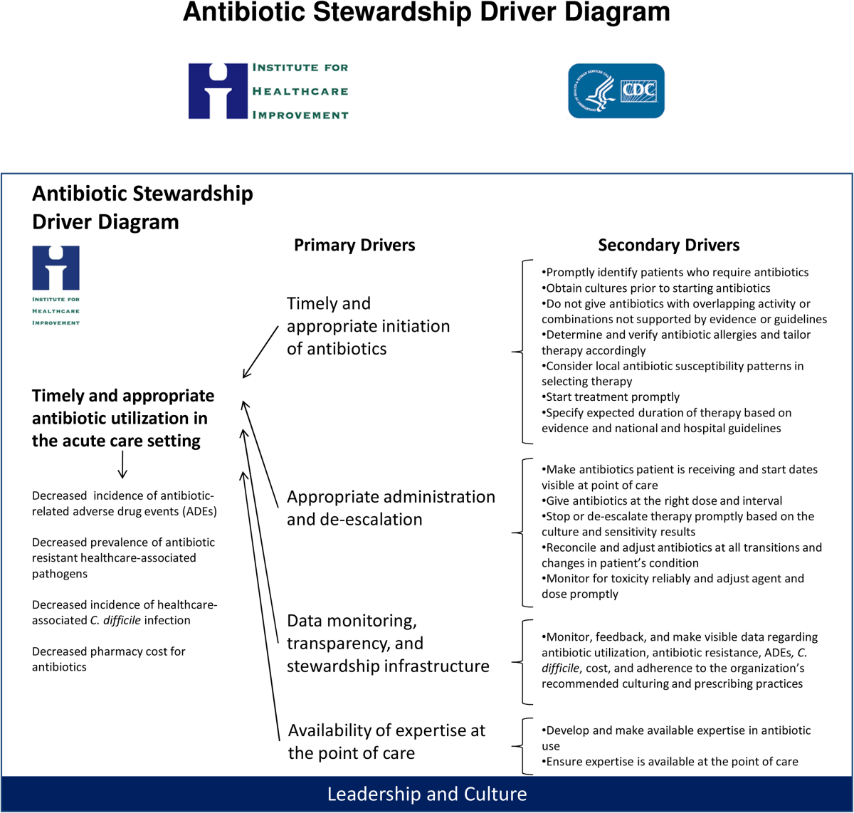
In response to the CDC's 2010 Get Smart for Healthcare campaign, which focused on stemming antimicrobial resistance and improving antimicrobial use, the Institute for Healthcare Improvement (IHI), in partnership with the CDC, brought together experts in the field to identify practical and feasible target practices for hospital‐based stewardship and created a Driver Diagram to guide implementation efforts (Figure 1). Rohde et al. described the initial pilot testing of these practices, the decision to more actively engage frontline providers, and the 3 key strategies identified as high‐yield improvement targets: enhancing the visibility of antimicrobial use at the point of care, creating easily accessible antimicrobial guidelines for common infections, and the implementation of a 72‐hour timeout after initiation of antimicrobials.[7]

In this article, we describe how, in partnership with the IHI and the CDC, the hospital medicine programs at 5 diverse hospitals iteratively tested these 3 strategies with a goal of identifying the barriers and facilitators to effective hospitalist‐led antimicrobial stewardship.
METHODS
Representatives from 5 hospital medicine programs, IHI, and the CDC attended a kick‐off meeting at the CDC in November 2012 to discuss the 3 proposed strategies, examples of prior testing, and ideas for implementation. Each hospitalist provided a high‐level summary of the current state of stewardship efforts at their respective institutions, identified possible future states related to the improvement strategies, and anticipated problems in achieving them. The 3 key strategies are described below.
Improved Documentation/Visibility at Points of Care
Making antimicrobial indication, day of therapy, and anticipated duration transparent in the medical record was the targeted improvement strategy to avoid unnecessary antimicrobial days that can result from provider uncertainty, particularly during patient handoffs. Daily hospitalist documentation was identified as a vehicle through which these aspects of antimicrobial use could be effectively communicated and propagated from provider to provider.
Stewardship educational sessions and/or awareness campaigns were hospitalist led, and were accompanied by follow‐up reminders in the forms of emails, texts, flyers, or conferences. Infectious disease physicians were not directly involved in education but were available for consultation if needed.
Improved Guideline Clarity and Accessibility
Enhancing the availability of guidelines for frequently encountered infections and clarifying key guideline recommendations such as treatment duration were identified as the improvement strategies to help make treatment regimens more appropriate and consistent across providers.
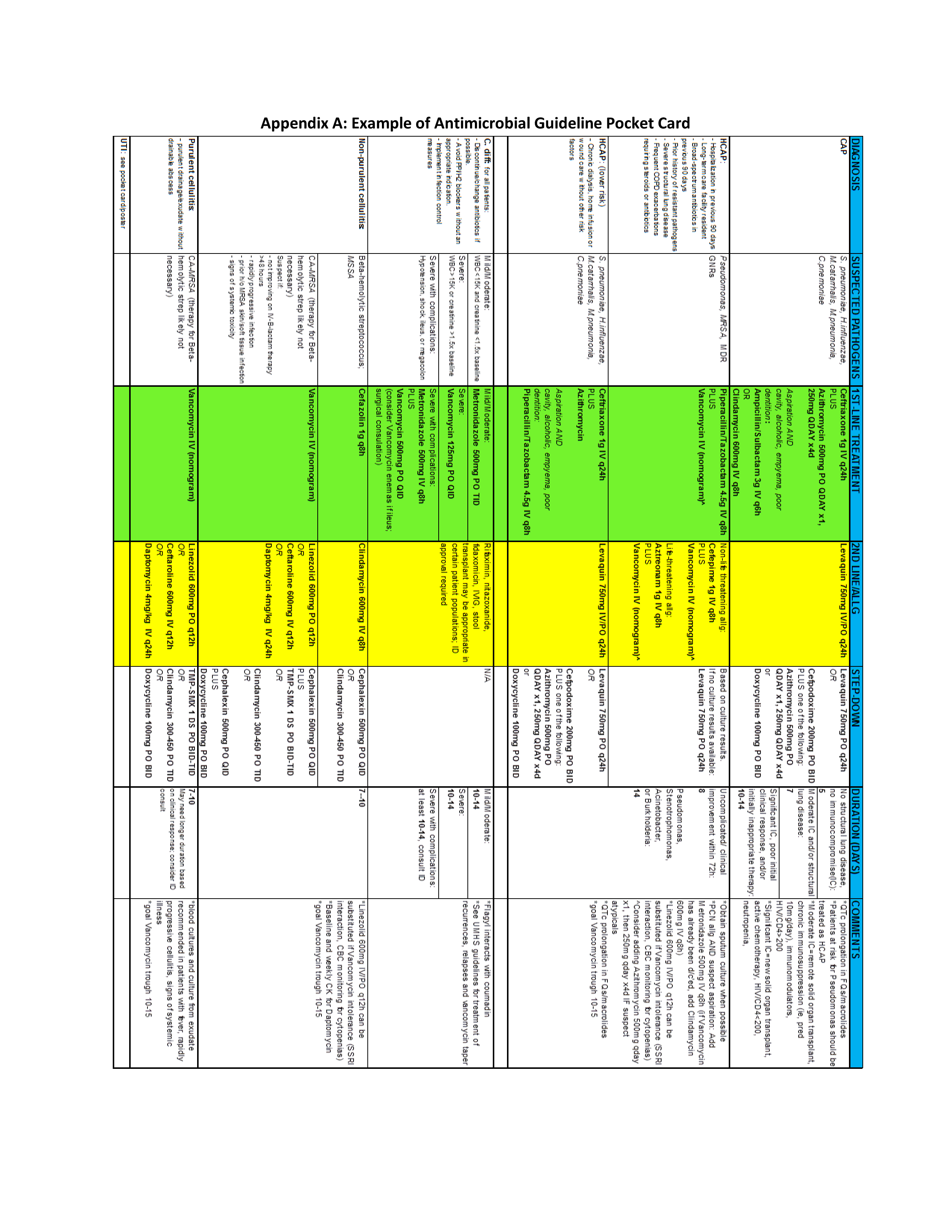
Interventions included designing simplified pocket cards for commonly encountered infections, (see Supporting Information, Appendix A, in the online version of this article), collaborating with infectious disease physicians on guideline development, and dissemination through email, smartphone, and wall flyers, and creation of a continuous medical education module focused on stewardship practices.
72‐Hour Antimicrobial Timeout
The 72‐hour antimicrobial timeout required that hospitalists routinely reassess antimicrobial use 72 hours following antimicrobial initiation, a time when most pertinent culture data had returned. Hospitalists partnered with clinical pharmacists at all sites, and addressed the following questions during each timeout: (1) Does the patient have a condition that requires continued use of antimicrobials? (2) Can the current antimicrobial regimen be tailored based on culture data? (3) What is the anticipated treatment duration? A variety of modifications occurred during timeouts, including broadening or narrowing the antimicrobial regimen based on culture data, switching to an oral antimicrobial, adjusting dose or frequency based on patient‐specific factors, as well as discontinuation of antimicrobials. Following the initial timeout, further adjustments were made as the clinical situation dictated; intermittent partnered timeouts continued during a patient's hospitalization on an individualized basis. Hospitalists were encouraged to independently review new diagnostic information daily and make changes as needed outside the dedicated time‐out sessions. All decisions to adjust antimicrobial regimens were provider driven; no hospitals employed automated antimicrobial discontinuation without provider input.
Implementation and Evaluation
Each site was tasked with conducting small tests of change aimed at implementing at least 1, and ideally all 3 strategies. Small, reasonably achievable interventions were preferred to large hospital‐wide initiatives so that key barriers and facilitators to the change could be quickly identified and addressed.
Methods of data collection varied across institutions and included anonymous physician survey, face‐to‐face physician interviews, and medical record review. Evaluations of hospital‐specific interventions utilized convenience samples to obtain real time, actionable data. Postintervention data were distributed through biweekly calls and compiled at the conclusion of the project. Barriers and facilitators of hospitalist‐centered antimicrobial stewardship collected over the course of the project were reviewed and used to identify common themes.
RESULTS
Participating hospitals included 1 community nonteaching hospital, 2 community teaching hospitals, and 2 academic medical centers. All hospitals used computerized order entry and had prior quality improvement experience; 4 out of 5 hospitals used electronic medical records. Postintervention data on antimicrobial documentation and timeouts were compiled, shared, and successes identified. For example, 2 hospitals saw an increase in complete antimicrobial documentation from 4% and 8% to 51% and 65%, respectively, of medical records reviewed over a 3‐month period. Additionally, cumulative timeout data across all hospitals showed that out of 726 antimicrobial timeouts evaluated, optimization or discontinuation occurred 218 times or 30% of the time.
Each site's key implementation barriers and facilitators were collected. Examples were compiled and common themes emerged (Table 1).
| ||
| Barriers: What impediments did we experience during our stewardship project? | Schedule and practice variability | Physician variability in structure of antimicrobial documentation |
| Prescribing etiquette: it's difficult to change course of treatment plan started by a colleague | ||
| Competing schedule demands of hospitalist and pharmacist | ||
| Skepticism of antimicrobial stewardship importance | Perception of incorporating stewardship practices into daily work as time consuming | |
| Improvement project fatigue from competing quality improvement initiatives | ||
| Unclear leadership buy‐in | ||
| Focusing too broadly | Choosing large initial interventions, which take significant time/effort to complete and quantify | |
| Setting unrealistic expectations (eg, expecting perfect adherence to documentation, guidelines, or timeout) | ||
| Facilitators: What countermeasures did we target to overcome barriers? | Engage the hospitalists | Establish a core part of the hospitalist group as stewardship champions |
| Speak 1‐on‐1 to colleagues about specific goals and ways to achieve them | ||
| Establish buy‐in from leadership | ||
| Encourage participation from a multidisciplinary team (eg, bedside nursing, clinical pharmacists) | ||
| Collect real time data and feedback | Utilize a data collection tool if possible/engage hospital coders to identify appropriate diagnoses | |
| Define your question and identify baseline data prior to intervention | ||
| Give rapid cycle feedback to colleagues that can impact antimicrobial prescribing in real time | ||
| Recognize and reward high performers | ||
| Limit scope | Start with small, quickly implementable interventions | |
| Identify interventions that are easy to integrate into hospitalist workflow | ||
DISCUSSION
We successfully brought together hospitalists from diverse institutions to undertake small tests of change aimed at 3 key antimicrobial use improvement strategies. Following our interventions, significant improvement in antimicrobial documentation occurred at 2 institutions focusing on this improvement strategy, and 72‐hour timeouts performed across all hospitals tailored antimicrobial use in 30% of the sessions. Through frequent collaborative discussions and information sharing, we were able to identify common barriers and facilitators to hospitalist‐centered stewardship efforts.
Each participating hospital medicine program noticed a gradual shift in thinking among their colleagues, from initial skepticism about embedding stewardship within their daily workflow, to general acceptance that it was a worthwhile and meaningful endeavor. We posited that this transition in belief and behavior evolved for several reasons. First, each group was educated about their own, personal prescribing practices from the outset rather than presenting abstract data. This allowed for ownership of the problem and buy‐in to improve it. Second, participants were able to experience the benefits at an individual level while the interventions were ongoing (eg, having other providers reciprocate structured documentation during patient handoffs, making antimicrobial plans clearer), reinforcing the achievability of stewardship practices within each group. Additionally, we focused on making small, manageable interventions that did not seem disruptive to hospitalists' daily workflow. For example, 1 group instituted antimicrobial timeouts during preexisting multidisciplinary rounds with clinical pharmacists. Last, project champions had both leadership and frontline roles within their groups and set the example for stewardship practices, which conveyed that this was a priority at the leadership level. These findings are in line with those of Charani et al., who evaluated behavior change strategies that influence antimicrobial prescribing in acute care. The authors found that behavioral determinants and social norms strongly influence prescribing practices in acute care, and that antimicrobial stewardship improvement projects should account for these influences.[8]
We also identified several barriers to antimicrobial stewardship implementation (Table 1) and proposed measures to address these barriers in future improvement efforts. For example, hospital medicine programs without a preexisting clinical pharmacy partnership asked hospitalist leadership for more direct clinical pharmacy involvement, recognizing the importance of a physician‐pharmacy alliance for stewardship efforts. To more effectively embed antimicrobial stewardship into daily routine, several hospitalists suggested standardized order sets for commonly encountered infections, as well as routine feedback on prescribing practices. Furthermore, although our simplified antimicrobial guideline pocket card enhanced access to this information, several colleagues suggested a smart phone application that would make access even easier and less cumbersome. Last, given the concern about the sustainability of antimicrobial stewardship initiatives, we recommended periodic reminders, random medical record review, and re‐education if necessary on our 3 strategies and their purpose.
Our study is not without limitations. Each participating hospitalist group enacted hospital‐specific interventions based on individual hospitalist program needs and goals, and although there was collective discussion, no group was tasked to undertake another group's initiative, thereby limiting generalizability. We did, however, identify common facilitators that could be adapted to a wide variety of hospitalist programs. We also note that our 3 main strategies were included in a recent review of quality indicators for measuring the success of antimicrobial stewardship programs; thus, although details of individual practice may vary, in principle these concepts can help identify areas for improvement within each unique stewardship program.[9] Importantly, we were unable to evaluate the impact of the 3 key improvement strategies on important clinical outcomes such as overall antimicrobial use, complications including CDI, and cost. However, others have found that improvement strategies similar to our 3 key processes are associated with meaningful improvements in clinical outcomes as well as reductions in healthcare costs.[10, 11] Last, long‐ term impact and sustainability were not evaluated. By choosing interventions that were viewed by frontline providers as valuable and attainable, however, we feel that each group will likely continue current practices beyond the initial evaluation timeframe.
Although these 5 hospitalist groups were able to successfully implement several aspects of the 3 key improvement strategies, we recognize that this is only the first step. Further effort is needed to quantify the impact of these improvement efforts on objective patient outcomes such as readmissions, length of stay, and antimicrobial‐related complications, which will better inform our local and national leaders on the inherent clinical and financial gains associated with hospitalist‐led stewardship work. Finally, creative ways to better integrate stewardship activities into existing provider workflows (eg, decision support and automation) will further accelerate improvement efforts.
In summary, hospitalists at 5 diverse institutions successfully implemented key antimicrobial improvement strategies and identified important implementation facilitators and barriers. Future efforts at hospitalist‐led stewardship should focus on strategies to scale‐up interventions and evaluate their impact on clinical outcomes and cost.
Acknowledgements
The authors thank Latoya Kuhn, MPH, for her assistance with statistical analyses. We also thank the clinical pharmacists at each institution for their partnership in stewardship efforts: Patrick Arnold, PharmD, and Matthew Tupps, PharmD, MHA, from University of Michigan Hospital and Health System; and Roland Tam, PharmD, from Emory Johns Creek Hospital.
Disclosures: Dr. Flanders reports consulting fees or honoraria from the Institute for Healthcare Improvement, has provided consultancy to the Society of Hospital Medicine, has served as a reviewer for expert testimony, received honoraria as a visiting lecturer to various hospitals, and has received royalties from publisher John Wiley & Sons. He has also received grant funding from Blue Cross Blue Shield of Michigan and the Agency for Healthcare Research and Quality. Dr. Ko reports consultancy for the American Hospital Association and the Society of Hospital Medicine involving work with catheter‐associated urinary tract infections. Ms. Jacobsen reports grant funding from the Institute for Healthcare Improvement. Dr. Rosenberg reports consultancy for Bristol‐Myers Squibb, Forest Pharmaceuticals, and Pfizer. The funding source for this collaborative was through the Institute for Healthcare Improvement and Centers for Disease Control and Prevention. Funding was provided by the Department of Health and Human Services, the Centers for Disease Control and Prevention, the National Center for Emerging Zoonotic and Infectious Diseases, and the Division of Healthcare Quality Promotion/Office of the Director. Avaris Concepts served as the prime contractor and the Institute for Healthcare Improvement as the subcontractor for the initiative. The findings and conclusions in this report represent the views of the authors and might not reflect the views of the Centers for Disease Control and Prevention. The authors report no conflicts of interest.
- , , . Clinical and economic burden of antimicrobial resistance. Expert Rev Anti Infect Ther. 2008;6(5):751–763.
- , , , et al. Hospital and societal costs of antimicrobial‐resistant infections in a Chicago teaching hospital: implications for antibiotic stewardship. Clin Infect Dis. 2009;49(8):1175–1184.
- , , , et al. Burden of Clostridium difficile infection in the United States. N Engl J Med. 2015;372(9):825–834.
- , , , et al.; Centers for Disease Control and Prevention (CDC). Vital signs: improving antibiotic use among hospitalized patients. MMWR Morb Mortal Wkly Rep. 2014;63(9):194–200.
- , , , et al.; Infectious Diseases Society of America; Society for Healthcare Epidemiology of America. Infectious Diseases Society of America and the Society for Healthcare Epidemiology of America guidelines for developing an institutional program to enhance antimicrobial stewardship. Clin Infect Dis. 2007;44(2):159–177.
- , , , et al.; Centers for Disease Control and Prevention Epicenters Program. Point‐of‐prescription interventions to improve antimicrobial stewardship. Clin Infect Dis. 2015;60(8):1252–1258.
- , , . Role of the hospitalist in antimicrobial stewardship: a review of work completed and description of a multisite collaborative. Clin Ther. 2013;35(6):751–757.
- , , , et al. Behavior change strategies to influence antimicrobial prescribing in acute care: a systematic review. Clin Infect Dis. 2011;53(7):651–662.
- , , , , . Quality indicators to measure appropriate antibiotic use in hospitalized adults. Clin Infect Dis. 2015;60(2):281–291.
- , . Application of antimicrobial stewardship to optimise management of community acquired pneumonia. Int J Clin Pract. 2011;65(7):775–783.
- , , , et al. Interventions to improve antibiotic prescribing practices for hospital inpatients. Cochrane Database Syst Rev. 2013;4:CD003543.
Inappropriate antimicrobial use in hospitalized patients is a well‐recognized driver for the development of drug‐resistant organisms and antimicrobial‐related complications such as Clostridium difficile infection (CDI).[1, 2] Infection with C difficile affects nearly 500,000 people annually resulting in higher healthcare expenditures, longer lengths of hospital stay, and nearly 15,000 deaths.[3] Data from the Centers for Disease Control and Prevention (CDC) suggest that a 30% reduction in the use of broad‐spectrum antimicrobials, or a 5% reduction in the proportion of hospitalized patients receiving antimicrobials, could equate to a 26% reduction in CDI.[4] It is estimated that up to 50% of antimicrobial use in the hospital setting may be inappropriate.[5]
Since the Infectious Diseases Society of America and the Society for Healthcare Epidemiology of America published guidelines for developing formal, hospital‐based antimicrobial stewardship programs in 2007, stewardship practices have been adapted by frontline providers to fit day‐to‐day inpatient care.[5] A recent review by Hamilton et al. described several studies in which stewardship practices were imbedded into daily workflows by way of checklists, education reminders, and periodic review of antimicrobial usage, as well as a multicenter pilot of point‐of‐care stewardship interventions successfully implemented by various providers including nursing, pharmacists, and hospitalists.[6]
In response to the CDC's 2010 Get Smart for Healthcare campaign, which focused on stemming antimicrobial resistance and improving antimicrobial use, the Institute for Healthcare Improvement (IHI), in partnership with the CDC, brought together experts in the field to identify practical and feasible target practices for hospital‐based stewardship and created a Driver Diagram to guide implementation efforts (Figure 1). Rohde et al. described the initial pilot testing of these practices, the decision to more actively engage frontline providers, and the 3 key strategies identified as high‐yield improvement targets: enhancing the visibility of antimicrobial use at the point of care, creating easily accessible antimicrobial guidelines for common infections, and the implementation of a 72‐hour timeout after initiation of antimicrobials.[7]
In this article, we describe how, in partnership with the IHI and the CDC, the hospital medicine programs at 5 diverse hospitals iteratively tested these 3 strategies with a goal of identifying the barriers and facilitators to effective hospitalist‐led antimicrobial stewardship.
METHODS
Representatives from 5 hospital medicine programs, IHI, and the CDC attended a kick‐off meeting at the CDC in November 2012 to discuss the 3 proposed strategies, examples of prior testing, and ideas for implementation. Each hospitalist provided a high‐level summary of the current state of stewardship efforts at their respective institutions, identified possible future states related to the improvement strategies, and anticipated problems in achieving them. The 3 key strategies are described below.
Improved Documentation/Visibility at Points of Care
Making antimicrobial indication, day of therapy, and anticipated duration transparent in the medical record was the targeted improvement strategy to avoid unnecessary antimicrobial days that can result from provider uncertainty, particularly during patient handoffs. Daily hospitalist documentation was identified as a vehicle through which these aspects of antimicrobial use could be effectively communicated and propagated from provider to provider.
Stewardship educational sessions and/or awareness campaigns were hospitalist led, and were accompanied by follow‐up reminders in the forms of emails, texts, flyers, or conferences. Infectious disease physicians were not directly involved in education but were available for consultation if needed.
Improved Guideline Clarity and Accessibility
Enhancing the availability of guidelines for frequently encountered infections and clarifying key guideline recommendations such as treatment duration were identified as the improvement strategies to help make treatment regimens more appropriate and consistent across providers.
Interventions included designing simplified pocket cards for commonly encountered infections, (see Supporting Information, Appendix A, in the online version of this article), collaborating with infectious disease physicians on guideline development, and dissemination through email, smartphone, and wall flyers, and creation of a continuous medical education module focused on stewardship practices.
72‐Hour Antimicrobial Timeout
The 72‐hour antimicrobial timeout required that hospitalists routinely reassess antimicrobial use 72 hours following antimicrobial initiation, a time when most pertinent culture data had returned. Hospitalists partnered with clinical pharmacists at all sites, and addressed the following questions during each timeout: (1) Does the patient have a condition that requires continued use of antimicrobials? (2) Can the current antimicrobial regimen be tailored based on culture data? (3) What is the anticipated treatment duration? A variety of modifications occurred during timeouts, including broadening or narrowing the antimicrobial regimen based on culture data, switching to an oral antimicrobial, adjusting dose or frequency based on patient‐specific factors, as well as discontinuation of antimicrobials. Following the initial timeout, further adjustments were made as the clinical situation dictated; intermittent partnered timeouts continued during a patient's hospitalization on an individualized basis. Hospitalists were encouraged to independently review new diagnostic information daily and make changes as needed outside the dedicated time‐out sessions. All decisions to adjust antimicrobial regimens were provider driven; no hospitals employed automated antimicrobial discontinuation without provider input.
Implementation and Evaluation
Each site was tasked with conducting small tests of change aimed at implementing at least 1, and ideally all 3 strategies. Small, reasonably achievable interventions were preferred to large hospital‐wide initiatives so that key barriers and facilitators to the change could be quickly identified and addressed.
Methods of data collection varied across institutions and included anonymous physician survey, face‐to‐face physician interviews, and medical record review. Evaluations of hospital‐specific interventions utilized convenience samples to obtain real time, actionable data. Postintervention data were distributed through biweekly calls and compiled at the conclusion of the project. Barriers and facilitators of hospitalist‐centered antimicrobial stewardship collected over the course of the project were reviewed and used to identify common themes.
RESULTS
Participating hospitals included 1 community nonteaching hospital, 2 community teaching hospitals, and 2 academic medical centers. All hospitals used computerized order entry and had prior quality improvement experience; 4 out of 5 hospitals used electronic medical records. Postintervention data on antimicrobial documentation and timeouts were compiled, shared, and successes identified. For example, 2 hospitals saw an increase in complete antimicrobial documentation from 4% and 8% to 51% and 65%, respectively, of medical records reviewed over a 3‐month period. Additionally, cumulative timeout data across all hospitals showed that out of 726 antimicrobial timeouts evaluated, optimization or discontinuation occurred 218 times or 30% of the time.
Each site's key implementation barriers and facilitators were collected. Examples were compiled and common themes emerged (Table 1).
| ||
| Barriers: What impediments did we experience during our stewardship project? | Schedule and practice variability | Physician variability in structure of antimicrobial documentation |
| Prescribing etiquette: it's difficult to change course of treatment plan started by a colleague | ||
| Competing schedule demands of hospitalist and pharmacist | ||
| Skepticism of antimicrobial stewardship importance | Perception of incorporating stewardship practices into daily work as time consuming | |
| Improvement project fatigue from competing quality improvement initiatives | ||
| Unclear leadership buy‐in | ||
| Focusing too broadly | Choosing large initial interventions, which take significant time/effort to complete and quantify | |
| Setting unrealistic expectations (eg, expecting perfect adherence to documentation, guidelines, or timeout) | ||
| Facilitators: What countermeasures did we target to overcome barriers? | Engage the hospitalists | Establish a core part of the hospitalist group as stewardship champions |
| Speak 1‐on‐1 to colleagues about specific goals and ways to achieve them | ||
| Establish buy‐in from leadership | ||
| Encourage participation from a multidisciplinary team (eg, bedside nursing, clinical pharmacists) | ||
| Collect real time data and feedback | Utilize a data collection tool if possible/engage hospital coders to identify appropriate diagnoses | |
| Define your question and identify baseline data prior to intervention | ||
| Give rapid cycle feedback to colleagues that can impact antimicrobial prescribing in real time | ||
| Recognize and reward high performers | ||
| Limit scope | Start with small, quickly implementable interventions | |
| Identify interventions that are easy to integrate into hospitalist workflow | ||
DISCUSSION
We successfully brought together hospitalists from diverse institutions to undertake small tests of change aimed at 3 key antimicrobial use improvement strategies. Following our interventions, significant improvement in antimicrobial documentation occurred at 2 institutions focusing on this improvement strategy, and 72‐hour timeouts performed across all hospitals tailored antimicrobial use in 30% of the sessions. Through frequent collaborative discussions and information sharing, we were able to identify common barriers and facilitators to hospitalist‐centered stewardship efforts.
Each participating hospital medicine program noticed a gradual shift in thinking among their colleagues, from initial skepticism about embedding stewardship within their daily workflow, to general acceptance that it was a worthwhile and meaningful endeavor. We posited that this transition in belief and behavior evolved for several reasons. First, each group was educated about their own, personal prescribing practices from the outset rather than presenting abstract data. This allowed for ownership of the problem and buy‐in to improve it. Second, participants were able to experience the benefits at an individual level while the interventions were ongoing (eg, having other providers reciprocate structured documentation during patient handoffs, making antimicrobial plans clearer), reinforcing the achievability of stewardship practices within each group. Additionally, we focused on making small, manageable interventions that did not seem disruptive to hospitalists' daily workflow. For example, 1 group instituted antimicrobial timeouts during preexisting multidisciplinary rounds with clinical pharmacists. Last, project champions had both leadership and frontline roles within their groups and set the example for stewardship practices, which conveyed that this was a priority at the leadership level. These findings are in line with those of Charani et al., who evaluated behavior change strategies that influence antimicrobial prescribing in acute care. The authors found that behavioral determinants and social norms strongly influence prescribing practices in acute care, and that antimicrobial stewardship improvement projects should account for these influences.[8]
We also identified several barriers to antimicrobial stewardship implementation (Table 1) and proposed measures to address these barriers in future improvement efforts. For example, hospital medicine programs without a preexisting clinical pharmacy partnership asked hospitalist leadership for more direct clinical pharmacy involvement, recognizing the importance of a physician‐pharmacy alliance for stewardship efforts. To more effectively embed antimicrobial stewardship into daily routine, several hospitalists suggested standardized order sets for commonly encountered infections, as well as routine feedback on prescribing practices. Furthermore, although our simplified antimicrobial guideline pocket card enhanced access to this information, several colleagues suggested a smart phone application that would make access even easier and less cumbersome. Last, given the concern about the sustainability of antimicrobial stewardship initiatives, we recommended periodic reminders, random medical record review, and re‐education if necessary on our 3 strategies and their purpose.
Our study is not without limitations. Each participating hospitalist group enacted hospital‐specific interventions based on individual hospitalist program needs and goals, and although there was collective discussion, no group was tasked to undertake another group's initiative, thereby limiting generalizability. We did, however, identify common facilitators that could be adapted to a wide variety of hospitalist programs. We also note that our 3 main strategies were included in a recent review of quality indicators for measuring the success of antimicrobial stewardship programs; thus, although details of individual practice may vary, in principle these concepts can help identify areas for improvement within each unique stewardship program.[9] Importantly, we were unable to evaluate the impact of the 3 key improvement strategies on important clinical outcomes such as overall antimicrobial use, complications including CDI, and cost. However, others have found that improvement strategies similar to our 3 key processes are associated with meaningful improvements in clinical outcomes as well as reductions in healthcare costs.[10, 11] Last, long‐ term impact and sustainability were not evaluated. By choosing interventions that were viewed by frontline providers as valuable and attainable, however, we feel that each group will likely continue current practices beyond the initial evaluation timeframe.
Although these 5 hospitalist groups were able to successfully implement several aspects of the 3 key improvement strategies, we recognize that this is only the first step. Further effort is needed to quantify the impact of these improvement efforts on objective patient outcomes such as readmissions, length of stay, and antimicrobial‐related complications, which will better inform our local and national leaders on the inherent clinical and financial gains associated with hospitalist‐led stewardship work. Finally, creative ways to better integrate stewardship activities into existing provider workflows (eg, decision support and automation) will further accelerate improvement efforts.
In summary, hospitalists at 5 diverse institutions successfully implemented key antimicrobial improvement strategies and identified important implementation facilitators and barriers. Future efforts at hospitalist‐led stewardship should focus on strategies to scale‐up interventions and evaluate their impact on clinical outcomes and cost.
Acknowledgements
The authors thank Latoya Kuhn, MPH, for her assistance with statistical analyses. We also thank the clinical pharmacists at each institution for their partnership in stewardship efforts: Patrick Arnold, PharmD, and Matthew Tupps, PharmD, MHA, from University of Michigan Hospital and Health System; and Roland Tam, PharmD, from Emory Johns Creek Hospital.
Disclosures: Dr. Flanders reports consulting fees or honoraria from the Institute for Healthcare Improvement, has provided consultancy to the Society of Hospital Medicine, has served as a reviewer for expert testimony, received honoraria as a visiting lecturer to various hospitals, and has received royalties from publisher John Wiley & Sons. He has also received grant funding from Blue Cross Blue Shield of Michigan and the Agency for Healthcare Research and Quality. Dr. Ko reports consultancy for the American Hospital Association and the Society of Hospital Medicine involving work with catheter‐associated urinary tract infections. Ms. Jacobsen reports grant funding from the Institute for Healthcare Improvement. Dr. Rosenberg reports consultancy for Bristol‐Myers Squibb, Forest Pharmaceuticals, and Pfizer. The funding source for this collaborative was through the Institute for Healthcare Improvement and Centers for Disease Control and Prevention. Funding was provided by the Department of Health and Human Services, the Centers for Disease Control and Prevention, the National Center for Emerging Zoonotic and Infectious Diseases, and the Division of Healthcare Quality Promotion/Office of the Director. Avaris Concepts served as the prime contractor and the Institute for Healthcare Improvement as the subcontractor for the initiative. The findings and conclusions in this report represent the views of the authors and might not reflect the views of the Centers for Disease Control and Prevention. The authors report no conflicts of interest.
- , , . Clinical and economic burden of antimicrobial resistance. Expert Rev Anti Infect Ther. 2008;6(5):751–763.
- , , , et al. Hospital and societal costs of antimicrobial‐resistant infections in a Chicago teaching hospital: implications for antibiotic stewardship. Clin Infect Dis. 2009;49(8):1175–1184.
- , , , et al. Burden of Clostridium difficile infection in the United States. N Engl J Med. 2015;372(9):825–834.
- , , , et al.; Centers for Disease Control and Prevention (CDC). Vital signs: improving antibiotic use among hospitalized patients. MMWR Morb Mortal Wkly Rep. 2014;63(9):194–200.
- , , , et al.; Infectious Diseases Society of America; Society for Healthcare Epidemiology of America. Infectious Diseases Society of America and the Society for Healthcare Epidemiology of America guidelines for developing an institutional program to enhance antimicrobial stewardship. Clin Infect Dis. 2007;44(2):159–177.
- , , , et al.; Centers for Disease Control and Prevention Epicenters Program. Point‐of‐prescription interventions to improve antimicrobial stewardship. Clin Infect Dis. 2015;60(8):1252–1258.
- , , . Role of the hospitalist in antimicrobial stewardship: a review of work completed and description of a multisite collaborative. Clin Ther. 2013;35(6):751–757.
- , , , et al. Behavior change strategies to influence antimicrobial prescribing in acute care: a systematic review. Clin Infect Dis. 2011;53(7):651–662.
- , , , , . Quality indicators to measure appropriate antibiotic use in hospitalized adults. Clin Infect Dis. 2015;60(2):281–291.
- , . Application of antimicrobial stewardship to optimise management of community acquired pneumonia. Int J Clin Pract. 2011;65(7):775–783.
- , , , et al. Interventions to improve antibiotic prescribing practices for hospital inpatients. Cochrane Database Syst Rev. 2013;4:CD003543.
Neuraminidase inhibition titer a better predictor of influenza protection
Neuraminidase inhibition (NAI) titer is a better predictor of protection against influenza infection than hemagglutination inhibition (HAI) titer, according to new research, which could have implications for future flu vaccine development.
Investigators at the National Institute of Allergy and Infectious Diseases (NIAID) and the University of Pennsylvania, Philadelphia, performed a healthy volunteer challenge study with a wild-type 2009 A(H1N1)pdm influenza A challenge virus at the NIH Clinical Center in Bethesda, Md., to evaluate two groups of participants with HAI titers of greater than 1:40 and less than 1:40. The primary objective was to determine whether participants with HAI titers of greater than 1:40 were less likely to develop mild to moderate influenza disease after intranasal inoculation

In a multiple regression analysis, researchers evaluated the independent effects of both HAI and NAI titers on four diseases severity measures. In all measures – duration of shedding (HAI: P = .164; NAI: P less than .001), duration of symptoms (HAI: P = .497; NAI: P = .011), number of symptoms (HAI: P = .533; NAI: P less than .001), and symptom severity score (HAI: P = .906; NAI: P less than .001) – increasing NAI titers showed a statistically significant independent effect of decreasing severity, while HAI titers showed no significant independent effect on any of the disease severity measures examined.
When grouped by baseline NAI titers, those participants with high titers (greater than or equal to 1:40) had only minimal increases in NAI after challenge, but unlike HAI titer, every cohort with a low NAI titer had a rise in NAI titer after challenge, regardless of the outcome.
“These data further suggest that NAI titer may play a more significant role as a correlate of protection than previously thought and that the role of neuraminidase immunity should be considered when studying influenza susceptibility after vaccination and as a critical target in future influenza vaccine platforms,” Dr. Jeffery K. Taubenberger of the NIAID and his coauthors concluded.
This study was the first time the current “gold standard” for evaluating influenza vaccines – a protective HAI titer of greater than 1:40 – has been evaluated in a well-controlled healthy volunteer challenge since the cutoff was established, and the first time NAI titer has been identified in a controlled trial to be an independent predictor of a reduction in all aspects of influenza. The authors declared no conflicts of interest.
Read the full study in mBio (doi: 10.1128/mBio.00417-16).
Neuraminidase inhibition (NAI) titer is a better predictor of protection against influenza infection than hemagglutination inhibition (HAI) titer, according to new research, which could have implications for future flu vaccine development.
Investigators at the National Institute of Allergy and Infectious Diseases (NIAID) and the University of Pennsylvania, Philadelphia, performed a healthy volunteer challenge study with a wild-type 2009 A(H1N1)pdm influenza A challenge virus at the NIH Clinical Center in Bethesda, Md., to evaluate two groups of participants with HAI titers of greater than 1:40 and less than 1:40. The primary objective was to determine whether participants with HAI titers of greater than 1:40 were less likely to develop mild to moderate influenza disease after intranasal inoculation
In a multiple regression analysis, researchers evaluated the independent effects of both HAI and NAI titers on four diseases severity measures. In all measures – duration of shedding (HAI: P = .164; NAI: P less than .001), duration of symptoms (HAI: P = .497; NAI: P = .011), number of symptoms (HAI: P = .533; NAI: P less than .001), and symptom severity score (HAI: P = .906; NAI: P less than .001) – increasing NAI titers showed a statistically significant independent effect of decreasing severity, while HAI titers showed no significant independent effect on any of the disease severity measures examined.
When grouped by baseline NAI titers, those participants with high titers (greater than or equal to 1:40) had only minimal increases in NAI after challenge, but unlike HAI titer, every cohort with a low NAI titer had a rise in NAI titer after challenge, regardless of the outcome.
“These data further suggest that NAI titer may play a more significant role as a correlate of protection than previously thought and that the role of neuraminidase immunity should be considered when studying influenza susceptibility after vaccination and as a critical target in future influenza vaccine platforms,” Dr. Jeffery K. Taubenberger of the NIAID and his coauthors concluded.
This study was the first time the current “gold standard” for evaluating influenza vaccines – a protective HAI titer of greater than 1:40 – has been evaluated in a well-controlled healthy volunteer challenge since the cutoff was established, and the first time NAI titer has been identified in a controlled trial to be an independent predictor of a reduction in all aspects of influenza. The authors declared no conflicts of interest.
Read the full study in mBio (doi: 10.1128/mBio.00417-16).
FROM MBIO

Slaying the dragon of false research
John Ioannidis was introduced as a rock star. To the congregation at the 2016 Lown Institute Conference in Chicago, that is what he is. The Stanford University professor is most famous for the heavily cited (over 2,000 citations so far) paper in PLoS Medicine, titled, “Why Most Published Research Findings Are False.”1 The cheering audience of clinicians, researchers, community organizers, and patient advocates were obviously familiar with the work. His message about the poor state of medical research and the inaccurate application of evidence-based medicine resonated with this audience. The conference’s emphasis was on Right Care, a balance between the many benefits of modern medical care and the harms of overdiagnosis and overtreatment. There are many similar initiatives in medicine. For instance, in 2012 the American Board of Internal Medicine Foundation launched Choosing Wisely “with a goal of advancing a national dialogue on avoiding wasteful or unnecessary medical tests, treatments, and procedures.”2 The U.S. Preventive Services Task Force has been issuing many recommendations recently. Several of those have counseled reducing screening and treatment rather than touting medical advances.
Pediatric hospital medicine (PHM) is a leader in this value medicine movement. Pediatricians are aware of the emotional trauma of procedures and hospitalizations, of the harm caused by radiation exposure, and of the phobia and dread of needle pokes. Primum non nocere.

The medical care system has contributed to the improvement in life expectancy over the last 50 years. The life expectancy for U.S. adults has increased about 3-4 years during that interval, although it is unclear what fraction of that is attributable to medical care. The per capita cost of the U.S. health care system is far greater than in other democratic developed countries, so we probably spend most of those extra 3-4 years working to pay for it. Government health care contributes heavily to the national debt. That is important because, despite all the technological advances in medicine, the socioeconomic determinants of health are far more important.3 So if we are wasting money on ineffective care, we can improve health by safely doing less.
A recently released article documents a salient example of inaccurate research leading to ineffective treatment. For over five winters, the pediatric hospitalist community has been debating whether nebulized hypertonic saline is beneficial for infants hospitalized with bronchiolitis. At a pro/con SmackDown debate at the 2011 PHM conference, both of the speakers were cautious in presenting their side of the debate. Nebulized hypertonic saline is effective for improving pulmonary clearance of the thick mucus of older children with cystic fibrosis. A few initial studies had shown conflicting but promising responses for infants with bronchiolitis, but the studies were underpowered and used disparate methods.
Since then, the accumulated evidence has been disappointing. Many additional small, underpowered studies were published in 2013-2014. A total of 18 studies were included in the latest meta-analysis by Brooks, Harrison, and Ralston, published online as “Association Between Hypertonic Saline and Hospital Length of Stay in Acute Viral Bronchiolitis: A Reanalysis of 2 Meta-analyses” in JAMA Pediatrics.4
Meta-analyses often obscure important differences among the studies they are combining, resulting in comparing apples and oranges, or worse creating fruit salad. But in a tour de force analysis, this article did not hand wave away all the typical statistical assumptions made by the average published meta-analysis. The intrepid authors examined the differences among the publications and calculated that those studies were too heterogeneous to be combined simply. When studied in more depth, two publications from one study population in China were outliers. The average length of stay was much longer in that study. Among the remaining papers, there was a residual correlation in which the studies leaning toward showing benefit had admitted the treatment population later in the course of the illness.
After adjustment for these anomalies, the conclusion was that the nebulized hypertonic saline treatment did not produce a clinically significant benefit. After years of debate on the hospitalist Listserv, this new article had one commentator pronouncing the end of using nebulized hypertonic saline for hospitalized infants with bronchiolitis and asserting that the issue could now rest in peace.
References
1. PLoS Med. 2005. doi: 10.1371/journal.pmed.0020124.
4. JAMA Pediatr. 2016 Apr 18. doi: 10.1001/jamapediatrics.2016.0079.
Dr. Powell is a pediatric hospitalist and clinical ethics consultant living in St. Louis. Dr. Powell said he had no relevant financial disclosures. E-mail him at [email protected].
John Ioannidis was introduced as a rock star. To the congregation at the 2016 Lown Institute Conference in Chicago, that is what he is. The Stanford University professor is most famous for the heavily cited (over 2,000 citations so far) paper in PLoS Medicine, titled, “Why Most Published Research Findings Are False.”1 The cheering audience of clinicians, researchers, community organizers, and patient advocates were obviously familiar with the work. His message about the poor state of medical research and the inaccurate application of evidence-based medicine resonated with this audience. The conference’s emphasis was on Right Care, a balance between the many benefits of modern medical care and the harms of overdiagnosis and overtreatment. There are many similar initiatives in medicine. For instance, in 2012 the American Board of Internal Medicine Foundation launched Choosing Wisely “with a goal of advancing a national dialogue on avoiding wasteful or unnecessary medical tests, treatments, and procedures.”2 The U.S. Preventive Services Task Force has been issuing many recommendations recently. Several of those have counseled reducing screening and treatment rather than touting medical advances.
Pediatric hospital medicine (PHM) is a leader in this value medicine movement. Pediatricians are aware of the emotional trauma of procedures and hospitalizations, of the harm caused by radiation exposure, and of the phobia and dread of needle pokes. Primum non nocere.
The medical care system has contributed to the improvement in life expectancy over the last 50 years. The life expectancy for U.S. adults has increased about 3-4 years during that interval, although it is unclear what fraction of that is attributable to medical care. The per capita cost of the U.S. health care system is far greater than in other democratic developed countries, so we probably spend most of those extra 3-4 years working to pay for it. Government health care contributes heavily to the national debt. That is important because, despite all the technological advances in medicine, the socioeconomic determinants of health are far more important.3 So if we are wasting money on ineffective care, we can improve health by safely doing less.
A recently released article documents a salient example of inaccurate research leading to ineffective treatment. For over five winters, the pediatric hospitalist community has been debating whether nebulized hypertonic saline is beneficial for infants hospitalized with bronchiolitis. At a pro/con SmackDown debate at the 2011 PHM conference, both of the speakers were cautious in presenting their side of the debate. Nebulized hypertonic saline is effective for improving pulmonary clearance of the thick mucus of older children with cystic fibrosis. A few initial studies had shown conflicting but promising responses for infants with bronchiolitis, but the studies were underpowered and used disparate methods.
Since then, the accumulated evidence has been disappointing. Many additional small, underpowered studies were published in 2013-2014. A total of 18 studies were included in the latest meta-analysis by Brooks, Harrison, and Ralston, published online as “Association Between Hypertonic Saline and Hospital Length of Stay in Acute Viral Bronchiolitis: A Reanalysis of 2 Meta-analyses” in JAMA Pediatrics.4
Meta-analyses often obscure important differences among the studies they are combining, resulting in comparing apples and oranges, or worse creating fruit salad. But in a tour de force analysis, this article did not hand wave away all the typical statistical assumptions made by the average published meta-analysis. The intrepid authors examined the differences among the publications and calculated that those studies were too heterogeneous to be combined simply. When studied in more depth, two publications from one study population in China were outliers. The average length of stay was much longer in that study. Among the remaining papers, there was a residual correlation in which the studies leaning toward showing benefit had admitted the treatment population later in the course of the illness.
After adjustment for these anomalies, the conclusion was that the nebulized hypertonic saline treatment did not produce a clinically significant benefit. After years of debate on the hospitalist Listserv, this new article had one commentator pronouncing the end of using nebulized hypertonic saline for hospitalized infants with bronchiolitis and asserting that the issue could now rest in peace.
References
1. PLoS Med. 2005. doi: 10.1371/journal.pmed.0020124.
4. JAMA Pediatr. 2016 Apr 18. doi: 10.1001/jamapediatrics.2016.0079.
Dr. Powell is a pediatric hospitalist and clinical ethics consultant living in St. Louis. Dr. Powell said he had no relevant financial disclosures. E-mail him at [email protected].

Rosacea Is a Risk Factor for Parkinson’s Disease
Rosacea is an independent risk factor for Parkinson’s disease, according to data published online ahead of print March 21 in JAMA Neurology. Shared pathogenic mechanisms involving elevated matrix metalloproteinase activity could explain the association, but its clinical consequences require further study, according to the authors.
In a 2001 study of 70 patients with Parkinson’s disease, researchers observed that 18.6% of the population had rosacea and that 31.4% of participants had facial flushing associated with temperature changes. This study prompted Alexander Egeberg, MD, PhD, of the Department of Dermato-Allergology at the University of Copenhagen’s Herlev and Gentofte Hospital, and colleagues to evaluate a potential association between rosacea and Parkinson’s disease in a nationwide cohort of the Danish population.

The investigators included in the study all Danish citizens who were age 18 or older from January 1, 1997, to December 31, 2011. Patients with prevalent rosacea or Parkinson’s disease at baseline, as well as those with a history of antiparkinson dopaminergic drug use, were excluded. The investigators identified patients with rosacea by the first documentation of a hospital diagnosis of rosacea or the filling of a second prescription of topical metronidazole. The study’s primary end point was a hospital diagnosis of idiopathic Parkinson’s disease. The secondary end point was the initiation of treatment with antiparkinson dopaminergic agents.
The study’s final cohort included 5,472,745 people with a maximum follow-up of 15 years. In all, 68,053 people had rosacea, and the investigators designated the group of participants without rosacea as the reference population. In all, 22,387 people (43.8% women; mean age, 75.9) received a diagnosis of Parkinson’s disease during the study period. In addition, 93,411 people in the reference population and 1,169 people with rosacea initiated treatment with antiparkinson dopaminergic agents.
The incidence rates of Parkinson’s disease per 10,000 person-years were 3.54 in the reference population and 7.62 in patients with rosacea. The incidence rates of treatment with antiparkinson dopaminergic agents were 15.03 and 32.17, respectively. Parkinson’s disease occurred approximately 2.4 years earlier in patients with rosacea. The incidence rate ratio of Parkinson’s disease, adjusted for age, sex, socioeconomic status, smoking, alcohol abuse, comorbidity, and medication, was 1.71 in patients with rosacea, compared with the reference population. The adjusted incidence rate ratio of treatment with antiparkinson dopaminergic agents was 1.59 in patients with rosacea, compared with the reference population.
Dr. Egeberg and colleagues found a reduced risk of Parkinson’s disease among patients who had filled prescriptions for a tetracycline. This finding suggests a need for randomized trials of this drug class in patients with Parkinson’s disease, they said.
In mouse models of Parkinson’s disease, increased expression of MMP-3 and MMP-9 has been implicated in the loss of dopaminergic neurons and nigrostriatal pathway degeneration. “Patients with rosacea also have increased activity of MMP-1, MMP-3, and MMP-9 in affected skin regions,” said the authors. “The recognition of neurogenic rosacea … lends further support to a pathogenic link between the two diseases. However, we emphasize that these findings are hypothesis generating; the basis for the pathogenic link between rosacea and Parkinson’s disease is unknown.”
—Erik Greb
Suggested Reading
Egeberg A, Hansen PR, Gislason GH, Thyssen JP. Exploring the association between rosacea and Parkinson disease: a Danish nationwide cohort study. JAMA Neurol. 2016 Mar 21 [Epub ahead of print].
Rosacea is an independent risk factor for Parkinson’s disease, according to data published online ahead of print March 21 in JAMA Neurology. Shared pathogenic mechanisms involving elevated matrix metalloproteinase activity could explain the association, but its clinical consequences require further study, according to the authors.
In a 2001 study of 70 patients with Parkinson’s disease, researchers observed that 18.6% of the population had rosacea and that 31.4% of participants had facial flushing associated with temperature changes. This study prompted Alexander Egeberg, MD, PhD, of the Department of Dermato-Allergology at the University of Copenhagen’s Herlev and Gentofte Hospital, and colleagues to evaluate a potential association between rosacea and Parkinson’s disease in a nationwide cohort of the Danish population.
The investigators included in the study all Danish citizens who were age 18 or older from January 1, 1997, to December 31, 2011. Patients with prevalent rosacea or Parkinson’s disease at baseline, as well as those with a history of antiparkinson dopaminergic drug use, were excluded. The investigators identified patients with rosacea by the first documentation of a hospital diagnosis of rosacea or the filling of a second prescription of topical metronidazole. The study’s primary end point was a hospital diagnosis of idiopathic Parkinson’s disease. The secondary end point was the initiation of treatment with antiparkinson dopaminergic agents.
The study’s final cohort included 5,472,745 people with a maximum follow-up of 15 years. In all, 68,053 people had rosacea, and the investigators designated the group of participants without rosacea as the reference population. In all, 22,387 people (43.8% women; mean age, 75.9) received a diagnosis of Parkinson’s disease during the study period. In addition, 93,411 people in the reference population and 1,169 people with rosacea initiated treatment with antiparkinson dopaminergic agents.
The incidence rates of Parkinson’s disease per 10,000 person-years were 3.54 in the reference population and 7.62 in patients with rosacea. The incidence rates of treatment with antiparkinson dopaminergic agents were 15.03 and 32.17, respectively. Parkinson’s disease occurred approximately 2.4 years earlier in patients with rosacea. The incidence rate ratio of Parkinson’s disease, adjusted for age, sex, socioeconomic status, smoking, alcohol abuse, comorbidity, and medication, was 1.71 in patients with rosacea, compared with the reference population. The adjusted incidence rate ratio of treatment with antiparkinson dopaminergic agents was 1.59 in patients with rosacea, compared with the reference population.
Dr. Egeberg and colleagues found a reduced risk of Parkinson’s disease among patients who had filled prescriptions for a tetracycline. This finding suggests a need for randomized trials of this drug class in patients with Parkinson’s disease, they said.
In mouse models of Parkinson’s disease, increased expression of MMP-3 and MMP-9 has been implicated in the loss of dopaminergic neurons and nigrostriatal pathway degeneration. “Patients with rosacea also have increased activity of MMP-1, MMP-3, and MMP-9 in affected skin regions,” said the authors. “The recognition of neurogenic rosacea … lends further support to a pathogenic link between the two diseases. However, we emphasize that these findings are hypothesis generating; the basis for the pathogenic link between rosacea and Parkinson’s disease is unknown.”
—Erik Greb
Suggested Reading
Egeberg A, Hansen PR, Gislason GH, Thyssen JP. Exploring the association between rosacea and Parkinson disease: a Danish nationwide cohort study. JAMA Neurol. 2016 Mar 21 [Epub ahead of print].
Suggested Reading
Egeberg A, Hansen PR, Gislason GH, Thyssen JP. Exploring the association between rosacea and Parkinson disease: a Danish nationwide cohort study. JAMA Neurol. 2016 Mar 21 [Epub ahead of print].

Remotely Collected Data From Patients With Parkinson’s Disease Now Publicly Available
Data from the mPower study of adults with Parkinson’s disease were published online March 3 in Scientific Data. A total of 9,520 participants, including patients and controls, consented to the observational study and agreed to share their data broadly with the research community. Participants downloaded an iPhone app that recorded data about their symptoms. The first six months of data from these participants were published.
“Our hope is that by sharing these data immediately, prior even to our own complete analysis, we will shorten the time to harnessing any utility that this study’s data may hold to improve the condition of patients who suffer from this disease,” said John Wilbanks and Stephen H. Friend, MD, PhD, both of Sage Bionetworks, in a commentary published in Nature Biotechnology.

Stephen H. Friend, MD, PhD
Sage Bionetworks initiated mPower in March 2015 to evaluate the feasibility of remotely collecting information about daily changes in symptom severity and their sensitivity to medication in Parkinson’s disease. Upon enrollment, participants were able to complete seven study tasks at any time. The first task was a baseline survey. Memory, tapping, voice, and walking tasks were to be completed three times per day. Participants also were asked to respond monthly to the Parkinson’s Disease Questionnaire-8 and a subset of questions from the Unified Parkinson’s Disease Rating Scale.
The memory, tapping, voice, and walking tasks were intended to be performed immediately before the patient took medication, after the patient took medication, and at another time. In the memory activity, patients watched a random series of squares in a grid light up one by one before being asked to touch the same squares in the correct order. In the tapping task, patients used two fingers to tap two stationary points on the screen for 20 seconds. During the voice activity, patients said, “Ah,” into a microphone at a steady volume for 10 seconds. In the walking activity, patients walked 20 steps in a straight line, turned around, and stood still for 30 seconds.
In all, 8,320 participants completed at least one survey or task after joining the study. A total of 6,805 participants completed the enrollment survey, and 1,087 of them reported having received a professional diagnosis of Parkinson’s disease. Follow-up was not consistent among participants, and 898 people contributed data on at least five days during the study’s first six months. The number of days that participants contributed data was similar between people with self-reported diagnosis of Parkinson’s disease and controls.
The mPower data may help establish the baseline variability of real-world activity measurement collected with mobile phones, and may help quantify fluctuation in the symptoms of Parkinson’s disease, said the authors.
—Erik Greb
Suggested Reading
Bot BM, Suver C, Neto EC, et al. The mPower study, Parkinson disease mobile data collected using ResearchKit. Sci Data. 2016;3:160011.
Wilbanks J, Friend SH. First, design for data sharing. Nat Biotechnol. 2016 Mar 3 [Epub ahead of print].
Data from the mPower study of adults with Parkinson’s disease were published online March 3 in Scientific Data. A total of 9,520 participants, including patients and controls, consented to the observational study and agreed to share their data broadly with the research community. Participants downloaded an iPhone app that recorded data about their symptoms. The first six months of data from these participants were published.
“Our hope is that by sharing these data immediately, prior even to our own complete analysis, we will shorten the time to harnessing any utility that this study’s data may hold to improve the condition of patients who suffer from this disease,” said John Wilbanks and Stephen H. Friend, MD, PhD, both of Sage Bionetworks, in a commentary published in Nature Biotechnology.
Stephen H. Friend, MD, PhD
Sage Bionetworks initiated mPower in March 2015 to evaluate the feasibility of remotely collecting information about daily changes in symptom severity and their sensitivity to medication in Parkinson’s disease. Upon enrollment, participants were able to complete seven study tasks at any time. The first task was a baseline survey. Memory, tapping, voice, and walking tasks were to be completed three times per day. Participants also were asked to respond monthly to the Parkinson’s Disease Questionnaire-8 and a subset of questions from the Unified Parkinson’s Disease Rating Scale.
The memory, tapping, voice, and walking tasks were intended to be performed immediately before the patient took medication, after the patient took medication, and at another time. In the memory activity, patients watched a random series of squares in a grid light up one by one before being asked to touch the same squares in the correct order. In the tapping task, patients used two fingers to tap two stationary points on the screen for 20 seconds. During the voice activity, patients said, “Ah,” into a microphone at a steady volume for 10 seconds. In the walking activity, patients walked 20 steps in a straight line, turned around, and stood still for 30 seconds.
In all, 8,320 participants completed at least one survey or task after joining the study. A total of 6,805 participants completed the enrollment survey, and 1,087 of them reported having received a professional diagnosis of Parkinson’s disease. Follow-up was not consistent among participants, and 898 people contributed data on at least five days during the study’s first six months. The number of days that participants contributed data was similar between people with self-reported diagnosis of Parkinson’s disease and controls.
The mPower data may help establish the baseline variability of real-world activity measurement collected with mobile phones, and may help quantify fluctuation in the symptoms of Parkinson’s disease, said the authors.
—Erik Greb
Suggested Reading
Bot BM, Suver C, Neto EC, et al. The mPower study, Parkinson disease mobile data collected using ResearchKit. Sci Data. 2016;3:160011.
Wilbanks J, Friend SH. First, design for data sharing. Nat Biotechnol. 2016 Mar 3 [Epub ahead of print].
Suggested Reading
Bot BM, Suver C, Neto EC, et al. The mPower study, Parkinson disease mobile data collected using ResearchKit. Sci Data. 2016;3:160011.
Wilbanks J, Friend SH. First, design for data sharing. Nat Biotechnol. 2016 Mar 3 [Epub ahead of print].

Essure<sup>®</sup>: Patient Education, Identification, and Counseling
Click here to download the PDF.
Female sterilization is the most widely used form of permanent birth control around the world, and for more than 3 decades, laparoscopic procedures have been the preferred intervention. This supplement covers a hysteroscopic sterilization option, which first became available in 2002.
Cindy M. Basinski, MD
Basinski and Juran, MDs, LLC
Newburgh, Indiana
Linda D. Bradley, MD
Cleveland Clinic
Cleveland, Ohio
Disclosures:
Dr. Basinski reports acting as a consultant for Bayer, including on Essure®.
Dr. Bradley reports acting as a consultant for Bayer, including on Essure®.
Supreme Court case could expand false claims liability
A case before the U.S. Supreme Court could expand physicians’ liability under the False Claims Act (FCA).
The case of Escobar v. Universal Health Services centers on the theory of implied certification and how that legal test should be used to determine whether a claim for payment is fraudulent.

The case “is an opportunity for the Supreme Court to figure out how far the False Claims Act is going to stretch,” said Lawrence M. Kraus, a Boston health law attorney who attended the April 19 oral arguments. “On the practical level, it may have an impact as to whether [such] cases get dismissed at an early stage or whether they go into the discovery phase, which can be quite long, unpleasant, and expensive.”
The Escobar case arises from the death of a patient who was treated at a Lawrence, Mass., mental health clinic operated by Universal Health Services. The patient died from an alleged adverse reaction to medication prescribed for her by clinic staff, according to allegations by her family. The patient’s father, Julio Escobar, later learned counselors and psychologists involved in his daughter’s treatment were not licensed, were not properly supervised by a physician, and had lied about their medical credentials, according to court documents.
The Massachusetts Department of Public Health found the clinic had violated 14 distinct regulations, including those relating to staff licensure and supervision. As a result of the investigation, the clinic entered into a correction plan with the agency and paid a civil fine.
Mr. Escobar and his wife then filed suit under the FCA and the Massachusetts False Claims Act, claiming that Universal had presented false claims to Medicaid by seeking payments for services provided by unlicensed, unsupervised health care providers. Although the reimbursement claims submitted to the government accurately described the services provided and cited the correct charges, the plaintiffs alleged that because the clinic’s operations violated state requirements to participate in Medicaid, Universal had also violated the FCA. The federal government intervened in the case on behalf of the Escobars.
Universal countered that the FCA suit was invalid because a reimbursement claim cannot be false unless its details are untrue or inaccurate.
The plaintiffs, however, contend that a claim does not have to include explicit false statements to be fraudulent. Rather, their complaint relies on “implied certification,” a theory holding that any submission for government payment includes an implicit certification that the health provider has complied with all applicable contract requirements, laws, and regulations that could be a condition of payment. Universal falsely claimed entitlement when it submitted reimbursement requests that did not conform to applicable laws, the plaintiffs argued.
The 1st U.S. Circuit Court of Appeals ruled in favor of Escobar, and Universal appealed to the Supreme Court.
Circuit courts across the country have split on the issue, Mr. Kraus noted.
“There have been a number of different approaches from appeals courts in the country,” he said. “This is not a new issue, but one that the Supreme Court found important enough to decide.”
Why should doctors care about this case?
A ruling for the plaintiff could increase the chances that physicians are accused of an FCA violation after submitting a claim for payment, said William W. Horton, a Birmingham, Ala., health law attorney and chair of the American Bar Association Health Law Section.

“The problem that this raises for health care providers is: There is an enormous web of laws and regulations out there, many of which don’t have anything to do with whether a particular service was rendered or not,” Mr. Horton said in an interview “If you adopt the implied certification theory and take a broad view, than you significantly enhance the scope of claims that could be pursued under the False Claims Act.”
Mr. Horton provides this example: Take a physician group that has an in-office lab, and assume that for some technical reason, the group doesn’t satisfy the Stark Law exception for in-office ancillary services. If a physician in the group refers a Medicare patient to the lab and the group bills Medicare, that’s a Stark Law violation because the group didn’t meet the Stark exception, even if there’s no dispute over whether the patient needed the test or whether the test was done correctly, or whether the Medicare claim accurately reflected the charges, he said. By broadly applying the implied certification theory to this scenario, a case could be made that the practice violated the FCA in submitting the claim because the group was implicitly certifying that the claim did not result from a referral that violated the Stark Law.
“The group could be found liable for the enormous penalties available under the False Claims Act even though the services rendered were medically necessary and appropriate, and even though the group did not expressly certify, in so many words, that the claim did not result from a referral that violated the Stark Law,” Mr. Horton said.
Medical associations, including the American Medical Association and American Hospital Association have weighed in on the case in favor of Universal Health Services. In its brief, the AMA said there is a “sharp distinction” between statutory, regulatory, or contractual violations and false or fraudulent claims.
“Implied certification claims find no support in the statute and do not resemble claims Congress had in mind when enacting or amending the FCA,” according to the brief. “They deprive contractors of their constitutional rights to have notice that they are engaging in conduct subject to heightened sanctions.”
How might the Supreme Court rule?
During oral arguments on April 19, some justices appeared to indicate which way they are leaning, Mr. Kraus said.
Chief Justice John Roberts seemed concerned about the reach of the FCA under the implied certification theory. He raised questions about how people conducting business with the government would know about each and every regulation that could apply as a condition of payment.
Associate Justice Sonia Sotomayer and Associate Justice Elena Kagan appeared in favor of implied certification, while Associate Justice Samuel Alito Jr., Associate Justice Clarence Thomas, and Associate Justice Ruth Bader-Ginsberg did not display a strong opinion either way, Mr. Kraus said. Associate Justice Stephen Breyer appeared to be conflicted, asking for guidance from Roy T. Englert, an attorney for Universal Health Services.
“I’m asking for advice from you, from your point of view,” Justice Breyer said to Mr. Englert. “What the sentence in the opinion should say that describes the circumstances under which the person who submits a form saying, ‘I want a thousand dollars. I just supplied the guns or the medical care.’ ... When has that person committed fraud? – Or that’s what I want. What is the sentence you want me to write?”
Justices could rule a number of ways. They could uphold the appeals court decision, which would affirm a broad interpretation of implied certification theory. They could rule that the implied certification theory is valid, but it cannot be stretched as far as the appeals court expanded it. Justices could choose to reject the implied certification theory altogether and decide that the government must expressly identify every condition of payment in which a health provider is certifying compliance when they submit a claim, either on the claim form or by regulation. The high court could also split on the issue four to four, leaving intact the range of circuit court interpretations on implied certification across the country.
“There’s a very real question as to whether they’re going to be able to get a majority on any of those decisions because this is not an easy question,” Mr. Horton said. “The court has a pretty wide range of potential rulings available to it, but I don’t know what they’re going to be able to majority around, if they’re going to be able get a majority around any result at all.”
A decision in the case is expected by June.
On Twitter @legal_med
A case before the U.S. Supreme Court could expand physicians’ liability under the False Claims Act (FCA).
The case of Escobar v. Universal Health Services centers on the theory of implied certification and how that legal test should be used to determine whether a claim for payment is fraudulent.
The case “is an opportunity for the Supreme Court to figure out how far the False Claims Act is going to stretch,” said Lawrence M. Kraus, a Boston health law attorney who attended the April 19 oral arguments. “On the practical level, it may have an impact as to whether [such] cases get dismissed at an early stage or whether they go into the discovery phase, which can be quite long, unpleasant, and expensive.”
The Escobar case arises from the death of a patient who was treated at a Lawrence, Mass., mental health clinic operated by Universal Health Services. The patient died from an alleged adverse reaction to medication prescribed for her by clinic staff, according to allegations by her family. The patient’s father, Julio Escobar, later learned counselors and psychologists involved in his daughter’s treatment were not licensed, were not properly supervised by a physician, and had lied about their medical credentials, according to court documents.
The Massachusetts Department of Public Health found the clinic had violated 14 distinct regulations, including those relating to staff licensure and supervision. As a result of the investigation, the clinic entered into a correction plan with the agency and paid a civil fine.
Mr. Escobar and his wife then filed suit under the FCA and the Massachusetts False Claims Act, claiming that Universal had presented false claims to Medicaid by seeking payments for services provided by unlicensed, unsupervised health care providers. Although the reimbursement claims submitted to the government accurately described the services provided and cited the correct charges, the plaintiffs alleged that because the clinic’s operations violated state requirements to participate in Medicaid, Universal had also violated the FCA. The federal government intervened in the case on behalf of the Escobars.
Universal countered that the FCA suit was invalid because a reimbursement claim cannot be false unless its details are untrue or inaccurate.
The plaintiffs, however, contend that a claim does not have to include explicit false statements to be fraudulent. Rather, their complaint relies on “implied certification,” a theory holding that any submission for government payment includes an implicit certification that the health provider has complied with all applicable contract requirements, laws, and regulations that could be a condition of payment. Universal falsely claimed entitlement when it submitted reimbursement requests that did not conform to applicable laws, the plaintiffs argued.
The 1st U.S. Circuit Court of Appeals ruled in favor of Escobar, and Universal appealed to the Supreme Court.
Circuit courts across the country have split on the issue, Mr. Kraus noted.
“There have been a number of different approaches from appeals courts in the country,” he said. “This is not a new issue, but one that the Supreme Court found important enough to decide.”
Why should doctors care about this case?
A ruling for the plaintiff could increase the chances that physicians are accused of an FCA violation after submitting a claim for payment, said William W. Horton, a Birmingham, Ala., health law attorney and chair of the American Bar Association Health Law Section.
“The problem that this raises for health care providers is: There is an enormous web of laws and regulations out there, many of which don’t have anything to do with whether a particular service was rendered or not,” Mr. Horton said in an interview “If you adopt the implied certification theory and take a broad view, than you significantly enhance the scope of claims that could be pursued under the False Claims Act.”
Mr. Horton provides this example: Take a physician group that has an in-office lab, and assume that for some technical reason, the group doesn’t satisfy the Stark Law exception for in-office ancillary services. If a physician in the group refers a Medicare patient to the lab and the group bills Medicare, that’s a Stark Law violation because the group didn’t meet the Stark exception, even if there’s no dispute over whether the patient needed the test or whether the test was done correctly, or whether the Medicare claim accurately reflected the charges, he said. By broadly applying the implied certification theory to this scenario, a case could be made that the practice violated the FCA in submitting the claim because the group was implicitly certifying that the claim did not result from a referral that violated the Stark Law.
“The group could be found liable for the enormous penalties available under the False Claims Act even though the services rendered were medically necessary and appropriate, and even though the group did not expressly certify, in so many words, that the claim did not result from a referral that violated the Stark Law,” Mr. Horton said.
Medical associations, including the American Medical Association and American Hospital Association have weighed in on the case in favor of Universal Health Services. In its brief, the AMA said there is a “sharp distinction” between statutory, regulatory, or contractual violations and false or fraudulent claims.
“Implied certification claims find no support in the statute and do not resemble claims Congress had in mind when enacting or amending the FCA,” according to the brief. “They deprive contractors of their constitutional rights to have notice that they are engaging in conduct subject to heightened sanctions.”
How might the Supreme Court rule?
During oral arguments on April 19, some justices appeared to indicate which way they are leaning, Mr. Kraus said.
Chief Justice John Roberts seemed concerned about the reach of the FCA under the implied certification theory. He raised questions about how people conducting business with the government would know about each and every regulation that could apply as a condition of payment.
Associate Justice Sonia Sotomayer and Associate Justice Elena Kagan appeared in favor of implied certification, while Associate Justice Samuel Alito Jr., Associate Justice Clarence Thomas, and Associate Justice Ruth Bader-Ginsberg did not display a strong opinion either way, Mr. Kraus said. Associate Justice Stephen Breyer appeared to be conflicted, asking for guidance from Roy T. Englert, an attorney for Universal Health Services.
“I’m asking for advice from you, from your point of view,” Justice Breyer said to Mr. Englert. “What the sentence in the opinion should say that describes the circumstances under which the person who submits a form saying, ‘I want a thousand dollars. I just supplied the guns or the medical care.’ ... When has that person committed fraud? – Or that’s what I want. What is the sentence you want me to write?”
Justices could rule a number of ways. They could uphold the appeals court decision, which would affirm a broad interpretation of implied certification theory. They could rule that the implied certification theory is valid, but it cannot be stretched as far as the appeals court expanded it. Justices could choose to reject the implied certification theory altogether and decide that the government must expressly identify every condition of payment in which a health provider is certifying compliance when they submit a claim, either on the claim form or by regulation. The high court could also split on the issue four to four, leaving intact the range of circuit court interpretations on implied certification across the country.
“There’s a very real question as to whether they’re going to be able to get a majority on any of those decisions because this is not an easy question,” Mr. Horton said. “The court has a pretty wide range of potential rulings available to it, but I don’t know what they’re going to be able to majority around, if they’re going to be able get a majority around any result at all.”
A decision in the case is expected by June.
On Twitter @legal_med

CDC reports hundreds of Zika virus cases in Puerto Rico
Since the beginning of the Zika virus outbreak in November 2015, Puerto Rico has had 683 cases of virus that have been laboratory-confirmed or presumed positive, according to new data from the Centers for Disease Control and Prevention.
Of the 683 Zika virus cases, 64% were found in women, with 65 cases occurring in pregnant women. The median age of patients was 34 years old. The most common symptoms were rash, myalgia, headache, fever, and arthralgia, all seen in more than 60% of patients. Hospitalization was required for 17 patients, and 1 death occurred due to complications from a severe thrombocytopenia. The data covers the time period of Nov. 1, 2015 to April 14, 2016.

Cases of Zika virus were negligible until the final week of November 2015, and rose slowly until spiking dramatically at the beginning of February 2016. Since early February, incidence of Zika has not fallen below 40 cases a week. Between Nov. 1, 2015 and April 14, 2016, there have been 110 cases of dengue and 61 cases of chikungunya reported, and neither had an incidence greater than 25 cases a week.
“Although Zika virus–associated deaths are rare, the first identified death in Puerto Rico highlights the possibility of severe cases, as well as the need for continued outreach to raise health care providers’ awareness of complications that might lead to severe disease or death,” the CDC investigators wrote. “To ensure continued blood safety, blood collection resumed with a donor screening program for Zika virus infection, and all units screened positive are removed.”
Find the full report in the CDC’s Morbidity and Mortality Weekly Report (doi: 10.15585/mmwr.mm6517e2)
Since the beginning of the Zika virus outbreak in November 2015, Puerto Rico has had 683 cases of virus that have been laboratory-confirmed or presumed positive, according to new data from the Centers for Disease Control and Prevention.
Of the 683 Zika virus cases, 64% were found in women, with 65 cases occurring in pregnant women. The median age of patients was 34 years old. The most common symptoms were rash, myalgia, headache, fever, and arthralgia, all seen in more than 60% of patients. Hospitalization was required for 17 patients, and 1 death occurred due to complications from a severe thrombocytopenia. The data covers the time period of Nov. 1, 2015 to April 14, 2016.
Cases of Zika virus were negligible until the final week of November 2015, and rose slowly until spiking dramatically at the beginning of February 2016. Since early February, incidence of Zika has not fallen below 40 cases a week. Between Nov. 1, 2015 and April 14, 2016, there have been 110 cases of dengue and 61 cases of chikungunya reported, and neither had an incidence greater than 25 cases a week.
“Although Zika virus–associated deaths are rare, the first identified death in Puerto Rico highlights the possibility of severe cases, as well as the need for continued outreach to raise health care providers’ awareness of complications that might lead to severe disease or death,” the CDC investigators wrote. “To ensure continued blood safety, blood collection resumed with a donor screening program for Zika virus infection, and all units screened positive are removed.”
Find the full report in the CDC’s Morbidity and Mortality Weekly Report (doi: 10.15585/mmwr.mm6517e2)
FROM MMWR

Migraine Treatments Previously Deemed Safe During Pregnancy Could Pose Serious Risks
Migraine treatments such as magnesium, ondansetron, acetaminophen, and butalbital could be harmful if taken during pregnancy, according to a study published in the April issue of Current Neurology and Neuroscience Reports. Having a history of migraine could have serious effects on the health of a pregnancy and perinatal outcomes, making safe and effective treatment important. “Patients and doctors need to be aware that concerns exist and they should carefully weigh the risks and benefits of these treatments,” said Rebecca Erwin Wells, MD, MPH, Assistant Professor of Neurology at Wake Forest Baptist Health in Winston-Salem, North Carolina, and colleagues.

Rebecca Erwin Wells, MD, MPH
Magnesium
Magnesium was formerly considered the only safe supplement for migraine during pregnancy, but its safety was recently challenged by the FDA. Eighteen case reports in the FDA’s Adverse Event Reporting System raised new concerns about potential risks such as low calcium, bone abnormalities, and rickets-like skeletal abnormalities in neonates exposed to IV magnesium in utero. Magnesium sulfate is still indicated for prevention and control of seizures in pre-eclampsia and eclampsia, however. The FDA reclassified magnesium sulfate injection as pregnancy category D because of its potential teratogenic effects. More research is needed to further understand the safety of magnesium in migraine prevention.
Ondansetron
Ondansetron is a popular antiemetic treatment for pregnant mothers, but recent findings have raised new concerns about fetal and maternal safety. The FDA released warnings about the risk of serotonin syndrome and serious dysrhythmias that the drug entails. In an observational study, 176 pregnant women exposed to ondansetron were compared with those exposed to other antiemetics and nonteratogens. No increased risks of major malformations with ondansetron were found. A case–control study in 2012 found that ondansetron increased the risk of cleft palate in offspring. A Danish investigation involving 897,018 women who were pregnant between 1997 and 2010 revealed a doubling in the prevalence of cardiac malformations with ondansetron use, but the data were not statistically significant. In a report published in the New England Journal of Medicine, Danish researchers examined 609,385 pregnancies from 2004 to 2011 and found no increased risks of adverse fetal outcomes associated with ondansetron.
Acetaminophen
Acetaminophen has been considered one of the safest analgesics to use during pregnancy, and more than 65% of pregnant women in the United States use it. Recent evidence, however, suggests possible links between maternal acetaminophen use and pediatric development of attention deficit hyperactivity disorder (ADHD) and wheezing. A Danish National Birth Cohort study from 1996 to 2002 involving 64,322 live-born child and mother pairs indicated that acetaminophen use during pregnancy was associated with an increased risk of ADHD-like behavior, a diagnosis of hyperkinetic disorder, or ADHD medication use at seven years. The greatest risk was associated with use during multiple trimesters and for more than 20 weeks. The Norwegian Mother and Child Cohort Study (1999–2008) involved 2,919 same-sex sibling pairs of mothers who were evaluated for paracetamol exposure and psychomotor and behavior development. Children with more than 28 days of prenatal exposure to paracetamol had poorer gross motor development, communication, and externalizing/internalizing behavior and higher activity levels.
Another prospective study, however, reported no significant relationship between maternal acetaminophen use in the first half of pregnancy and child IQ or attention. Other studies have linked paracetamol exposure to an increased risk of pediatric wheezing and asthma. However, in an Italian prospective birth cohort study involving 3,538 children, researchers found that all such associations were explained by confounding factors such as age at delivery, smoking, siblings, and asthma or asthmatic bronchitis.
Butalbital
Butalbital has been considered an abortive treatment of choice for migraine during pregnancy. According to the National Birth Defects Prevention Study, however, butalbital is associated with a potential increase in congenital heart defects. In this study, which involved 21,090 infants exposed to butalbital and 8,373 unaffected controls, periconceptual butalbital use was linked to tetralogy of Fallot, pulmonary valve stenosis, and secundum-type atrial septum defect. The small sample size limited the study’s power.
Nonpharmacologic and Procedure-Based Treatments
Pregnant women who are concerned about the risks associated with magnesium, ondansetron, acetaminophen, butalbital, or other pharmacologic treatments can try nonpharmacologic treatments. Therapies such as relaxation training, biofeedback, and physical therapy can be helpful for treatment of migraines. Healthy habits such as having a balanced diet, avoiding alcohol, limiting caffeine consumption, staying properly hydrated, and getting adequate sleep and exercise can also aid in preventing migraines.
Procedure-based migraine treatment options include craniosacral therapy and acupuncture. Craniosacral therapy involves gentle maneuvers to address restrictions in the craniosacral system. Evidence for this treatment’s efficacy in migraine, however, is limited. Acupuncture is as effective as prophylactic drug treatment for preventing migraine, and could help minimize nausea and vomiting that accompany headaches. If migraines persist with nonpharmacologic treatments, other recommended pharmacologic options such as beta-blockers, tricyclic antidepressants, riboflavin, coenzyme Q10, and pyridoxine could be effective and safe treatments.
More research is necessary to understand all the potential risks for migraine treatments and to find the safest treatment options. Pregnant mothers are advised to consult their doctor before starting any migraine treatment.
—Erica Robinson
Suggested Reading
Wells RE, Turner DP, Lee M, et al. Managing migraine during pregnancy and lactation. Curr Neurol Neurosci Rep. 2016;16(4):40.
Migraine treatments such as magnesium, ondansetron, acetaminophen, and butalbital could be harmful if taken during pregnancy, according to a study published in the April issue of Current Neurology and Neuroscience Reports. Having a history of migraine could have serious effects on the health of a pregnancy and perinatal outcomes, making safe and effective treatment important. “Patients and doctors need to be aware that concerns exist and they should carefully weigh the risks and benefits of these treatments,” said Rebecca Erwin Wells, MD, MPH, Assistant Professor of Neurology at Wake Forest Baptist Health in Winston-Salem, North Carolina, and colleagues.
Rebecca Erwin Wells, MD, MPH
Magnesium
Magnesium was formerly considered the only safe supplement for migraine during pregnancy, but its safety was recently challenged by the FDA. Eighteen case reports in the FDA’s Adverse Event Reporting System raised new concerns about potential risks such as low calcium, bone abnormalities, and rickets-like skeletal abnormalities in neonates exposed to IV magnesium in utero. Magnesium sulfate is still indicated for prevention and control of seizures in pre-eclampsia and eclampsia, however. The FDA reclassified magnesium sulfate injection as pregnancy category D because of its potential teratogenic effects. More research is needed to further understand the safety of magnesium in migraine prevention.
Ondansetron
Ondansetron is a popular antiemetic treatment for pregnant mothers, but recent findings have raised new concerns about fetal and maternal safety. The FDA released warnings about the risk of serotonin syndrome and serious dysrhythmias that the drug entails. In an observational study, 176 pregnant women exposed to ondansetron were compared with those exposed to other antiemetics and nonteratogens. No increased risks of major malformations with ondansetron were found. A case–control study in 2012 found that ondansetron increased the risk of cleft palate in offspring. A Danish investigation involving 897,018 women who were pregnant between 1997 and 2010 revealed a doubling in the prevalence of cardiac malformations with ondansetron use, but the data were not statistically significant. In a report published in the New England Journal of Medicine, Danish researchers examined 609,385 pregnancies from 2004 to 2011 and found no increased risks of adverse fetal outcomes associated with ondansetron.
Acetaminophen
Acetaminophen has been considered one of the safest analgesics to use during pregnancy, and more than 65% of pregnant women in the United States use it. Recent evidence, however, suggests possible links between maternal acetaminophen use and pediatric development of attention deficit hyperactivity disorder (ADHD) and wheezing. A Danish National Birth Cohort study from 1996 to 2002 involving 64,322 live-born child and mother pairs indicated that acetaminophen use during pregnancy was associated with an increased risk of ADHD-like behavior, a diagnosis of hyperkinetic disorder, or ADHD medication use at seven years. The greatest risk was associated with use during multiple trimesters and for more than 20 weeks. The Norwegian Mother and Child Cohort Study (1999–2008) involved 2,919 same-sex sibling pairs of mothers who were evaluated for paracetamol exposure and psychomotor and behavior development. Children with more than 28 days of prenatal exposure to paracetamol had poorer gross motor development, communication, and externalizing/internalizing behavior and higher activity levels.
Another prospective study, however, reported no significant relationship between maternal acetaminophen use in the first half of pregnancy and child IQ or attention. Other studies have linked paracetamol exposure to an increased risk of pediatric wheezing and asthma. However, in an Italian prospective birth cohort study involving 3,538 children, researchers found that all such associations were explained by confounding factors such as age at delivery, smoking, siblings, and asthma or asthmatic bronchitis.
Butalbital
Butalbital has been considered an abortive treatment of choice for migraine during pregnancy. According to the National Birth Defects Prevention Study, however, butalbital is associated with a potential increase in congenital heart defects. In this study, which involved 21,090 infants exposed to butalbital and 8,373 unaffected controls, periconceptual butalbital use was linked to tetralogy of Fallot, pulmonary valve stenosis, and secundum-type atrial septum defect. The small sample size limited the study’s power.
Nonpharmacologic and Procedure-Based Treatments
Pregnant women who are concerned about the risks associated with magnesium, ondansetron, acetaminophen, butalbital, or other pharmacologic treatments can try nonpharmacologic treatments. Therapies such as relaxation training, biofeedback, and physical therapy can be helpful for treatment of migraines. Healthy habits such as having a balanced diet, avoiding alcohol, limiting caffeine consumption, staying properly hydrated, and getting adequate sleep and exercise can also aid in preventing migraines.
Procedure-based migraine treatment options include craniosacral therapy and acupuncture. Craniosacral therapy involves gentle maneuvers to address restrictions in the craniosacral system. Evidence for this treatment’s efficacy in migraine, however, is limited. Acupuncture is as effective as prophylactic drug treatment for preventing migraine, and could help minimize nausea and vomiting that accompany headaches. If migraines persist with nonpharmacologic treatments, other recommended pharmacologic options such as beta-blockers, tricyclic antidepressants, riboflavin, coenzyme Q10, and pyridoxine could be effective and safe treatments.
More research is necessary to understand all the potential risks for migraine treatments and to find the safest treatment options. Pregnant mothers are advised to consult their doctor before starting any migraine treatment.
—Erica Robinson
Suggested Reading
Wells RE, Turner DP, Lee M, et al. Managing migraine during pregnancy and lactation. Curr Neurol Neurosci Rep. 2016;16(4):40.
Suggested Reading
Wells RE, Turner DP, Lee M, et al. Managing migraine during pregnancy and lactation. Curr Neurol Neurosci Rep. 2016;16(4):40.

Lamotrigine May Not Increase Fetal Malformation Risk
A new analysis of registry data from European countries does not support an association between prenatal exposure to lamotrigine monotherapy and risk of orofacial cleft and clubfoot, in contrast to signals from previous studies of the antiepileptic drug. The analysis was published online ahead of print April 6 in Neurology.
Helen Dolk, DrPH, Professor of Epidemiology and Health Services Research and Head of the Center for Maternal, Fetal, and Infant Research at the University of Ulster in Coleraine, Northern Ireland, and her colleagues analyzed data from 10.1 million births exposed to antiepileptic drugs, including lamotrigine as a monotherapy, during the first trimester between 1995 and 2011. The births were recorded in 21 population-based registries from 16 European countries. The outcomes of interest were major congenital malformations in general, as well as orofacial clefts and clubfoot.

Helen Dolk, DrPH
Assessment of all antiepileptic drug-exposed congenital malformation registrations revealed that 12% of pregnant registrants were exposed to lamotrigine monotherapy. An additional 7% were exposed to lamotrigine as part of polytherapy. A total of 77.1% of pregnant women exposed to lamotrigine monotherapy had a diagnosis of epilepsy. The proportion of lamotrigine monotherapy exposures increased during the study period.
A total of 147 babies exposed to lamotrigine monotherapy with congenital malformations not attributable to chromosomal irregularities were identified from the total sample. The odds ratio for having a child with orofacial clefts after exposure to lamotrigine monotherapy was 1.31. Based on these data, the authors estimated that exposure to lamotrigine would result in orofacial clefts in fewer than one in every 550 exposed babies.
The odds ratio for having a child with clubfoot after exposure to lamotrigine monotherapy was 1.83. Although the study results confirmed the statistically significant signal for an overall excess risk of clubfoot found in a previous study that analyzed births during 1995–2005, the investigators could not reproduce this result in an independent study population of 6.3 million births during 2005–2011. Lamotrigine monotherapy was not associated with significant differences in the risk for developing any other congenital malformations, the investigators said.
—William Perlman
Suggested Reading
Dolk H, Wang H, Loane M, et al. Lamotrigine use in pregnancy and risk of orofacial cleft and other congenital anomalies. Neurology. 2016 Apr 6 [Epub ahead of print].
Cunnington MC, Weil JG, Messenheimer JA, et al. Final results from 18 years of the International Lamotrigine Pregnancy Registry. Neurology. 2011;76(21):1817-1823.
Vaida FJ, D’Brien TJ, Graham JE, et al. Dose dependence of fetal malformations associated with valproate. Neurology. 2013;81(11):999-1003.
A new analysis of registry data from European countries does not support an association between prenatal exposure to lamotrigine monotherapy and risk of orofacial cleft and clubfoot, in contrast to signals from previous studies of the antiepileptic drug. The analysis was published online ahead of print April 6 in Neurology.
Helen Dolk, DrPH, Professor of Epidemiology and Health Services Research and Head of the Center for Maternal, Fetal, and Infant Research at the University of Ulster in Coleraine, Northern Ireland, and her colleagues analyzed data from 10.1 million births exposed to antiepileptic drugs, including lamotrigine as a monotherapy, during the first trimester between 1995 and 2011. The births were recorded in 21 population-based registries from 16 European countries. The outcomes of interest were major congenital malformations in general, as well as orofacial clefts and clubfoot.
Helen Dolk, DrPH
Assessment of all antiepileptic drug-exposed congenital malformation registrations revealed that 12% of pregnant registrants were exposed to lamotrigine monotherapy. An additional 7% were exposed to lamotrigine as part of polytherapy. A total of 77.1% of pregnant women exposed to lamotrigine monotherapy had a diagnosis of epilepsy. The proportion of lamotrigine monotherapy exposures increased during the study period.
A total of 147 babies exposed to lamotrigine monotherapy with congenital malformations not attributable to chromosomal irregularities were identified from the total sample. The odds ratio for having a child with orofacial clefts after exposure to lamotrigine monotherapy was 1.31. Based on these data, the authors estimated that exposure to lamotrigine would result in orofacial clefts in fewer than one in every 550 exposed babies.
The odds ratio for having a child with clubfoot after exposure to lamotrigine monotherapy was 1.83. Although the study results confirmed the statistically significant signal for an overall excess risk of clubfoot found in a previous study that analyzed births during 1995–2005, the investigators could not reproduce this result in an independent study population of 6.3 million births during 2005–2011. Lamotrigine monotherapy was not associated with significant differences in the risk for developing any other congenital malformations, the investigators said.
—William Perlman
Suggested Reading
Dolk H, Wang H, Loane M, et al. Lamotrigine use in pregnancy and risk of orofacial cleft and other congenital anomalies. Neurology. 2016 Apr 6 [Epub ahead of print].
Cunnington MC, Weil JG, Messenheimer JA, et al. Final results from 18 years of the International Lamotrigine Pregnancy Registry. Neurology. 2011;76(21):1817-1823.
Vaida FJ, D’Brien TJ, Graham JE, et al. Dose dependence of fetal malformations associated with valproate. Neurology. 2013;81(11):999-1003.
