User login
Pruritic hyperpigmented patch on back
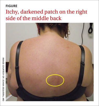
A 60-year-old woman visited our clinic complaining of an area on the right side of her middle back that was itchy, and had been bothering her for the past 10 years. She said her symptoms began without a trigger, and that a darkened area had appeared in the location of the itch. She had already been prescribed topical corticosteroids and antifungals and had tried over-the counter aids, but nothing relieved the itch. The patient had a history of cervical radiculopathy and was morbidly obese at the time of the visit.
On examination, the pruritic area consisted of a hyperpigmented, non-infiltrated 7-cm patch that was lateral to the vertebral column and within the dermatomes T4 to T6 (FIGURE). The patient also had hyperesthesia to light touch in this region and scratch abrasions.
WHAT IS YOUR DIAGNOSIS?
HOW WOULD YOU TREAT THIS PATIENT?
Diagnosis: Notalgia paresthetica
The location of the pruritic area and the patient’s clinical presentation led us to diagnose notalgia paresthetica. NP is a common dermatologic complaint characterized by unilateral pruritus that is medial or inferior to the scapula with dermatomal distribution.
The etiology of NP remains unknown, although it is thought to be a neuropathic itch caused by afferent nerve entrapment. The dorsal rami of the thoracic spinal nerves T2 to T6 are considered to be responsible for these symptoms. NP is not only a skin disease, but a cutaneous sign of an underlying spinal condition, including degenerative cervical spine disease.1-3
NP is a clinical diagnosis. There is typically a history of localized pruritus on the unilateral infrascapula area and there are few or no visible signs of disease. Patients frequently report a spider-bite sensation, prickly feelings, and/or an indescribable itch sensation. In addition, they may experience dysesthesia with diffuse mild burning, some surface numbness, and “under the skin” discomfort.
On physical examination, the patient may have a unilateral and ill-defined tan, pink, or hyperpigmented nonindurated patch on the infrascapular back that is a result of long-time scratching. Secondary skin changes such as lichenification, excoriations, eczema, xerosis, and infection often occur. Mild sensory alterations to light touch, vibration, and pin pricks may round out the clinical picture.1-3 Atypical forms of NP include localized pruritus on the upper back, neck, scalp, or shoulder.
Pruritus without other skin lesions can help pinpoint the Dx
The differential diagnosis for NP includes atopic dermatitis, contact dermatitis, drug eruptions, herpes zoster, idiopathic pruritus and systemic disease (such as renal, cholestatic, or hematologic pruritus, or pruritus associated with malignancy), tinea corporis, tinea versicolor, and xerosis.
Clues in the history. The chronic evolution of pruritus without other skin lesions, like vesicles or squamous areas, and the location of a hyperpigmented patch near the scapula region in a midlife patient, should prompt you to consider NP. A biopsy may show signs of post-inflammatory infiltrate of the papillary dermis with dermal melanophages.4,5
Although imaging tests are not required for a diagnosis of NP, basic cervical and possibly thoracic radiographs or magnetic resonance imaging (MRI) may be helpful in patients with symptoms of spine pain, tenderness, spasms, decreased range of motion, or any history of spinal trauma or injury. The images may reveal spinal disorders, including osteoarthritic lesions such as kyphosis, kyphoscoliosis, and vertebral hyperostosis.4
The exact cause of NP is unclear, but the evidence suggests that it results from damage to the cutaneous branches of the posterior divisions of the spinal nerves. This can occur by either impingement from degenerative changes in the spine or by spasms in the paraspinal musculature.2
The itch is neuropathic; antihistamines, steroids won’t help
It is difficult to treat NP without treating the underlying disease, which is usually spinal damage.4 Little has been published on the treatment of NP, and most of the literature on the subject involves case reports. Because the pruritus in NP is neuropathic, antihistamines and topical steroids are ineffective.4
The most commonly used treatment for NP among dermatologists is capsaicin as a 0.025% cream or 8% patch. One study with 20 patients reported improvement of pruritus in 70% of patients at 2 weeks, with some relapsing in about a month.6
Another treatment that has been used is cutaneous botulinum toxin type A injections, but its use is controversial. This strategy was proposed by Weinfeld7 after successful treatment of 2 patients. However, other studies have had variable outcomes with no resolution of pruritus.8
Other treatments include gabapentin,9 transcutaneous electrical nerve stimulation,10 and narrow-band ultraviolet-B.11 It is appropriate to consider surgical decompression or neurolysis of the nerve when other forms of treatment fail.12
Our patient was treated with topical capsaicin cream 0.25 mg/g, which lessened the intensity of her itching. After 2 months, the patient reported improvement of her symptoms.
CORRESPONDENCE
Joana Sequeira, MD, Estrada da Mata nº56, Leiria, Portugal; [email protected].
1. Stumpf A, Ständer S. Neuropathic itch: diagnosis and management. Dermatol Ther. 2013;26:104-109.
2. Ellis C. Notalgia paresthetica: the unreachable itch. Dermatol Pract Concept. 2013;3:3-6.
3. Bolognia JL, Jorizzo JL, Schaffer JV, eds. Pruritus and dysesthesia. In: Dermatology. 3rd ed. Philadelphia, PA: Elsevier Saunders;2012:121.
4. Raison-Peyron N, Meunier L, Acevedo M, et al. Notalgia paresthetica: clinical, physiopathological and therapeutic aspects. A study of 12 cases. J Eur Acad Dermatol Venereol. 1999;12:215-221.
5. Savk O, Savk E. Investigation of spinal pathology in notalgia paresthetica. J Am Acad Dermatol. 2005;52:1085-1087.
6. Wallengren J, Klinker M. Successful treatment of notalgia paresthetica with topical capsaicin: vehicle-controlled, double-blind, crossover study. J Am Acad Dermatol. 1995;32:287-289.
7. Weinfeld PK. Successful treatment of notalgia paresthetica with botulinum toxin type A. Arch Dermatol. 2007;143:980-982.
8. Pérez-Pérez L, García-Gavín J, Allegue F, et al. Notalgia paresthetica: treatment using intradermal botulinum toxin A. Actas Dermosifiliogr. 2014;105:74-77.
9. Maciel AA, Cunha PR, Laraia IO, et al. Efficacy of gabapentin in the improvement of pruritus and quality of life of patients with nostalgia paresthetica. An Bras Dermatol. 2014;89:570-575.
10. Savk E, Savk O, Sendur F. Transcutaneous electrical nerve stimulation offers partial relief in notalgia paresthetica patients with a relevant spinal pathology. J Dermatol. 2007;34:315-319.
11. Pérez-Pérez L, Allegue F, Fabeiro JM, et al. Notalgia paresthesica successfully treated with narrow-band UVB: report of five cases. J Eur Acad Dermatol Venereol. 2010;24:730-732.
12. Williams EH, Rosson GD, Elsamanoudi I, et al. Surgical decompression for notalgia paresthetica: a case report. Microsurgery. 2010;30:70-72.
A 60-year-old woman visited our clinic complaining of an area on the right side of her middle back that was itchy, and had been bothering her for the past 10 years. She said her symptoms began without a trigger, and that a darkened area had appeared in the location of the itch. She had already been prescribed topical corticosteroids and antifungals and had tried over-the counter aids, but nothing relieved the itch. The patient had a history of cervical radiculopathy and was morbidly obese at the time of the visit.
On examination, the pruritic area consisted of a hyperpigmented, non-infiltrated 7-cm patch that was lateral to the vertebral column and within the dermatomes T4 to T6 (FIGURE). The patient also had hyperesthesia to light touch in this region and scratch abrasions.
WHAT IS YOUR DIAGNOSIS?
HOW WOULD YOU TREAT THIS PATIENT?
Diagnosis: Notalgia paresthetica
The location of the pruritic area and the patient’s clinical presentation led us to diagnose notalgia paresthetica. NP is a common dermatologic complaint characterized by unilateral pruritus that is medial or inferior to the scapula with dermatomal distribution.
The etiology of NP remains unknown, although it is thought to be a neuropathic itch caused by afferent nerve entrapment. The dorsal rami of the thoracic spinal nerves T2 to T6 are considered to be responsible for these symptoms. NP is not only a skin disease, but a cutaneous sign of an underlying spinal condition, including degenerative cervical spine disease.1-3
NP is a clinical diagnosis. There is typically a history of localized pruritus on the unilateral infrascapula area and there are few or no visible signs of disease. Patients frequently report a spider-bite sensation, prickly feelings, and/or an indescribable itch sensation. In addition, they may experience dysesthesia with diffuse mild burning, some surface numbness, and “under the skin” discomfort.
On physical examination, the patient may have a unilateral and ill-defined tan, pink, or hyperpigmented nonindurated patch on the infrascapular back that is a result of long-time scratching. Secondary skin changes such as lichenification, excoriations, eczema, xerosis, and infection often occur. Mild sensory alterations to light touch, vibration, and pin pricks may round out the clinical picture.1-3 Atypical forms of NP include localized pruritus on the upper back, neck, scalp, or shoulder.
Pruritus without other skin lesions can help pinpoint the Dx
The differential diagnosis for NP includes atopic dermatitis, contact dermatitis, drug eruptions, herpes zoster, idiopathic pruritus and systemic disease (such as renal, cholestatic, or hematologic pruritus, or pruritus associated with malignancy), tinea corporis, tinea versicolor, and xerosis.
Clues in the history. The chronic evolution of pruritus without other skin lesions, like vesicles or squamous areas, and the location of a hyperpigmented patch near the scapula region in a midlife patient, should prompt you to consider NP. A biopsy may show signs of post-inflammatory infiltrate of the papillary dermis with dermal melanophages.4,5
Although imaging tests are not required for a diagnosis of NP, basic cervical and possibly thoracic radiographs or magnetic resonance imaging (MRI) may be helpful in patients with symptoms of spine pain, tenderness, spasms, decreased range of motion, or any history of spinal trauma or injury. The images may reveal spinal disorders, including osteoarthritic lesions such as kyphosis, kyphoscoliosis, and vertebral hyperostosis.4
The exact cause of NP is unclear, but the evidence suggests that it results from damage to the cutaneous branches of the posterior divisions of the spinal nerves. This can occur by either impingement from degenerative changes in the spine or by spasms in the paraspinal musculature.2
The itch is neuropathic; antihistamines, steroids won’t help
It is difficult to treat NP without treating the underlying disease, which is usually spinal damage.4 Little has been published on the treatment of NP, and most of the literature on the subject involves case reports. Because the pruritus in NP is neuropathic, antihistamines and topical steroids are ineffective.4
The most commonly used treatment for NP among dermatologists is capsaicin as a 0.025% cream or 8% patch. One study with 20 patients reported improvement of pruritus in 70% of patients at 2 weeks, with some relapsing in about a month.6
Another treatment that has been used is cutaneous botulinum toxin type A injections, but its use is controversial. This strategy was proposed by Weinfeld7 after successful treatment of 2 patients. However, other studies have had variable outcomes with no resolution of pruritus.8
Other treatments include gabapentin,9 transcutaneous electrical nerve stimulation,10 and narrow-band ultraviolet-B.11 It is appropriate to consider surgical decompression or neurolysis of the nerve when other forms of treatment fail.12
Our patient was treated with topical capsaicin cream 0.25 mg/g, which lessened the intensity of her itching. After 2 months, the patient reported improvement of her symptoms.
CORRESPONDENCE
Joana Sequeira, MD, Estrada da Mata nº56, Leiria, Portugal; [email protected].
1. Stumpf A, Ständer S. Neuropathic itch: diagnosis and management. Dermatol Ther. 2013;26:104-109.
2. Ellis C. Notalgia paresthetica: the unreachable itch. Dermatol Pract Concept. 2013;3:3-6.
3. Bolognia JL, Jorizzo JL, Schaffer JV, eds. Pruritus and dysesthesia. In: Dermatology. 3rd ed. Philadelphia, PA: Elsevier Saunders;2012:121.
4. Raison-Peyron N, Meunier L, Acevedo M, et al. Notalgia paresthetica: clinical, physiopathological and therapeutic aspects. A study of 12 cases. J Eur Acad Dermatol Venereol. 1999;12:215-221.
5. Savk O, Savk E. Investigation of spinal pathology in notalgia paresthetica. J Am Acad Dermatol. 2005;52:1085-1087.
6. Wallengren J, Klinker M. Successful treatment of notalgia paresthetica with topical capsaicin: vehicle-controlled, double-blind, crossover study. J Am Acad Dermatol. 1995;32:287-289.
7. Weinfeld PK. Successful treatment of notalgia paresthetica with botulinum toxin type A. Arch Dermatol. 2007;143:980-982.
8. Pérez-Pérez L, García-Gavín J, Allegue F, et al. Notalgia paresthetica: treatment using intradermal botulinum toxin A. Actas Dermosifiliogr. 2014;105:74-77.
9. Maciel AA, Cunha PR, Laraia IO, et al. Efficacy of gabapentin in the improvement of pruritus and quality of life of patients with nostalgia paresthetica. An Bras Dermatol. 2014;89:570-575.
10. Savk E, Savk O, Sendur F. Transcutaneous electrical nerve stimulation offers partial relief in notalgia paresthetica patients with a relevant spinal pathology. J Dermatol. 2007;34:315-319.
11. Pérez-Pérez L, Allegue F, Fabeiro JM, et al. Notalgia paresthesica successfully treated with narrow-band UVB: report of five cases. J Eur Acad Dermatol Venereol. 2010;24:730-732.
12. Williams EH, Rosson GD, Elsamanoudi I, et al. Surgical decompression for notalgia paresthetica: a case report. Microsurgery. 2010;30:70-72.
CMS proposes bundled payments for AMI, CABG
The Centers for Medicare & Medicaid Services is proposing new bundled payment models for acute myocardial infarction and coronary artery bypass grafting, and a separate payment to incentivize the use of cardiac rehabilitation.
As part of the proposal, CMS also is developing a pathway that would allow the bundle to be recognized as an advanced alternative payment model under the Medicare Access and CHIP Reauthorization Act and qualify the physicians and clinicians being paid through the model for the 5% incentive payment.

The proposed bundled payment model would place patient care accountability for 90 days after discharge on the hospital where acute myocardial infarction care or coronary artery bypass grafting occurred. Beginning July 1, 2017, hospitals in 98 randomly selected metropolitan statistical areas would be placed under this model and monitored for a 5-year period to test whether the model leads to improved outcomes and generates cost savings.
The proposed rule can be seen here and an advanced notice is expected to be published on the Federal Register website on July 26. CMS will be accepting comments on the proposal for 60 days following official publication in the Federal Register.
“In 2014, more than 200,000 Medicare beneficiaries were hospitalized for heart attack treatment or underwent bypass surgery, costing Medicare over $6 billion. But the cost of treating patients varied by 50% across hospitals, and the share of patients readmitted to the hospitals within 30 days varied by more than 50%. And patient experience also varies,” CMS Acting Principal Deputy Administrator and Chief Medical Officer Patrick Conway, MD, said during a July 25 press teleconference introducing the proposal. “In some cases, hospitals, doctors, and rehabilitation facilities work together to support a patient from heart attack or surgery all the way through recovery. But in other cases, coordination breaks down, especially when a patient leaves the hospital. By structuring a payment around a patient’s total experience of care, bundled payments support better care coordination and ultimately better outcomes for patients.”
The hospital would be paid a fixed target price for each care episode, with hospitals delivering higher-quality care receiving a higher target price. The hospital would either keep the savings achieved or, if the costs exceeded the target pricing, have to repay Medicare the difference.
Target prices will be based on historical cost data beginning with hospitalization and extending out 90 days following discharge and adjusted based on the complexity of treatment required. For the 18 months of the program (July 1, 2017, through Dec. 31, 2018) target prices would be based on a blend of two-thirds participant-specific data and one-third regional data. In the third performance year (2019), the mix would move to one-third participant data and two-thirds regional data. Beginning in 2020, only regional data would be used to set target prices.
For heart attacks, the following quality measures are being proposed: Hospital 30-day, all-cause, risk-standardized mortality following acute myocardial infarction hospitalization; excess days in acute care after hospitalization for acute myocardial infarction; Hospital Consumer Assessment of Healthcare Providers and Systems (HCAHPS) survey scores; and voluntary hybrid hospital 30-day, all-cause, risk-standardized mortality eMeasure data submission.
For bypass surgery, the quality measures will be the hospital 30-day all-cause, risk-standardized mortality rate following coronary artery bypass graft; and HCAHPS survey scores.
“CMS’s evaluation ... will examine quality during the episode period, after the episode period ends, and for longer durations such as 1-year mortality rates,” the agency said in a fact sheet describing the proposal. “CMS will examine outcomes and patient experience measures such as mortality, readmissions, complications, and other clinically relevant outcomes.”
Separately, the agency is proposing to test a cardiac rehabilitation incentive payment. The two-part cardiac rehabilitation incentive payment would be paid retrospectively based on the total cardiac rehabilitation use of beneficiaries attributable to participant hospitals.
“Currently, only 15% of heart attack patients receive cardiac rehabilitation, even though completing a rehabilitation program can lower the risk of the second heart attack or death,” Dr. Conway said. “Patients who receive cardiac rehabilitation are assigned a team of health care professionals such as cardiologists, dietitians, and physical therapists who help the patient to recover and regain cardiovascular fitness.”
The initial payment would be $25 per cardiac rehabilitation service for each of the first 11 services paid for by Medicare during the 90-day care period for a heart attack or bypass surgery. After 11 services, the payment would increase to $175 during the care period.
The number of sessions would be limited to two 1-hour sessions per day up to 36 sessions over up to 36 weeks, with the option for an additional 36 sessions over an extended period if approved by the local Medicare contractor. Intensive cardiac rehabilitation program sessions would be limited to 72 1-hour sessions, up to six sessions per day, over 18 weeks.
While officials from the American College of Cardiology said that the organization supports the concepts of value-based care, “it is important that bundled care models be carried out in such a way that clinicians are given the time and tools to truly impact patient care in the best ways possible. Changes in payment structures in health care can pose significant challenges to clinicians and must be driven by clinical practices that improve patient outcomes,” ACC President Richard A. Chazal, MD, said in a statement. “We are optimistic that CMS will listen to comments, incorporate feedback from clinicians, and provide ample time for implementation of these new payment models. Our ultimate goal is to improve patient care and to improve heart health.”
The Centers for Medicare & Medicaid Services is proposing new bundled payment models for acute myocardial infarction and coronary artery bypass grafting, and a separate payment to incentivize the use of cardiac rehabilitation.
As part of the proposal, CMS also is developing a pathway that would allow the bundle to be recognized as an advanced alternative payment model under the Medicare Access and CHIP Reauthorization Act and qualify the physicians and clinicians being paid through the model for the 5% incentive payment.
The proposed bundled payment model would place patient care accountability for 90 days after discharge on the hospital where acute myocardial infarction care or coronary artery bypass grafting occurred. Beginning July 1, 2017, hospitals in 98 randomly selected metropolitan statistical areas would be placed under this model and monitored for a 5-year period to test whether the model leads to improved outcomes and generates cost savings.
The proposed rule can be seen here and an advanced notice is expected to be published on the Federal Register website on July 26. CMS will be accepting comments on the proposal for 60 days following official publication in the Federal Register.
“In 2014, more than 200,000 Medicare beneficiaries were hospitalized for heart attack treatment or underwent bypass surgery, costing Medicare over $6 billion. But the cost of treating patients varied by 50% across hospitals, and the share of patients readmitted to the hospitals within 30 days varied by more than 50%. And patient experience also varies,” CMS Acting Principal Deputy Administrator and Chief Medical Officer Patrick Conway, MD, said during a July 25 press teleconference introducing the proposal. “In some cases, hospitals, doctors, and rehabilitation facilities work together to support a patient from heart attack or surgery all the way through recovery. But in other cases, coordination breaks down, especially when a patient leaves the hospital. By structuring a payment around a patient’s total experience of care, bundled payments support better care coordination and ultimately better outcomes for patients.”
The hospital would be paid a fixed target price for each care episode, with hospitals delivering higher-quality care receiving a higher target price. The hospital would either keep the savings achieved or, if the costs exceeded the target pricing, have to repay Medicare the difference.
Target prices will be based on historical cost data beginning with hospitalization and extending out 90 days following discharge and adjusted based on the complexity of treatment required. For the 18 months of the program (July 1, 2017, through Dec. 31, 2018) target prices would be based on a blend of two-thirds participant-specific data and one-third regional data. In the third performance year (2019), the mix would move to one-third participant data and two-thirds regional data. Beginning in 2020, only regional data would be used to set target prices.
For heart attacks, the following quality measures are being proposed: Hospital 30-day, all-cause, risk-standardized mortality following acute myocardial infarction hospitalization; excess days in acute care after hospitalization for acute myocardial infarction; Hospital Consumer Assessment of Healthcare Providers and Systems (HCAHPS) survey scores; and voluntary hybrid hospital 30-day, all-cause, risk-standardized mortality eMeasure data submission.
For bypass surgery, the quality measures will be the hospital 30-day all-cause, risk-standardized mortality rate following coronary artery bypass graft; and HCAHPS survey scores.
“CMS’s evaluation ... will examine quality during the episode period, after the episode period ends, and for longer durations such as 1-year mortality rates,” the agency said in a fact sheet describing the proposal. “CMS will examine outcomes and patient experience measures such as mortality, readmissions, complications, and other clinically relevant outcomes.”
Separately, the agency is proposing to test a cardiac rehabilitation incentive payment. The two-part cardiac rehabilitation incentive payment would be paid retrospectively based on the total cardiac rehabilitation use of beneficiaries attributable to participant hospitals.
“Currently, only 15% of heart attack patients receive cardiac rehabilitation, even though completing a rehabilitation program can lower the risk of the second heart attack or death,” Dr. Conway said. “Patients who receive cardiac rehabilitation are assigned a team of health care professionals such as cardiologists, dietitians, and physical therapists who help the patient to recover and regain cardiovascular fitness.”
The initial payment would be $25 per cardiac rehabilitation service for each of the first 11 services paid for by Medicare during the 90-day care period for a heart attack or bypass surgery. After 11 services, the payment would increase to $175 during the care period.
The number of sessions would be limited to two 1-hour sessions per day up to 36 sessions over up to 36 weeks, with the option for an additional 36 sessions over an extended period if approved by the local Medicare contractor. Intensive cardiac rehabilitation program sessions would be limited to 72 1-hour sessions, up to six sessions per day, over 18 weeks.
While officials from the American College of Cardiology said that the organization supports the concepts of value-based care, “it is important that bundled care models be carried out in such a way that clinicians are given the time and tools to truly impact patient care in the best ways possible. Changes in payment structures in health care can pose significant challenges to clinicians and must be driven by clinical practices that improve patient outcomes,” ACC President Richard A. Chazal, MD, said in a statement. “We are optimistic that CMS will listen to comments, incorporate feedback from clinicians, and provide ample time for implementation of these new payment models. Our ultimate goal is to improve patient care and to improve heart health.”

Routine cardiac screening in spondyloarthritis shown unneeded
DENVER – The largest controlled assessment of structural cardiac disease in patients with axial spondyloarthritis (SpA) failed to show any excess above a matched healthy sample, a finding that boosts recent guidelines that recommended against routine cardiac assessments in these patients.
“These findings provide the first evidence to support current recommendations against routine echocardiographic screening in asymptomatic patients with axial spondyloarthritis,” Risheen Reejhsinghani, MD, said at the annual meeting of the Spondyloarthritis Research and Treatment Network.

“If a patient has symptoms of cardiac disease – uncontrolled hypertension, dyspnea, or chest pain – then you would do the appropriate testing as you would for anyone,” said Dr. Reejhsinghani, a cardiologist at the University of California, San Francisco.
Past reports from small and often uncontrolled groups of patients with ankylosing spondylitis (AS) had suggested a possible link between the disease and structural heart disease. The 2015 treatment recommendations for AS and axial SpA from the American College of Rheumatology and other organizations included a “strong” recommendation against screening for cardiac conduction defects or for valvular heart disease (Arthritis Rheum. 2016 Feb;68[2]:282-98).
The prospective study by Dr. Reejhsinghani and her associates enrolled 154 patients diagnosed with axial SpA and 51 age-matched controls recruited from Health eHeart participants, a community-based heart disease study run from San Francisco. Additional matching by hypertension status refined the population to 133 patients with axial SpA matched with 51 healthy controls. The researchers also did another prespecified analysis that compared the 51 controls with 94 age- and hypertension-matched patients who fulfilled the 1984 modified New York criteria for a specific diagnosis of AS. Overall, nearly two-thirds of the people in the study were men, they averaged about 43 years old, and the average duration from AS diagnosis was 18 years.
The researchers performed systematic cardiac examination by transthoracic echo in all 205 participants that examined them for aortic root size, aortic regurgitation, diastolic dysfunction, and size of the aortic annulus, sinotubular junction, and ascending aorta. These examinations identified an unusually large aortic root diameter in about 5% of the cases and 2% of the controls. Aortic insufficiency (regurgitation) occurred in about 40% of the cases and 50% of the controls. An analysis of the cases and controls that was matched for age and hypertension showed a diastolic dysfunction prevalence of 17% in the cases and 27% in the controls. None of the between-group differences were statistically significant. The results were similar for the entire age-matched group studied, the age- and hypertension-matched subgroup, and the AS subgroup.
Future studies need to examine the possible impact that various treatments, including biologics, have on the prevalence of cardiac disorders, Dr. Reejhsinghani said.
On Twitter @mitchelzoler
Some patients with ankylosing spondylitis develop aortic insufficiency and need aortic valve replacements. Historically, clinicians worried about this risk and this led to a debate when an American College of Rheumatology panel recently developed ankylosing spondylitis treatment recommendations. This committee, on which I participated, decided to strongly recommend against routine screening with ECG or echocardiography. When we made that decision, we did not have these new data; we based our recommendation largely on our concerns about the cost efficacy of routine screening of asymptomatic patients.

|
Dr. David T.Y. Yu |
The new findings reported by Dr. Reejhsinghani show for certain that we do not need routine structural screening on these patients. It’s certainly appropriate to assess a patient with ECG and echo if they have persistent hypertension, dyspnea, or other cardiac symptoms. I would also do further testing in an ankylosing spondylitis patient with a heart murmur, and it is also appropriate to keep in mind a possible increased risk in spondyloarthritis patients for ischemic cardiovascular diseases.
The data reported by Dr. Reejhsinghani are convincing, especially because of the matching she did to control for age and presence of hypertension.
David T.Y. Yu, MD, is a rheumatologist at the University of California, Los Angeles. He made these comments in an interview. He had no disclosures.
Some patients with ankylosing spondylitis develop aortic insufficiency and need aortic valve replacements. Historically, clinicians worried about this risk and this led to a debate when an American College of Rheumatology panel recently developed ankylosing spondylitis treatment recommendations. This committee, on which I participated, decided to strongly recommend against routine screening with ECG or echocardiography. When we made that decision, we did not have these new data; we based our recommendation largely on our concerns about the cost efficacy of routine screening of asymptomatic patients.
|
|
Dr. David T.Y. Yu |
The new findings reported by Dr. Reejhsinghani show for certain that we do not need routine structural screening on these patients. It’s certainly appropriate to assess a patient with ECG and echo if they have persistent hypertension, dyspnea, or other cardiac symptoms. I would also do further testing in an ankylosing spondylitis patient with a heart murmur, and it is also appropriate to keep in mind a possible increased risk in spondyloarthritis patients for ischemic cardiovascular diseases.
The data reported by Dr. Reejhsinghani are convincing, especially because of the matching she did to control for age and presence of hypertension.
David T.Y. Yu, MD, is a rheumatologist at the University of California, Los Angeles. He made these comments in an interview. He had no disclosures.
DENVER – The largest controlled assessment of structural cardiac disease in patients with axial spondyloarthritis (SpA) failed to show any excess above a matched healthy sample, a finding that boosts recent guidelines that recommended against routine cardiac assessments in these patients.
“These findings provide the first evidence to support current recommendations against routine echocardiographic screening in asymptomatic patients with axial spondyloarthritis,” Risheen Reejhsinghani, MD, said at the annual meeting of the Spondyloarthritis Research and Treatment Network.
“If a patient has symptoms of cardiac disease – uncontrolled hypertension, dyspnea, or chest pain – then you would do the appropriate testing as you would for anyone,” said Dr. Reejhsinghani, a cardiologist at the University of California, San Francisco.
Past reports from small and often uncontrolled groups of patients with ankylosing spondylitis (AS) had suggested a possible link between the disease and structural heart disease. The 2015 treatment recommendations for AS and axial SpA from the American College of Rheumatology and other organizations included a “strong” recommendation against screening for cardiac conduction defects or for valvular heart disease (Arthritis Rheum. 2016 Feb;68[2]:282-98).
The prospective study by Dr. Reejhsinghani and her associates enrolled 154 patients diagnosed with axial SpA and 51 age-matched controls recruited from Health eHeart participants, a community-based heart disease study run from San Francisco. Additional matching by hypertension status refined the population to 133 patients with axial SpA matched with 51 healthy controls. The researchers also did another prespecified analysis that compared the 51 controls with 94 age- and hypertension-matched patients who fulfilled the 1984 modified New York criteria for a specific diagnosis of AS. Overall, nearly two-thirds of the people in the study were men, they averaged about 43 years old, and the average duration from AS diagnosis was 18 years.
The researchers performed systematic cardiac examination by transthoracic echo in all 205 participants that examined them for aortic root size, aortic regurgitation, diastolic dysfunction, and size of the aortic annulus, sinotubular junction, and ascending aorta. These examinations identified an unusually large aortic root diameter in about 5% of the cases and 2% of the controls. Aortic insufficiency (regurgitation) occurred in about 40% of the cases and 50% of the controls. An analysis of the cases and controls that was matched for age and hypertension showed a diastolic dysfunction prevalence of 17% in the cases and 27% in the controls. None of the between-group differences were statistically significant. The results were similar for the entire age-matched group studied, the age- and hypertension-matched subgroup, and the AS subgroup.
Future studies need to examine the possible impact that various treatments, including biologics, have on the prevalence of cardiac disorders, Dr. Reejhsinghani said.
On Twitter @mitchelzoler
AT THE 2016 SPARTAN ANNUAL MEETING
Key clinical point: Patients with axial spondyloarthritis or ankylosing spondylitis had no excess prevalence of structural cardiac abnormalities in the largest controlled study yet reported.
Major finding: Aortic insufficiency occurred in about 40% of the axial spondyloarthritis cases and 50% of the matched controls.
Data source: A prospective, matched case and control study with 205 total subjects from a single U.S. center.
Disclosures: Dr. Reejhsinghani had no disclosures.

VIDEO: Loss of function in Rab10 gene cuts Alzheimer’s risk by up to 40%
TORONTO – A rare loss of function in a gene that influences gamma secretase trafficking has been found to reduce the risk of Alzheimer’s by up to 40%, even in people who are homozygous carriers of the apolipoprotein E (apo E) epsilon 4 allele.
The gene – Rab10 – is a member of a family of about 60 genes involved in intracellular protein transport, Keoni Kauwe, PhD, said at the Alzheimer’s Association International Conference 2016. In addition to influencing endocytosis, ciliary transport, phagosome maturation, and insulin transport, Rab10 appears to assist in the transport of gamma secretase to the cell membrane.
By studying a population of elders who appear resistant to developing Alzheimer’s, Dr. Kauwe of Brigham Young University, Provo, Utah, and his associates determined that many of them share a natural inhibition of Rab10 – a unique characteristic that strongly contributes to their resistance to Alzheimer’s, he said. This knocked-down function would decrease the amount of gamma secretase to reach the cell membrane, Dr. Kauwe reasoned. Therefore, the proteolytic pathway that creates amyloid beta (Abeta) should be attenuated, allowing much less amyloid beta to be created and, presumably, be around to initiate the amyloid cascade that sparks Alzheimer’s disease.
His study cohort was drawn from two large data sets of cognitively normal elders, including many who were apo E epsilon 4 homozygotes. About 5% of the sample showed the loss-of-function Rab10 variant, which Dr. Kauwe said conferred a 20%-40% risk reduction for developing Alzheimer’s disease.
To understand the gene’s effects on amyloid beta production, he both overexpressed it and silenced it in cell lines. Overexpression resulted in a significant increase in the Abeta42/Abeta40 ratio (a riskier Alzheimer’s profile), while silencing it resulted in a significant decrease in the Abeta42/Abeta40 ratio (a more favorable profile).
Rab10 could be a therapeutic target, similar to the PCSK9 gene that influences low-density lipoprotein creation, Dr. Kauwe said. Monoclonal antibodies that block PCSK9 have recently been developed after a similar observation that naturally occurring loss of functions in the gene was associated with lower LDL levels.
“We’re going to be looking at the same thing with Rab10,” he said. “We do think that Rab10 inhibition could be a big story. It’s a high-risk venture, certainly, but it could also be high reward.”
Dr. Kauwe discussed his findings in a video interview at the conference.
The video associated with this article is no longer available on this site. Please view all of our videos on the MDedge YouTube channel
On Twitter @alz_gal
AT AAIC 2016

ACOG: Offer immediate postpartum LARC as option
Use of long-acting reversible contraception immediately post partum can help reduce the risk of unintended and short-interval pregnancy, according to a new policy statement from the American College of Obstetricians and Gynecologists.
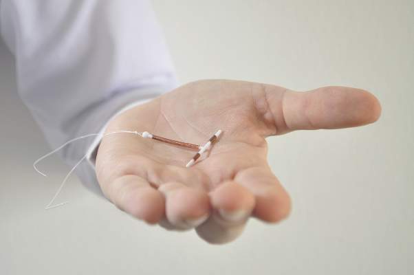
“Immediate postpartum LARC should be offered as an effective option for postpartum contraception; there are few contraindications to postpartum IUDs and implants,” according to the statement issued by ACOG’s Committee on Obstetric Practice (Obstet Gynecol. 2016;128:e32-37).

Unintended pregnancies account for approximately 45% of pregnancies in the United States overall, and at least 70% of pregnancies in the first year post partum, the committee noted.
The statement recommends prenatal counseling about the risks and benefits of LARCs, along with alternatives, to help patients make informed decisions. The committee emphasized that health care providers “should counsel women about the convenience and effectiveness of immediate postpartum LARC, as well as the benefits of reducing unintended pregnancy and lengthening pregnancy intervals.”
However, immediate postpartum use of IUDs is contraindicated in women with intrauterine infection at delivery, postpartum hemorrhage, and puerperal sepsis.
The committee called for systems to provide LARC at the comprehensive postpartum visit if necessary, and recommended stocking LARC devices in labor and delivery units for immediate postpartum placement. In addition, coding and reimbursement strategies are needed to support immediate postpartum LARC, according to the statement.
The policy statement was endorsed by the American College of Nurse-Midwives and the Society for Maternal-Fetal Medicine.
Use of long-acting reversible contraception immediately post partum can help reduce the risk of unintended and short-interval pregnancy, according to a new policy statement from the American College of Obstetricians and Gynecologists.
“Immediate postpartum LARC should be offered as an effective option for postpartum contraception; there are few contraindications to postpartum IUDs and implants,” according to the statement issued by ACOG’s Committee on Obstetric Practice (Obstet Gynecol. 2016;128:e32-37).
Unintended pregnancies account for approximately 45% of pregnancies in the United States overall, and at least 70% of pregnancies in the first year post partum, the committee noted.
The statement recommends prenatal counseling about the risks and benefits of LARCs, along with alternatives, to help patients make informed decisions. The committee emphasized that health care providers “should counsel women about the convenience and effectiveness of immediate postpartum LARC, as well as the benefits of reducing unintended pregnancy and lengthening pregnancy intervals.”
However, immediate postpartum use of IUDs is contraindicated in women with intrauterine infection at delivery, postpartum hemorrhage, and puerperal sepsis.
The committee called for systems to provide LARC at the comprehensive postpartum visit if necessary, and recommended stocking LARC devices in labor and delivery units for immediate postpartum placement. In addition, coding and reimbursement strategies are needed to support immediate postpartum LARC, according to the statement.
The policy statement was endorsed by the American College of Nurse-Midwives and the Society for Maternal-Fetal Medicine.
FROM OBSTETRICS & GYNECOLOGY
CDC Updates Zika Guidance for Managing Pregnant Women
Updated guidelines released by the Centers for Disease Control and Prevention outline how to diagnose suspected cases of Zika virus infection among pregnant women based on how quickly they present with symptoms.
The guidance was updated in light of “the emerging data indicating that Zika virus RNA can be detected for prolonged periods in some pregnant women,” the CDC wrote in the July 25 issue of the Morbidity and Mortality Weekly Report (doi: 10.15585/mmwr.mm6529e1).

All pregnant women should be assessed for Zika virus at each of their prenatal care visits, regardless of their recent travel history or exposure to mosquitoes, by being evaluated for signs and symptoms of infection, such as fever, rash, and arthralgia. From there, management can take one of two directions.
The first direction involves women who are tested within 2 weeks of either symptom onset, or their suspected exposure to the virus. Pregnant women who are asymptomatic and do not live in an area with an ongoing Zika virus outbreak, as well as women who are symptomatic, should have their serum and urine analyzed using a real-time reverse transcription–polymerase chain reaction (rRT-PCR) test. If the result of this test is positive, it should be considered as confirmation that the woman has a “recent Zika virus infection.”
However, if the test results are negative, symptomatic women should undergo immunoglobin M testing for both Zika virus and dengue virus, while asymptomatic women should undergo just Zika virus IgM testing within 2-12 weeks of the possible exposure. In either case, if the tests come back negative, then the patient can be definitively cleared of any recent Zika virus infection. However, if either the Zika virus or dengue virus tests are positive or equivocal, then there is a “presumptive recent Zika virus or dengue virus or Flavivirus infection” in the woman, at which point plaque reduction neutralization testing (PRNT) must be conducted.
The second direction for management involves pregnant women who are initially tested within 2-12 weeks of either symptom onset or suspected exposure to the virus. Asymptomatic women who do not live in an area with active Zika virus transmission, or do live in an area with ongoing Zika virus cases but are already in the first or second trimester of their pregnancy – along with any women who are symptomatic – should have their serum analyzed through IgM testing for both Zika virus and dengue virus.
If both results are negative, then there is no Zika virus infection. If the Zika virus test is negative but the dengue virus test is either positive or equivocal, the woman has a “presumptive dengue virus infection” and should undergo PRNT. If the Zika virus test is either positive or equivocal, then the woman has a “presumptive recent Zika virus or Flavivirus infection,” regardless of the dengue virus IgM result. In the latter case, the next step is to conduct a reflex Zika virus rRT-PCR test on both serum and urine. A negative result on the serum test should be followed by PRNT; a positive result should be taken as proof of a “recent Zika virus infection.”
For any diagnostic chain that ends with a PRNT, the results of that test can be interpreted in one of three ways. If the Zika virus PRNT result is at least 10 and the dengue virus result is less than 10, then there is a recent Zika virus infection. If both the Zika virus and dengue virus PRNT results are 10 or greater, than there is a Flavivirus infection, but the specific one cannot be determined. Finally, if both results are less than 10, then there is no evidence of a recent Zika virus infection.
“For symptomatic and asymptomatic pregnant women with possible Zika virus exposure who seek care [more than] 12 weeks after symptom onset or possible exposure, IgM antibody testing might be considered,” the CDC wrote. “If fetal abnormalities are present, rRT-PCR testing should also be performed on maternal serum and urine [but] a negative IgM antibody test or rRT-PCR result [more than] 12 weeks after symptom onset or possible exposure does not rule out recent Zika virus infection.”
For pregnant women with a diagnosis of a confirmed or presumptive Flavivirus infection, regardless of whether it’s specifically Zika virus or not, the CDC recommends getting serial ultrasounds every 3-4 weeks during pregnancy in order to monitor the fetus’s development, while “decisions regarding amniocentesis should be individualized for each clinical circumstance.” After the child’s birth, rRT-PCR should be conducted on the child’s cord blood and serum; Zika virus and dengue virus IgM are also recommended.
Pregnant women without a diagnosis of either Zika virus or dengue virus should have a prenatal ultrasound; if abnormalities are found in the fetus, CDC recommends that they have a repeat Zika virus rRT-PCR and IgM test and that clinicians base management on corresponding laboratory results. If nothing is found, however, then standard care can resume with ongoing vigilance to avoid Zika virus infections. Women with dengue virus diagnoses should follow the guidance that is currently in place.
“Pregnant women with laboratory evidence of confirmed or possible Zika virus infection who experience a fetal loss or stillbirth should be offered pathology testing for Zika virus infection,” the CDC added. “This testing might provide insight into the etiology of the fetal loss, which could inform a woman’s future pregnancy planning.”
The CDC also issued updated guidance for preventing sexual transmission of Zika virus and that applies to all men and women who have traveled to or reside in areas with active Zika virus transmission and their sex partners (MMWR. ePub: 2016 25 Jul. doi: 10.15585/mmwr.mm6529e2). The CDC advises couples in which a woman is pregnant to use barriers methods or abstain from sex for the duration of the pregnancy. For couples not planning pregnancy and in which there is a male partner with confirmed Zika virus infection or symptoms of infection, the men should use barrier methods or abstain from sex for at least 6 months after the onset of illness. For women with Zika virus infection, they should use barrier protection or abstain from sex for at least 8 weeks after the onset of illness.
Updated guidelines released by the Centers for Disease Control and Prevention outline how to diagnose suspected cases of Zika virus infection among pregnant women based on how quickly they present with symptoms.
The guidance was updated in light of “the emerging data indicating that Zika virus RNA can be detected for prolonged periods in some pregnant women,” the CDC wrote in the July 25 issue of the Morbidity and Mortality Weekly Report (doi: 10.15585/mmwr.mm6529e1).
All pregnant women should be assessed for Zika virus at each of their prenatal care visits, regardless of their recent travel history or exposure to mosquitoes, by being evaluated for signs and symptoms of infection, such as fever, rash, and arthralgia. From there, management can take one of two directions.
The first direction involves women who are tested within 2 weeks of either symptom onset, or their suspected exposure to the virus. Pregnant women who are asymptomatic and do not live in an area with an ongoing Zika virus outbreak, as well as women who are symptomatic, should have their serum and urine analyzed using a real-time reverse transcription–polymerase chain reaction (rRT-PCR) test. If the result of this test is positive, it should be considered as confirmation that the woman has a “recent Zika virus infection.”
However, if the test results are negative, symptomatic women should undergo immunoglobin M testing for both Zika virus and dengue virus, while asymptomatic women should undergo just Zika virus IgM testing within 2-12 weeks of the possible exposure. In either case, if the tests come back negative, then the patient can be definitively cleared of any recent Zika virus infection. However, if either the Zika virus or dengue virus tests are positive or equivocal, then there is a “presumptive recent Zika virus or dengue virus or Flavivirus infection” in the woman, at which point plaque reduction neutralization testing (PRNT) must be conducted.
The second direction for management involves pregnant women who are initially tested within 2-12 weeks of either symptom onset or suspected exposure to the virus. Asymptomatic women who do not live in an area with active Zika virus transmission, or do live in an area with ongoing Zika virus cases but are already in the first or second trimester of their pregnancy – along with any women who are symptomatic – should have their serum analyzed through IgM testing for both Zika virus and dengue virus.
If both results are negative, then there is no Zika virus infection. If the Zika virus test is negative but the dengue virus test is either positive or equivocal, the woman has a “presumptive dengue virus infection” and should undergo PRNT. If the Zika virus test is either positive or equivocal, then the woman has a “presumptive recent Zika virus or Flavivirus infection,” regardless of the dengue virus IgM result. In the latter case, the next step is to conduct a reflex Zika virus rRT-PCR test on both serum and urine. A negative result on the serum test should be followed by PRNT; a positive result should be taken as proof of a “recent Zika virus infection.”
For any diagnostic chain that ends with a PRNT, the results of that test can be interpreted in one of three ways. If the Zika virus PRNT result is at least 10 and the dengue virus result is less than 10, then there is a recent Zika virus infection. If both the Zika virus and dengue virus PRNT results are 10 or greater, than there is a Flavivirus infection, but the specific one cannot be determined. Finally, if both results are less than 10, then there is no evidence of a recent Zika virus infection.
“For symptomatic and asymptomatic pregnant women with possible Zika virus exposure who seek care [more than] 12 weeks after symptom onset or possible exposure, IgM antibody testing might be considered,” the CDC wrote. “If fetal abnormalities are present, rRT-PCR testing should also be performed on maternal serum and urine [but] a negative IgM antibody test or rRT-PCR result [more than] 12 weeks after symptom onset or possible exposure does not rule out recent Zika virus infection.”
For pregnant women with a diagnosis of a confirmed or presumptive Flavivirus infection, regardless of whether it’s specifically Zika virus or not, the CDC recommends getting serial ultrasounds every 3-4 weeks during pregnancy in order to monitor the fetus’s development, while “decisions regarding amniocentesis should be individualized for each clinical circumstance.” After the child’s birth, rRT-PCR should be conducted on the child’s cord blood and serum; Zika virus and dengue virus IgM are also recommended.
Pregnant women without a diagnosis of either Zika virus or dengue virus should have a prenatal ultrasound; if abnormalities are found in the fetus, CDC recommends that they have a repeat Zika virus rRT-PCR and IgM test and that clinicians base management on corresponding laboratory results. If nothing is found, however, then standard care can resume with ongoing vigilance to avoid Zika virus infections. Women with dengue virus diagnoses should follow the guidance that is currently in place.
“Pregnant women with laboratory evidence of confirmed or possible Zika virus infection who experience a fetal loss or stillbirth should be offered pathology testing for Zika virus infection,” the CDC added. “This testing might provide insight into the etiology of the fetal loss, which could inform a woman’s future pregnancy planning.”
The CDC also issued updated guidance for preventing sexual transmission of Zika virus and that applies to all men and women who have traveled to or reside in areas with active Zika virus transmission and their sex partners (MMWR. ePub: 2016 25 Jul. doi: 10.15585/mmwr.mm6529e2). The CDC advises couples in which a woman is pregnant to use barriers methods or abstain from sex for the duration of the pregnancy. For couples not planning pregnancy and in which there is a male partner with confirmed Zika virus infection or symptoms of infection, the men should use barrier methods or abstain from sex for at least 6 months after the onset of illness. For women with Zika virus infection, they should use barrier protection or abstain from sex for at least 8 weeks after the onset of illness.
FROM MMWR

CDC updates Zika guidance for managing pregnant women
Updated guidelines released by the Centers for Disease Control and Prevention outline how to diagnose suspected cases of Zika virus infection among pregnant women based on how quickly they present with symptoms.
The guidance was updated in light of “the emerging data indicating that Zika virus RNA can be detected for prolonged periods in some pregnant women,” the CDC wrote in the July 25 issue of the Morbidity and Mortality Weekly Report (doi: 10.15585/mmwr.mm6529e1).

All pregnant women should be assessed for Zika virus at each of their prenatal care visits, regardless of their recent travel history or exposure to mosquitoes, by being evaluated for signs and symptoms of infection, such as fever, rash, and arthralgia. From there, management can take one of two directions.
The first direction involves women who are tested within 2 weeks of either symptom onset, or their suspected exposure to the virus. Pregnant women who are asymptomatic and do not live in an area with an ongoing Zika virus outbreak, as well as women who are symptomatic, should have their serum and urine analyzed using a real-time reverse transcription–polymerase chain reaction (rRT-PCR) test. If the result of this test is positive, it should be considered as confirmation that the woman has a “recent Zika virus infection.”
However, if the test results are negative, symptomatic women should undergo immunoglobin M testing for both Zika virus and dengue virus, while asymptomatic women should undergo just Zika virus IgM testing within 2-12 weeks of the possible exposure. In either case, if the tests come back negative, then the patient can be definitively cleared of any recent Zika virus infection. However, if either the Zika virus or dengue virus tests are positive or equivocal, then there is a “presumptive recent Zika virus or dengue virus or Flavivirus infection” in the woman, at which point plaque reduction neutralization testing (PRNT) must be conducted.
The second direction for management involves pregnant women who are initially tested within 2-12 weeks of either symptom onset or suspected exposure to the virus. Asymptomatic women who do not live in an area with active Zika virus transmission, or do live in an area with ongoing Zika virus cases but are already in the first or second trimester of their pregnancy – along with any women who are symptomatic – should have their serum analyzed through IgM testing for both Zika virus and dengue virus.
If both results are negative, then there is no Zika virus infection. If the Zika virus test is negative but the dengue virus test is either positive or equivocal, the woman has a “presumptive dengue virus infection” and should undergo PRNT. If the Zika virus test is either positive or equivocal, then the woman has a “presumptive recent Zika virus or Flavivirus infection,” regardless of the dengue virus IgM result. In the latter case, the next step is to conduct a reflex Zika virus rRT-PCR test on both serum and urine. A negative result on the serum test should be followed by PRNT; a positive result should be taken as proof of a “recent Zika virus infection.”
For any diagnostic chain that ends with a PRNT, the results of that test can be interpreted in one of three ways. If the Zika virus PRNT result is at least 10 and the dengue virus result is less than 10, then there is a recent Zika virus infection. If both the Zika virus and dengue virus PRNT results are 10 or greater, than there is a Flavivirus infection, but the specific one cannot be determined. Finally, if both results are less than 10, then there is no evidence of a recent Zika virus infection.
“For symptomatic and asymptomatic pregnant women with possible Zika virus exposure who seek care [more than] 12 weeks after symptom onset or possible exposure, IgM antibody testing might be considered,” the CDC wrote. “If fetal abnormalities are present, rRT-PCR testing should also be performed on maternal serum and urine [but] a negative IgM antibody test or rRT-PCR result [more than] 12 weeks after symptom onset or possible exposure does not rule out recent Zika virus infection.”
For pregnant women with a diagnosis of a confirmed or presumptive Flavivirus infection, regardless of whether it’s specifically Zika virus or not, the CDC recommends getting serial ultrasounds every 3-4 weeks during pregnancy in order to monitor the fetus’s development, while “decisions regarding amniocentesis should be individualized for each clinical circumstance.” After the child’s birth, rRT-PCR should be conducted on the child’s cord blood and serum; Zika virus and dengue virus IgM are also recommended.
Pregnant women without a diagnosis of either Zika virus or dengue virus should have a prenatal ultrasound; if abnormalities are found in the fetus, CDC recommends that they have a repeat Zika virus rRT-PCR and IgM test and that clinicians base management on corresponding laboratory results. If nothing is found, however, then standard care can resume with ongoing vigilance to avoid Zika virus infections. Women with dengue virus diagnoses should follow the guidance that is currently in place.
“Pregnant women with laboratory evidence of confirmed or possible Zika virus infection who experience a fetal loss or stillbirth should be offered pathology testing for Zika virus infection,” the CDC added. “This testing might provide insight into the etiology of the fetal loss, which could inform a woman’s future pregnancy planning.”
The CDC also issued updated guidance for preventing sexual transmission of Zika virus and that applies to all men and women who have traveled to or reside in areas with active Zika virus transmission and their sex partners (MMWR. ePub: 2016 25 Jul. doi: 10.15585/mmwr.mm6529e2). The CDC advises couples in which a woman is pregnant to use barriers methods or abstain from sex for the duration of the pregnancy. For couples not planning pregnancy and in which there is a male partner with confirmed Zika virus infection or symptoms of infection, the men should use barrier methods or abstain from sex for at least 6 months after the onset of illness. For women with Zika virus infection, they should use barrier protection or abstain from sex for at least 8 weeks after the onset of illness.
Updated guidelines released by the Centers for Disease Control and Prevention outline how to diagnose suspected cases of Zika virus infection among pregnant women based on how quickly they present with symptoms.
The guidance was updated in light of “the emerging data indicating that Zika virus RNA can be detected for prolonged periods in some pregnant women,” the CDC wrote in the July 25 issue of the Morbidity and Mortality Weekly Report (doi: 10.15585/mmwr.mm6529e1).
All pregnant women should be assessed for Zika virus at each of their prenatal care visits, regardless of their recent travel history or exposure to mosquitoes, by being evaluated for signs and symptoms of infection, such as fever, rash, and arthralgia. From there, management can take one of two directions.
The first direction involves women who are tested within 2 weeks of either symptom onset, or their suspected exposure to the virus. Pregnant women who are asymptomatic and do not live in an area with an ongoing Zika virus outbreak, as well as women who are symptomatic, should have their serum and urine analyzed using a real-time reverse transcription–polymerase chain reaction (rRT-PCR) test. If the result of this test is positive, it should be considered as confirmation that the woman has a “recent Zika virus infection.”
However, if the test results are negative, symptomatic women should undergo immunoglobin M testing for both Zika virus and dengue virus, while asymptomatic women should undergo just Zika virus IgM testing within 2-12 weeks of the possible exposure. In either case, if the tests come back negative, then the patient can be definitively cleared of any recent Zika virus infection. However, if either the Zika virus or dengue virus tests are positive or equivocal, then there is a “presumptive recent Zika virus or dengue virus or Flavivirus infection” in the woman, at which point plaque reduction neutralization testing (PRNT) must be conducted.
The second direction for management involves pregnant women who are initially tested within 2-12 weeks of either symptom onset or suspected exposure to the virus. Asymptomatic women who do not live in an area with active Zika virus transmission, or do live in an area with ongoing Zika virus cases but are already in the first or second trimester of their pregnancy – along with any women who are symptomatic – should have their serum analyzed through IgM testing for both Zika virus and dengue virus.
If both results are negative, then there is no Zika virus infection. If the Zika virus test is negative but the dengue virus test is either positive or equivocal, the woman has a “presumptive dengue virus infection” and should undergo PRNT. If the Zika virus test is either positive or equivocal, then the woman has a “presumptive recent Zika virus or Flavivirus infection,” regardless of the dengue virus IgM result. In the latter case, the next step is to conduct a reflex Zika virus rRT-PCR test on both serum and urine. A negative result on the serum test should be followed by PRNT; a positive result should be taken as proof of a “recent Zika virus infection.”
For any diagnostic chain that ends with a PRNT, the results of that test can be interpreted in one of three ways. If the Zika virus PRNT result is at least 10 and the dengue virus result is less than 10, then there is a recent Zika virus infection. If both the Zika virus and dengue virus PRNT results are 10 or greater, than there is a Flavivirus infection, but the specific one cannot be determined. Finally, if both results are less than 10, then there is no evidence of a recent Zika virus infection.
“For symptomatic and asymptomatic pregnant women with possible Zika virus exposure who seek care [more than] 12 weeks after symptom onset or possible exposure, IgM antibody testing might be considered,” the CDC wrote. “If fetal abnormalities are present, rRT-PCR testing should also be performed on maternal serum and urine [but] a negative IgM antibody test or rRT-PCR result [more than] 12 weeks after symptom onset or possible exposure does not rule out recent Zika virus infection.”
For pregnant women with a diagnosis of a confirmed or presumptive Flavivirus infection, regardless of whether it’s specifically Zika virus or not, the CDC recommends getting serial ultrasounds every 3-4 weeks during pregnancy in order to monitor the fetus’s development, while “decisions regarding amniocentesis should be individualized for each clinical circumstance.” After the child’s birth, rRT-PCR should be conducted on the child’s cord blood and serum; Zika virus and dengue virus IgM are also recommended.
Pregnant women without a diagnosis of either Zika virus or dengue virus should have a prenatal ultrasound; if abnormalities are found in the fetus, CDC recommends that they have a repeat Zika virus rRT-PCR and IgM test and that clinicians base management on corresponding laboratory results. If nothing is found, however, then standard care can resume with ongoing vigilance to avoid Zika virus infections. Women with dengue virus diagnoses should follow the guidance that is currently in place.
“Pregnant women with laboratory evidence of confirmed or possible Zika virus infection who experience a fetal loss or stillbirth should be offered pathology testing for Zika virus infection,” the CDC added. “This testing might provide insight into the etiology of the fetal loss, which could inform a woman’s future pregnancy planning.”
The CDC also issued updated guidance for preventing sexual transmission of Zika virus and that applies to all men and women who have traveled to or reside in areas with active Zika virus transmission and their sex partners (MMWR. ePub: 2016 25 Jul. doi: 10.15585/mmwr.mm6529e2). The CDC advises couples in which a woman is pregnant to use barriers methods or abstain from sex for the duration of the pregnancy. For couples not planning pregnancy and in which there is a male partner with confirmed Zika virus infection or symptoms of infection, the men should use barrier methods or abstain from sex for at least 6 months after the onset of illness. For women with Zika virus infection, they should use barrier protection or abstain from sex for at least 8 weeks after the onset of illness.
FROM MMWR

ASCO: Always Screen Cancer Survivors for Chronic Pain
All adult cancer survivors should be screened for chronic pain at every visit, according to the American Society of Clinical Oncology’s first clinical practice guideline for managing this patient population, published July 25 in the Journal of Clinical Oncology.
An estimated 14 million adults with a history of cancer are living in the United States alone, and the prevalence of chronic pain related to their malignancy is reported to be as high as 40%. Yet most health care providers “haven’t been trained to recognize or treat long-term pain associated with cancer,” Judith A. Paice, PhD, RN, said in a press statement accompanying the release of the new guideline.
Existing guidelines focus on the acute pain of cancer and its treatment, or on acute pain related to advanced cancer, said Dr. Paice, cochair of the expert panel that developed the guideline and research professor of hematology/oncology at Northwestern University, Chicago.

The chronic pain addressed in the new guideline document can be related to the malignancy itself or to its treatment. Surgery, chemotherapy, hormone therapy, radiation treatment, and stem-cell transplantation can induce dozens of pain-producing complications, including osteonecrosis, bone fractures, peripheral or central nerve damage, fistulas, lymphedema, arthralgias, and myelopathy. The pain can develop months or years after diagnosis, significantly impacting patients’ quality of life well after treatment is completed.
“This guideline will help clinicians identify pain early and develop comprehensive treatment plans using a broad range of approaches,” Dr. Paice said in the statement.
The panel that developed the guideline comprised experts in medical oncology, hematology, pain medicine, palliative care, hospice, radiation oncology, social work, rehabilitation, psychology, and anesthesiology. They performed a systematic review of the literature and based their recommendations on 35 meta-analyses, 9 randomized controlled trials, 19 comparative studies, and expert consensus.
Some key recommendations include the following:
• Screen all survivors of adult cancers for chronic pain using both a physical exam and an in-depth interview that covers physical, functional, psychological, social, and spiritual aspects of pain, as well as the patient’s treatment history and comorbid conditions. The guideline includes a list of 36 common pain syndromes resulting from cancer treatments.
• Monitor patients at every visit for late-onset treatment effects that can produce pain, just as they are monitored for recurrent disease and secondary malignancy. Some cancer survivors may not recognize that their current pain is related to past disease or its treatments, or may consider it an untreatable complication that they have to endure.
• Consider the entire range of pain medicines – not just opioid analgesics – including NSAIDs, acetaminophen, and selected antidepressants and anticonvulsants with analgesic efficacy. The panel noted that some cancer survivors with chronic pain may benefit from other drugs, including muscle relaxants, benzodiazepines, N-methyl-D-aspartate receptor blockers, alpha-2 agonists, and neutraceuticals and botanicals. But they urged caution since the efficacy of these agents has not been fully established.
Consider nonmedical treatments such as physical, occupational, and recreation therapy; exercise; orthotics; ultrasound; massage; acupuncture; cognitive behavioral therapy; relaxation therapy; and TENS or other types of nerve stimulation.
Consider medical cannabis or cannabinoids as an adjuvant but not a first-line pain treatment, being sure to follow specific state regulations.
Consider a trial of opioids only for carefully selected patients who don’t respond to more conservative management. Weigh potential risks against benefits and employ universal precautions to minimize abuse and addiction, and be cautious in coprescribing other centrally-acting agents (J Clin Oncol. 2016 July 25. doi: 10.1200/JCO.2016.68.5206).
The full clinical practice guideline and other clinical tools and resources are available at www.asco.org/chronic-pai-guideline.
The guideline development process was supported by ASCO. Dr. Paice reported having no relevant financial disclosures; some of her associates reported financial ties to industry sources.
All adult cancer survivors should be screened for chronic pain at every visit, according to the American Society of Clinical Oncology’s first clinical practice guideline for managing this patient population, published July 25 in the Journal of Clinical Oncology.
An estimated 14 million adults with a history of cancer are living in the United States alone, and the prevalence of chronic pain related to their malignancy is reported to be as high as 40%. Yet most health care providers “haven’t been trained to recognize or treat long-term pain associated with cancer,” Judith A. Paice, PhD, RN, said in a press statement accompanying the release of the new guideline.
Existing guidelines focus on the acute pain of cancer and its treatment, or on acute pain related to advanced cancer, said Dr. Paice, cochair of the expert panel that developed the guideline and research professor of hematology/oncology at Northwestern University, Chicago.
The chronic pain addressed in the new guideline document can be related to the malignancy itself or to its treatment. Surgery, chemotherapy, hormone therapy, radiation treatment, and stem-cell transplantation can induce dozens of pain-producing complications, including osteonecrosis, bone fractures, peripheral or central nerve damage, fistulas, lymphedema, arthralgias, and myelopathy. The pain can develop months or years after diagnosis, significantly impacting patients’ quality of life well after treatment is completed.
“This guideline will help clinicians identify pain early and develop comprehensive treatment plans using a broad range of approaches,” Dr. Paice said in the statement.
The panel that developed the guideline comprised experts in medical oncology, hematology, pain medicine, palliative care, hospice, radiation oncology, social work, rehabilitation, psychology, and anesthesiology. They performed a systematic review of the literature and based their recommendations on 35 meta-analyses, 9 randomized controlled trials, 19 comparative studies, and expert consensus.
Some key recommendations include the following:
• Screen all survivors of adult cancers for chronic pain using both a physical exam and an in-depth interview that covers physical, functional, psychological, social, and spiritual aspects of pain, as well as the patient’s treatment history and comorbid conditions. The guideline includes a list of 36 common pain syndromes resulting from cancer treatments.
• Monitor patients at every visit for late-onset treatment effects that can produce pain, just as they are monitored for recurrent disease and secondary malignancy. Some cancer survivors may not recognize that their current pain is related to past disease or its treatments, or may consider it an untreatable complication that they have to endure.
• Consider the entire range of pain medicines – not just opioid analgesics – including NSAIDs, acetaminophen, and selected antidepressants and anticonvulsants with analgesic efficacy. The panel noted that some cancer survivors with chronic pain may benefit from other drugs, including muscle relaxants, benzodiazepines, N-methyl-D-aspartate receptor blockers, alpha-2 agonists, and neutraceuticals and botanicals. But they urged caution since the efficacy of these agents has not been fully established.
Consider nonmedical treatments such as physical, occupational, and recreation therapy; exercise; orthotics; ultrasound; massage; acupuncture; cognitive behavioral therapy; relaxation therapy; and TENS or other types of nerve stimulation.
Consider medical cannabis or cannabinoids as an adjuvant but not a first-line pain treatment, being sure to follow specific state regulations.
Consider a trial of opioids only for carefully selected patients who don’t respond to more conservative management. Weigh potential risks against benefits and employ universal precautions to minimize abuse and addiction, and be cautious in coprescribing other centrally-acting agents (J Clin Oncol. 2016 July 25. doi: 10.1200/JCO.2016.68.5206).
The full clinical practice guideline and other clinical tools and resources are available at www.asco.org/chronic-pai-guideline.
The guideline development process was supported by ASCO. Dr. Paice reported having no relevant financial disclosures; some of her associates reported financial ties to industry sources.
FROM THE JOURNAL OF CLINICAL ONCOLOGY

New HER2-testing guidelines result in more women eligible for HER2-directed treatment
New guidelines for immunohistochemistry (IHC) and fluorescent in situ hybridization (FISH) pathology testing categorize more breast cancers as “equivocal” regarding HER2 positivity and ultimately lead to identifying more of them as HER2 positive, investigators reported online in Journal of Clinical Oncology.
The American Society of Clinical Oncology and the College of American Pathologists released updated IHC and FISH guidelines in 2013, after the Food and Drug Administration approved the initial set of such guidelines in 1998 and ASCO/CAP issued their first such guidelines in 2007. “The intent of the 2013 guidelines was to decrease the number of equivocal [cancers],” said Mithun Vinod Shah, MD, PhD, of the Mayo Clinic, Rochester Minn., and his associates.
To assess the impact of implementing the new guidelines, the investigators analyzed 2,851 breast cancer samples sent to their cytogenetics laboratory by 139 medical centers for HER2 testing during a 1-year period. They compared the three sets of testing criteria – the FDA, the 2007, and the 2013 guidelines.
According to the 2013 guidelines, 69.7% of the tumors were classified as HER2 negative, 16.1% as HER2 positive, and 14.2% as equivocal. In contrast, the 2007 guidelines classified 85.1% as negative, 11.0% as positive, and 3.9% as equivocal, while the FDA guidelines classified 13.1% as positive and 86.9% as negative (there is no “equivocal” category in the FDA guidelines). Thus, “the final FISH interpretations indicate that by using 2013 guidelines, 358 additional patients were interpreted as positive, compared with the 2007 guidelines and that 298 additional patients were considered positive, compared with the FDA criteria,” Dr. Shah and his associates said.
The 2013 guidelines recommend additional chromosome 17 probe testing (among other strategies) to resolve equivocal results, so the investigators did so with the 405 samples classified as equivocal by the 2013 criteria. This resulted in 52.3% of the 405 equivocal tumors being reclassified as HER2 positive, 8.9% being reclassified as HER2 negative, and 38.8% remaining equivocal. Thus, HER2 positivity in the overall cohort rose significantly to 23.6% using the newest guidelines.
These findings demonstrate that using the 2013 guidelines for IHC and FISH pathology testing identifies more women who are eligible for HER2-directed therapy, the investigators said (J Clin Oncol. 2016 Jul 25. doi: 10.1200/JCO.2015.61.8983).
FROM JOURNAL OF CLINICAL ONCOLOGY
Key clinical point: New IHC and FISH pathology guidelines categorize more breast cancers as “equivocal” regarding HER2 positivity and ultimately lead to identifying more of them as HER2 positive.
Major finding: By using 2013 guidelines, 358 additional tumors were interpreted as positive, compared with the 2007 guidelines and 298 additional tumors were considered positive, compared with the FDA criteria.
Data source: A cohort study involving 2,851 breast cancer samples analyzed according to three different pathology guidelines during a 1-year period.
Disclosures: This study was supported by the Mayo Clinic. Dr. Shah reported having no relevant financial disclosures; his associates reported ties to Merck, Hospira, Ariad Pharmaceuticals, Abbott Molecular, and Genome Diagnostics.
Study shows faster increase in obesity prevalence among cancer survivors
From 1997 to 2014, cancer survivors had a significantly faster increase in obesity prevalence compared with adults without a history of cancer, investigators found.
Furthermore, the elevated annual increase in rates of obesity was more pronounced in women, in breast and colorectal cancer survivors, and in non-Hispanic blacks.

Cancer patients may be at an increased risk for weight gain caused by specific cancer treatments such as chemotherapy, steroid modifications, and various hormone therapies, especially in “hormonally and metabolically driven cancers such as breast and colorectal cancer,” wrote Heather Greenlee, ND, PhD, of Columbia University, New York, and her associates (J Clin Oncol. 2016 July 25. doi: 10.1200/JCO.2016.66.4391). “To better inform future research on obesity and cancer survival and to inform the planning and implementation of weight loss interventions in cancer survivors, we compared trends from 1997 to 2014 in obesity prevalence in U.S. adults with and without a history of cancer,” the researchers explained.
Obesity prevalence and trends were evaluated through the National Health Interview Survey, an ongoing cross-sectional survey of the health status, health care access, and behaviors of the U.S. civilian population conducted by the National Center for Health Statistics.
For the current study, surveys from a total of 538,969 adults aged 18-85 years with (n = 32,447) or without (n = 506,522) a history of cancer were analyzed. Participants provided self-reported height and weight measures from which body mass index was calculated. Obesity was defined as a BMI of 30 kg/m2 or higher for non-Asians and 27.5 kg/m2 or higher for Asians. Participants also self-reported sex, race, and cancer history.
Overall, the prevalence of obesity consistently increased from 1997 to 2014. Among cancer survivors, the prevalence of obesity increased from 22.4% to 31.7% while in adults without a history of cancer, prevalence increased from 20.9% to 29.5%.
The annual increase in the rate of obesity was significantly greater in both women and men with a history of cancer (2.9% and 2.8% respectively) compared with those without a history of cancer (2.3% and 2.4%, P less than .001 for all).
The elevated annual increase in rates of obesity was even higher in colorectal (3.1% in women and 3.7% in men) and breast (3.0%) cancer survivors compared with adults without a history of cancer, but was lower in prostate cancer survivors (2.1%; P = .001 for all).
“These findings call for public health planning of effective and scalable weight management and control programs for cancer survivors, especially for breast and colorectal cancer survivors and for non-Hispanic blacks,” the investigators concluded.
The National Cancer Institute funded the study. Dr. Greenlee and one other investigator reported serving in advisory roles for EHE International.
On Twitter @jessnicolecraig
From 1997 to 2014, cancer survivors had a significantly faster increase in obesity prevalence compared with adults without a history of cancer, investigators found.
Furthermore, the elevated annual increase in rates of obesity was more pronounced in women, in breast and colorectal cancer survivors, and in non-Hispanic blacks.
Cancer patients may be at an increased risk for weight gain caused by specific cancer treatments such as chemotherapy, steroid modifications, and various hormone therapies, especially in “hormonally and metabolically driven cancers such as breast and colorectal cancer,” wrote Heather Greenlee, ND, PhD, of Columbia University, New York, and her associates (J Clin Oncol. 2016 July 25. doi: 10.1200/JCO.2016.66.4391). “To better inform future research on obesity and cancer survival and to inform the planning and implementation of weight loss interventions in cancer survivors, we compared trends from 1997 to 2014 in obesity prevalence in U.S. adults with and without a history of cancer,” the researchers explained.
Obesity prevalence and trends were evaluated through the National Health Interview Survey, an ongoing cross-sectional survey of the health status, health care access, and behaviors of the U.S. civilian population conducted by the National Center for Health Statistics.
For the current study, surveys from a total of 538,969 adults aged 18-85 years with (n = 32,447) or without (n = 506,522) a history of cancer were analyzed. Participants provided self-reported height and weight measures from which body mass index was calculated. Obesity was defined as a BMI of 30 kg/m2 or higher for non-Asians and 27.5 kg/m2 or higher for Asians. Participants also self-reported sex, race, and cancer history.
Overall, the prevalence of obesity consistently increased from 1997 to 2014. Among cancer survivors, the prevalence of obesity increased from 22.4% to 31.7% while in adults without a history of cancer, prevalence increased from 20.9% to 29.5%.
The annual increase in the rate of obesity was significantly greater in both women and men with a history of cancer (2.9% and 2.8% respectively) compared with those without a history of cancer (2.3% and 2.4%, P less than .001 for all).
The elevated annual increase in rates of obesity was even higher in colorectal (3.1% in women and 3.7% in men) and breast (3.0%) cancer survivors compared with adults without a history of cancer, but was lower in prostate cancer survivors (2.1%; P = .001 for all).
“These findings call for public health planning of effective and scalable weight management and control programs for cancer survivors, especially for breast and colorectal cancer survivors and for non-Hispanic blacks,” the investigators concluded.
The National Cancer Institute funded the study. Dr. Greenlee and one other investigator reported serving in advisory roles for EHE International.
On Twitter @jessnicolecraig
FROM THE JOURNAL OF CLINICAL ONCOLOGY
Key clinical point: Cancer survivors had a significantly faster increase in obesity prevalence compared with adults without a history of cancer.
Major finding: The annual increase in the rate of obesity was significantly higher in both women and men with a history of cancer (2.9% and 2.8% respectively) compared with those without a history of cancer (2.3% and 2.4%, P less than .001 for all).
Data source: National Health Interview Survey responses from 538,969 adults aged 18-85 years with (n = 32,447) or without (n = 506,522) a history of cancer.
Disclosures: The National Cancer Institute funded the study. Dr. Greenlee and one other investigator reported serving in advisory roles for EHE International.
