User login
Extended pneumococcal vaccination schedule boosts early immunity for preemies
A randomized clinical trial evaluating three dosing strategies for 13-valent pneumococcal vaccine (PCV13) in preterm infants found that more widely spaced priming vaccinations resulted in higher immunoglobulin G (IgG) during the first 12 months of life, but reduced the immune response seen after the 12-month booster was given.
After the primary schedule, the percent of infants lacking seroprotection for more than one half of the serotypes in the PCV13 formulation was 25% on a reduced two-dose schedule, 12% on an accelerated schedule, and 3% on an extended schedule (P less than .001).
Conversely, “A reduced priming schedule of PCV13 resulted in higher post-booster IgG concentrations but lower post-primary concentrations,” wrote Alison Kent, MBChB, and her coinvestigators in the PUNS (Prems Under New Schedule) Study Group (Pediatrics. 2016;138[3]:e20153945).

“Infants who received the extended schedule had lower fold increases in concentrations after booster vaccination than the other groups,” wrote Dr. Kent of the Pediatric Infectious Diseases Research Group and Vaccine Institute, St. George’s, University of London, and her collaborators. Participants receiving the extended schedule had lower geometric mean concentrations (GMCs) of antibodies than did those on the reduced schedule for nine serotypes and those on the accelerated schedule for four serotypes.
The study enrolled 210 premature infants in a phase IV, controlled, open-label trial at 12 sites in the United Kingdom. Infants of less than 35 weeks gestation, and between 7 and 12 weeks of age, were randomly assigned to receive PCV13 on one of three schedules. The reduced schedule gave two priming doses at 2 and 4 months of age; the accelerated schedule gave the doses at 2, 3, and 4 months of age; and the extended schedule gave doses at 2, 4, and 6 months of age. All infants received a booster vaccination at 12 or 13 months of age, and all received a standard suite of childhood immunizations for other diseases. The entire study was completed by 194 patients.
Serotype-specific IgG concentrations were obtained pre-vaccination, 1 month after the primary vaccination, and before and 1 month after the booster vaccination was given. IgG levels were reported for each PCV serotype; “there was considerable variation between serotypes,” ranging from 0.16 ng/mL for serotype 6b on the reduced schedule to 8.49 ng/mL for serotype 14 on the extended schedule, the investigators said.
Dr. Kent and her collaborators also used logistic regression analysis to explore how the vaccine’s effectiveness was affected by a number of factors. These included gestational length, the receipt of blood transfusions or pre- or post-natal steroids, BCG vaccination, early postvaccination acetaminophen, and the presence of chronic lung disease.
Later gestation was associated with increased seroprotection for four serotypes at 2 months of age, and with an increase in post-primary vaccination IgG concentrations for three others (P-values ranging from P less than .001 to P = .021).
No other factors were associated with protective IgG levels at any point, except that receipt of prenatal steroids had a negative association with seroprotection for several serotypes. “At no time points were antenatal steroids associated with higher antibody concentrations,” wrote the investigators.
Most studies of immunogenicity of infant vaccination schedules have been completed using term infants, with limited knowledge about efficacy in preterm infants. Previous work had shown that preterm infants had lower IgG concentrations after the primary and booster vaccinations for eight serotypes of PCV, compared with term infants. “The lower immunogenicity ... is concerning because premature infants are also less likely to benefit from the protective maternal antibodies transferred during late pregnancy,” Dr. Kent and her coauthors wrote.
The lower booster immunogenicity after the extended schedule is an effect that has been previously observed with other vaccinations and may be related to the formation of immune complexes with previously existing antibodies with the vaccine antigen, said Dr. Kent and her coauthors. The variation in immunogenicity timing for the various priming schedules, they said, will be helpful for those caring for preterm infants, enabling them “to consider this finding in the context of their own immunization programs and epidemiologic situations.”
The study was funded by Pfizer as an investigator-led study, without Pfizer’s input on the conduct of the trial, analysis of data, interpretation of results, or the preparation of this manuscript. Pfizer manufactures Prevnar 13.
On Twitter @karioakes
The needs of varying populations, the prevalence of various serotypes, and other local epidemiologic and economic factors all influence vaccination schedules. For PCV, the present study showed how widely seroprotection varied between serotypes and between different priming schedules.
Invasive pneumococcal disease (IPD) can be devastating in the vulnerable preterm population, as can pneumococcal pneumonia. Though the current vaccination schedule recommendations in the United States takes into account age-related changes in the immune system, truly optimized vaccine delivery for all populations, including this vulnerable one, is still more a goal than a reality.
However, each of the schedules examined in this study have been studied in areas where they are in clinical use, and all are generally protective of IPD. The timing of other vaccinations, as well as economic and logistic realities, will also affect vaccination schedules, and must be taken into account.
The findings of this study show that no one schedule is best for all populations, and also highlight why those making vaccine policy around the globe will continue to arrive at varying answers when considering the needs of their populations.
Mark H. Sawyer, MD, is a professor of pediatrics at the University of California, San Diego. Mobeen Rathore, MD, is director of the University of Florida Center for HIV/AIDS Research, Education and Service. They had no conflicts of interest to declare. Their remarks are drawn from a companion commentary in Pediatrics (Pediatrics. 2016;138[3]:e20160975).
A randomized clinical trial evaluating three dosing strategies for 13-valent pneumococcal vaccine (PCV13) in preterm infants found that more widely spaced priming vaccinations resulted in higher immunoglobulin G (IgG) during the first 12 months of life, but reduced the immune response seen after the 12-month booster was given.
After the primary schedule, the percent of infants lacking seroprotection for more than one half of the serotypes in the PCV13 formulation was 25% on a reduced two-dose schedule, 12% on an accelerated schedule, and 3% on an extended schedule (P less than .001).
Conversely, “A reduced priming schedule of PCV13 resulted in higher post-booster IgG concentrations but lower post-primary concentrations,” wrote Alison Kent, MBChB, and her coinvestigators in the PUNS (Prems Under New Schedule) Study Group (Pediatrics. 2016;138[3]:e20153945).
“Infants who received the extended schedule had lower fold increases in concentrations after booster vaccination than the other groups,” wrote Dr. Kent of the Pediatric Infectious Diseases Research Group and Vaccine Institute, St. George’s, University of London, and her collaborators. Participants receiving the extended schedule had lower geometric mean concentrations (GMCs) of antibodies than did those on the reduced schedule for nine serotypes and those on the accelerated schedule for four serotypes.
The study enrolled 210 premature infants in a phase IV, controlled, open-label trial at 12 sites in the United Kingdom. Infants of less than 35 weeks gestation, and between 7 and 12 weeks of age, were randomly assigned to receive PCV13 on one of three schedules. The reduced schedule gave two priming doses at 2 and 4 months of age; the accelerated schedule gave the doses at 2, 3, and 4 months of age; and the extended schedule gave doses at 2, 4, and 6 months of age. All infants received a booster vaccination at 12 or 13 months of age, and all received a standard suite of childhood immunizations for other diseases. The entire study was completed by 194 patients.
Serotype-specific IgG concentrations were obtained pre-vaccination, 1 month after the primary vaccination, and before and 1 month after the booster vaccination was given. IgG levels were reported for each PCV serotype; “there was considerable variation between serotypes,” ranging from 0.16 ng/mL for serotype 6b on the reduced schedule to 8.49 ng/mL for serotype 14 on the extended schedule, the investigators said.
Dr. Kent and her collaborators also used logistic regression analysis to explore how the vaccine’s effectiveness was affected by a number of factors. These included gestational length, the receipt of blood transfusions or pre- or post-natal steroids, BCG vaccination, early postvaccination acetaminophen, and the presence of chronic lung disease.
Later gestation was associated with increased seroprotection for four serotypes at 2 months of age, and with an increase in post-primary vaccination IgG concentrations for three others (P-values ranging from P less than .001 to P = .021).
No other factors were associated with protective IgG levels at any point, except that receipt of prenatal steroids had a negative association with seroprotection for several serotypes. “At no time points were antenatal steroids associated with higher antibody concentrations,” wrote the investigators.
Most studies of immunogenicity of infant vaccination schedules have been completed using term infants, with limited knowledge about efficacy in preterm infants. Previous work had shown that preterm infants had lower IgG concentrations after the primary and booster vaccinations for eight serotypes of PCV, compared with term infants. “The lower immunogenicity ... is concerning because premature infants are also less likely to benefit from the protective maternal antibodies transferred during late pregnancy,” Dr. Kent and her coauthors wrote.
The lower booster immunogenicity after the extended schedule is an effect that has been previously observed with other vaccinations and may be related to the formation of immune complexes with previously existing antibodies with the vaccine antigen, said Dr. Kent and her coauthors. The variation in immunogenicity timing for the various priming schedules, they said, will be helpful for those caring for preterm infants, enabling them “to consider this finding in the context of their own immunization programs and epidemiologic situations.”
The study was funded by Pfizer as an investigator-led study, without Pfizer’s input on the conduct of the trial, analysis of data, interpretation of results, or the preparation of this manuscript. Pfizer manufactures Prevnar 13.
On Twitter @karioakes
FROM PEDIATRICS
Key clinical point: More widely-spaced pneumococcal vaccinations boosted early immunity but reduced the effectiveness of a 12-month booster in preterm infants.
Major finding: Of preterm infants on an extended pneumococcal conjugate vaccine (PCV13) schedule, just 3% lacked seroprotection for over half of the serotypes.
Data source: Randomized, placebo-controlled, open-label study of 210 preterm infants receiving PCV13vaccination on one of three dosing schedules.
Disclosures: The study was funded by Pfizer as an investigator-led study, without Pfizer’s input on the conduct of the trial, analysis of data, interpretation of results, or the preparation of this manuscript. Pfizer manufactures Prevnar 13.

Hemophilia may not be protective against CVD
ORLANDO – Conventional wisdom holds that persons with hemophilia are at lower risk for death from cardiovascular events than the general population, but there are conflicting data from studies attempting to confirm or refute this notion.
In a prospective, multicenter European study of more than 500 patients with hemophilia, the predicted cardiovascular disease (CVD) event rate was less than half of what was expected, reported Paul R. van der Valk, MD, from University Medical Center in Utrecht, the Netherlands, and colleagues.
In patients with hemophilia, “the leading cause of death seems to be malignancy related and intracranial hemorrhage. Atrial fibrillation is quite common,” they wrote in a scientific poster presented at the World Federation of Hemophilia World Congress.
But a second study of 100 patients with hemophilia showed that the bleeding disorder did not appear to offer protection against cardiovascular or thrombotic adverse events, Beatriz Delgado, MD, and colleagues at the Centro Hospitalar São João in Porto, Portugal, also presented in a poster session.
Risk scores considered
The prospective study by Dr. van der Valk and colleagues was designed to assess the incidence of CVD in patients with hemophilia in the Netherlands and the United Kingdom, compared with the general population. They used the QRISK2 cardiovascular risk score (2011 version). This instrument is used to calculate risk of CVD events in adults from 30 to 84 with no history of CVD or statin use.
Among 557 patients with a calculable QRISK2 score at 2 years of follow-up, there were four CVD events, including two cases of ischemic heart disease, one ischemic stroke, and one intracranial hemorrhage. The median 2-year QRISK2 score was 1.7%, and the 10-year risk was 9.0%, which translated in to an expected 9.5 CVD events during 2 years of follow-up.
Among the additional 104 patients who could not have a QRISK2 score calculated because of age over 84, statin use, or history of CVD, there were four CVD events: ischemic heart disease, transient ischemic attack/carotid artery stenosis, myocardial infarction, and intracranial hemorrhage.
Other CVD events occurring in both groups included atrial fibrillation in eight patients, other arrhythmia in three, and, in one patient each, aortic valve pathology, pericarditis, and carotid artery plaque.
Portuguese experience
Dr, Delgado and colleagues noted that although hemophilia has traditionally been regarded as protective against ischemic heart disease or thrombotic events, modern management “has prolonged life expectancy of people with hemophilia and consequently, an associated increase in incidence and prevalence of conditions that affect the general older population, such as cardiovascular disease and hypertension, are expected.”
They conducted a retrospective analysis of 100 adults (69 with hemophilia A, and 31 with hemophilia B) treated at their center. Overall, 29 patients had severe hemophilia A, 20 moderate hemophilia A, and 20 mild hemophilia A. Among patients with hemophilia B, 11 had severe, 15 moderate, and 5 mild disease.
Two patients (one with moderate and one with severe hemophilia) had angina episodes with coronary artery disease and underwent multiple coronary bypass grafts. The patient with severe disease had a disabling ischemic stroke during recovery from bypass surgery, and the patient with moderate hemophilia had an acute myocardial infarction prior to bypass surgery.
Two other patients had transient ischemic strokes from which they recovered without disability.
The main cardiovascular risk factors identified in this population were hypertension, diabetes mellitus, heavy smoking, obesity, and dyslipidemia. All patients were screened for prothrombotic risk, and all patients had negative test results.
“Hemophilia does not seem to have a protective effect against thrombotic/cardiovascular events, mainly in [patients with] coronary artery disease,” the investigators wrote.
Endothelial function

In a study presented in an oral abstract session, Shannon Jackson, MD, of the University of British Columbia in Vancouver found that men with hemophilia appear to have better macrovascular endothelial function but worse microvascular endothelial function than did men with normal clotting parameters.
“Based on endothelial function, contemporary hemophilia cohorts may have a different profile of cardiovascular risk than healthy controls,” she said.
Although they do not have an obvious mechanistic explanation for the differences in microvascular function among patients with hemophilia vs. healthy controls, there is evidence to show that microvascular function is predictive of cardiovascular events independent of other risk factors in otherwise healthy people. Their findings indicate that further study of the association between endothelial function, cardiovascular risk factors, and atherosclerosis among patients with hemophilia is warranted, she said.
Dr. van der Valk and colleagues and Dr. Delgado and colleagues did not disclose funding or conflicts of interest. The study by Dr. Jackson and colleagues was supported by the Canadian Hemophilia Society and Pfizer. She disclosed honoraria for speaking engagements for Biogen, Baxalta, and Pfizer and for participating in scientific advisory boards for Bayer, Pfizer, Baxalta, CLS Behring, and Novo Nordisk.
ORLANDO – Conventional wisdom holds that persons with hemophilia are at lower risk for death from cardiovascular events than the general population, but there are conflicting data from studies attempting to confirm or refute this notion.
In a prospective, multicenter European study of more than 500 patients with hemophilia, the predicted cardiovascular disease (CVD) event rate was less than half of what was expected, reported Paul R. van der Valk, MD, from University Medical Center in Utrecht, the Netherlands, and colleagues.
In patients with hemophilia, “the leading cause of death seems to be malignancy related and intracranial hemorrhage. Atrial fibrillation is quite common,” they wrote in a scientific poster presented at the World Federation of Hemophilia World Congress.
But a second study of 100 patients with hemophilia showed that the bleeding disorder did not appear to offer protection against cardiovascular or thrombotic adverse events, Beatriz Delgado, MD, and colleagues at the Centro Hospitalar São João in Porto, Portugal, also presented in a poster session.
Risk scores considered
The prospective study by Dr. van der Valk and colleagues was designed to assess the incidence of CVD in patients with hemophilia in the Netherlands and the United Kingdom, compared with the general population. They used the QRISK2 cardiovascular risk score (2011 version). This instrument is used to calculate risk of CVD events in adults from 30 to 84 with no history of CVD or statin use.
Among 557 patients with a calculable QRISK2 score at 2 years of follow-up, there were four CVD events, including two cases of ischemic heart disease, one ischemic stroke, and one intracranial hemorrhage. The median 2-year QRISK2 score was 1.7%, and the 10-year risk was 9.0%, which translated in to an expected 9.5 CVD events during 2 years of follow-up.
Among the additional 104 patients who could not have a QRISK2 score calculated because of age over 84, statin use, or history of CVD, there were four CVD events: ischemic heart disease, transient ischemic attack/carotid artery stenosis, myocardial infarction, and intracranial hemorrhage.
Other CVD events occurring in both groups included atrial fibrillation in eight patients, other arrhythmia in three, and, in one patient each, aortic valve pathology, pericarditis, and carotid artery plaque.
Portuguese experience
Dr, Delgado and colleagues noted that although hemophilia has traditionally been regarded as protective against ischemic heart disease or thrombotic events, modern management “has prolonged life expectancy of people with hemophilia and consequently, an associated increase in incidence and prevalence of conditions that affect the general older population, such as cardiovascular disease and hypertension, are expected.”
They conducted a retrospective analysis of 100 adults (69 with hemophilia A, and 31 with hemophilia B) treated at their center. Overall, 29 patients had severe hemophilia A, 20 moderate hemophilia A, and 20 mild hemophilia A. Among patients with hemophilia B, 11 had severe, 15 moderate, and 5 mild disease.
Two patients (one with moderate and one with severe hemophilia) had angina episodes with coronary artery disease and underwent multiple coronary bypass grafts. The patient with severe disease had a disabling ischemic stroke during recovery from bypass surgery, and the patient with moderate hemophilia had an acute myocardial infarction prior to bypass surgery.
Two other patients had transient ischemic strokes from which they recovered without disability.
The main cardiovascular risk factors identified in this population were hypertension, diabetes mellitus, heavy smoking, obesity, and dyslipidemia. All patients were screened for prothrombotic risk, and all patients had negative test results.
“Hemophilia does not seem to have a protective effect against thrombotic/cardiovascular events, mainly in [patients with] coronary artery disease,” the investigators wrote.
Endothelial function
In a study presented in an oral abstract session, Shannon Jackson, MD, of the University of British Columbia in Vancouver found that men with hemophilia appear to have better macrovascular endothelial function but worse microvascular endothelial function than did men with normal clotting parameters.
“Based on endothelial function, contemporary hemophilia cohorts may have a different profile of cardiovascular risk than healthy controls,” she said.
Although they do not have an obvious mechanistic explanation for the differences in microvascular function among patients with hemophilia vs. healthy controls, there is evidence to show that microvascular function is predictive of cardiovascular events independent of other risk factors in otherwise healthy people. Their findings indicate that further study of the association between endothelial function, cardiovascular risk factors, and atherosclerosis among patients with hemophilia is warranted, she said.
Dr. van der Valk and colleagues and Dr. Delgado and colleagues did not disclose funding or conflicts of interest. The study by Dr. Jackson and colleagues was supported by the Canadian Hemophilia Society and Pfizer. She disclosed honoraria for speaking engagements for Biogen, Baxalta, and Pfizer and for participating in scientific advisory boards for Bayer, Pfizer, Baxalta, CLS Behring, and Novo Nordisk.
At WFH 2016 World Congress
Key clinical point: Hemophilia has traditionally been considered protective against CVD but data presented at the World Federation of Hemophilia World Congress show conflicting results.Major finding: The CVD event rate in one study was lower than predicted by risk scores, but a separate study showed a higher rate of events. A third study suggests that men with hemophilia have worse microvascular endothelial function than men without bleeding disorders.
Data source: Two prospective and one retrospective study of CVD in patients with hemophilia.
Disclosures: Dr. van der Valk and colleagues and Dr. Delgado and colleagues did not disclose funding or conflicts of interest. The study by Dr. Jackson and colleagues was supported by the Canadian Hemophilia Society and Pfizer. She disclosed honoraria for speaking engagements for Biogen, Baxalta, and Pfizer and for participating in scientific advisory boards for Bayer, Pfizer, Baxalta, CLS Behring, and Novo Nordisk.
Age, not infusion frequency, affects hemophilia prophylaxis adherence
ORLANDO – The frequency of prophylactic clotting factor infusions does not appear to adversely affect adherence to hemophilia therapy in children, but age seems to play a role in compliance to prescribed regimens, investigators in two separate studies report.
A study of pediatric patients with moderate to severe hemophilia A or B on bleeding prophylaxis showed no significant differences in adherence between patients receiving two clotting factor infusions per week and those receiving three or four per week, reported Karen Strike, a physiotherapist at McMaster Children’s Hospital in Hamilton, Ont., and colleagues.

“This study demonstrates that our patients have a very high degree of adherence, and it doesn’t appear to be related to infusion frequency,” she said in an interview at a scientific poster session at the World Federation of Hemophilia World Congress.
Although their sample size was small – just 23 patients – the median levels of adherence were high for both twice-weekly infusers (99.5%) and 3-4 times per week infusers (96%; P = .053).
Ms. Strike acknowledges that the borderline P value could be due to the small sample size instead of a lack of association. Additionally, because the patients are managed by a regional hemophilia treatment center, they tend to be more engaged and more likely to cooperate with clinician instructions, she said.
“Basically, patients are either going to do what’s prescribed or they’re not. What that actual prescription is doesn’t seem to matter. If they prescribe you twice a week or they prescribe me every other day, you’re going to be adherent to twice a week or you’re not.
The investigators have expanded their study to include adult patients on primary prophylaxis and to look at additional co-variates that might have an effect on adherence, including interactions with a health care team, geographical distance from a hemophilia treatment center, joint health status, and infusion delivery method (peripheral vs. port).
Age may be a factor
In a separate study, German investigators report that adherence appears to vary by age.
Wolfgang Miesbach, MD, from Goethe University Hospital in Frankfurt, Germany, and colleagues asked all members of the German hemophilia patient organization with moderate or severe hemophilia to fill out the VERITAS-PRO (Validated Hemophilia Regimen Treatment Adherence Scale-Prophylaxis) questionnaire and compared scores across age groups.
Data were available on a total of 364 patients. The investigators found that among 131 children from birth to age 19, adherence to prescribed regimens was 100%. In contrast, the adherence rate among 189 adults aged 20-59 was 88.1%. After age 59, adherence rates began to improve as those 60 and older (44 patients) reported 93.9% adherence.
“Within the patients aged 20+, care by a hemophilia center was the only significant indicator for better adherence. The tendency of better adherence of patients aged greater than 60 compared to patients 20-59 may be explained by the significant association of the occurrence of pain with increasing age although a significant influence of pain on the adherence levels could not be demonstrated,” the investigators wrote.
Their findings point the way to possible interventions to facilitate adherence to prophylaxis among patients with hemophilia, they wrote.
Both studies were internally funded. The authors reported no relevant disclosures.
ORLANDO – The frequency of prophylactic clotting factor infusions does not appear to adversely affect adherence to hemophilia therapy in children, but age seems to play a role in compliance to prescribed regimens, investigators in two separate studies report.
A study of pediatric patients with moderate to severe hemophilia A or B on bleeding prophylaxis showed no significant differences in adherence between patients receiving two clotting factor infusions per week and those receiving three or four per week, reported Karen Strike, a physiotherapist at McMaster Children’s Hospital in Hamilton, Ont., and colleagues.
“This study demonstrates that our patients have a very high degree of adherence, and it doesn’t appear to be related to infusion frequency,” she said in an interview at a scientific poster session at the World Federation of Hemophilia World Congress.
Although their sample size was small – just 23 patients – the median levels of adherence were high for both twice-weekly infusers (99.5%) and 3-4 times per week infusers (96%; P = .053).
Ms. Strike acknowledges that the borderline P value could be due to the small sample size instead of a lack of association. Additionally, because the patients are managed by a regional hemophilia treatment center, they tend to be more engaged and more likely to cooperate with clinician instructions, she said.
“Basically, patients are either going to do what’s prescribed or they’re not. What that actual prescription is doesn’t seem to matter. If they prescribe you twice a week or they prescribe me every other day, you’re going to be adherent to twice a week or you’re not.
The investigators have expanded their study to include adult patients on primary prophylaxis and to look at additional co-variates that might have an effect on adherence, including interactions with a health care team, geographical distance from a hemophilia treatment center, joint health status, and infusion delivery method (peripheral vs. port).
Age may be a factor
In a separate study, German investigators report that adherence appears to vary by age.
Wolfgang Miesbach, MD, from Goethe University Hospital in Frankfurt, Germany, and colleagues asked all members of the German hemophilia patient organization with moderate or severe hemophilia to fill out the VERITAS-PRO (Validated Hemophilia Regimen Treatment Adherence Scale-Prophylaxis) questionnaire and compared scores across age groups.
Data were available on a total of 364 patients. The investigators found that among 131 children from birth to age 19, adherence to prescribed regimens was 100%. In contrast, the adherence rate among 189 adults aged 20-59 was 88.1%. After age 59, adherence rates began to improve as those 60 and older (44 patients) reported 93.9% adherence.
“Within the patients aged 20+, care by a hemophilia center was the only significant indicator for better adherence. The tendency of better adherence of patients aged greater than 60 compared to patients 20-59 may be explained by the significant association of the occurrence of pain with increasing age although a significant influence of pain on the adherence levels could not be demonstrated,” the investigators wrote.
Their findings point the way to possible interventions to facilitate adherence to prophylaxis among patients with hemophilia, they wrote.
Both studies were internally funded. The authors reported no relevant disclosures.
AT WFH 2016 WORLD CONGRESS
Key clinical point: Adherence to hemophilia prophylaxis regimens may be influenced by age but not frequency of infusions.
Major finding: Infusion frequency did not make a difference in adherence rates, but young and middle-aged adults reported lower adherence than did children or seniors.
Data source: A study of 23 pediatric hemophilia patients in Canada, and a separate study of 364 children and adults with moderate to severe hemophilia in Germany.
Disclosures: Both studies were internally funded. The authors reported no relevant disclosures.
Blood viral RNA may indicate severity of MERS coronavirus clinical course
The presence of blood viral RNA in patients presenting with possible Middle East respiratory syndrome coronavirus (MERS-CoV) may be a very reliable indicator of the severity of the infection’s clinical course, according to a new study published in Emerging Infectious Diseases.
“Our study aimed to evaluate the diagnostic utility of blood specimens for MERS-CoV infection by using large numbers of patients with a single viral origin and to determine the relationship between blood viral detection and clinical characteristics,” wrote the authors, led by So Yeon Kim, MD, of the National Medical Center in Seoul, South Korea.

The investigators recruited 21 MERS-CoV patients within South Korea, all of whom had been previously diagnosed by the Korea Centers for Disease Control and Prevention via respiratory samples and were of “a single viral origin.” All subjects contributed ethylenediaminetetraacetic acid (EDTA)-treated whole blood and serum specimens, from which viral RNA was extracted (Emerg Infect Dis. 2016 Oct 15;22[10]. doi: 10.3201/eid2210.160218).
Viral RNA was detected in 6 of 21 whole blood samples and 6 of 21 serum samples at hospital admission. However, because two patients were viral positive in either specimen subtype of EDTA-treated whole blood or serum, the overall detection rate for the population was 7 of 21 (33%). Being positive for blood viral RNA at admission was found to be associated with a fever of higher than 37.5 °C (99.5 °F) on the date of sample collection (P = .007), being placed on mechanical ventilation at some point during the clinical course (P = .003), extracorporeal membrane oxygenation (P = .025), and death (P = .025).
However, “between the blood viral RNA-positive and -negative groups, we found no differences in age, duration from symptom onset to diagnosis of MERS-CoV infection, or an invasive procedure before the specimens were obtained,” the investigators noted.
The takeaway, the authors underscore, is that although early blood viral RNA presence may not be a useful diagnostic tool, it “might be a good prognostic indicator of severe outcome” due to its high association with worse clinical course.
The research was funded by the National Medical Center Research Institute.
The presence of blood viral RNA in patients presenting with possible Middle East respiratory syndrome coronavirus (MERS-CoV) may be a very reliable indicator of the severity of the infection’s clinical course, according to a new study published in Emerging Infectious Diseases.
“Our study aimed to evaluate the diagnostic utility of blood specimens for MERS-CoV infection by using large numbers of patients with a single viral origin and to determine the relationship between blood viral detection and clinical characteristics,” wrote the authors, led by So Yeon Kim, MD, of the National Medical Center in Seoul, South Korea.
The investigators recruited 21 MERS-CoV patients within South Korea, all of whom had been previously diagnosed by the Korea Centers for Disease Control and Prevention via respiratory samples and were of “a single viral origin.” All subjects contributed ethylenediaminetetraacetic acid (EDTA)-treated whole blood and serum specimens, from which viral RNA was extracted (Emerg Infect Dis. 2016 Oct 15;22[10]. doi: 10.3201/eid2210.160218).
Viral RNA was detected in 6 of 21 whole blood samples and 6 of 21 serum samples at hospital admission. However, because two patients were viral positive in either specimen subtype of EDTA-treated whole blood or serum, the overall detection rate for the population was 7 of 21 (33%). Being positive for blood viral RNA at admission was found to be associated with a fever of higher than 37.5 °C (99.5 °F) on the date of sample collection (P = .007), being placed on mechanical ventilation at some point during the clinical course (P = .003), extracorporeal membrane oxygenation (P = .025), and death (P = .025).
However, “between the blood viral RNA-positive and -negative groups, we found no differences in age, duration from symptom onset to diagnosis of MERS-CoV infection, or an invasive procedure before the specimens were obtained,” the investigators noted.
The takeaway, the authors underscore, is that although early blood viral RNA presence may not be a useful diagnostic tool, it “might be a good prognostic indicator of severe outcome” due to its high association with worse clinical course.
The research was funded by the National Medical Center Research Institute.
FROM EMERGING INFECTIOUS DISEASES
Key clinical point: Checking for blood viral RNA at hospital admission may be a reliable indicator of the severity of MERS coronavirus infection clinical course.
Major finding: Blood viral RNA positivity at admission was associated with fever higher than 37.5 °C on the sampling date (P = .007), requirement for mechanical ventilation during the following clinical course (P = .003), extracorporeal membrane oxygenation (P = .025), and patient death (P = .025).
Data source: Prospective analysis of 21 patients with Middle East respiratory syndrome coronavirus (MERS-CoV).
Disclosures: The research was funded by the National Medical Center Research Institute.

HIV chemoprophylaxis shown effective in 15-year-olds
DURBAN, SOUTH AFRICA – Oral emtricitabine/tenofovir for pre-exposure prophylaxis against HIV acquisition in high-risk 15- to 17-year-old males proved safe and effective in the first clinical trial looking at the drug’s effects in a population so young, Sybil Hosek, PhD, reported at the 21st International AIDS Conference.
Based upon these encouraging findings, the drug’s manufacturer, Gilead Sciences, plans to file a request for the Food and Drug Administration to grant an expanded indication for emtricitabine/tenofovir (Truvada) for pre-exposure prophylaxis (PrEP) against HIV infection in teens as young as 15 years. The drug is currently approved for use only in patients aged 18 and up because there were no data in younger patients, said Dr. Hosek of John H. Stroger, Jr. Hospital, Chicago.

The prospect of an expanded indication in younger adolescents is most welcome news, she added.
“I really want to strongly, strongly, strongly say that adolescents need access to PrEP,” Dr. Hosek declared. “This is one of the best prevention options we’ve had in a long time.”
Co-investigator Craig M. Wilson, MD, concurred. “The epicenter of the HIV/AIDS epidemic in the U.S. is in 13- to 24-year-old males who have sex with males, particular MSM of color,” noted Dr. Wilson, professor of epidemiology, pediatrics, and director of the Sparkman Center for Global Health at the University of Alabama, Birmingham.
Dr. Hosek reported on 77 male teens ages 15-17 at high self-reported risk for HIV infection because of behaviors such as condomless anal intercourse with an HIV-positive or unknown-status partner. All 77 were negative for HIV at enrollment, which didn’t require parental permission. Prior to embarking on 48 months of once-daily, open-label emtricitabine/tenofovir for PrEP in this multicenter U.S. trial, they received personalized risk reduction, adherence, and behavior counseling. As part of the study protocol they had clinic visits monthly for the first 12 weeks. At that point the visits, which included testing for HIV and other STIs as well as measurement of blood drug levels as an indicator of adherence, were scaled back to once every 3 months.

The PrEP was safe and well tolerated. No one discontinued treatment because of side effects. The only adverse event of note was weight loss of 10%-19% in two patients. New STIs were diagnosed and treated in 12.3% of participants in the first 24 weeks of the study and in 10.6% in the next 24 weeks.
Three patients seroconverted during the 48-week study, for a hefty HIV infection rate of 6.41% per year. One of these patients never took the PrEP medication, the other two did so on and off but had no or very low blood levels of the drug at the time of seroconversion.
Adherence was a major issue, according to Dr. Hosek. She deemed adherence to be “really good” during the first 12 weeks of the study. During that period, the majority of participants had blood levels indicating they were taking their medication at least 4 days per week, providing high-level protection. More than 95% of subjects had detectable levels of drug, indicating they were at least trying to keep up with their medication schedule. However, once the clinic visits were scaled back from monthly to quarterly, adherence fell off drastically.
Audience member Carlos del Rio, MD, commented that he found the poor adherence over time to be really discouraging.
“The adolescent challenge is tremendous. All the studies show us that this group isn’t getting any protection. Are we trying to fit a square peg in a round hole? Is this something that’s just not going to happen, so we should look at alternatives such as long-acting injectables? It looks to me like we’re not going to get the adherence we need in adolescents with any of the things that are out there at this moment,” said Dr. del Rio, professor and chair of the department of global health and codirector of the center for AIDS research at Emory University in Atlanta.
Dr. Hosek replied that she found heartening the “outstanding” treatment adherence rate when patients were being seen monthly.
“Young people need more time,” Dr. Hosek observed. “And if they need that time from us, we have to give it to them. If they need to see us more frequently, if they need to text with us, if they need interim phone calls, a peer support group, an adherence club – whatever they need, if they want PrEP and they want to make it work, then we need to help them make it work. That’s our responsibility, to give them the time and attention they need.”
Loss of bone mineral density with PrEP
Dr. Wilson said an issue that bears watching, assuming a large increase in the use of emtricitabine/tenofovir for HIV PrEP in adolescents is in store in the near future, is drug-related loss in bone mineral density.
He presented data on changes in bone mineral density (BMD) as measured by dual-energy x-ray absorptiometry with results assessed at a core laboratory every 6 months in a companion study to the one presented by Dr. Hosek, this one involving 72 high-HIV-risk patients aged 18-22 years on 48 weeks of open-label emtricitabine/tenofovir followed by 48 weeks off PrEP.
Consistent with what’s been seen in studies of adults on emtricitabine/tenofovir, statistically significant decreases in mean Z-scores adjusted for age, sex, and race were seen at the hip and lumbar spine in this younger population between baseline and week 48 of PrEP. The reductions in BMD were in the range of 0.1-0.2 standard deviation. That’s noteworthy because up until age 20, people are supposed to be accruing bone mineralization, he observed.
During the subsequent 48 weeks off-PrEP patients showed evidence of partial but not full remineralization.
“There’s nothing here to indicate we should stop using PrEP in this age group, but given that we’d like to see high-risk young patients remain on therapy for longer than in this 48-week study, I think it would be smart to get longer-term exposure data to ensure that we still believe it’s safe,” the pediatrician commented.
Reassuringly, there is no evidence of an increase in fractures or complaints of bone pain in any studies of HIV-positive patients on tenofovir, he observed.
Because it’s unrealistic to expect to be able to routinely do serial DEXA scans in young patients on emtricitabine/tenofovir once PrEP is ramped up to the scale HIV specialists are hoping for, Dr. Wilson said he and his coinvestigators are now looking at potential biomarkers of clinically significant bone loss in young patients on chemoprophylaxis.
Dr. Wilson drew attention to the disturbingly high HIV seroconversion rate of 7.2% per year following discontinuation of PrEP after 48 weeks.
“Remember, this is a population that had already gone through extensive counseling, behavioral interventions, and personalized prevention and adherence support during the 48 weeks they were on the study drug, so they had been informed as to what the risks were. Yet we still end up with one of the highest seroconversion rates observed in any PrEP study. That tells us we still have a lot of work to do in these particular young populations,” according to Dr. Wilson.
These clinical trials of PrEP in 15- to 17- and 18- to 22-year-olds were carried out by the Adolescent Medicine Trials Network for HIV/AIDS Interventions with funding from the National Institutes of Health. Dr. Husek and Dr. Wilson reported having no financial conflicts.
DURBAN, SOUTH AFRICA – Oral emtricitabine/tenofovir for pre-exposure prophylaxis against HIV acquisition in high-risk 15- to 17-year-old males proved safe and effective in the first clinical trial looking at the drug’s effects in a population so young, Sybil Hosek, PhD, reported at the 21st International AIDS Conference.
Based upon these encouraging findings, the drug’s manufacturer, Gilead Sciences, plans to file a request for the Food and Drug Administration to grant an expanded indication for emtricitabine/tenofovir (Truvada) for pre-exposure prophylaxis (PrEP) against HIV infection in teens as young as 15 years. The drug is currently approved for use only in patients aged 18 and up because there were no data in younger patients, said Dr. Hosek of John H. Stroger, Jr. Hospital, Chicago.
The prospect of an expanded indication in younger adolescents is most welcome news, she added.
“I really want to strongly, strongly, strongly say that adolescents need access to PrEP,” Dr. Hosek declared. “This is one of the best prevention options we’ve had in a long time.”
Co-investigator Craig M. Wilson, MD, concurred. “The epicenter of the HIV/AIDS epidemic in the U.S. is in 13- to 24-year-old males who have sex with males, particular MSM of color,” noted Dr. Wilson, professor of epidemiology, pediatrics, and director of the Sparkman Center for Global Health at the University of Alabama, Birmingham.
Dr. Hosek reported on 77 male teens ages 15-17 at high self-reported risk for HIV infection because of behaviors such as condomless anal intercourse with an HIV-positive or unknown-status partner. All 77 were negative for HIV at enrollment, which didn’t require parental permission. Prior to embarking on 48 months of once-daily, open-label emtricitabine/tenofovir for PrEP in this multicenter U.S. trial, they received personalized risk reduction, adherence, and behavior counseling. As part of the study protocol they had clinic visits monthly for the first 12 weeks. At that point the visits, which included testing for HIV and other STIs as well as measurement of blood drug levels as an indicator of adherence, were scaled back to once every 3 months.
The PrEP was safe and well tolerated. No one discontinued treatment because of side effects. The only adverse event of note was weight loss of 10%-19% in two patients. New STIs were diagnosed and treated in 12.3% of participants in the first 24 weeks of the study and in 10.6% in the next 24 weeks.
Three patients seroconverted during the 48-week study, for a hefty HIV infection rate of 6.41% per year. One of these patients never took the PrEP medication, the other two did so on and off but had no or very low blood levels of the drug at the time of seroconversion.
Adherence was a major issue, according to Dr. Hosek. She deemed adherence to be “really good” during the first 12 weeks of the study. During that period, the majority of participants had blood levels indicating they were taking their medication at least 4 days per week, providing high-level protection. More than 95% of subjects had detectable levels of drug, indicating they were at least trying to keep up with their medication schedule. However, once the clinic visits were scaled back from monthly to quarterly, adherence fell off drastically.
Audience member Carlos del Rio, MD, commented that he found the poor adherence over time to be really discouraging.
“The adolescent challenge is tremendous. All the studies show us that this group isn’t getting any protection. Are we trying to fit a square peg in a round hole? Is this something that’s just not going to happen, so we should look at alternatives such as long-acting injectables? It looks to me like we’re not going to get the adherence we need in adolescents with any of the things that are out there at this moment,” said Dr. del Rio, professor and chair of the department of global health and codirector of the center for AIDS research at Emory University in Atlanta.
Dr. Hosek replied that she found heartening the “outstanding” treatment adherence rate when patients were being seen monthly.
“Young people need more time,” Dr. Hosek observed. “And if they need that time from us, we have to give it to them. If they need to see us more frequently, if they need to text with us, if they need interim phone calls, a peer support group, an adherence club – whatever they need, if they want PrEP and they want to make it work, then we need to help them make it work. That’s our responsibility, to give them the time and attention they need.”
Loss of bone mineral density with PrEP
Dr. Wilson said an issue that bears watching, assuming a large increase in the use of emtricitabine/tenofovir for HIV PrEP in adolescents is in store in the near future, is drug-related loss in bone mineral density.
He presented data on changes in bone mineral density (BMD) as measured by dual-energy x-ray absorptiometry with results assessed at a core laboratory every 6 months in a companion study to the one presented by Dr. Hosek, this one involving 72 high-HIV-risk patients aged 18-22 years on 48 weeks of open-label emtricitabine/tenofovir followed by 48 weeks off PrEP.
Consistent with what’s been seen in studies of adults on emtricitabine/tenofovir, statistically significant decreases in mean Z-scores adjusted for age, sex, and race were seen at the hip and lumbar spine in this younger population between baseline and week 48 of PrEP. The reductions in BMD were in the range of 0.1-0.2 standard deviation. That’s noteworthy because up until age 20, people are supposed to be accruing bone mineralization, he observed.
During the subsequent 48 weeks off-PrEP patients showed evidence of partial but not full remineralization.
“There’s nothing here to indicate we should stop using PrEP in this age group, but given that we’d like to see high-risk young patients remain on therapy for longer than in this 48-week study, I think it would be smart to get longer-term exposure data to ensure that we still believe it’s safe,” the pediatrician commented.
Reassuringly, there is no evidence of an increase in fractures or complaints of bone pain in any studies of HIV-positive patients on tenofovir, he observed.
Because it’s unrealistic to expect to be able to routinely do serial DEXA scans in young patients on emtricitabine/tenofovir once PrEP is ramped up to the scale HIV specialists are hoping for, Dr. Wilson said he and his coinvestigators are now looking at potential biomarkers of clinically significant bone loss in young patients on chemoprophylaxis.
Dr. Wilson drew attention to the disturbingly high HIV seroconversion rate of 7.2% per year following discontinuation of PrEP after 48 weeks.
“Remember, this is a population that had already gone through extensive counseling, behavioral interventions, and personalized prevention and adherence support during the 48 weeks they were on the study drug, so they had been informed as to what the risks were. Yet we still end up with one of the highest seroconversion rates observed in any PrEP study. That tells us we still have a lot of work to do in these particular young populations,” according to Dr. Wilson.
These clinical trials of PrEP in 15- to 17- and 18- to 22-year-olds were carried out by the Adolescent Medicine Trials Network for HIV/AIDS Interventions with funding from the National Institutes of Health. Dr. Husek and Dr. Wilson reported having no financial conflicts.
AT AIDS 2016
Key clinical point: The licensed indication for daily emtricitabine/tenofovir for prevention of HIV infection might be expanded to include high-risk patients ages 15 and older based upon new study results.
Major finding: Emtricitabine/tenofovir was safe and well-tolerated for pre-exposure prophylaxis against HIV acquisition in teen males ages 15-17; however, adherence was a problem.
Data source: This prospective, open-label study included 77 male 15- to 17-year-olds at high risk for HIV infection who were placed on daily oral emtricitabine/tenofovir for chemoprophylaxis for 48 weeks.
Disclosures: The study was carried out by the Adolescent Medicine Trials Network for HIV/AIDS Interventions with funding from the National Institutes of Health. The presenter reported having no financial conflicts.

Spinal stimulator devices becoming more versatile
LAKE BUENA VISTA, FLA. – Innovation in the design of spinal cord stimulators for control of chronic pain syndromes includes wireless devices to eliminate surgical implantation of the battery and devices with high frequency conduction to reduce paresthesias, according to an update at the Pain Care for Primary Care meeting.
“Paresthesias have been poorly tolerated by some patients, but this effect was not observed in a trial with high frequency stimulation,” explained Paul J. Cristo, MD, of the department of anesthesiology and critical care medicine, Johns Hopkins University, Baltimore.
Spinal cord stimulation for chronic pain control is not a new concept. The first device received Food and Drug Administration approval in 1989. Typically, these devices involve implantation of electrodes in the epidural space to deliver a low level of electricity that for many, but not all, individuals alleviates pain. With traditional devices, a battery and programmable generator is implanted in the buttock or abdomen to supply impulses.
Innovations since their introduction have included incremental reductions in the size of the generator and battery, facilitating implantation, and batteries that function longer or can be recharged. Some batteries now deliver stimulation for 10 or more years before they must be surgically replaced. In addition, several manufacturers now market devices that do not interfere with MRI.
The mechanism by which these devices relieve pain remains incompletely understood, according to Dr. Cristo. He cited animal studies that support several mechanisms, such as stimulation of A alpha and A beta afferent fibers to interrupt pain signals and release of serotonin and norepinephrine to inhibit pain transmission.
High-frequency stimulation
Up until last year, all devices provided neurostimulation through low frequency pulses. One of the major adverse effects has been a tingling sensation that some patients tolerate poorly. The first device to deliver high frequency pulses was approved by the FDA last year. According to Dr. Cristo, the advantage of high frequency energy is that it eliminates the risk of paresthesias.
In the registration trial with the high frequency device, which is now marketed under the name Senza by the Nevro Company, 198 chronic pain patients were randomized to an experimental or traditional device (Anesthesiology. 2015 Oct;123[4]:851-60). With the novel device, the spinal cord stimulation was delivered at a frequency of 10 kHz. Conventional spinal stimulators operate at frequencies of less than 100 Hz.
At 3 months, the proportion of patients classified as responders was greater for both back pain (84.5% vs. 43.8%) and leg pain (83.1% vs. 55.5%) when the novel and traditional devices were compared, but paresthesias, which were commonly reported on the traditional device, were not reported at all with the high frequency device.
Wireless stimulators
Wireless spinal stimulators are another relatively recent innovation with the potential to expand the proportion of chronic pain patients who may benefit from these devices. The first such device, marketed by Stimwave under the brand name Freedom SCS System, with a sister device for peripheral nerve stimulation called the StimQ PNS System, still requires implantation of a wire to deliver electrical stimulation, but the power source is external.
“The patient wears a belt that is equipped with a wireless programming device,” Dr. Cristo explained. He suggested that some may find the belt cumbersome, but a less invasive procedure is particularly attractive for patients with multiple comorbidities that make them poor candidates for device implantation.
Candidates for spinal cord stimulation are typically offered a trial to gauge response and tolerability before the device is implanted, according to Dr. Cristo. He said that pain relief of 50% or greater is generally considered a criterion for long-term benefit, but clinicians should also consider favorable effects on other outcomes, such as quality of sleep or mood.
“For some patients who do not respond to first-line options, spinal cord stimulation can substantially reduce pain levels,” Dr. Cristo said. “I use these in my own practice most commonly for failed back pain syndrome, pelvic pain, and complex regional pain syndromes.”
Although spinal cord stimulation is not first-line, Dr. Cristo also cautioned that success appears to be better when this treatment is initiated within a reasonable time frame after chronic pain has been diagnosed.
“Specifically, there is an 85% success rate if the pain has been diagnosed within 2 years. If we wait more than 15 years after the chronic pain has been diagnosed, the success rate drops to 8%,” reported Dr. Cristo, citing a published analysis.
For primary care physicians with chronic pain patients, Dr. Cristo said, “I think they should be aware of this option, because it can be effective, but they should also be aware of the time frame in which it is most likely to offer relief.”
Dr. Cristo reports financial relationships with Algiarty, Grünenthal, Quest Diagnostics, and Recro Pharma. The meeting was held by the American Pain Society and Global Academy for Medical Education. Global Academy and this news organization are owned the same company.
EXPERT ANALYSIS FROM PAIN CARE FOR PRIMARY CARE

10 Strategies for Delivering a Great Presentation
It’s noon on Tuesday, and James, a new PGY-2 resident, begins his presentation on COPD. After five minutes, you notice half of the residents playing Words with Friends, the “ortho-bound” medical student talking with a buddy in the back, and the attendings looking on with innate skepticism.

Your talk on atrial fibrillation is next month, and just watching James brings on palpitations of your own. So what do you do?
Introduction
Public speaking is a near certainty for most of us regardless of training stage. A well-executed presentation establishes the clinician as an institutional authority, adroitly educating anyone around you.

So how can you deliver that killer update on atrial fibrillation? Here, we provide you with 10 tips for preparing and delivering a great presentation.
Preparation
1. Consider the audience and what they already know. No matter how interesting we think we are, if we don’t present with the audience’s needs in mind, we might as well be talking to an empty room. Consider what the audience may or may not know about the topic; this allows you to decide whether to give a comprehensive didactic on atrial fibrillation for trainees or an anticoagulation update for cardiologists. Great presenters survey their audience early on with a question such as, “How many of you here know the results of the AFFIRM trial?” This allows you to make small alterations to meet the needs of your audience.
2. Visualize the stage and setting. Understanding the stage helps you anticipate and address barriers to learning. Imagine for a moment the difference in these two scenarios: a discussion of hyponatremia with a group of medical students at 4 p.m. in a dark room versus a discussion on principles of atrial fibrillation management at 11 a.m. in an auditorium. Both require interaction, although an auditorium-based presentation requires testing your audio-visual equipment in advance.
3. Determine your objectives. To determine your objectives, begin with the end in mind. If you were to visualize your audience members at the end of the talk, what would they know (knowledge), be able to do (behavior), or have a new outlook on (attitude)? The objectives will determine the content you deliver and the activities for learning. For a one-hour presentation, identifying three to five objectives is a good rule of thumb.
4. Build your presentation. Whether using PowerPoint, Prezi, or a white board, “build” the presentation from the objectives. Table 1 outlines one example format; Figure 1 outlines some best practices of PowerPoint.
Humans evolved to interpret visual imagery, not read text, so try to use pictures instead of bullet points. Consider first building slides with text and then using an internet search engine to convert words to pictures. For example, “atrial rate 200 bpm” is better displayed with an actual ECG.
5. Practice. Practicing helps you become more comfortable with the content itself as well as how to present that content. If you can, practice with a colleague and receive feedback to sharpen your material. No time to spare? Practice the introduction and any major point that you want to get across. Audiences decide within the first five minutes whether your talk is worth listening to before pulling out their cellphones to open up Facebook.

Delivery
1. Confront nervousness. Many of us become nervous when speaking in front of an audience. To address this, it’s perfectly reasonable to rehearse a presentation at home or in a quiet call room ahead of time. If you feel extremely nervous, breathe deeply for five- to 10-second intervals. During the presentation itself, find friendly or familiar faces in the audience and look them in the eyes as you speak. This eases nerves and improves your technique.
2. Hook your audience. The purpose of the hook is to “grab” the attention of the audience. The best presenters intrigue the audience with a story or problem at the outset and use the presentation to address that problem. Consider the differences in these openings:
- “As of 2010, atrial fibrillation has affected 33.5 million Americans each year, with a reported prevalence of stroke of 2.8% to 24.2%.”
- “Sarah is a 67-year-old woman with a history of atrial fibrillation who loved to play the piano until she experienced a stroke, paralysis of her left arm, and the end of her career as a pianist. Today, I’m going to teach you how to reduce the risk of stroke in your patients with atrial fibrillation.”
Then as the presentation proceeds, develop the case to keep the audience thinking about their differential diagnosis or management strategy.
3. Speak clearly. We all use fillers such as “uh” and “um” without noticing. To learn to speak well, practice as much as possible and ask for feedback on your diction. Consider watching TED Talks, short clips of fascinating stories whose presenters are highly coached in public speaking. Use specific statements to key in the audience on important points, such as, “If you remember anything from this talk, I want you to remember …”
Remember, too, that public speaking requires enthusiasm. There’s nothing worse than beginning with, “I know that you all have heard about atrial fibrillation 500 times, so let’s just get through this.” The energy of the audience reflects the energy of the speaker.
4. Facilitate learning. Don’t do all of the talking; in fact, let the audience talk for you. For audience members to learn, they must engage with the material. Use a question/answer forum such as polleverywhere.com, where the audience responds in real time. Alternatively, pose a scenario to discuss using a pair-share technique. For a talk on atrial fibrillation, give direction to “turn to a neighbor and discuss anticoagulation for Mr. Jones, a 66-year-old man with cirrhosis, CVA, and hypertension admitted with atrial fibrillation.” Debrief this activity to solicit thoughts from the audience and then address the scenario.
5. Break the glass. Don’t hide behind the podium! “Breaking the glass” means stepping away from the podium to create an experience more akin to a dialogue. Remember, the audience is interested in hearing what you have to say—otherwise, they would have read about atrial fibrillation from UpToDate. Stepping away from the podium breaks the expected monotony and can help burn nervous energy.

Bottom Line
A fantastic presentation requires preparation and a thoughtful delivery. Spend the time to prepare. After all, that upcoming presentation on atrial fibrillation is only one month away. It will arrive sooner than you think. TH
Dr. Rendon is associate program director and Dr. Roesch is an assistant professor in the Division of Hospital Medicine at the University of New Mexico Hospital in Albuquerque. They are co-directors of the medical student clinical reasoning course. Both are members of SHM’s Physicians in Training Committee.
References
- Anderson C. How to give a killer presentation. Harvard Business Review. June 2013.
- Covey C. The 7 Habits of Highly Effective People. Franklin Covey Co.; 2004.
- Ganz L. Epidemiology of and risk factors for atrial fibrillation. Updated October 15, 2015.
- Sharpe B. How to give a great talk. Presented as part of SHM national conference; 2014; Las Vegas.
- Skeff K, Stratos G. Methods for Teaching Medicine. Philadelphia: ACP Press; 2010.
It’s noon on Tuesday, and James, a new PGY-2 resident, begins his presentation on COPD. After five minutes, you notice half of the residents playing Words with Friends, the “ortho-bound” medical student talking with a buddy in the back, and the attendings looking on with innate skepticism.
Your talk on atrial fibrillation is next month, and just watching James brings on palpitations of your own. So what do you do?
Introduction
Public speaking is a near certainty for most of us regardless of training stage. A well-executed presentation establishes the clinician as an institutional authority, adroitly educating anyone around you.
So how can you deliver that killer update on atrial fibrillation? Here, we provide you with 10 tips for preparing and delivering a great presentation.
Preparation
1. Consider the audience and what they already know. No matter how interesting we think we are, if we don’t present with the audience’s needs in mind, we might as well be talking to an empty room. Consider what the audience may or may not know about the topic; this allows you to decide whether to give a comprehensive didactic on atrial fibrillation for trainees or an anticoagulation update for cardiologists. Great presenters survey their audience early on with a question such as, “How many of you here know the results of the AFFIRM trial?” This allows you to make small alterations to meet the needs of your audience.
2. Visualize the stage and setting. Understanding the stage helps you anticipate and address barriers to learning. Imagine for a moment the difference in these two scenarios: a discussion of hyponatremia with a group of medical students at 4 p.m. in a dark room versus a discussion on principles of atrial fibrillation management at 11 a.m. in an auditorium. Both require interaction, although an auditorium-based presentation requires testing your audio-visual equipment in advance.
3. Determine your objectives. To determine your objectives, begin with the end in mind. If you were to visualize your audience members at the end of the talk, what would they know (knowledge), be able to do (behavior), or have a new outlook on (attitude)? The objectives will determine the content you deliver and the activities for learning. For a one-hour presentation, identifying three to five objectives is a good rule of thumb.
4. Build your presentation. Whether using PowerPoint, Prezi, or a white board, “build” the presentation from the objectives. Table 1 outlines one example format; Figure 1 outlines some best practices of PowerPoint.
Humans evolved to interpret visual imagery, not read text, so try to use pictures instead of bullet points. Consider first building slides with text and then using an internet search engine to convert words to pictures. For example, “atrial rate 200 bpm” is better displayed with an actual ECG.
5. Practice. Practicing helps you become more comfortable with the content itself as well as how to present that content. If you can, practice with a colleague and receive feedback to sharpen your material. No time to spare? Practice the introduction and any major point that you want to get across. Audiences decide within the first five minutes whether your talk is worth listening to before pulling out their cellphones to open up Facebook.
Delivery
1. Confront nervousness. Many of us become nervous when speaking in front of an audience. To address this, it’s perfectly reasonable to rehearse a presentation at home or in a quiet call room ahead of time. If you feel extremely nervous, breathe deeply for five- to 10-second intervals. During the presentation itself, find friendly or familiar faces in the audience and look them in the eyes as you speak. This eases nerves and improves your technique.
2. Hook your audience. The purpose of the hook is to “grab” the attention of the audience. The best presenters intrigue the audience with a story or problem at the outset and use the presentation to address that problem. Consider the differences in these openings:
- “As of 2010, atrial fibrillation has affected 33.5 million Americans each year, with a reported prevalence of stroke of 2.8% to 24.2%.”
- “Sarah is a 67-year-old woman with a history of atrial fibrillation who loved to play the piano until she experienced a stroke, paralysis of her left arm, and the end of her career as a pianist. Today, I’m going to teach you how to reduce the risk of stroke in your patients with atrial fibrillation.”
Then as the presentation proceeds, develop the case to keep the audience thinking about their differential diagnosis or management strategy.
3. Speak clearly. We all use fillers such as “uh” and “um” without noticing. To learn to speak well, practice as much as possible and ask for feedback on your diction. Consider watching TED Talks, short clips of fascinating stories whose presenters are highly coached in public speaking. Use specific statements to key in the audience on important points, such as, “If you remember anything from this talk, I want you to remember …”
Remember, too, that public speaking requires enthusiasm. There’s nothing worse than beginning with, “I know that you all have heard about atrial fibrillation 500 times, so let’s just get through this.” The energy of the audience reflects the energy of the speaker.
4. Facilitate learning. Don’t do all of the talking; in fact, let the audience talk for you. For audience members to learn, they must engage with the material. Use a question/answer forum such as polleverywhere.com, where the audience responds in real time. Alternatively, pose a scenario to discuss using a pair-share technique. For a talk on atrial fibrillation, give direction to “turn to a neighbor and discuss anticoagulation for Mr. Jones, a 66-year-old man with cirrhosis, CVA, and hypertension admitted with atrial fibrillation.” Debrief this activity to solicit thoughts from the audience and then address the scenario.
5. Break the glass. Don’t hide behind the podium! “Breaking the glass” means stepping away from the podium to create an experience more akin to a dialogue. Remember, the audience is interested in hearing what you have to say—otherwise, they would have read about atrial fibrillation from UpToDate. Stepping away from the podium breaks the expected monotony and can help burn nervous energy.
Bottom Line
A fantastic presentation requires preparation and a thoughtful delivery. Spend the time to prepare. After all, that upcoming presentation on atrial fibrillation is only one month away. It will arrive sooner than you think. TH
Dr. Rendon is associate program director and Dr. Roesch is an assistant professor in the Division of Hospital Medicine at the University of New Mexico Hospital in Albuquerque. They are co-directors of the medical student clinical reasoning course. Both are members of SHM’s Physicians in Training Committee.
References
- Anderson C. How to give a killer presentation. Harvard Business Review. June 2013.
- Covey C. The 7 Habits of Highly Effective People. Franklin Covey Co.; 2004.
- Ganz L. Epidemiology of and risk factors for atrial fibrillation. Updated October 15, 2015.
- Sharpe B. How to give a great talk. Presented as part of SHM national conference; 2014; Las Vegas.
- Skeff K, Stratos G. Methods for Teaching Medicine. Philadelphia: ACP Press; 2010.
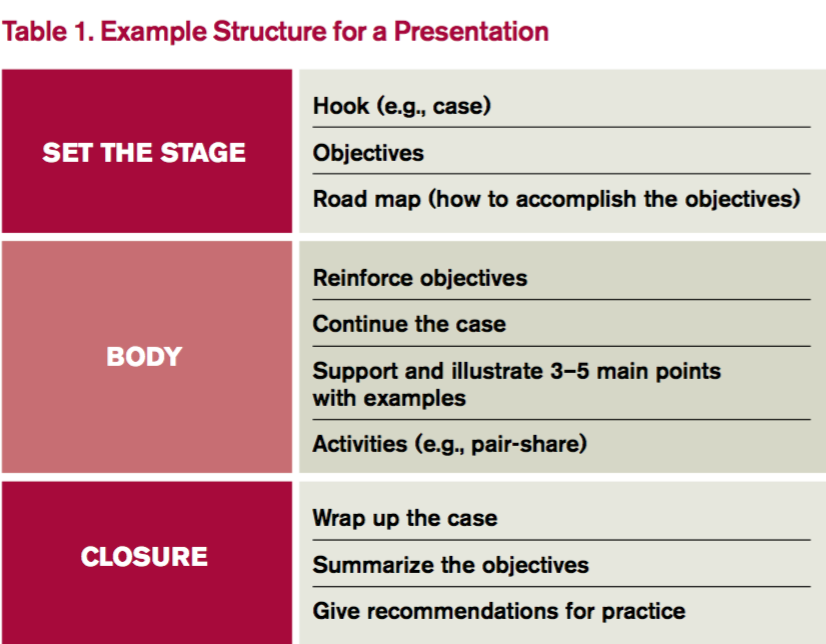
Products granted orphan designation for use in HSCT
The European Commission has granted orphan drug designation for the T-cell therapy product candidate BPX-501 and the small molecule rimiducid.
BPX-501 consists of genetically modified donor T cells incorporating the CaspaCIDe safety switch, which is designed to eliminate the T cells in the event of toxicity.
Rimiducid is used to activate the CaspaCIDe safety switch, which consists of the CID-binding domain coupled to the signaling domain of caspase-9, an enzyme that is part of the apoptotic pathway.
The goal of this therapy is to allow physicians to more safely perform haploidentical hematopoietic stem cell transplant (haplo-HSCT).
Haplo-HSCT recipients receive BPX-501 to speed immune reconstitution and provide control over viral infections. And rimiducid is used to eliminate BPX-501 alloreactive T cells if severe graft-vs-host disease (GVHD) occurs.
If a patient develops severe GVHD, rimiducid is used to trigger activation of the domain of caspase-9, which leads to selective apoptosis of the CaspaCIDe-containing cells.
About orphan designation
Orphan drug designation from the European Commission provides regulatory and financial incentives for companies to develop and market therapies that treat serious or life-threatening conditions that affect no more than 5 in 10,000 people in the European Union (EU), and where no treatment is currently approved.
In addition to a 10-year period of marketing exclusivity in the EU upon product approval, orphan drug designation provides fee waivers, protocol assistance, and marketing authorization under the centralized procedure granting approval in all EU countries.
BPX-501/rimiducid development
BPX-501 and rimiducid are being developed by Bellicum Pharmaceuticals.
The company has met with regulatory authorities in Europe to discuss the potential approval pathway for BPX-501 and rimiducid for the treatment of immunodeficiency and GVHD following haplo-HSCT in pediatric patients with leukemias, lymphomas, and rare inherited blood diseases who do not have a matched donor.
These discussions have resulted in an initial agreement regarding the company’s development plans, subject to further refinement in a formal protocol assistance process that is available for orphan drug products.
Based on regulatory discussions, Bellicum believes that data from the European arm of its BP-004 trial, with a 6-month follow-up time and expanded to enroll additional patients, could form the basis of marketing authorization applications for BPX-501 and rimiducid.
The European Medicines Agency’s Committee for Medicinal Products for Human Use has agreed that review and approval under “exceptional circumstances” may be suitable, recognizing that a randomized trial may not be feasible in the pediatric setting. In place of a randomized trial, Bellicum intends to collect data from a concurrent observational study of allogeneic HSCT outcomes in the pediatric setting.
The European Medicines Agency can grant early market authorization to orphan drug products under exceptional circumstances. Exceptional circumstances can be granted for medicines that treat very rare diseases or where controlled studies are impractical or not consistent with accepted principles of medical ethics.
BP-004 trial
BP-004 is a phase 1/2 dose-escalation trial of BPX-501 and rimiducid in pediatric patients with malignant and nonmalignant diseases. Interim results from this trial were reported in 2 presentations at the 42nd Annual Meeting of the European Society for Blood and Marrow Transplantation in April 2016.
One presentation involved patients with acute leukemia who received BPX-501 after haplo-HSCT. At a median follow-up of 7 months, 16 of the 17 patients were alive and disease-free. There were several cases of GVHD, but nearly all were resolved.
The other presentation covered patients with nonmalignant disorders who received BPX-501 after haplo-HSCT. At a median follow-up of 7 months, all 24 patients studied were still alive and disease-free. The incidence of GVHD was considered “very low.” ![]()
Core curriculum for opioid prescribing preempts certification
LAKE BUENA VISTA, FLA. – If the Food and Drug Administration moves to require some form of certification to prescribe opioids, an educational program based on the FDA’s own risk evaluation and mitigation strategy (REMS) report of 2012 is already in place to fulfill that purpose, according to a pain specialist who explained the principles of this program at Pain Care for Primary Care.
Some form of certification process for opioid prescribing has been discussed for many years, explained David E. J. Bazzo, MD, clinical professor of family medicine at the University of California, San Diego, but “there has been renewed interest as a result of the continuing public health crisis regarding opioid overdoses.”
The core curriculum provided by the Collaborative for REMS Education (CO*RE) curriculum is based on clinical competencies to meet the requirements of the FDA REMS blueprint, which was issued in July 2012. The focus of REMS is on opioids with an extended-release (ER) or long-acting (LA) formulation. It is not the only such program available, but Dr. Bazzo noted that CO*RE was actually initiated in 2010 and provided some of the basis for the FDA REMS that followed 2 years later.
The CO*RE program, which can be completed at no cost, is available online. Those who complete the program are provided with a completer certificate, which Dr. Bazzo expects to be honored by the FDA if it does decide to require proof of competency. The program involves the participation of 13 organizations, such as the American Pain Society (APS), the American Society of Addiction Medicine (ASAM), and the American College of Emergency Physicians (ACEP).
There is a substantial possibility that the FDA will move to require competency for opioid prescribers. At a recent joint meeting of the FDA’s Drug Safety and Risk Management Advisory Committee and the Anesthetic and Analgesic Drug Products Advisory Committee in which modifications in the REMS were discussed, the majority of those on both advisory committees endorsed mandatory training.
“Why is education so important? It is because we are in a super tough position,” Dr. Bazzo explained. He suggested that physicians need effective agents for controlling pain, but the mortality and morbidity associated with opioids has steadily increased despite the FDA REMS.
“Deaths due to drug abuse or poisoning are just the beginning. For every death, there are 10 treatment admissions for abuse, and 130 people who are addicted,” said Dr. Bazzo, citing data from the Centers for Disease Control and Prevention (CDC).
In addition to an introduction, which outlines the goals of appropriate opioid prescribing, the CO*RE curriculum consists of six units. The first focuses on assessing candidates for opioid therapy, which includes instructions on history taking and documenting the findings. Tools for assessing risks posed by opioids, particularly for abuse, are also outlined.
The second unit describes best practices for initiating therapy, modifying doses, and discontinuing opioids, the third unit provides information on monitoring adherence and aberrant behavior in patients, and the fourth unit provides information about how to counsel patients about what constitutes appropriate use of opioids and the risks of inappropriate use. The fifth unit is largely an overview of key information in the previous four units and the sixth unit outlines the specific features of currently available opioids.
“The bottom line for the program overall is learning how to balance risks and benefits,” Dr. Bazzo explained. He suggested that many clinicians do not fully grasp that opioids are not stand-alone medications but a tool within a more comprehensive strategy for improving function.
“When we give analgesics for chronic pain, the goal is not to eliminate pain. If this is your goal, you will fail 99.9999% of the time. The goal is to improve function,” Dr. Bazzo said. He characterized treatment of chronic pain “as a team sport” that involves a collaboration between specialists and patients to achieve specific endpoints.
“It is a little like treating hypertension. You adjust medications until you get to the goal,” Dr. Bazzo said. This involves defining the goal before the treatment is initiated and then documenting progress toward that goal to guide treatment strategies.
The CO*RE REMS program is consistent with the CDC Guideline for Prescribing Opioids for Chronic Pain (MMWR. 65[1];1–49). It is part of a nationwide effort to reduce deaths related to opioid use.
“The problems with opioids can be greatly reduced if clinicians recognize and adhere to best practices,” Dr. Bazzo maintained.
Dr. Bazzo reports no potential financial conflicts of interest. The meeting was held by the APS and Global Academy for Medical Education. Global Academy and this news organization are owned by the same company.
EXPERT ANALYSIS FROM PAIN CARE FOR PRIMARY CARE

For Neuropathic Pain, Marijuana Yields Modest Benefits
LAKE BUENA VISTA, FLA. – As medical marijuana penetrates mainstream medical practice in the United States and elsewhere, awareness of the potential benefits, potential risks, and local laws governing its use for treatment of chronic pain gain importance, according to a specialist reviewing available data at Pain Care for Primary Care.
For many specific types of chronic pain, more data are needed to judge the benefit-to-risk ratio of marijuana relative to other options, but there are reasonable data suggesting both acceptable safety and meaningful efficacy of this analgesic in neuropathic pain, according to Mark A. Ware, MBBS, associate professor in the departments of anesthesia and family medicine, McGill University, Montreal.

In neuropathic pain, the evidence includes at least five randomized trials, according to Dr. Ware. In a recently published review article for which Dr. Ware served as senior author, the degree of neuropathic pain reductions were characterized as being on an order similar to those achieved with opioids and anticonvulsants (J Pain. 2016 Jun;17[6]:654-68). In one study, the number needed to treat (NNT) for a 50% pain reduction was just 2.
In Canada, marijuana is now available for at least some medical uses in every province. In the United States, 23 states have passed laws permitting clinical use of marijuana, according to Dr. Ware. He suggested that legalization of marijuana has fueled a growing acceptance of marijuana as a treatment option whether or not it is prescribed. For this reason, it’s necessary to examine the objective evidence to provide appropriate counseling.
“I think we are past the point where this option can simply be ignored,” Dr. Ware said. Even if they do not intend to prescribe marijuana for chronic pain, “clinicians should become familiar with the evidence regarding benefit and safety as well as the laws regarding its use.” In a study of long-term safety led by Dr. Ware, a standardized cannabis product containing 12.5% tetrahydrocannabinol was dispensed to 215 current or prior users of marijuana with a non-cancer chronic pain syndrome (J Pain. 2015 Dec;16[12]:1233-42). Followed for 1 year, adverse events in this group were compared with 216 control patients who also had chronic pain but were not using cannabis.
The odds ratio (OR) of non-serious adverse events in the categories of respiratory disorders (OR, 1.77) infectious disorders (OR, 1.51), nervous system disorders (OR, 1.77), and psychiatric disorders (OR, 2.74) were all significantly higher in the group treated with cannabis, but almost all were judged to be of mild to moderate severity. There was no significant difference in the risk of serious AEs. Dr. Ware also reported there was no difference between the cannabis group and controls for neurocognitive testing at baseline, 6 months, or the end of 1 year.
Pain control was also monitored over the course of the study. According to Dr. Ware, average pain scores in the cannabis group fell modestly but consistently over the course of the study. Over the same period, the pain scores rose slightly in the control group.
There is a long list of unanswered questions regarding effective use of marijuana in the control of chronic pain, he said. For example, Dr. Ware noted that the optimal composition of cannabinoids has yet to be determined. He noted that more than one of the complex constituents may contribute to pain control, and these constituents are not necessarily the same as those most favored by recreational users seeking a euphoric “high.”
There is also a long list of unanswered questions about safety. Dr. Ware reviewed some evidence that inhaled vaporized marijuana may be safer than traditional smoked marijuana due to a reduced exposure to toxins, but he suggested more rigorous studies are needed to generate objective data that can better quantify the benefit-to-risk of this and other methods of marijuana delivery.
Despite unanswered questions, marijuana is widely available and likely to be considered by patients for chronic pain whether or not it is recommended by physicians. It is for this reason that clinicians need to become familiar with both its potential risks and benefits.
“It will be helpful to patients if you can provide them practical and accurate information about what is and is not known about this treatment,” Dr. Ware suggested.
Dr. Ware reported a financial relationship with CanniMed. The meeting was held by the American Pain Society and Global Academy for Medical Education. Global Academy and this news organization are owned by the same company.
LAKE BUENA VISTA, FLA. – As medical marijuana penetrates mainstream medical practice in the United States and elsewhere, awareness of the potential benefits, potential risks, and local laws governing its use for treatment of chronic pain gain importance, according to a specialist reviewing available data at Pain Care for Primary Care.
For many specific types of chronic pain, more data are needed to judge the benefit-to-risk ratio of marijuana relative to other options, but there are reasonable data suggesting both acceptable safety and meaningful efficacy of this analgesic in neuropathic pain, according to Mark A. Ware, MBBS, associate professor in the departments of anesthesia and family medicine, McGill University, Montreal.
In neuropathic pain, the evidence includes at least five randomized trials, according to Dr. Ware. In a recently published review article for which Dr. Ware served as senior author, the degree of neuropathic pain reductions were characterized as being on an order similar to those achieved with opioids and anticonvulsants (J Pain. 2016 Jun;17[6]:654-68). In one study, the number needed to treat (NNT) for a 50% pain reduction was just 2.
In Canada, marijuana is now available for at least some medical uses in every province. In the United States, 23 states have passed laws permitting clinical use of marijuana, according to Dr. Ware. He suggested that legalization of marijuana has fueled a growing acceptance of marijuana as a treatment option whether or not it is prescribed. For this reason, it’s necessary to examine the objective evidence to provide appropriate counseling.
“I think we are past the point where this option can simply be ignored,” Dr. Ware said. Even if they do not intend to prescribe marijuana for chronic pain, “clinicians should become familiar with the evidence regarding benefit and safety as well as the laws regarding its use.” In a study of long-term safety led by Dr. Ware, a standardized cannabis product containing 12.5% tetrahydrocannabinol was dispensed to 215 current or prior users of marijuana with a non-cancer chronic pain syndrome (J Pain. 2015 Dec;16[12]:1233-42). Followed for 1 year, adverse events in this group were compared with 216 control patients who also had chronic pain but were not using cannabis.
The odds ratio (OR) of non-serious adverse events in the categories of respiratory disorders (OR, 1.77) infectious disorders (OR, 1.51), nervous system disorders (OR, 1.77), and psychiatric disorders (OR, 2.74) were all significantly higher in the group treated with cannabis, but almost all were judged to be of mild to moderate severity. There was no significant difference in the risk of serious AEs. Dr. Ware also reported there was no difference between the cannabis group and controls for neurocognitive testing at baseline, 6 months, or the end of 1 year.
Pain control was also monitored over the course of the study. According to Dr. Ware, average pain scores in the cannabis group fell modestly but consistently over the course of the study. Over the same period, the pain scores rose slightly in the control group.
There is a long list of unanswered questions regarding effective use of marijuana in the control of chronic pain, he said. For example, Dr. Ware noted that the optimal composition of cannabinoids has yet to be determined. He noted that more than one of the complex constituents may contribute to pain control, and these constituents are not necessarily the same as those most favored by recreational users seeking a euphoric “high.”
There is also a long list of unanswered questions about safety. Dr. Ware reviewed some evidence that inhaled vaporized marijuana may be safer than traditional smoked marijuana due to a reduced exposure to toxins, but he suggested more rigorous studies are needed to generate objective data that can better quantify the benefit-to-risk of this and other methods of marijuana delivery.
Despite unanswered questions, marijuana is widely available and likely to be considered by patients for chronic pain whether or not it is recommended by physicians. It is for this reason that clinicians need to become familiar with both its potential risks and benefits.
“It will be helpful to patients if you can provide them practical and accurate information about what is and is not known about this treatment,” Dr. Ware suggested.
Dr. Ware reported a financial relationship with CanniMed. The meeting was held by the American Pain Society and Global Academy for Medical Education. Global Academy and this news organization are owned by the same company.
EXPERT ANALYSIS FROM PAIN CARE FOR PRIMARY CARE