User login
Therapy increases factor IX activity in hemophilia B

SCOTTSDALE, ARIZONA—The investigational therapy SPK-9001 delivers “consistent and sustained” increases in factor IX (FIX) activity in patients with hemophilia B, according to researchers.
All 10 subjects who received SPK-9001 in a phase 1/2 trial have consistently achieved the targeted therapeutic range of FIX activity.
There have been no confirmed bleeds, and 9 of the subjects have not received any infusions of FIX therapy since receiving SPK-9001.
SPK-9001 reduced the patients’ annualized bleeding rate by 96% and the annualized infusion rate by 99%.
Adam Cuker, MD, of the Perelman School of Medicine at the University of Pennsylvania in Philadelphia, and his colleagues presented these results at the Hemostasis and Thrombosis Research Society 2017 Scientific Symposium.
The trial is sponsored by Spark Therapeutics and Pfizer, the companies developing SPK-9001.
SPK-9001 is a bio-engineered adeno-associated virus capsid expressing a codon-optimized, high-activity human FIX variant enabling endogenous production of FIX.
In the phase 1/2 trial, 10 hemophilia B patients received a single administration of SPK-9001 (5 x 10¹¹ vector genomes/kg body weight).
As of the data cutoff (March 24, 2017), 9 of the 10 patients have not taken FIX concentrates to prevent or control bleeding events since they received SPK-9001.
One patient with severe joint disease has self-administered infusions. The patient self-infused FIX therapy for a suspected ankle bleed on day 2 after receiving SPK-9001 and self-administered precautionary infusions another 9 times between December 2, 2016, and January 2, 2017, for persistent knee pain. The patient has not used additional FIX therapy since January 2.
For all 10 patients, the mean steady-state FIX activity level 12 weeks after SPK-9001 administration was a sustained 33% (range as of the data cutoff: 14% to 81%).
Based on individual patient history prior to the study, the annualized bleeding rate was reduced by 96% to a mean of 0.39 annual bleeds, compared with 9.2 bleeds before SPK-9001 administration.
The annualized infusion rate was reduced 99% to a mean of 0.98 annual infusions, compared with 68.5 infusions before SPK-9001 administration.
To date, no serious adverse events have been reported, including no FIX inhibitors and no thrombotic events.
Two of the 10 patients experienced an asymptomatic, transient elevation in liver enzymes, or decline in FIX activity, potentially indicative of an immune response to the Spark100 vector capsid, that occurred several weeks post-infusion.
Both patients received a tapering dose of oral corticosteroids, after which their alanine aminotransferase levels returned to baseline.
The FIX activity level of one of these patients has stabilized at approximately 15% for more than 9 weeks post-corticosteroid use. The other patient had a FIX activity level between 70% and 80% upon completion of steroid use. ![]()
CAR T-cell therapy demonstrates efficacy in mice with MM

WASHINGTON, DC—A chimeric antigen receptor (CAR) T-cell therapy is active against multiple myeloma (MM), according to preclinical research.
The therapy, known as P-BCMA-101, demonstrated persistent anti-tumor activity in a mouse model of MM.
Treatment with P-BCMA-101 eliminated tumors after relapse and prolonged survival in the mice, when compared to other CAR-T cell therapies.
These results were presented in a poster at the AACR Annual Meeting 2017 (abstract 3759).
The research was conducted by employees of Poseida Therapeutics Inc., the company developing P-BCMA-101, as well as others.
About P-BCMA-101
The researchers explained that P-BCMA-101 employs a B-cell maturation antigen (BCMA)-specific Centyrin™ rather than a single-chain variable fragment (scFv) for antigen detection.
Centyrins™ are fully human and have similar binding affinities as scFvs. However, Centyrins are smaller, more thermostable, and predicted to be less immunogenic.
P-BCMA-101 is engineered using the PiggyBac™ DNA modification system. PiggyBac eliminates the need to use lentivirus or gamma-retrovirus as a gene delivery mechanism, resulting in improved manufacturing and cost savings.
In addition, the increased cargo capacity of PiggyBac allows for the incorporation of a safety switch and a selectable gene. The safety switch can be “flipped” to enable depletion in case adverse events occur. And the selectable gene allows for enrichment of CARTyrin+ cells using a non-genotoxic drug.
Findings
The researchers found that more than 70% of P-BCMA-101 cells possessed a stem cell memory phenotype, creating a significant population of self-renewing, multipotent progenitors capable of reconstituting the entire spectrum of memory and effector T-cell subsets required to prevent cancer relapse. Similar competitor products typically report 0% to 20% stem cell memory phenotype.
In addition, P-BCMA-101 was enriched with more than 95% of T cells successfully modified, which compares favorably to the roughly 30% to 50% commonly expected with clinical manufacture using lentivirus.
P-BCMA-101 did not exhibit effects of CAR-mediated tonic signaling, a common cause of T-cell exhaustion that leads to poor durability. Tonic signaling is caused by oligomerization of unstable binding domains commonly seen with traditional scFv CARs.
The researchers tested P-BCMA-101 in NSG mice bearing luciferase+ MM.1S cells. The mice received a single administration of either 4 x 10⁶ or 12 x 106 P-BCMA-101 cells.
P-BCMA-101 treatment reduced tumor burden to the limit of detection within 7 days. Conversely, all untreated control mice succumbed to MM within 4 weeks.
P-BCMA-101 expanded and persisted in the treated mice, eliminated tumors following relapse, and prolonged survival.
Most treated mice survived 110 days, and none of them died from tumor burden during the study. This compares favorably to lentivirus-based products that have shown roughly 50-day survival in the same model.
Based on these results, Poseida plans to initiate a phase 1 trial of P-BCMA-101 in patients with relapsed or refractory MM. ![]()
FDA: REMS for ESAs no longer needed, though risks persist

The US Food and Drug Administration (FDA) has determined that the risk evaluation and mitigation strategy (REMS) for erythropoiesis-stimulating agents (ESAs) is no longer necessary.
The REMS was limited to the use of epoetin alfa (marketed as Epogen and Procrit) and darbepoetin alfa (marketed as Aranesp) to treat patients with anemia due to myelosuppressive chemotherapy.
The FDA said the REMS is no longer necessary to ensure that the benefits of Epogen/Procrit and Aranesp outweigh the risks these drugs pose, which include shortened overall survival and an increased risk of tumor progression or recurrence in patients with cancer.
The FDA has released the REMS requirements for these ESAs and said the risks the drugs pose can be communicated by the current product prescribing information.
The FDA decided the REMS is no longer needed based on its own analyses and an evaluation of the REMS assessment submitted by Amgen, Inc., the company that markets Epogen/Procrit and Aranesp.
Details on the analyses and evaluation are available from the following page on the FDA website: Information on Erythropoiesis-Stimulating Agents (ESA) Epoetin alfa (marketed as Procrit, Epogen), Darbepoetin alfa (marketed as Aranesp). ![]()
It’s been a good year for heart failure research ... mostly
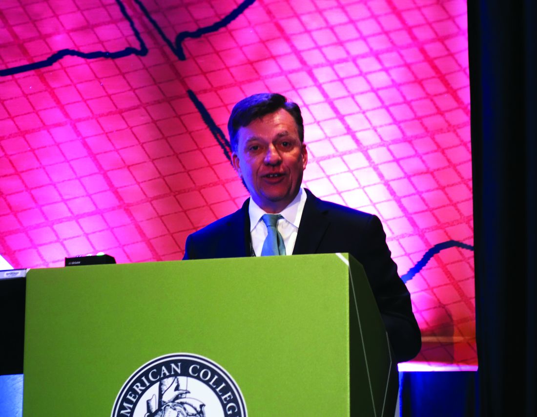
WASHINGTON – It’s been a “relatively positive” year for heart failure research and advances in patient care, Christopher M. O’Connor, MD, said at the annual meeting of the American College of Cardiology.
“Having been in this field for 30 years looking at clinical trials, generally for every 10 important trials done in a year, 1 has been positive and 9 have been negative; if we look at the past year, it’s more like 5 and 6. So, not a bad year for cardiomyopathy,” declared Dr. O’Connor, CEO and executive director of the Inova Heart and Vascular Institute in Falls Church, Va., and president-elect of the Heart Failure Society of America.
The good news
• Empagliflozin (Jardiance) earns FDA approval for reduction in risk of cardiovascular death in type 2 diabetes patients. “This is one of the most amazing stories in heart failure,” said Dr. O’Connor, who is also professor of medicine at Duke University in Durham, N.C.
The pivotal EMPA-REG OUTCOME study showed a highly significant 35% reduction in the secondary endpoint of risk of hospitalization for heart failure, as well the decrease in cardiovascular mortality which was the primary endpoint and proved persuasive to the FDA (N Engl J Med. 2015 Nov 26;373[22]:2117-28).
“It was a remarkable development. Because of this trial, there are now a number of ongoing phase III clinical trials looking at this class of drugs in heart failure patients with and without diabetes, which makes this a very important research movement. We are now looking deeper at phenotypes and trying to get more specific with these drug therapies,” he said.
• A new and improved LVAD. This fully magnetically levitated centrifugal-flow pump type of left ventricular assist device for advanced heart failure showed superior event-free survival, compared with a commercially available axial continuous-flow pump LVAD in the randomized MOMENTUM-3 trial (N Engl J Med. 2017 Feb 2;376[5]:440-50).
The novel pump was designed to overcome a significant problem with axial continuous-flow LVADs: a proclivity for pump thrombosis. The magnetically levitated centrifugal-flow pump proved a smashing success in this regard, with zero cases of pump thrombosis occurring during the 6-month study.
“This may be the first time in the history of heart failure research that the engineers have beaten the biologists in important clinical outcomes,” the cardiologist quipped.
• Omecamtiv mecarbil successfully addresses impaired contractility in heart failure with reduced ejection fraction (HFrEF). This drug, a selective cardiac myosin activator, resulted in increased duration of systole and improved stroke volume accompanied by reductions in heart rate, left ventricular end-diastolic and -systolic dimensions, and NT-proBNP in the 87-site, 13-country, phase II COSMIC-HF study (Lancet. 2016 Dec 10;388[10062]:2895-903).
“This is probably the most novel new drug mechanism out there in clinical trials,” according to Dr. O’Connor.
On the basis of the highly encouraging results for the surrogate endpoints assessed in COSMIC-HF, a large phase III clinical trial known as GALACTIC is underway.
• Palliative care gets a welcome boost. Dr. O’Connor was a coinvestigator in PAL-HF, a single-center study presented at the 2016 annual meeting of the Heart Failure Society of America.
“This is a very important trial of palliative care in advanced heart failure. We probably don’t have as much evidence in this space as we should,” he observed. “This was a multidisciplinary intervention in which we gave the patients a medical tool kit to alleviate pain, dyspnea, and discomfort. The tool kit included benzodiazepines, sleep medications, sublingual nitroglycerin, and morphinelike products, all very carefully monitored by staff coordinators.”
The primary outcome was change in two validated heart failure quality of life measures. Both instruments documented significant improvement compared with usual care.
“There was no decrease in mortality, which wasn’t a goal in this advanced heart failure population, and no reduction in heart failure hospitalizations, but there were significant reductions in depression and anxiety,” Dr. O’Connor said.
• Vericiguat. This oral soluble cyclic guanylate cyclase stimulator missed its primary endpoint in the phase II dose-escalation SOCRATES-REDUCED trial in patients with HFrEF (JAMA. 2015 Dec 1;314[21]:2251-62), but showed an impressive improvement in quality of life. It is now the subject of the ongoing, randomized, phase III VICTORIA trial involving a planned 4,000 patients with HFrEF with the composite primary endpoint of cardiovascular death or heart failure hospitalization.
The phase II SOCRATES-PRESERVED trial also missed its primary endpoint but showed a clinically meaningful improvement in quality of life in patients with heart failure with preserved ejection fraction (HFpEF) (Eur Heart J. 2017 Mar 22. doi: 10.1093/eurheartj/ehw593). Discussions are ongoing as to whether the next step should be a confirmatory phase II study or a move straight to phase III.
The bad news
• NSAIDs linked to increased risk of heart failure. European investigators analyzed five population-based databases totaling more than 8.3 million individuals and determined that current use of any of more than two dozen NSAIDs was associated with significantly increased risk of hospital admission for heart failure. The risk appeared to be dose dependent and varied between individual agents, ranging from a 16% increased risk with naproxen to an 83% increase with ketorolac (Toradol) (BMJ. 2016 Sep 28. doi: 10.1136/bmj.4857).
• Therapeutic natriuretic peptides hit bottom. The negative results for the investigational agent ularitide in patients with acute decompensated heart failure in the large phase III TRUE-AHF trial presented at the 2016 meeting of the American Heart Association, following upon an earlier negative study of the related drug nesiritide (Natrecor) in more than 7,100 acute heart failure patients (N Engl J Med. 2011 Jul 7; 365:32-43), probably spells the end of the line for this strategy of boosting outcomes in acute heart failure, according to Dr. O’Connor.
Moreover, Novartis has announced that the phase III RELAX-AHF-2 trial of serelaxin in 6,600 patients with acute heart failure failed to meet its primary endpoints of reduced cardiovascular deaths or reduced worsening of heart failure. The trial will be formally presented later this year.
“Ularitide seemed to show an early improvement in heart failure events that was not sustained in-hospital, and there was absolutely no difference in mortality. The drug probably acts like a pharmacologic tourniquet, in my view. So I think this field of therapeutic natriuretic peptides is probably closed,” he said.
• ICDs don’t reduce mortality in patients with nonischemic heart failure. This was the conclusion reached in the DANISH trial, in which more than 1,100 patients with symptomatic systolic heart failure were randomized to an ICD or usual care (N Engl J Med. 2016 Sep 29;375[13]:1221-30).
“This study really shook up the field, raising the question, ‘Are we using defibrillators too frequently in this population?’ It has stimulated a lot of discussion, including within the guidelines committee,” according to Dr. O’Connor.
• Tolvaptan nixed for acute decompensated heart failure. The TACTICS-HF trial studied the use of tolvaptan (Samsca), an oral vasopressin-2 receptor antagonist, to reduce dyspnea in patients hospitalized with acute decompensated heart failure. Dr. O’Connor was a coinvestigator in the study, which showed that tolvaptan was no better than placebo at 8 and 24 hours (J Am Coll Cardiol. 2017 Mar 21;69[11]:1399-406).
“For now, the routine use of vasopressin antagonists in acute heart failure is not to be encouraged, although there may still be subsets where it’s worth trying – certainly in severe hyponatremia,” the cardiologist said.
• GUIDE-IT gets lost. This was a roughly 1,000-patient randomized trial of a treatment strategy aimed at improving clinical outcomes by aggressively titrating evidence-based heart failure therapies in order to suppress natriuretic peptide biomarkers. GUIDE-IT was stopped early by the data safety monitoring board due to a lack of discernible difference in outcomes, compared with usual care. Dr. O’Connor, a coinvestigator, termed the soon-to-be-published study “a disappointment.”
Sleep apnea studies show silver lining
Sleep apnea is common in patients with heart failure and is associated with worse clinical heart failure outcomes. But in the large, randomized SERVE-HF trial, investigators showed that treatment of central sleep apnea with adaptive servo-ventilation in patients with HFrEF unexpectedly increased mortality, compared with optimal medical therapy (N Engl J Med. 2015 Sep 17;373:1095-105). In a more recent secondary analysis, however, the SERVE-HF investigators found that the increased risk for cardiovascular death associated with adaptive servo-ventilation was actually restricted to patients with a very poor left ventricular ejection fraction of 30% or less, and there was a signal of possible benefit in patients in the top tier of LVEF, at 36%-45% (Lancet Respir Med. 2016 Nov; 4[11]:873-81).
This observation dovetails nicely with the findings of the CAT-HF trial presented by Dr. O’Connor at the 2016 World Congress on Heart Failure. In this phase II study, 215 patients with acute decompensated heart failure and either obstructive or central sleep apnea were randomized to optimal medical therapy alone or in combination with adaptive servo-ventilation. There was no overall difference in outcome between the two groups; however, in the subgroup of patients with HFpEF, the risk of the primary composite endpoint was reduced by 62% with adaptive servo-ventilation.
As a result of these intriguing findings from SERVE-HF and CAT-HF, a registry and/or randomized clinical trial of adaptive servo-ventilation in patients with HFpEF is now being considered under NIH sponsorship, according to Dr. O’Connor.
He reported having no financial conflicts.
EXPERT ANALYSIS FROM ACC 17
Postsurgical O2 may cut AHI events
Postoperative oxygen therapy in patients with previously undetected obstructive sleep apnea (OSA) led to a reduction in apnea-hypopnea index (AHI) events per hour with no increase in apnea-hypopnea event duration.
The results suggest that postoperative oxygen could be useful in patients with OSA who refuse continuous positive airway pressure (CPAP) therapy, those with newly diagnosed OSA, and those with suspected OSA.
The researchers set out to determine if postoperative oxygen therapy could improve oxygenation in patients with previously undiagnosed OSA, reasoning that the intervention could reduce adverse events.
The study, published in CHEST (2017 March;151[3]:597-611), provided generally good news, but with a caveat: “Essentially we are saying, yes, if you give supplemental oxygen, you improve oxygenation of the patient. But overall we have to be careful because a significant number of patients have significant carbon dioxide retention when receiving supplemental oxygen. So we have to monitor patients – not just oxygen, but we may have to monitor carbon dioxide levels, too,” said lead study author Frances Chung, MBBS, professor of anesthesiology at the University of Toronto and Toronto Western Hospital.
The researchers randomized 123 patients with an AHI of at least five events per hour to postoperative oxygen (3 L/min for 3 nights via nasal prongs) or no postoperative oxygen.
On the third night, the oxygen group had a higher average oxygen saturation than controls (95.2% plus or minus 3.2% vs. 91.4% plus or minus 3.5%; P less than .0001) and a lower oxygen desaturation index (median, 2.3 events per hour vs. median, 18.5; P less than .0001).
A lower number of AHI events per hour occurred in the oxygen group (median, 8.0) than in the control group (median, 15.6; P = .016).
On average, the longest apnea-hypopnea event (median, 33.8 seconds) was shorter for a patient on oxygen, compared with a patient who did not receive oxygen (median, 49.6 seconds; P = .002).
But one finding surprised the researchers and led to some concern: Across both groups, 11.4% of patients experienced substantial CO2 retention. Specifically, for at least 10% of 1 of the nights, these patients had a partial pressure of CO2 of at least 55 mm Hg, according to measurements taken with a transcutaneous CO2 monitor. Of the 14 patients who experienced this event, 13 were receiving oxygen.
Dr. Chung said the results argue strongly for postsurgical oxygen in patients with OSA, who are known to be at increased risk for complications. “We are not doing something about it, and we should be doing something. Because one death from a complication is too many,” she said.
The study was funded by the University Health Network Foundation, Toronto, and the University of Toronto. Dr. Chung reported receiving research grant support from Ontario Ministry of Health Innovation Grant, University Health Network Foundation, ResMed Foundation, Acacia, and Medtronic.
FROM CHEST
Key clinical point: Postsurgical oxygen may reduce complications in patients with obstructive sleep apnea.
Major finding: For patients receiving oxygen, the longest apnea-hypopnea event duration was 33.8 seconds versus 49.6 for controls.
Data source: A randomized, placebo-controlled study of 123 patients.
Disclosures: The study was funded by the University Health Network Foundation, Toronto, and the University of Toronto. Dr. Chung reported receiving research grant support from various entities.
New topical field therapies on the way for actinic keratoses
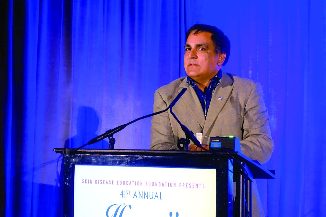
WAILEA, HAWAII – The search is on for new topical therapies for actinic keratoses (AKs) that patients will find more appealing than what’s now available, according to Neal Bhatia, MD.
Compliance with current topical field agents for actinic keratoses is not great. Many patients balk at the intense local skin reactions these agents elicit. There is a misplaced sense, especially among patients who don’t grasp the relationship between AKs and squamous cell carcinoma, that the treatment is worse than the disease, Dr. Bhatia said at the Hawaii Dermatology Seminar provided by Global Academy for Medical Education/Skin Disease Research Foundation.
One that he said he is particularly excited about is a novel formulation of 4% 5-fluorouracil (5-FU) in an aqueous vehicle cream containing peanut oil. In a recently published, double-blind, multicenter clinical trial involving 841 patients, once-daily application of this product for 4 weeks provided better outcomes and superior tolerability, compared with the old-school regimen of 5% 5-FU cream twice daily.
One hundred percent of patients on 4% 5-FU in peanut oil achieved at least 75% clearance of AKs, as did 95% of those on twice daily 5% 5-FU. The incidence of application site skin irritation in the group on the novel product was only 30%, compared with 60% in the comparison group (J Drugs Dermatol. 2016 Oct 1;15[10]:1218-24).
The peanut oil provided a moisturizing effect and was safe even in patients with peanut allergy, Dr. Bhatia noted.
Another product in development as a topical field therapy for AKs is ingenol disoxate gel, a relative of ingenol mebutate (Picato). Unlike ingenol mebutate, ingenol disoxate remains stable without refrigeration. It’s also a more potent activator of protein kinase C. In mouse models, it shows significantly more cytotoxic potency than does ingenol mebutate.
Based upon the favorable results of a short-term, double-blind phase II study, Leo Pharma now has ingenol disoxate in larger clinical trials as field therapy for AKs on the full face, scalp, or chest, with treatment of larger surface areas than those for which ingenol mebutate is approved (J Dermatolog Treat. 2017 Apr 4:1-7).
Actikerall is also in the developmental pipeline. It consists of 0.5% 5-FU, in combination with 10% salicylic acid, in a film-forming base. Developed by Almirall, it is marketed in Canada by Cipher Pharmaceuticals.
SR-T100 gel utilizes as its active ingredient an antiproliferative extract of Solanum lycocarpum, the Brazilian wolf apple. Taiwan-based G & E Herbal Biotechnology is developing the product, which is in an ongoing phase II clinical trial.
Other novel agents for topical field therapy in phase II studies are KX2-391 ointment, a dual Src kinase/tubulin polymerization inhibitor that causes apoptosis of hyperproliferating cells, under development by Athenex, and Vidac Pharma’s VDA-1102 ointment, which selectively triggers apoptosis in neoplastic cells by modulating voltage-dependent anion channel 1/hexokinase enzyme 2, with minimal impact upon surrounding normal cells.
The principle underlying topical field therapy, compared with simply freezing AKs once they arise, is straightforward, Dr. Bhatia stressed. “It’s the difference between treating only what we can see and treating what’s also on the way.”
Dr. Bhatia reported having financial relationships with more than two dozen pharmaceutical companies. SDEF and this news organization are owned by the same parent company.
EXPERT ANALYSIS FROM SDEF HAWAII DERMATOLOGY SEMINAR
Shared-savings ESRD organization reduced missed treatments, hospitalizations
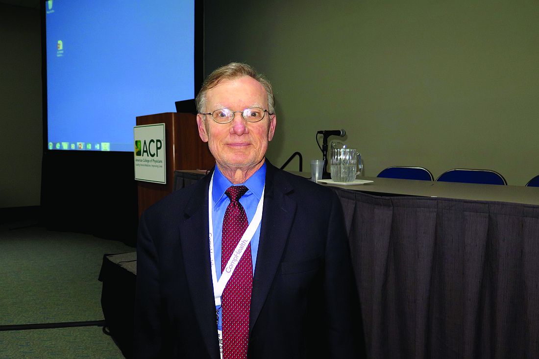
SAN DIEGO – In the first year since Delaware Valley Nephrology joined an end–stage renal disease seamless care organization, patient hospitalizations have declined by 40% and rehospitalizations by 20%, according to Edward R. Jones, MD.
At the annual meeting of the American College of Physicians, Dr. Jones, a nephrologist at a Philadelphia-based practice, described his experience with the alternative payment model, which focuses on managing costs while providing better health outcomes.
More than 15 months ago, Dr. Jones and seven other nephrologists in his practice partnered with Fresenius Medical Care to form an ESCO with two other nephrology groups, amounting to 45 physicians who work out of 30 dialysis units in 14 hospitals and care for 1,000 patients.
It became one of 13 ESCOs that have been operating nationwide since October 2015. The ESCOs share savings with the Centers for Medicare & Medicaid Services if matched beneficiaries’ expenditures decrease and quality is maintained or improved, and it shares losses if beneficiaries’ expenditures increase.
To be eligible for participation in the ESCO, patients must be enrolled in Medicare Parts A and B and have Medicare as their primary payer. They are ineligible if they are enrolled in a Medicare Advantage plan. Patients must not have received a kidney transplant in the last 12 months and must receive at least 51% of annual dialysis services in the ESCO’s market area.
“The governing structure of the ESCO was critical as we put it together,” Dr. Jones said. “We insisted that a nephrologist be at the table of the governing body, as well as a consumer advocate, and no one controls more than 50% of the vote.
“The concept here is that a benchmark for spending is set,” he explained. “If your spending is less than the benchmark expenditure, there are shared savings. The opposite occurs if there are losses, so it’s a fairly straightforward program.”
To illustrate how the ESCO works, he discussed a hypothetical model in which the ESCO used $50 million of expenditures from 2012 to 2015.
“If the actual ESCO expenditures are $47 million, there are $3 million in potential shared savings,” Dr. Jones explained. “It’s potential because two things are necessary. First, you have to have hit at least 1% savings or 1% loss. This allows for any random outcomes. Of $3 million of potential shared savings, CMS keeps 25%.
“The remaining shared savings depends on where you hit on the metric performance,” he continued. “In the first year, it’s a pay for reporting [system], so all we had to do is hit three measures. So, the first year was great.”
In the second year of ESCO participation, centers have to perform in the 90th percentile on 16 metrics to get full shared savings. Drawn from Medicare claims and medical records, the list of metrics includes diabetic foot exam, medication reconciliation, and advanced care planning, and it accounts for 50% of the score. Surveys of person- and caregiver-centered experience outcomes compose 33% of the score.
“These metrics are compared to all other metrics throughout the country,” Dr. Jones said. “Wherever you fall, you generate a certain number of points, between 0 and 2. It’s tied to quality and outcomes. If you’re in the 90th percentile you get all 2 points. If you’re below the 50th percentile, you get no points.”
The goal of the ESCO is to achieve the minimal savings rate, to maximize achievement of quality metrics, and to reduce expenses below the set benchmark. In Dr. Jones’ practice area, ESRD care costs about $87,000 per patient per year – 34% are related to inpatient costs, and 30% are related to dialysis costs.
Keys to a successful ESCO include “a cooperative and engaged nephrologist” and the ability to gather analytics.
“There is no way that a practice of my size has the capability of generating the analytics to be able to risk-stratify patients,” he said. “Who is likely to be readmitted? We put all of our effort into those folks. Medication reconciliation accounts for a lot of rehospitalization. We spend a lot of time on that.”
There is also increased use of care navigators. “Our goal is for patients to call the care navigators before they call the nephrologist,” Dr. Jones said. “We have safety net clinics that are open 24/7. So, if a patient comes in on a Saturday or Sunday, we can divert the patient to those areas.”
In the first year since joining the ESCO, patient hospitalizations at Dr. Jones’ practice have declined by 40%, and rehospitalizations have dropped by 20%.
“Importantly, we know that when a patient is not in his dialysis chair within 15 minutes, the care navigator calls him,” he said. “The number of missed treatments is down dramatically.”
Dr. Jones reported owning stock in Cytosorbents. He also serves as a consultant for Fresenius Medical Care, Fresenius Medical Services, Physicians Choice Dialysis, and Reliant Renal Care. He is also an advisory board member for Cytosorbents and Fresenius.
EXPERT ANALYSIS AT ACP INTERNAL MEDICINE
Obstacles plague prenatal cell-free DNA testing use
A recent California study examining cell-free DNA (cfDNA) testing suggests that the test generally performs well as a second-line screen for fetal aneuploidy. However, while the test itself is reliable, some physicians say gaps in patient education, limited availability of genetic counseling, and a lack of coordinated research and data collection are all standing in the way of its optimal utilization.
In some of the most recent data presented on cfDNA test performance, researchers in California found that the test had few false positives and was best at predicting Down syndrome, with a positive predictive value of 98.6%.
California offers prenatal screening as a public health program, with a structured system of screening tests and follow-up services, according to Dr. Currier, who is chief of the evaluation section of the state’s Genetic Disease Screening Program. First trimester screening includes screens for trisomies 18 and 21. During the second trimester, the screening adds on checks for neural tube defects and Smith-Lemli-Opitz syndrome, also known as 7-dehydrocholesterol reductase deficiency.

Overall, 20,852 patients were offered cfDNA screening during this period, and 63% (n = 12,960) went on to have the screen. Dr. Currier and his colleagues found that about a third of patients with a first- or second-trimester positive cfDNA test opted for diagnostic testing with either chorionic villus sampling or amniocentesis. If patients had a known karyotype, most were in agreement with the trisomy or sex-chromosome aneuploidies that had been picked up by cfDNA testing.
There were a small number of false-positive cfDNA results seen in the California data, Dr. Currier noted. Performance was best for Down syndrome, where cfDNA had a positive predictive value of 98.6%. Here, 214 of the 611 cfDNA positive results received diagnostic confirmation. Of those, 197 were true positives and 3 were false positives, while a karyotype other than trisomy 21 was revealed in 14 cases. Positive predictive values were lower in the less-common aneuploidies, he said.
A small number of test failures also occurred. Of the 148 failures reported, just 40 were followed by successful repeat tests.
“Cell-free DNA is a good second-tier screening test, but it does have false-positive and false-negative results, so pre- and post-test counseling addressing these limitations are essential,” Dr. Currier said.
Nancy Rose, MD, agrees with that assessment of the limitations of the cfDNA test. She helped write the American College of Obstetricians and Gynecologists 2015 opinion on cell-free DNA screening for aneuploidy, which states that conventional screening methods “remain the most appropriate choice for first-line screening for most women in the general obstetric population.”
While it is “perfectly reasonable” for general ob.gyns. to offer cfDNA screening, in her practice, “genetic counselors call out all the results for general providers because patients don’t really understand what a screen negative result is,” Dr. Rose said in an interview. “Certainly, screen positive results should go to a genetic counselor for sure.”
However, that level of follow-up may be hard to implement. “One issue that we’re all facing right now is that genetic counselors are a scarce commodity because of the competition with them working for labs or insurance companies,” said Dr. Rose, a maternal-fetal medicine specialist in Salt Lake City.
If the lack of appropriate counseling is an issue, are patients sometimes jumping the gun? The immediate past president of the Society for Maternal-Fetal Medicine, Mary Norton, MD, said maybe so.
When asked whether she thinks pregnancies are being terminated on the basis of cfDNA results alone, Dr. Norton said, “I absolutely think that is happening. How many is hard to know. I do know that we have many patients that we see who say, ‘My result here says that the chance of Down syndrome is greater than 99%,’ but that is not what that number means, and people don’t understand that.”
She added, “In some cases there are also ultrasound abnormalities, but there is no question that there is misunderstanding leading to the loss of normal fetuses.”
Patient misunderstandings about what’s entailed in first- and second-line screening tests persist, said Dr. Norton, professor of obstetrics and gynecology and reproductive sciences at the University of California, San Francisco. One example is ongoing hesitancy about amniocentesis. “Amnio has become very, very safe,” she said. “It’s much safer than it used to be. The miscarriage rate is nearly nonexistent.”
The care team needs to work to find time to educate patients about both older technologies and noninvasive prenatal testing (NIPT), Dr. Rose and Dr. Norton stressed.
“Even though ‘NIPT’ is easy to say – it rolls off the tongue – we really shouldn’t call it that. It’s misleading, because all of the other screening tests are noninvasive too,” Dr. Norton said. “I’ve had many patients who declined diagnostic testing, even though they want as much information as possible about what they should do.”
Dr. Rose added, “It’s a screening test; it’s not perfect. But screening tests are really what obstetricians do. They do pap smears; they do breast exams. So I think that isn’t the issue so much as that there is no time in an obstetrician’s office to really educate women about what they are accepting.”
Additional ethical issues revolving around the mother may arise in rare cases. Only about 10% of the DNA sampled is fetal, meaning that the rest is maternal DNA, Dr. Norton explained. “And they’re not separated,” she said. “So, the issue that is coming up is that they are finding things in the mother that are unanticipated, and women aren’t told that ahead of time.”
Some of these thorny – and still evolving – legal and ethical questions were addressed during a workshop at the 2017 Pregnancy Meeting. A working group that met there is developing a summary of their discussion for future publication.
For Dr. Rose, the larger issue is the lack of coordinated data collection and quality initiatives. She pointed out that the California outcomes study may not be generalizable to most of the rest of the country because most states don’t have such a coordinated public health approach to prenatal testing and data collection.
This reality is hampering knowledge advancement in the field, she said. When she was asked about terminations on the basis of cfDNA alone, Dr. Rose said, “Because there’s no outcome data on these patients, because there are no unbiased studies that cross through these companies, we actually don’t really know that on a national level. There are no unbiased outcome data. There may be small studies, but I think there are no national data to answer that question at the moment.”
Overall, Dr. Rose said that cfDNA screening is fairly reliable. “This is no different than what has been done before with serum analyte screening, and it’s probably a little bit better,” she said. “My feeling is the tragedy is really in the lack of companies either being able to work together or to have some unbiased funding so you could really actually know what test performance means.”
Dr. Norton reported receiving research funding from Natera and Ultragenyx. Dr. Rose reported no relevant disclosures. Dr. Currier’s study was funded by the California Department of Public Health, where he is employed.
[email protected]
On Twitter @karioakes
A recent California study examining cell-free DNA (cfDNA) testing suggests that the test generally performs well as a second-line screen for fetal aneuploidy. However, while the test itself is reliable, some physicians say gaps in patient education, limited availability of genetic counseling, and a lack of coordinated research and data collection are all standing in the way of its optimal utilization.
In some of the most recent data presented on cfDNA test performance, researchers in California found that the test had few false positives and was best at predicting Down syndrome, with a positive predictive value of 98.6%.
California offers prenatal screening as a public health program, with a structured system of screening tests and follow-up services, according to Dr. Currier, who is chief of the evaluation section of the state’s Genetic Disease Screening Program. First trimester screening includes screens for trisomies 18 and 21. During the second trimester, the screening adds on checks for neural tube defects and Smith-Lemli-Opitz syndrome, also known as 7-dehydrocholesterol reductase deficiency.
Overall, 20,852 patients were offered cfDNA screening during this period, and 63% (n = 12,960) went on to have the screen. Dr. Currier and his colleagues found that about a third of patients with a first- or second-trimester positive cfDNA test opted for diagnostic testing with either chorionic villus sampling or amniocentesis. If patients had a known karyotype, most were in agreement with the trisomy or sex-chromosome aneuploidies that had been picked up by cfDNA testing.
There were a small number of false-positive cfDNA results seen in the California data, Dr. Currier noted. Performance was best for Down syndrome, where cfDNA had a positive predictive value of 98.6%. Here, 214 of the 611 cfDNA positive results received diagnostic confirmation. Of those, 197 were true positives and 3 were false positives, while a karyotype other than trisomy 21 was revealed in 14 cases. Positive predictive values were lower in the less-common aneuploidies, he said.
A small number of test failures also occurred. Of the 148 failures reported, just 40 were followed by successful repeat tests.
“Cell-free DNA is a good second-tier screening test, but it does have false-positive and false-negative results, so pre- and post-test counseling addressing these limitations are essential,” Dr. Currier said.
Nancy Rose, MD, agrees with that assessment of the limitations of the cfDNA test. She helped write the American College of Obstetricians and Gynecologists 2015 opinion on cell-free DNA screening for aneuploidy, which states that conventional screening methods “remain the most appropriate choice for first-line screening for most women in the general obstetric population.”
While it is “perfectly reasonable” for general ob.gyns. to offer cfDNA screening, in her practice, “genetic counselors call out all the results for general providers because patients don’t really understand what a screen negative result is,” Dr. Rose said in an interview. “Certainly, screen positive results should go to a genetic counselor for sure.”
However, that level of follow-up may be hard to implement. “One issue that we’re all facing right now is that genetic counselors are a scarce commodity because of the competition with them working for labs or insurance companies,” said Dr. Rose, a maternal-fetal medicine specialist in Salt Lake City.
If the lack of appropriate counseling is an issue, are patients sometimes jumping the gun? The immediate past president of the Society for Maternal-Fetal Medicine, Mary Norton, MD, said maybe so.
When asked whether she thinks pregnancies are being terminated on the basis of cfDNA results alone, Dr. Norton said, “I absolutely think that is happening. How many is hard to know. I do know that we have many patients that we see who say, ‘My result here says that the chance of Down syndrome is greater than 99%,’ but that is not what that number means, and people don’t understand that.”
She added, “In some cases there are also ultrasound abnormalities, but there is no question that there is misunderstanding leading to the loss of normal fetuses.”
Patient misunderstandings about what’s entailed in first- and second-line screening tests persist, said Dr. Norton, professor of obstetrics and gynecology and reproductive sciences at the University of California, San Francisco. One example is ongoing hesitancy about amniocentesis. “Amnio has become very, very safe,” she said. “It’s much safer than it used to be. The miscarriage rate is nearly nonexistent.”
The care team needs to work to find time to educate patients about both older technologies and noninvasive prenatal testing (NIPT), Dr. Rose and Dr. Norton stressed.
“Even though ‘NIPT’ is easy to say – it rolls off the tongue – we really shouldn’t call it that. It’s misleading, because all of the other screening tests are noninvasive too,” Dr. Norton said. “I’ve had many patients who declined diagnostic testing, even though they want as much information as possible about what they should do.”
Dr. Rose added, “It’s a screening test; it’s not perfect. But screening tests are really what obstetricians do. They do pap smears; they do breast exams. So I think that isn’t the issue so much as that there is no time in an obstetrician’s office to really educate women about what they are accepting.”
Additional ethical issues revolving around the mother may arise in rare cases. Only about 10% of the DNA sampled is fetal, meaning that the rest is maternal DNA, Dr. Norton explained. “And they’re not separated,” she said. “So, the issue that is coming up is that they are finding things in the mother that are unanticipated, and women aren’t told that ahead of time.”
Some of these thorny – and still evolving – legal and ethical questions were addressed during a workshop at the 2017 Pregnancy Meeting. A working group that met there is developing a summary of their discussion for future publication.
For Dr. Rose, the larger issue is the lack of coordinated data collection and quality initiatives. She pointed out that the California outcomes study may not be generalizable to most of the rest of the country because most states don’t have such a coordinated public health approach to prenatal testing and data collection.
This reality is hampering knowledge advancement in the field, she said. When she was asked about terminations on the basis of cfDNA alone, Dr. Rose said, “Because there’s no outcome data on these patients, because there are no unbiased studies that cross through these companies, we actually don’t really know that on a national level. There are no unbiased outcome data. There may be small studies, but I think there are no national data to answer that question at the moment.”
Overall, Dr. Rose said that cfDNA screening is fairly reliable. “This is no different than what has been done before with serum analyte screening, and it’s probably a little bit better,” she said. “My feeling is the tragedy is really in the lack of companies either being able to work together or to have some unbiased funding so you could really actually know what test performance means.”
Dr. Norton reported receiving research funding from Natera and Ultragenyx. Dr. Rose reported no relevant disclosures. Dr. Currier’s study was funded by the California Department of Public Health, where he is employed.
[email protected]
On Twitter @karioakes
FROM THE PREGNANCY MEETING
Where we go from here: Successes and opportunities in the medtech arena
BOSTON – An array of potentially transformative technological innovations in gastroenterology with related effects on systemic health – including metabolic diseases – are showing promise at different stages of clinical testing, and gastroenterologists need to make sure they are not left behind in this opportunity to make a positive impact on patient health, according to an overview of recent medtech successes and coming trends presented at the 2017 AGA Tech Summit.
“Innovation in GI tech is continuing at a substantial pace because of the sustained interest among investors, entrepreneurs, and established industry players and the many opportunities and unmet needs,” Jay Pasricha, MD, MBBS, director of the Johns Hopkins Center for Neurogastroenterology, Baltimore, said in an interview. The obstacle for gastroenterologists is willingness to adapt.
The rapid, transformative changes in bariatric endoscopy provide an immediate example, according to Dr. Pasricha. There has been enormous innovation in this area with more devices coming. He suggested that meaningful participation in this growing field not only involves a commitment to mastering the technical challenges of available devices but also to providing comprehensive care.
“There needs to be a commitment to what might be described as the bookends of treatment. This means screening and appropriate selection of candidates for specific procedures and providing the follow-up and supportive care that lead to good outcomes. In other words, do it right or don’t do it,” Dr. Pasricha said.
He considers endoscopic approaches to weight loss — balloons, gastric restriction, or other strategies — emblematic of a flexion point in gastroenterology that has the potential to result in increasing polarization in the specialty. Those who adapt to deliver the advances in care permitted by new technology will participate in advancing the field. Those who do not may cede advanced procedures to those specialists who step in.
The AGA Obesity Practice Guide, for example, provides gastroenterologists with a comprehensive, multi-disciplinary process to personalize innovative obesity care for safe and effective weight management, including the use of endoscopic and surgical procedures. The accompanying episode-of-care framework helps gastroenterologists understand how to operationalize obesity management for financial success (www.gastro.org/obesity).
Endoscopic mucosal resection of localized cancers, an area in which technological innovations are also promising, is another opportunity for gastroenterologists. According to Dr. Pasricha, “Innovations with the potential to leapfrog over traditional procedures are very feasible from a technical perspective, but which specialists will be the ones who collaborate on these interventions to make them a reality?”
While acknowledging that the current reimbursement structure does not incentivize complex and potentially time-consuming procedures over bread-and-butter procedures such as screening colonoscopy, Dr. Pasricha maintained that innovation in these areas represents the future. Eventually, advances in technology will simplify complex procedures, but Dr. Pasricha suggested that gastroenterologists should be leading disruptive technologies to ensure a place in delivery of next generation solutions.
In a broad survey of the likely directions, Dr. Pasricha provided examples of several frontiers in which technological innovation has a potential role. Efforts to harness the microbiome, for example, include devices with the potential to improve bowel function. Vagal stimulators in various forms have the potential to address pathology along the gut-brain axis.
“Devices designed to modify gut-brain signaling are not just relevant to GI diseases but are being pursued for much broader indications, such as those relating to mood and cognitive function,” Dr. Pasricha reported.
Casting a large net to touch on the science and economics of medtech innovations across a wide variety of unmet needs in gastroenterology, Dr. Pasricha described a common barrier. At the stage when new devices are introduced, innovators are faced with obstacles, including uncertain reimbursement and potential medico-legal risk, yet innovations depend on finding a path.
“Who is going to make the leap?” asked Dr. Pasricha, describing several areas of device innovation in which “people are testing the waters with no one yet willing to jump in.” He made the point that in fields relevant to their expertise, gastroenterologists should be participating in opportunities that may lead to better patient care.
“Otherwise, we may end up in an unhappy compromise – a small subset of technically advanced GI docs will compete with a larger and growing cadre of surgeons who are becoming increasingly skilled in flexible endoscopy,” Dr. Pasricha explained.
“But, in either scenario, the cognitive components of what represents best care for a given patient, which is the epitome of personalized medicine, may get short shrift,” he added. He envisions a worst case scenario in which screening colonoscopy is replaced by a pill or a fecal test, “and our specialty is not ready for taking care of all the other unmet needs because we have been complacent.”
Still, there are large areas in which opportunities abound, such as motility and functional disorders, that represent a huge socioeconomic and clinical burden, according to Dr. Pasricha, who believes advances in this field should be embraced.
For entrepreneurs pursuing medtech overall and in GI diseases specifically, Dr. Pasricha had some specific advice. He suggested that efforts should not be restricted to the development of new devices or other solutions but should include developing a context for the innovation, such as training of those who will employ the innovation, creating awareness among physicians who will refer, and helping to ensure reimbursement.
“All stakeholders need to invest in the growth of their markets and not just in the technology,” Dr. Pasricha said. He emphasized that one of the most important roadblocks are the gatekeepers who fail to refer patients to specialists capable of implementing a new procedure.
However, he believes that there is enormous room for successful innovation in GI medtech.
“The future remains bright. The gut, the gut-brain axis, and the gut-metabolic axis are all keys to unlocking the potential for endoscopic interventions to impact so many chronic diseases,” Dr. Pasricha maintained. “It’s up to us to recognize this and take ownership of this space. This conference as always serves to highlight these opportunities and potential road maps to that future.”
FROM THE 2017 AGA TECH SUMMIT
21st Century Cures Act creates opportunities, risks
BOSTON – The recently passed 21st Century Cures Act has large implications for the pharmaceutical and medical device industry even if the impact will be better understood through the actions of the new presidential administration, according to the 2017 AGA Tech Summit keynote speaker.
“The Cures Act is a huge document with an array of provisions that seeks, among other goals, to create a pathway to streamline the [Food and Drug Administration] and the regulatory process,” Herbert Lerner, MD, senior director of regulatory and clinical sciences at the law firm Hogan Lovells in Washington, explained in an interview.

In his keynote address at the meeting, sponsored by the AGA Center for GI Innovation and Technology, Dr. Lerner described the 21st Century Cures Act with an emphasis on its intent relative to regulatory compliance by the device industry. As law, the provisions of the Act are relevant to planning for regulatory review of new products, but Dr. Lerner acknowledged that the health care industry should be considering both the intent of the Act and how it may unfold under the budgetary cuts planned by the current administration.
“One consequence of the current hiring freeze may result in personnel shortages that could substantially impact the ability of the FDA to perform its functions,” Dr. Lerner cautioned. In addition, he cited the emphasis placed by the administration on reducing the burden of regulation that surpasses the scope of the Cures Act.
“The new President has asked that two regulations be eliminated for any new regulation that is proposed. It is hard to predict how this would impact the FDA if it was enforced,” Dr. Lerner reported.
Passed after 2 years of political wrangling, the Cures Act addresses a wide spectrum of health policy, including increasing funding for cancer research, care of the mentally ill, and treatment of opioid addiction, but Dr. Lerner concentrated on the portions most likely to affect the medical tech industry. For example, he dissected provisions that have renewed the emphasis on the selection of experts serving on advisory panels that provide recommendations to the FDA on new product approvals, the language that has addressed the idea of centralized institutional review boards for clinical trials for device manufacturers, and new regulations outlining provisions that may affect how the industry addresses changes to devices that have already been marketed.
“There are important changes in a number of aspects of the regulatory process mandated by the Cures Act that will impose a meaningful effect on how the medical device and drug industry pursue new products or execute regulatory compliance with existing products,” Dr. Lerner said. The Cures Act also provides language to cover new directions in health care such as the exploding use of mobile medical apps to monitor health.
Understanding the intent of the Cures Act, which had substantial support by both major political parties, is important even if it is now unclear whether all parts of the Act will be implemented as planned under a new administration, according to Dr. Lerner. He suggested that support for a streamlined and more efficient FDA was the force behind the changes in regulatory review included in this new Act. Change is underway even if a complete understanding of how these changes will be enacted remains unclear.
Based on current staff shortages and expected budget cuts at the FDA, it does appear that the review process for submissions of devices will grow longer even if the Cures Act has been designed to streamline the process. Dr. Lerner, acknowledging this paradox, suggested that companies planning to submit an application for a new device to the FDA be particularly rigorous in ensuring that the application is complete.
“Spend a little extra time to make sure that what you are sending in is as good as it can be,” Dr. Lerner offered to industry representatives working in the evolving environment. He cautioned that with the expected additional time taken for regulatory review, revisions and resubmissions are likely to compound delays.
AGA supported passage of the 21st Century Cures Act and will continue to work with Congress to help ensure the regulations continue encouraging innovation, while also helping the gastroenterology community determine which treatments work best for patients.
BOSTON – The recently passed 21st Century Cures Act has large implications for the pharmaceutical and medical device industry even if the impact will be better understood through the actions of the new presidential administration, according to the 2017 AGA Tech Summit keynote speaker.
“The Cures Act is a huge document with an array of provisions that seeks, among other goals, to create a pathway to streamline the [Food and Drug Administration] and the regulatory process,” Herbert Lerner, MD, senior director of regulatory and clinical sciences at the law firm Hogan Lovells in Washington, explained in an interview.
In his keynote address at the meeting, sponsored by the AGA Center for GI Innovation and Technology, Dr. Lerner described the 21st Century Cures Act with an emphasis on its intent relative to regulatory compliance by the device industry. As law, the provisions of the Act are relevant to planning for regulatory review of new products, but Dr. Lerner acknowledged that the health care industry should be considering both the intent of the Act and how it may unfold under the budgetary cuts planned by the current administration.
“One consequence of the current hiring freeze may result in personnel shortages that could substantially impact the ability of the FDA to perform its functions,” Dr. Lerner cautioned. In addition, he cited the emphasis placed by the administration on reducing the burden of regulation that surpasses the scope of the Cures Act.
“The new President has asked that two regulations be eliminated for any new regulation that is proposed. It is hard to predict how this would impact the FDA if it was enforced,” Dr. Lerner reported.
Passed after 2 years of political wrangling, the Cures Act addresses a wide spectrum of health policy, including increasing funding for cancer research, care of the mentally ill, and treatment of opioid addiction, but Dr. Lerner concentrated on the portions most likely to affect the medical tech industry. For example, he dissected provisions that have renewed the emphasis on the selection of experts serving on advisory panels that provide recommendations to the FDA on new product approvals, the language that has addressed the idea of centralized institutional review boards for clinical trials for device manufacturers, and new regulations outlining provisions that may affect how the industry addresses changes to devices that have already been marketed.
“There are important changes in a number of aspects of the regulatory process mandated by the Cures Act that will impose a meaningful effect on how the medical device and drug industry pursue new products or execute regulatory compliance with existing products,” Dr. Lerner said. The Cures Act also provides language to cover new directions in health care such as the exploding use of mobile medical apps to monitor health.
Understanding the intent of the Cures Act, which had substantial support by both major political parties, is important even if it is now unclear whether all parts of the Act will be implemented as planned under a new administration, according to Dr. Lerner. He suggested that support for a streamlined and more efficient FDA was the force behind the changes in regulatory review included in this new Act. Change is underway even if a complete understanding of how these changes will be enacted remains unclear.
Based on current staff shortages and expected budget cuts at the FDA, it does appear that the review process for submissions of devices will grow longer even if the Cures Act has been designed to streamline the process. Dr. Lerner, acknowledging this paradox, suggested that companies planning to submit an application for a new device to the FDA be particularly rigorous in ensuring that the application is complete.
“Spend a little extra time to make sure that what you are sending in is as good as it can be,” Dr. Lerner offered to industry representatives working in the evolving environment. He cautioned that with the expected additional time taken for regulatory review, revisions and resubmissions are likely to compound delays.
AGA supported passage of the 21st Century Cures Act and will continue to work with Congress to help ensure the regulations continue encouraging innovation, while also helping the gastroenterology community determine which treatments work best for patients.
FROM THE 2017 AGA TECH SUMMIT