User login
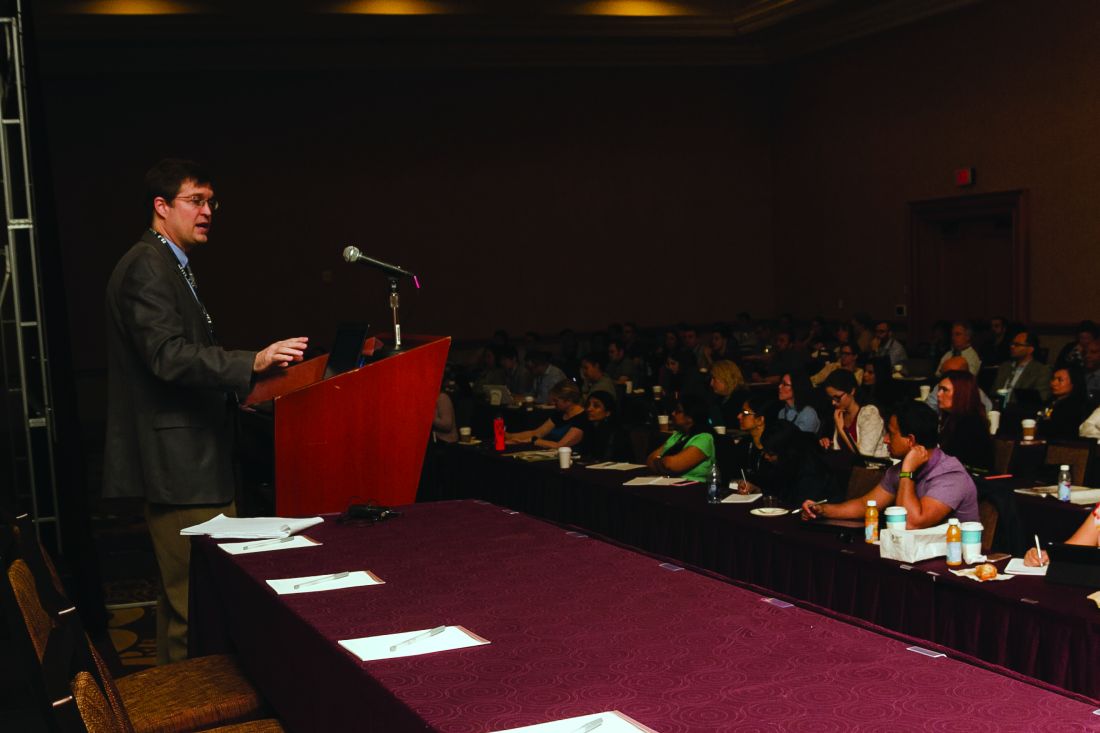
Trends in value-based care favor hospitalists, expert says
Value-based care’s ascendancy equals increased opportunities for hospitalists who know how to wield informatics, speak the language of systems improvement, and have an ability to function as a commodity within a health system, according to the dean of hospital medicine.
“If I were a betting man, I’d say there is no way value-based care can happen without hospitalists,” Robert Wachter, MD, MHM, said during a leadership summit at this year’s annual meeting of the Society of Hospital Medicine. Dr. Wachter is the New York Times bestselling author of “The Digital Doctor: Hope, Hype, and Harm at the Dawn of Medicine’s Computer Age.” He also is a professor and chair of the department of medicine at the University of California, San Francisco.

“There will be talk about closing hospitals and building competencies in ambulatory care. That’s the first 5 minutes, but then there is the realization people are still going to get sick, representing a massive amount of cost, and as far as I can tell, there is no alternative to having hospitals for this,” Dr. Wachter said. “Hospitalists will come out just fine.”
This will be true particularly for hospitalists who understand that electronic health records companies have helped health systems collect massive amounts of data but do not offer help for how those data can be applied to drive better outcomes and improve performance ratings, according to Dr. Wachter.
“These companies have no competencies in data analytics or data visualization,” Dr. Wachter said. “I am hopeful because Silicon Valley has woken up to the possibilities of this and are looking for ways to partner with health care. … The world of improving value, safety, and patient experience is going to be a world enabled by thoughtful use of informatics. So, if you don’t have those competencies, I think it’s important to grow them over time.”
Speaking the language of “lean” or that of other systematic strategies for improvement will also increase the value a hospitalist can bring to an organization, according to Dr. Wachter, who said that as hospital senior leadership and frontline personnel work together to implement system improvement, new leadership roles for hospitalists, ranging from chief patient experience officer to chief quality officers, and everything in between, are being created.
Another “hopeful trend” for hospitalists is the growing focus on the provider experience, said Dr. Wachter, since improving systems is not possible if staff are not engaged. “It’s impossible to believe you’re going to accomplish your goals of improving care and improving patient experience, and efficiencies, with a cadre of burned-out doctors,” he said.
Dr. Wachter said that to be indispensable, hospitalists need to understand they often are commoditized, offered by their senior leadership as bargaining chips when deals are made with surrounding health systems seeking to outsource some of their hospitalist functions.
“Managing a large community-affiliated network was not a core competency in the past,” Dr Wachter said. “But it will become increasingly important.”
Value-based care’s ascendancy equals increased opportunities for hospitalists who know how to wield informatics, speak the language of systems improvement, and have an ability to function as a commodity within a health system, according to the dean of hospital medicine.
“If I were a betting man, I’d say there is no way value-based care can happen without hospitalists,” Robert Wachter, MD, MHM, said during a leadership summit at this year’s annual meeting of the Society of Hospital Medicine. Dr. Wachter is the New York Times bestselling author of “The Digital Doctor: Hope, Hype, and Harm at the Dawn of Medicine’s Computer Age.” He also is a professor and chair of the department of medicine at the University of California, San Francisco.
“There will be talk about closing hospitals and building competencies in ambulatory care. That’s the first 5 minutes, but then there is the realization people are still going to get sick, representing a massive amount of cost, and as far as I can tell, there is no alternative to having hospitals for this,” Dr. Wachter said. “Hospitalists will come out just fine.”
This will be true particularly for hospitalists who understand that electronic health records companies have helped health systems collect massive amounts of data but do not offer help for how those data can be applied to drive better outcomes and improve performance ratings, according to Dr. Wachter.
“These companies have no competencies in data analytics or data visualization,” Dr. Wachter said. “I am hopeful because Silicon Valley has woken up to the possibilities of this and are looking for ways to partner with health care. … The world of improving value, safety, and patient experience is going to be a world enabled by thoughtful use of informatics. So, if you don’t have those competencies, I think it’s important to grow them over time.”
Speaking the language of “lean” or that of other systematic strategies for improvement will also increase the value a hospitalist can bring to an organization, according to Dr. Wachter, who said that as hospital senior leadership and frontline personnel work together to implement system improvement, new leadership roles for hospitalists, ranging from chief patient experience officer to chief quality officers, and everything in between, are being created.
Another “hopeful trend” for hospitalists is the growing focus on the provider experience, said Dr. Wachter, since improving systems is not possible if staff are not engaged. “It’s impossible to believe you’re going to accomplish your goals of improving care and improving patient experience, and efficiencies, with a cadre of burned-out doctors,” he said.
Dr. Wachter said that to be indispensable, hospitalists need to understand they often are commoditized, offered by their senior leadership as bargaining chips when deals are made with surrounding health systems seeking to outsource some of their hospitalist functions.
“Managing a large community-affiliated network was not a core competency in the past,” Dr Wachter said. “But it will become increasingly important.”
VIDEO: Dr. Lisa Newman on triple negative breast cancer in African American women
LAS VEGAS – The heavy burden of triple negative and other aggressive breast cancers among African American women cannot be simplified to socioeconomic factors alone.
International investigations by Lisa Newman, MD, director of the Breast Oncology Program at the Henry Ford Health System, Detroit, and other researchers are making it clear that genetic factors play a significant role.
She explained the latest findings and what they mean for screening, genetic referral, and treatment in an interview at the American Society of Breast Surgeons annual meeting.
The video associated with this article is no longer available on this site. Please view all of our videos on the MDedge YouTube channel
AT ASBS 2017

Attendees drill down on infections at ID boot camp
No hands went up when Glenn Wortmann, MD, chief of infectious diseases at MedStar Washington Hospital Center, asked whether any of the hospitalists in front of him had handled any cases of Candida auris, a kind of yeast that is highly resistant to several potent antifungals and, in some cases, has been found to be resistant to every antifungal thrown at it.
They might not have seen this dreadful bug, first identified in Japan in 2009, yet. But they will soon, Dr. Wortmann said.
James Pile, MD, SFHM, a pre-course director and associate professor of internal medicine at Case Western Reserve University, Cleveland, said he expected that most of those who signed up already knew quite a bit about I.D. issues, but the point of the course was to go deeper.
“Our goal was to find the sweet spot in what they don’t know or need a refresher on,” he said.
In his talk, he focused on infectious disease emergencies – such as infective endocarditis – which hospitalists may not see as often but that “we just have to get right when we do see them because the stakes are very high.”
He covered spinal epidural abscess, bacterial meningitis, and soft tissue necrotizing infections, saying that a crucial element in managing these cases is to seriously consider them a possibility in the first place. He also recommended having a low threshold for obtaining a surgical consultation, a CT scan, or both in patients with what appears to be severe cellulitis.
John Sanders, MD, MPH, head of infectious diseases at Wake Forest Baptist Health, Winston-Salem, N.C., dug into the nitty-gritty of C. difficile infections, touching on the possible role of acid suppressants, especially proton pump inhibitors, in the increasing incidence of these infections. The news wasn’t all bad: He also discussed emerging therapies, such as CRS3123 – a narrow-spectrum antibiotic that inhibits protein synthesis, toxin production, and sporulation – and monoclonal antibodies, which have been shown to lower recurrence rates when used with antibiotics.
The keys to controlling C. diff infections, he said, are hand-washing, remembering that the spores are resistant to ethanol, limiting fluoroquinolone use, and isolating patients with active infections.
Ultraviolet lighting for C. diff control is a gray area, he said.
“The data [are] mixed,” he said. “I think it’s a good idea to do it, and it’s certainly being pushed. But it’s somewhat controversial as to whether it’s cost effective.”

Presidential address focused on learning and teams
Thoralf M. Sundt III, MD, titled his AATS Presidential Address, “Ancora Imparo: Always Learning,” to emphasize the fact that learning is at the core of team development and of dealing with a complex world.
The world once seemed simple, deterministic – a Newtonian world of individual performance, he said. But in fact, it is a complex, unpredictable world, where the solutions of a previous generation of leaders that relied on individual knowledge and authority no longer apply.
“Complex is not the same as complicated,” said Dr. Sundt. “A watch is complicated, but it is entirely predictable. Complexity is different.” Complex systems are composed of many diverse autonomous parts. They are independent, they are linked as a system, and most important – they adapt. They change in response to the environment and to their own component parts.
How they change depends critically on the nature of the interactions among those components, allowing them to demonstrate emergent properties. And they are unpredictable too, said Dr. Sundt.
This unpredictability poses problems, he added, pointing to the inevitability of systems failures in truly complex endeavors from nuclear power to petrochemical plants.
“The implication of this is that we must think beyond error prevention because we simply cannot prevent them all,” said Dr. Sundt. “That is why checklists alone will not solve our problems.” The focus, instead, he suggested, needs to be on “error management” in order to include detection and recovery.
This is all relevant to surgeons because “our world, our patients, the procedures we perform, and the institutions in which we performed them – are increasingly complex.” Therefore, “no matter how much we care, and no matter how much we try, accidents, mistakes and mishaps will occur – individual effort is not enough.”
But complexity is only half the problem, he said. “The other half is us.”
Dr. Sundt discussed two ways of thinking – fast and slow – with fast thinking grabbing on to patterns and making quick, seemingly intuitive decisions, such as those made by “experts,” and the slow following a deliberative and logical path.
Both types of thinking have their benefits and lacks, which points to how important it is to have access to each type in order to maximize success. Thus, he pointed out, there must be a focus on teams and on a diversity of views, because diversity increases the chances of finding the best good solution.
So diversity is necessary, but is it sufficient? Two other things are necessary, according to Dr. Sundt.
The first is synergy, which is the difference between another useless administrative meeting and an exciting, generative one during which everyone shares opinions, changes their views a bit, and collectively comes up with an entirely original idea.
“But the key to transforming a workgroup to a high-performing team is learning,” he said. “It is an active process that requires both cognitive, and - unfortunately for us task-oriented personalities – affective skills. The aim is to create a ‘learning organization’ – a culture that encourages learning through open interactions in a psychologically safe space.
“It is entirely within our power to change the nature of the interactions we have within our teams, and we can do it today. No hospital committee or departmental approval is required. You cannot solve all of the problems, but you do have an enormous impact on those individuals around you.”
As the leader of a team, “everyone is watching you.” he added.
Dr. Sundt concluded by pointing out that it is also extremely important to confront inevitable failure as a learning experience. “You must wring every drop of learning out of it,” he counseled. And ultimately, “trust your team - you need them. The patient needs them,” he concluded.
Routine surveillance adds cost but little benefit following a pediatric tumor
MONTREAL – It can be a tough sell to worried patients or their caregivers, but surveillance imaging in children with a history of some treated solid tumors may not add anything to care except costs, a team of investigators suggested.
A study of the relationship between surveillance imaging and outcomes in patients who were followed after therapy for solid tumors found that “off therapy surveillance imaging did not affect outcomes in our relapsed solid tumor patients,” wrote Jacob Zimmerli and his coinvestigators at the University of Utah Huntsman Cancer Institute, Salt Lake City.
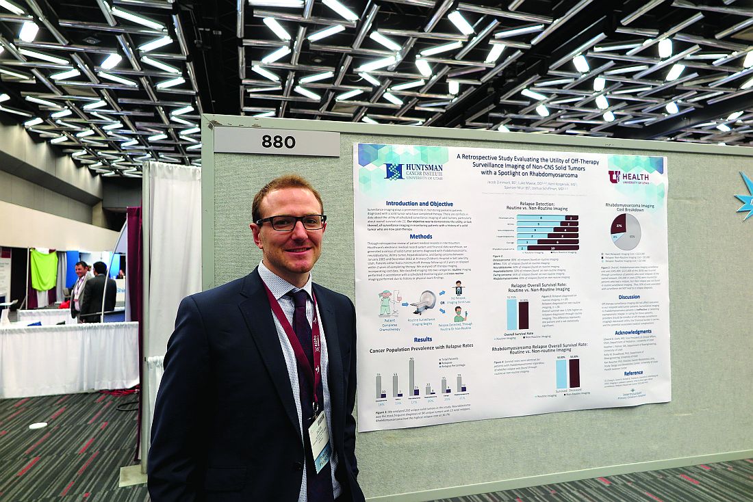
“In the case of rhabdomyosarcoma, we had a 31% relapse rate of our 39 unique cancer diagnoses that we looked at over a 10-year period. We had 12 relapses, and, of those relapses, 10 were found through nonroutine imaging,” Mr. Zimmerli said in an interview.
Survival rates for patients with rhabdomyosarcoma were identical whether their relapsed disease was detected via surveillance imaging or imaging performed when relapse was directly suspected because of clinical or physical findings, he said.
Surveillance imaging also came at a significant financial cost. Of the $345,000 total expenditure for imaging of rhabdomyosarcoma, $223,000 was spent on imaging for patients who never experienced a relapse. Additionally, $95,000 was spent on imaging for patients who had a relapse but whose relapses were not detected by routine surveillance imaging. Therefore, the investigators calculated, 92% of costs associated with surveillance did not lead to a relapse diagnosis.
“This finding in itself will lead us to look at a few more specific cancer subgroups and maybe do more cost analysis to see if there is a better way to treat these patients,” Mr. Zimmerli said.
To see whether surveillance imaging was associated with outcomes, the investigators performed a retrospective review of data on 292 patients treated at Primary Children’s Hospital in Salt Lake City for osteosarcoma, Wilms’ tumor, neuroblastoma, hepatoblastoma, Ewing sarcoma , or rhabdomyosarcoma.
All patients had either completed a minimum of 2 years of posttherapy follow-up or had a disease relapse within 2 years of completing therapy.
The investigators found that 8 of 10 osteosarcoma relapses, five of seven Wilms’ tumor relapses, and 9 of 15 neuroblastoma relapses were detected on routine imaging.
In contrast, only two of four hepatoblastoma relapses, three of nine Ewings sarcoma relapses, and 2 of 12 rhabdomyosarcoma relapses were found on routine imaging.
The overall survival rate for patients with relapses diagnosed on routine imaging was 51.72%, compared with 50% for patients whose relapses were diagnosed on imaging following the appearance of symptoms. The difference represented only one patient and was not statistically significant.
Mr. Zimmerli acknowledged that many routine posttherapy imaging studies are performed to assuage patient concerns and that it may be difficult to convince parents of children with a history of cancer that routine imaging of some tumors may not offer the assurances of relapse-free survival that they seek.
The study was supported by Intermountain Healthcare. The investigators reported no conflicts of interest.
MONTREAL – It can be a tough sell to worried patients or their caregivers, but surveillance imaging in children with a history of some treated solid tumors may not add anything to care except costs, a team of investigators suggested.
A study of the relationship between surveillance imaging and outcomes in patients who were followed after therapy for solid tumors found that “off therapy surveillance imaging did not affect outcomes in our relapsed solid tumor patients,” wrote Jacob Zimmerli and his coinvestigators at the University of Utah Huntsman Cancer Institute, Salt Lake City.
“In the case of rhabdomyosarcoma, we had a 31% relapse rate of our 39 unique cancer diagnoses that we looked at over a 10-year period. We had 12 relapses, and, of those relapses, 10 were found through nonroutine imaging,” Mr. Zimmerli said in an interview.
Survival rates for patients with rhabdomyosarcoma were identical whether their relapsed disease was detected via surveillance imaging or imaging performed when relapse was directly suspected because of clinical or physical findings, he said.
Surveillance imaging also came at a significant financial cost. Of the $345,000 total expenditure for imaging of rhabdomyosarcoma, $223,000 was spent on imaging for patients who never experienced a relapse. Additionally, $95,000 was spent on imaging for patients who had a relapse but whose relapses were not detected by routine surveillance imaging. Therefore, the investigators calculated, 92% of costs associated with surveillance did not lead to a relapse diagnosis.
“This finding in itself will lead us to look at a few more specific cancer subgroups and maybe do more cost analysis to see if there is a better way to treat these patients,” Mr. Zimmerli said.
To see whether surveillance imaging was associated with outcomes, the investigators performed a retrospective review of data on 292 patients treated at Primary Children’s Hospital in Salt Lake City for osteosarcoma, Wilms’ tumor, neuroblastoma, hepatoblastoma, Ewing sarcoma , or rhabdomyosarcoma.
All patients had either completed a minimum of 2 years of posttherapy follow-up or had a disease relapse within 2 years of completing therapy.
The investigators found that 8 of 10 osteosarcoma relapses, five of seven Wilms’ tumor relapses, and 9 of 15 neuroblastoma relapses were detected on routine imaging.
In contrast, only two of four hepatoblastoma relapses, three of nine Ewings sarcoma relapses, and 2 of 12 rhabdomyosarcoma relapses were found on routine imaging.
The overall survival rate for patients with relapses diagnosed on routine imaging was 51.72%, compared with 50% for patients whose relapses were diagnosed on imaging following the appearance of symptoms. The difference represented only one patient and was not statistically significant.
Mr. Zimmerli acknowledged that many routine posttherapy imaging studies are performed to assuage patient concerns and that it may be difficult to convince parents of children with a history of cancer that routine imaging of some tumors may not offer the assurances of relapse-free survival that they seek.
The study was supported by Intermountain Healthcare. The investigators reported no conflicts of interest.
FROM ASPHO
Key clinical point: Routine surveillance was less effective at detecting relapse of some pediatric solid tumors than imaging performed to confirm a clinical finding.
Major finding: Using imaging performed after symptoms appeared, 10 of 12 rhabdomyosarcoma relapses were detected.
Data source: A retrospective review of outcomes following routine and nonroutine imaging among children followed after treatment for one of six solid tumor types.
Disclosures: The study was supported by Intermountain Healthcare. The investigators reported no conflicts of interest.
5-Fluorouracil failed four separate measures of photoaging
PORTLAND – A standard course of topical 5-fluorouracil (5-FU) does not noticeably improve visual signs of facial photoaging, such as forehead lines and crow’s feet, according to the results of a blinded, controlled study of 281 elderly white men.
Four validated photonumeric measures revealed no statistically significant differences between the intervention and vehicle control arms at 6, 12, or 18 months’ follow-up, Kaveri Korgavkar, MD, said at the annual meeting of the Society for Investigative Dermatology. “This might be a true lack of impact, or current scales may not be sensitive enough to capture aspects of aging that are improved by 5-fluorouracil,” commented Dr. Korgavkar, who presented the findings on behalf of the VAKCCT (the Veterans Affairs Keratinocyte Carcinoma Chemoprevention Trial) work group.
The treatment and control groups resembled each other demographically and clinically at baseline. Participants averaged 71.5 years of age (standard deviation, 0.57 years), 97% were male, 99% were white, and all had clinically meaningful histories of sun damage with at least two keratinocyte carcinomas in the previous 2 years, including at least one lesion on the face or ears. Previously, the VAKCCT investigators reported positive results for 5-FU as a chemopreventive – for example, it was associated with about a 60% reduction in actinic keratoses, compared with placebo, and the effects persisted for up to 3 years.
However, none of the four photonumeric scales of photoaging uncovered significant differences between the treatment and control groups at 6, 12, or 18 months’ follow-up, Dr. Korgavkar reported. That finding belies the results of two other previous studies, but they were small and uncontrolled, she added. One study of 19 patients reported statistically significant improvements over time in wrinkling, hyperpigmentation, lentigines, and sallowness based on the Griffith’s scale, while a second prospective study of 32 patients reported significant improvements in visual signs of photoaging on the forearms, with a corresponding rise in levels of procollagen 1 and a decrease in dermal elastosis at 1 month.
Existing scales might more effectively capture some aspects of photoaging – such as wrinkles or crow’s feet – than others, Dr. Korgavkar said in an interview. Therefore, she and her associates are working to construct more sensitive and comprehensive visual scales of photoaging, she said.
The VAKCCT was sponsored by the VA Office of Research and Development. Dr. Korgavkar had no conflicts of interest.
AT SID 2017
Key clinical point: A standard topical course of 5-fluorouracil did not noticeably improve visual signs of photoaging, such as forehead lines and crow’s feet.
Major finding: Four validated photonumeric measures of photoaging revealed no statistically significant differences between the intervention and the vehicle control at 6, 12, or 18 months’ follow-up.
Data source: An analysis of data from 281 participants in the Veterans Affairs Keratinocyte Carcinoma Chemoprevention trial (VAKCCT).
Disclosures: The VAKCCT was sponsored by the VA Office of Research and Development. Dr. Korgavkar had no conflicts of interest.
Durvalumab approved for advanced urothelial carcinoma
for patients with locally advanced or metastatic urothelial carcinoma who have disease progression after prior treatment with a platinum-containing chemotherapy.
The agency also approved a complementary diagnostic for the assessment of the PD-L1 protein the tumor tissue.
The most common adverse reactions were fatigue, musculoskeletal pain, constipation, decreased appetite, nausea, peripheral edema, and urinary tract infection. Pneumonitis, hepatitis, colitis, thyroid disease, adrenal insufficiency, and diabetes also occurred in patients taking durvalumab.
The recommended dose of durvalumab is 10 mg/kg IV over a period of 60 minutes, every 2 weeks, until disease progression or unacceptable toxicity occurs. Full prescribing information is available here.
Durvalumab is marketed as Imfinzi by AstraZeneca. The complementary diagnostic is the Ventana PD-L1 (SP263) Assay from Ventana Medical Systems.
Spontaneous coronary artery dissection: New insights
Washington – Patients with spontaneous coronary artery dissection as their presentation of acute coronary syndrome have a markedly better long-term survival rate than do patients with ACS in its various other forms, Rahul Potluri, MD, reported at the annual meeting of the American College of Cardiology.
In his retrospective cohort study of 182 U.K. patients diagnosed with spontaneous coronary artery dissection (SCAD) as the cause of their ACS and 32,981 controls with ACS without SCAD, the 15-year all-cause mortality rate was 10.4% in the SCAD group vs. 32.1% in the controls.
Although SCAD was first described in 1931, half of the roughly 1,500 cases reported in the medical literature have been published within the past 5 years. That makes this series of 182 SCAD patients with 15-year outcome data unique in providing the longest follow-up to date reported in a sizable patient series, said Dr. Potluri of the University of Alberta, Edmonton, and Aston University in Birmingham, England.

SCAD is a rupture in a coronary artery wall not related to atherosclerotic heart disease, trauma, or iatrogenic causes. The dissection is thought to result from an intimal tear or medial hemorrhage from the vasa vasorum, with subsequent accumulation of blood in a false lumen.
It’s not entirely unexpected that patients with SCAD have a better long-term prognosis than do those with ACS without SCAD, Dr. Potluri said. After all, patients with SCAD tend to be considerably younger: In Dr. Potluri’s series, the average age was younger than 52 years, vs. 66 years in controls. Plus they had lower levels of cardiovascular risk factors, and by definition they were free of atherosclerotic heart disease. But SCAD often occurs in conjunction with fibromuscular dysplasia, and the long-term health implications of that combination have not been well studied.
In a multivariate logistic regression analysis adjusted for age, sex, ethnicity, and comorbid conditions, the likelihood of dying within 5 years was 89% greater in the non-SCAD group. During follow-up, the non-SCAD ACS group was 4.1-fold more likely to have another ACS and 32% more likely to be diagnosed with heart failure.
SCAD was typically managed conservatively in Dr. Potluri’s series: In the SCAD group, 11% underwent PCI and 2.7% had CABG, compared with 51% and 10.7%, respectively, of controls.
“Conservative treatment appears to be safe in the SCAD patient population,” he said.
The prevalence of SCAD in his series of more than 33,000 patients admitted for ACS to U.K. hospitals in 2000-2014 was 0.54%. Dr. Potluri said that low figure likely reflects considerable underreporting due to the fact that some data were from the early 2000s, when SCAD was still largely below physicians’ radar.
Dr. Potluri is an authority on big data analytics in medical research. He formed his 182-patient SCAD series using ACALM (Algorithm for Comorbidity, Associations, Length of Stay, and Mortality), an analytic tool he developed years ago as a medical student. At the time of his SCAD study, the ACALM registry included anonymous, deidentified data on 1.8 million U.K. patients. The ACALM methodology utilizes a form of artificial intelligence known as novel field derivation to analyze huge amalgamated data sets. This is made possible by the fact that all U.K. hospitals report patient data in an identical standardized way, which is not the case in the United States.
Dr. Potluri has previously applied the ACALM methodology to a variety of other health care issues, including studies of an association between hyperlipidemia and breast cancer, the relationship between cardiovascular disease and mental health, and the so-called weekend effect, whereby U.K. patients hospitalized for cardiovascular reasons on the weekend have higher mortality than do those admitted on weekdays.
THE ACALM registry now exceeds 4 million patients. Dr. Potluri said he plans to analyze this full group to develop a large, comprehensive SCAD patient registry. This will enable investigators to evaluate potential psychosocial risk factors for SCAD, the role of fibromuscular dysplasia and connective tissue diseases, and other practical questions.
He reported having no financial conflicts regarding his study.
Washington – Patients with spontaneous coronary artery dissection as their presentation of acute coronary syndrome have a markedly better long-term survival rate than do patients with ACS in its various other forms, Rahul Potluri, MD, reported at the annual meeting of the American College of Cardiology.
In his retrospective cohort study of 182 U.K. patients diagnosed with spontaneous coronary artery dissection (SCAD) as the cause of their ACS and 32,981 controls with ACS without SCAD, the 15-year all-cause mortality rate was 10.4% in the SCAD group vs. 32.1% in the controls.
Although SCAD was first described in 1931, half of the roughly 1,500 cases reported in the medical literature have been published within the past 5 years. That makes this series of 182 SCAD patients with 15-year outcome data unique in providing the longest follow-up to date reported in a sizable patient series, said Dr. Potluri of the University of Alberta, Edmonton, and Aston University in Birmingham, England.
SCAD is a rupture in a coronary artery wall not related to atherosclerotic heart disease, trauma, or iatrogenic causes. The dissection is thought to result from an intimal tear or medial hemorrhage from the vasa vasorum, with subsequent accumulation of blood in a false lumen.
It’s not entirely unexpected that patients with SCAD have a better long-term prognosis than do those with ACS without SCAD, Dr. Potluri said. After all, patients with SCAD tend to be considerably younger: In Dr. Potluri’s series, the average age was younger than 52 years, vs. 66 years in controls. Plus they had lower levels of cardiovascular risk factors, and by definition they were free of atherosclerotic heart disease. But SCAD often occurs in conjunction with fibromuscular dysplasia, and the long-term health implications of that combination have not been well studied.
In a multivariate logistic regression analysis adjusted for age, sex, ethnicity, and comorbid conditions, the likelihood of dying within 5 years was 89% greater in the non-SCAD group. During follow-up, the non-SCAD ACS group was 4.1-fold more likely to have another ACS and 32% more likely to be diagnosed with heart failure.
SCAD was typically managed conservatively in Dr. Potluri’s series: In the SCAD group, 11% underwent PCI and 2.7% had CABG, compared with 51% and 10.7%, respectively, of controls.
“Conservative treatment appears to be safe in the SCAD patient population,” he said.
The prevalence of SCAD in his series of more than 33,000 patients admitted for ACS to U.K. hospitals in 2000-2014 was 0.54%. Dr. Potluri said that low figure likely reflects considerable underreporting due to the fact that some data were from the early 2000s, when SCAD was still largely below physicians’ radar.
Dr. Potluri is an authority on big data analytics in medical research. He formed his 182-patient SCAD series using ACALM (Algorithm for Comorbidity, Associations, Length of Stay, and Mortality), an analytic tool he developed years ago as a medical student. At the time of his SCAD study, the ACALM registry included anonymous, deidentified data on 1.8 million U.K. patients. The ACALM methodology utilizes a form of artificial intelligence known as novel field derivation to analyze huge amalgamated data sets. This is made possible by the fact that all U.K. hospitals report patient data in an identical standardized way, which is not the case in the United States.
Dr. Potluri has previously applied the ACALM methodology to a variety of other health care issues, including studies of an association between hyperlipidemia and breast cancer, the relationship between cardiovascular disease and mental health, and the so-called weekend effect, whereby U.K. patients hospitalized for cardiovascular reasons on the weekend have higher mortality than do those admitted on weekdays.
THE ACALM registry now exceeds 4 million patients. Dr. Potluri said he plans to analyze this full group to develop a large, comprehensive SCAD patient registry. This will enable investigators to evaluate potential psychosocial risk factors for SCAD, the role of fibromuscular dysplasia and connective tissue diseases, and other practical questions.
He reported having no financial conflicts regarding his study.
At ACC 17
Key clinical point:
Major finding: The 15-year all-cause mortality rate was 10% in patients with spontaneous coronary artery dissection, compared with 32% in those with ACS without spontaneous coronary artery dissection.
Data source: A retrospective cohort study including 15-year outcomes for 182 patients with spontaneous coronary artery dissection and more than 32,000 controls with ACS without dissection.
Disclosures: The study presenter reported having no financial conflicts.
Student-Resident Luncheon offers relaxed networking
The ritual of networking at medical conferences often involves waiting to chat with a speaker after a session or finessing an introduction in a busy hallway. That’s why, at the Student-Resident Luncheon, organized by SHM’s Physicians-in-Training Committee, Tuesday, May 2, at noon, trainees will have a chance to interact with experts in the field in a much more relaxed setting.
The free luncheon will include several tables, each dedicated to a specific topic, such as pediatric hospital medicine, with an experienced hospitalist at each one. The residents and students who attend can choose their table and will have a chance to sit at two different tables. There will also be an “open forum” segment at the end, when the trainees can seek out other experts, said Darlene Tad-y, MD, committee chair and assistant professor of medicine at the University of Colorado at Denver, Aurora.
“The purpose behind it was to bring our trainees together so that they can meet each other and to bring some of the SHM leaders to them so that they can learn about what [the leaders] do in hospital medicine and see the breadth of work that hospitalists are doing around the country,” she said.
Pediatric hospital medicine, medical education, and global health are three of the confirmed topics that will be covered at the luncheon, Dr. Tad-y said. There will be a maximum of 10 people at each table, including the expert.
“We wanted it to be very immediate for the students and residents who are going to be there,” she said. “It’s a pretty small group setting.”
The event has been getting more popular each year and is now in its third year. SHM said 500 students and residents participated last year. Residents and students who register for the annual meeting receive an invitation to attend the luncheon. Those who decide on-site that they want to attend will have the ability to do so, Dr. Tad-y said.
Such close interaction with people whose literature they may have read can be very helpful for trainees, she said. They can “sit at a table with them and hear their story and learn how they got to where they were. I think it’s quite impactful.
Knee osteoarthritis linked to premature mortality
LAS VEGAS – Knee osteoarthritis was independently associated with increased risk of premature mortality in the largest-ever meta-analysis to examine the issue, Kirsten M. Leyland, PhD, reported at the World Congress on Osteoarthritis.
The meta-analysis used individual patient-level data on 11,954 participants in six prospective observational cohort studies conducted in four countries. The key finding: Subjects with symptomatic and radiographically confirmed knee OA were at 17% greater risk of premature mortality, compared with pain-free, radiographically negative participants, independent of age, sex, and race, according to Dr. Leyland, a musculoskeletal epidemiologist at the University of Oxford (England).
The six prospective cohort studies included in the new analysis are well known in the field of osteoarthritis research: the Framingham (Mass.) Osteoarthritis Study, the Rotterdam (the Netherlands) Study, the Multicenter Osteoarthritis Study (MOST), the Johnston County (N.C.) Osteoarthritis Project, the Tasmanian Older Adult Cohort Study, and the Chingford (U.K.) 1000 Women Study. These studies feature 7.4-23.7 years of prospective follow-up.
In an interview, Dr. Leyland said that the current analysis uses all-cause mortality as the outcome because causes of death are recorded differently in the countries where the studies are taking place. It will take several more years for statisticians to harmonize the cause-of-death data and draw definitive conclusions; however, preliminary analysis suggests the causes of premature mortality in the knee OA patients are disproportionately cardiovascular, she added.
Dr. Leyland reported having no financial conflicts regarding the study, which was supported by Arthritis Research UK.
AT OARSI 2017
Key clinical point:
Major finding: Knee osteoarthritis is independently associated with a 17% increased likelihood of premature mortality.
Data source: This meta-analysis harmonized individual patient-level data drawn from six major prospective, population-based cohort studies worldwide.
Disclosures: The presenter reported having no financial conflicts regarding the study, which was supported by Arthritis Research UK.