User login
FDA approves abaloparatide for postmenopausal osteoporosis
The Food and Drug Administration has approved abaloparatide (Tymlos) for postmenopausal women with osteoporosis at high risk for fracture.
Abaloparatide was approved based on 18-month results from the ACTIVE trial and 6-month results from the ACTIVExtend trial. Patients in the ACTIVE trial showed an relative risk reduction of 86% for new vertebral fractures and 43% for nonvertebral fractures, compared with placebo, according to a statement from manufacturer Radius Health. Results were similar regardless of age, years since menopause, presence or absence of prior fracture (vertebral or nonvertebral), and bone mineral density at baseline.
Find the full statement on the Radius Health website.
On the big stage, SHM leaders discuss pressing issues
When Society of Hospital Medicine president Brian Harte, MD, SFHM, made remarks last year as president-elect, he outlined four areas that call for attention and action.
On Tuesday, as the outgoing president making remarks at the opening plenary session, he traced the progress in those areas, while also airing some concerns as the society moves forward.
On the “absolute necessity” for SHM to reach out and connect with all practicing hospitalists, he reported that the society continued to expand its footprint, making contact with 50,000 hospitalists.
“SHM continues to be a strong professional organization,” Dr. Harte said, noting how the society cleared the 15,000 mark in membership last year. He also emphasized the “big tent” concept – making SHM the home for practitioners in many disciplines – and the importance of leadership.
“Sometimes it feels like everyone thinks of themselves as someone that we have to report to and therefore leadership development continues to be an important driver for our activities,” said Dr. Harte, president of Cleveland Clinic’s Akron General Hospital.

On the need to continue to focus on patient and family-centered care, he said, the curriculum at this meeting and past meetings shows a recognition of how important communication and empathy are.
“By doing so, we support a culture and environment wherein patients and families can actively participate in their care,” he said.
On being involved in shaping the changing healthcare landscape, the SHM board last fall held a retreat with hospitalist leaders to outline a framework for SHM to take advantage of members’ experience and expertise in this effort. The society is also working with the American College of Surgeons on designing an alternative payment model that could be more favorable for hospitals and hospitalists.
On the push for recognition of the hospitalist specialty, the new C6 Medicare billing code for hospitalists was a big step forward.
There are also some “things that keep me up at night,” he said.
“Having to prove our value continuously is absolutely essential, and it worries me that we may not always have this at the fore of our minds,” he said. Things that help him “get back to sleep,” he said, are the youthfulness and forward-thinking nature of hospitalists and the strength of the society.
“While respecting our past,” Dr. Harte said, “we can only be successful in moving forward if we refuse to be too beholden to it.”
President-elect Ron Greeno, MD, MHM, senior adviser for medical affairs at Team Health, in brief remarks, reminded the audience about how hospital medicine itself was a reform intended to deliver better care at lower prices, and that it therefore makes perfect sense for hospitalists to be involved in this latest wave of reform.
He made an enthusiastic call for more hospitalists to be involved.
“We need more – this is a big challenge,” he said. “At the end of the day, it’s going to take more than us knowing how to take care of patients at the bedside. We have to get involved in designing the new delivery system if we’re going to make sure that we actually have a say in the kind of care that our patients get.”
When Society of Hospital Medicine president Brian Harte, MD, SFHM, made remarks last year as president-elect, he outlined four areas that call for attention and action.
On Tuesday, as the outgoing president making remarks at the opening plenary session, he traced the progress in those areas, while also airing some concerns as the society moves forward.
On the “absolute necessity” for SHM to reach out and connect with all practicing hospitalists, he reported that the society continued to expand its footprint, making contact with 50,000 hospitalists.
“SHM continues to be a strong professional organization,” Dr. Harte said, noting how the society cleared the 15,000 mark in membership last year. He also emphasized the “big tent” concept – making SHM the home for practitioners in many disciplines – and the importance of leadership.
“Sometimes it feels like everyone thinks of themselves as someone that we have to report to and therefore leadership development continues to be an important driver for our activities,” said Dr. Harte, president of Cleveland Clinic’s Akron General Hospital.
On the need to continue to focus on patient and family-centered care, he said, the curriculum at this meeting and past meetings shows a recognition of how important communication and empathy are.
“By doing so, we support a culture and environment wherein patients and families can actively participate in their care,” he said.
On being involved in shaping the changing healthcare landscape, the SHM board last fall held a retreat with hospitalist leaders to outline a framework for SHM to take advantage of members’ experience and expertise in this effort. The society is also working with the American College of Surgeons on designing an alternative payment model that could be more favorable for hospitals and hospitalists.
On the push for recognition of the hospitalist specialty, the new C6 Medicare billing code for hospitalists was a big step forward.
There are also some “things that keep me up at night,” he said.
“Having to prove our value continuously is absolutely essential, and it worries me that we may not always have this at the fore of our minds,” he said. Things that help him “get back to sleep,” he said, are the youthfulness and forward-thinking nature of hospitalists and the strength of the society.
“While respecting our past,” Dr. Harte said, “we can only be successful in moving forward if we refuse to be too beholden to it.”
President-elect Ron Greeno, MD, MHM, senior adviser for medical affairs at Team Health, in brief remarks, reminded the audience about how hospital medicine itself was a reform intended to deliver better care at lower prices, and that it therefore makes perfect sense for hospitalists to be involved in this latest wave of reform.
He made an enthusiastic call for more hospitalists to be involved.
“We need more – this is a big challenge,” he said. “At the end of the day, it’s going to take more than us knowing how to take care of patients at the bedside. We have to get involved in designing the new delivery system if we’re going to make sure that we actually have a say in the kind of care that our patients get.”
VIDEO: Low-tech system tweaks help hospitalists minimize workflow disruptions
What are some of the most common interruptions physicians face, and what simple solutions exist to help minimize the breaks in workflow?
Physicians are interrupted, on average, 15 times an hour, according to Roberta Himebaugh, a senior vice president at TeamHealth in Pleasanton, Calif., but as she explains in this video recorded at HM17, there are some simple, low-tech – and other – solutions that health systems can use to help hospitalists streamline workflow.
Urgent care sites cater to cancer patients, letting them check some worries at door
On an afternoon a few weeks ago, Faithe Craig noticed that her temperature had spiked to just above 100 degrees. For most people, the change might not be cause for alarm, but Ms. Craig is being treated for stage III breast cancer, and any temperature change could signal a serious problem.
She called her nurse at the hospital clinic where she gets care at the University of Texas Southwestern Medical Center, Dallas, who told her to come in immediately for cancer urgent-care services at the hospital’s hematology-oncology clinic.
Clinicians had details of her cancer care at their fingertips. “They already knew my story and knew everything about me,” she said. The blood work showed she had severe anemia, requiring a blood transfusion.
It’s been more than a year since the medical center began providing same-day urgent care services to cancer patients. It’s an effort to help them avoid the emergency department and hospital admissions, said Thomas Froehlich, MD, medical director of the center’s cancer clinics.
Cancer treatment “clearly carries a lot of side effects and toxicity, and there are also the complications of dealing with the cancer,” Dr. Froehlich said. “Many of these things, if you can intervene early, you keep patients at home and out of the hospital.”
UT Southwestern isn’t alone. A small but growing number of hospitals and oncology practices are incorporating urgent care aimed specifically at cancer patients, in which specialists are available for same-day appointments, often with extended hours, sometimes 24/7.
Keeping cancer patients out of the ED makes sense not only because many of them have compromised immune systems that put them at risk in a waiting room full of sick people but also in providing them the most efficient and appropriate care.
“What we hear from cancer physicians and administrators is that in the emergency department, not all emergency physicians and nurses feel equally confident in their ability to treat cancer patients,” said Lindsay Conway, managing director of research at the Advisory Board, a health care research and consulting firm. “So they may admit them when it’s not necessary.”
Severe pain, nausea, fever and dehydration are not uncommon side effects of traditional chemotherapy. Newer immunotherapy treatments that activate the immune system to fight cancer can cause serious and sudden reactions if the body instead attacks healthy organs and tissues.
It can be difficult for non–cancer specialists to evaluate what these symptoms mean. “Targeted therapies are wonderful, but if you don’t know the drug, you’re going to have a hard time managing the person,” said Barbara McAneny, MD, CEO of New Mexico Oncology Hematology Consultants in Albuquerque, which operates three cancer centers in New Mexico that together provide same-day urgent care services for more than a dozen cancer patients daily.
Offering same-day services fits in with a broader shift in oncology toward patient-centered care, said J. Leonard Lichtenfeld, MD, deputy chief medical officer of the American Cancer Society.
“There’s a general sense within the practice of oncology that we need to do a better job of managing pain and side effects, and we need to provide a higher level of care,” Dr. Lichtenfeld said.
The Centers for Medicare & Medicaid Services is encouraging these efforts through new payment and delivery models designed to reward quality cancer care, Dr. Lichtenfeld said. In addition, starting in 2020, hospitals may be penalized financially if patients who are receiving outpatient chemotherapy visit the ED or are admitted to the hospital, according to a final rule issued in November.
Avoiding the ED makes financial sense for patients and insurers, too.
Johns Hopkins Hospital in Baltimore opened a six-bed urgent care center next to its infusion center a couple of years ago. Of the patients who land there, about 80% are discharged home, at an average total hospital charge of $1,600, said Sharon Krumm, PhD, director of nursing at the Johns Hopkins Kimmel Cancer Center. Only 20% of cancer patients who visit the hospital’s emergency department are discharged home; those who are have an average total hospital charge of $2,300. Patients who are admitted face ED charges plus the hefty cost of a hospital admission.
Rebecca Cohen has been a frequent visitor to the Johns Hopkins urgent care center. Diagnosed more than 2 years ago with stage IV lung cancer, Ms. Cohen, 68, is receiving immunotherapy. She’s been treated or checked for dehydration, electrolyte abnormalities, low hemoglobin, low sodium, blood clots, and infection, among other things.
Before she started going to the cancer urgent care center, “you sat in the waiting room at the emergency room with people who had the most extraordinary diseases,” she said. “Having stage IV lung cancer, the thought of being exposed to pneumonia or bronchitis is more than scary.”
Kaiser Health News is a national health policy news service that is part of the nonpartisan Henry J. Kaiser Family Foundation.
Sutureless, guided lumpectomy produced clean margins in majority
LAS VEGAS – An automated, minimally invasive, stereotactic-guided lumpectomy performed well in an outpatient setting, with no sutures required, potentially decreasing patient morbidity, according to Pat Whitworth, MD.
Pain scores were low, and all high-risk lesions were successfully removed.
The new technology is part of the natural progression of cancer resection, said Dr. Whitworth, director of the Nashville (Tenn.) Breast Center, at the annual meeting of the American Society of Breast Surgeons.
“We went from radical mastectomy, to modified radical mastectomy, to lumpectomy. This is just part of that progression where we match what we do to what the patient really needs,” he said.
Dr. Whitworth said he believes that radiologists will soon be using such technology to treat breast cancer, which puts the onus on breast cancer surgeons to adopt it themselves. “I think it’s important for breast surgeons to acquire the necessary skill and techniques to use the same tools and work collaboratively with radiologists, because this is coming,” he said.
To see if it were possible to achieve results similar to those with lumpectomy, Dr. Whitworth analyzed data from 279 women who had a small ductal carcinoma in situ (DCIS), invasive carcinoma, or high-risk lesion removed using a 15- or 20-mm radiofrequency basket capture with imaging guidance (lumpectomy). Patients who received a cancer diagnosis underwent a second, 20-mm basket capture to obtain shaved margins.
The procedure was conducted under local anesthesia and sedation, and the incisions were closed using Steri-Strip skin closures. The average pain score was 1.55 out of 10 (range, 0-7).
Of 125 patients found to have DCIS (n = 52) and invasive lesions (n = 73), the first capture achieved clear margins in 69 cases, and the shaved margin capture achieved clean margins in another 33 cases.
The remaining 23 patients (18%) had a positive margin by histologic standards following lumpectomy and shaved margin. Of the 22 with reported results, 17 (77%) had no residual lesion following open surgery.
The results convinced Dr. Wentworth of the technique’s utility, particularly in patients who may have a heightened surgical risk, he said. “We think this can replace open lumpectomy in selected patients, with favorable margin clearance.”
The approach is fairly simple, but Dr. Wentworth recommended beginning with stereotactic-guided technology before attempting the ultrasound-guided version, which requires a little more skill. “The biggest challenge is learning that you have to use a lot more local anesthetic. When this technology first came out, people tried to use the same amount of local anesthetic that they used for standard vacuum-assisted core biopsy, and it’s very painful unless you put 30 or 40 cc of dilute anesthetic in there,” said Dr. Wentworth.
The study was funded by Medtronic, which markets the technology. Dr. Whitworth is a principal at Targeted Medical Education, which receives funding from Medtronic.
AT ASBS 2017
Key clinical point: The technique could replace standard lumpectomy in patients at high surgical risk.
Major finding: Clean margins were obtained in 102 of 125 women with diagnosed cancer.
Data source: A retrospective analysis of 279 patients.
Disclosures: The study was funded by Medtronic, which markets the technology. Dr. Whitworth is a principal at Targeted Medical Education, which receives funding from Medtronic.
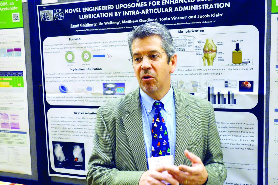
Novel agent brings clinically meaningful improvements in knee osteoarthritis
LAS VEGAS – The investigational agent FX006 brought improvements in pain and function in patients with knee osteoarthritis that were not only statistically significant but also clinically meaningful by three different yardsticks, according to Scott Kelley, MD.
FX006 (Zilretta) is an extended-release, microsphere-based formulation of triamcinolone acetonide for intra-articular injection now under review for marketing approval by the Food and Drug Administration, which has said it will render a decision in October, Dr. Kelley reported at the World Congress on Osteoarthritis.
He presented a post hoc pooled analysis of the 798 patients with knee OA who participated in two phase II randomized, double-blind clinical trials and in an identically designed pivotal phase III trial of a single 40-mg intra-articular injection of FX006. The three studies included 324 patients who got FX006, 212 who received a single 40-mg intra-articular injection of standard triamcinolone acetonide crystalline suspension, and 262 who got a placebo injection of 5 mL of saline.
All participants had radiographically documented symptomatic knee OA with baseline daily pain scores of 6-9 on a 0-10 scale, corresponding to pain in the moderate-to-severe range. Patients telephoned in their average daily pain scores every day during the studies, one of which lasted 12 weeks while the other two ran for 24 weeks. Patients also visited their clinician every 4 weeks for Western Ontario and McMaster Universities Osteoarthritis Index (WOMAC) assessments of pain, stiffness, and physical function.
This secondary analysis was undertaken to demonstrate that FX006 distinguishes itself from current nonsurgical treatments for knee OA, which typically achieve clinical effects that are small, transient, and not clinically relevant, Dr. Kelley said at the congress sponsored by the Osteoarthritis Research Society International.
“What’s different about this product formulation is it maintains a prolonged release of triamcinolone acetonide inside the synovial fluid. The pharmacology has demonstrated prolonged residence time in synovial fluid out to at least 12 weeks after intra-articular administration. In addition, it mitigates the peak plasma level after intra-articular administration, so it blunts the level of corticosteroid in plasma and has a lower systemic exposure over time,” he said.
The clinical relevance of the outcomes achieved in the pooled studies was assessed using three different metrics: the American Academy of Orthopaedic Surgeons (AAOS) criteria for determining whether a result represents a minimal clinically important improvement; the Outcomes Measures in Rheumatology–Osteoarthritis Research Society International (OMERACT-OARSI) “strict responder” definition; and the Initiative on Methods, Measurement, and Pain Assessment in Clinical Trials (IMMPACT)–defined proportion of patients achieving substantial improvement in pain.
At the 4- and 8-week follow-up visits post injection, only the FX006 group achieved the AAOS threshold of improvement that’s deemed both statistically and clinically significant. Standard triamcinolone couldn’t reach that bar, even at week 4. This makes FX006 the first intra-articular treatment for knee OA to achieve this stringent standard for a clinically meaningful effect. By week 12, the improvement in pain in the FX006 group had slipped to the level of ‘‘statistically and possibly clinically significant.” At weeks 16-24 following a single injection, the FX006 results were no longer statistically or clinically significant.
A significantly greater percentage of the FX006 group met the OMERACT-OARSI strict responder definition of substantial pain improvement at weeks 4, 8, and 12, compared with the standard triamcinolone and placebo groups. The degree of pain improvement in the FX006 group remained superior to placebo through week 16. The OMERACT-OARSI definition of substantial improvement requires at least a 50% improvement in the WOMAC-A pain score plus an absolute improvement of 20 points or more on the WOMAC-A pain score or WOMAC-C physical function 100-point normalized scale.
Substantial clinical improvement as defined by the IMMPACT criteria requires at least a 50% improvement from baseline in the WOMAC-A pain score. Roughly 60% and 50% of the FX006 group met that standard at weeks 4 and 8, respectively, rates that were significantly higher than for standard triamcinolone. The FX006 group’s IMMPACT substantial improvement rate significantly exceeded that of placebo-treated controls throughout all 24 weeks.
These studies as well as the pooled analysis were funded by Flexion Therapeutics.
FX006 is a type of steroid for joint injection that is formulated to stay in the joint longer than typical steroids through the use of microsphere technology. Typical intra-articular steroid injections tend to provide benefit over the short term, and as described by the Cochrane review, the greatest effect is only moderate at 1-2 weeks and decreases from there, with small effects at 13 weeks and none at 26 weeks (Cochrane Database Syst Rev. 2015;10:CD005328).

As we have previously reported (Semin Arthritis Rheum. 2014;43:701-12), intra-articular corticosteroids are widely recommended by professional societies for the management of knee OA, although a specific formulation is not generally given. This compound may fill an important treatment gap by extending the efficacy of a treatment that is generally accepted as useful and safe for this common painful condition. Also, given the controversy surrounding other intra-articular therapies, it may provide a safe and effective alternative for patients who have not benefited from, or not tolerated, one or more traditional intra-articular steroid injection(s).
Amanda E. Nelson, MD, is with the division of rheumatology, allergy, and immunology and the Thurston Arthritis Research Center at the University of North Carolina, Chapel Hill. She is a consultant to GlaxoSmithKline and has received honoraria for online presentations for MedScape and QuantiaMD regarding OA treatment guidelines.
FX006 is a type of steroid for joint injection that is formulated to stay in the joint longer than typical steroids through the use of microsphere technology. Typical intra-articular steroid injections tend to provide benefit over the short term, and as described by the Cochrane review, the greatest effect is only moderate at 1-2 weeks and decreases from there, with small effects at 13 weeks and none at 26 weeks (Cochrane Database Syst Rev. 2015;10:CD005328).
As we have previously reported (Semin Arthritis Rheum. 2014;43:701-12), intra-articular corticosteroids are widely recommended by professional societies for the management of knee OA, although a specific formulation is not generally given. This compound may fill an important treatment gap by extending the efficacy of a treatment that is generally accepted as useful and safe for this common painful condition. Also, given the controversy surrounding other intra-articular therapies, it may provide a safe and effective alternative for patients who have not benefited from, or not tolerated, one or more traditional intra-articular steroid injection(s).
Amanda E. Nelson, MD, is with the division of rheumatology, allergy, and immunology and the Thurston Arthritis Research Center at the University of North Carolina, Chapel Hill. She is a consultant to GlaxoSmithKline and has received honoraria for online presentations for MedScape and QuantiaMD regarding OA treatment guidelines.
LAS VEGAS – The investigational agent FX006 brought improvements in pain and function in patients with knee osteoarthritis that were not only statistically significant but also clinically meaningful by three different yardsticks, according to Scott Kelley, MD.
FX006 (Zilretta) is an extended-release, microsphere-based formulation of triamcinolone acetonide for intra-articular injection now under review for marketing approval by the Food and Drug Administration, which has said it will render a decision in October, Dr. Kelley reported at the World Congress on Osteoarthritis.
He presented a post hoc pooled analysis of the 798 patients with knee OA who participated in two phase II randomized, double-blind clinical trials and in an identically designed pivotal phase III trial of a single 40-mg intra-articular injection of FX006. The three studies included 324 patients who got FX006, 212 who received a single 40-mg intra-articular injection of standard triamcinolone acetonide crystalline suspension, and 262 who got a placebo injection of 5 mL of saline.
All participants had radiographically documented symptomatic knee OA with baseline daily pain scores of 6-9 on a 0-10 scale, corresponding to pain in the moderate-to-severe range. Patients telephoned in their average daily pain scores every day during the studies, one of which lasted 12 weeks while the other two ran for 24 weeks. Patients also visited their clinician every 4 weeks for Western Ontario and McMaster Universities Osteoarthritis Index (WOMAC) assessments of pain, stiffness, and physical function.
This secondary analysis was undertaken to demonstrate that FX006 distinguishes itself from current nonsurgical treatments for knee OA, which typically achieve clinical effects that are small, transient, and not clinically relevant, Dr. Kelley said at the congress sponsored by the Osteoarthritis Research Society International.
“What’s different about this product formulation is it maintains a prolonged release of triamcinolone acetonide inside the synovial fluid. The pharmacology has demonstrated prolonged residence time in synovial fluid out to at least 12 weeks after intra-articular administration. In addition, it mitigates the peak plasma level after intra-articular administration, so it blunts the level of corticosteroid in plasma and has a lower systemic exposure over time,” he said.
The clinical relevance of the outcomes achieved in the pooled studies was assessed using three different metrics: the American Academy of Orthopaedic Surgeons (AAOS) criteria for determining whether a result represents a minimal clinically important improvement; the Outcomes Measures in Rheumatology–Osteoarthritis Research Society International (OMERACT-OARSI) “strict responder” definition; and the Initiative on Methods, Measurement, and Pain Assessment in Clinical Trials (IMMPACT)–defined proportion of patients achieving substantial improvement in pain.
At the 4- and 8-week follow-up visits post injection, only the FX006 group achieved the AAOS threshold of improvement that’s deemed both statistically and clinically significant. Standard triamcinolone couldn’t reach that bar, even at week 4. This makes FX006 the first intra-articular treatment for knee OA to achieve this stringent standard for a clinically meaningful effect. By week 12, the improvement in pain in the FX006 group had slipped to the level of ‘‘statistically and possibly clinically significant.” At weeks 16-24 following a single injection, the FX006 results were no longer statistically or clinically significant.
A significantly greater percentage of the FX006 group met the OMERACT-OARSI strict responder definition of substantial pain improvement at weeks 4, 8, and 12, compared with the standard triamcinolone and placebo groups. The degree of pain improvement in the FX006 group remained superior to placebo through week 16. The OMERACT-OARSI definition of substantial improvement requires at least a 50% improvement in the WOMAC-A pain score plus an absolute improvement of 20 points or more on the WOMAC-A pain score or WOMAC-C physical function 100-point normalized scale.
Substantial clinical improvement as defined by the IMMPACT criteria requires at least a 50% improvement from baseline in the WOMAC-A pain score. Roughly 60% and 50% of the FX006 group met that standard at weeks 4 and 8, respectively, rates that were significantly higher than for standard triamcinolone. The FX006 group’s IMMPACT substantial improvement rate significantly exceeded that of placebo-treated controls throughout all 24 weeks.
These studies as well as the pooled analysis were funded by Flexion Therapeutics.
AT OARSI 2017
Key clinical point:
Major finding: The investigational extended-release formulation of triamcinolone, known as FX006 (Zilretta), is the first intra-articular treatment for knee OA to meet the American Academy of Orthopaedic Surgeons’ stringent standard for a clinically meaningful, as opposed to merely statistically significant, effect.
Data source: A post hoc pooled analysis of three randomized, double-blind clinical trials involving nearly 800 patients with knee osteoarthritis.
Disclosures: The pooled analysis was funded by Flexion Therapeutics and presented by a company officer.

Monday’s Lillehei Forum highlighted basic research
The Lillehei Forum is a series of presentations on basic research by residents in the field of cardiothoracic surgery.
A targeted near-infrared contrast agent specific for lung adenocarcinomas was assessed for effectiveness during intraoperative molecular imaging (IMI) in animal models. Use of the contrast agent proved significantly more effective at detecting positive margins than did surgery alone, according to a study presented by Jarrod D. Predina, MD.
Initial human experiences with IMI for NSCLC using a fluorescent contrast agent have been limited by technical hurdles including high background noise in inflammatory tissues and low signal output, according to Dr. Predina. As an alternative, he and his colleagues at the University of Pennsylvania hypothesized that a targeted near-infrared contrast agent specific for lung adenocarcinomas would improve sensitivity and specificity during surgery.
Using a mouse surgical model of non–small cell lung cancer (NSCLC) that recapitulates local and systemic post-operative recurrences, Dr. Predina and his colleagues at the University of Pennsylvania injected 140 mice intravenously prior to resection with a near-infrared imaging agent (OTL0038). This agent is specific for pulmonary adenocarcinomas due to its high affinity binding of the folate receptor alpha. Tumor-bearing mice were randomized to surgery with or without IMI. Suspicious residual disease was resected and analyzed by immunohistochemistry, flow cytometry, and immunofluorescence. Based on this data, OTL0038 was tested in a pilot study of five canines with spontaneously occurring lung cancer.
In a local recurrence model system, of 80 mice assessed, surgeons identified 10 positive margins in those randomized to imaging with IMI vs. three positive margins in mice undergoing surgery alone, a significant difference. In systemic recurrence models (60 mice), the mean number of pulmonary nodules located with IMI was 7.2 vs. 3.4 in controls, also a significant difference, according to Dr. Predina.
In five canines with a presumed diagnosis of NSCLC, no toxicity was observed. Four of five canines had fluorescent tumors; the non-fluorescing tumor was discovered to be a metastatic mammary tumor on final pathologic analysis. In one canine, an otherwise undetectable 8-mm pulmonary adenocarcinoma was discovered with IMI.
“Our data suggest that a targeted near-infrared contrast agent may improve IMI technology. Ultimately, this will enable accurate identification of residual disease that may otherwise be overlooked. These results are the basis of an ongoing phase I human trial,” concluded Dr. Predina.
The complexity of the lung has limited the prospects of bioengineering strategies utilizing stem cells and fully decellularized or bioartificial scaffolds, according to Brandon A. Guenthart, MD, of Columbia University.
Using standard protocols, Dr. Guenthart and his colleagues procured human lungs rejected for transplantation on the basis of standard clinical criteria. Lungs were placed on a custom-built EVLP system, ventilated and perfused. Utilizing video bronchoscopy, real-time transpleural imaging, and a custom designed micro-catheter delivery and occlusion system, the team delivered a decellularization solution into targeted lung regions that resulted in the selective removal of airway epithelium.
Human airway epithelial cells or human-derived alveolar progenitor cells were labeled (with a quantum dot or a near-infrared dye) and delivered into decellularized lung regions. Following delivery, EVLP was continued for 4-6 hours to allow for cell engraftment. Lung wedge samples were collected at each time point for histologic analysis.
Following decellularization, hematoxylin and eosin staining confirmed removal of pseudostratified and columnar epithelium in proximal airways and removal of type I and II pneumocytes in the distal lung. Delivered cells were retained in the lung after EVLP and fixation, and cellular morphology and distribution within the alveoli indicated early engraftment.
“Bioengineering human lungs utilizing advanced therapeutic interventions such as cell replacement may help combat the critical shortage of transplantable lungs,” said Dr. Guenthart. “Additionally, in the future, targeted intervention and patient-specific cell replacement strategies may have translational applications in vivo and eliminate the need for transplantation in select patients,” he concluded.
They used a previously validated porcine lung injury model of intravenous lipopolysaccharide (LPS) to induce a systemic inflammatory response and subsequent severe ARDS requiring ECMO support. A total of eight mature adult swine were administered LPS via the external jugular vein followed by sternotomy and central ECMO cannulation (right atrium to ascending aorta).
The left pulmonary artery (inflow) and left superior and inferior pulmonary veins (outflow) were dissected out and cannulated to isolate the left lung, which then underwent 4 hours normorthermic IVLP with Steen Solution followed by 4 hours of lung reperfusion after IVLP decannulation.
Dr. Mehaffey and his colleagues then compared the right (LPS control) and left lungs (LPS+IVLP) of the same animal.
All animals demonstrated a significant reduction in PaO2/FiO2 ratio and total lung compliance 2 hours after the start of LPS infusion. During IVLP, the left (treated) pulmonary vein oxygenation was superior to right (control) pulmonary vein oxygenation. After reperfusion and IVLP decannulation, six (75%) animals had improved lung function allowing for ECMO decannulation.
The left lung showed significant improvement of lung-specific oxygenation compared to the right control at 4 hours of reperfusion. Similarly, total lung compliance improved after targeted rehabilitation of the left lung, according to Dr. Mehaffey. In addition, the wet-to-dry ratio of lung tissue demonstrated significantly reduced edema in the rehabilitated left lungs compared to right controls.
Dr. Mehaffey and his colleagues concluded that IVLP may reduce the duration of ECMO and ventilator support resulting in major reduction in morbidity, mortality, and health care–related costs in patients with ARDS.
Monday’s Plenary tackled key issues
Presentations of the latest research in cardiothoracic surgery are the hallmark of the AATS Plenary session and this year’s session did not disappoint.
When repairing large paraesophageal hernias, transthoracic Belsey Mark IV fundoplication led to lower rates of leaks and reoperations than did the widely used laparoscopic Nissen fundoplication, according to a study presented by Danuel Laan, MD.
Dr. Laan, a resident at the Mayo Clinic in Rochester, Minn., and his colleagues compared outcomes after Belsey Mark IV fundoplication with those after laparoscopic Nissen fundoplication.
The researchers performed a retrospective review of prospectively collected data on all Mayo patients from 2002 to 2011 who underwent repair of a large paraesophageal hernia, defined as having more than 50% of the stomach within the chest. The analysis excluded patients who had already undergone a fundoplication.
Matching 118 Belsey fundoplication patients 1:1 with 118 Nissen fundoplication patients, the investigators compared the two groups for recurrence, need for reoperation, and perioperative outcomes. Nearly 30% of patients in the Belsey and Nissen groups were male. Mean age was 68.7 years and 69.8 years, respectively. Body mass index was 30.3 kg/m2 and 28.8 kg/m2, respectively.
Ten Belsey patients (8.4%) had a hernia recurrence, as did 19 Nissen patients (16.1%). Rates of leak and reoperation were greater in Nissen fundoplication patients than in Belsey patients. There were no leaks in the Belsey patients, compared with eight leaks (6.8%) in the Nissen patients, a statistically significant difference. Reoperation was necessary in 3 Belsey patients (2.5%), significantly fewer than the 11 Nissen patients (9.3%) who underwent reoperation.
Among 77 patients in each group followed for 5 years or less, there was no difference in symptoms. However, in 41 patients in each group followed for more than 5 years, symptoms were excellent or good in 85% of Belsey patients, compared with 56% of Nissen patients.
AATS: Surgical intervention relatively safe for AAOCA in youth
It’s relatively safe to surgically treat anomalous aortic origin of a coronary artery in children and adolescents, with essentially all patients cleared for full activity after 3 months, according to a study presented by Carlos M. Mery, MD.
Anomalous aortic origin of a coronary artery (AAOCA) is the second leading cause of sudden cardiac death (SCD) in young people, noted Dr. Mery of Texas Children’s Hospital.
He and his colleagues prospectively analyzed outcomes for 44 AAOCA patients who underwent surgical intervention as part of a standardized management program. The researchers included all AAOCA patients aged 2-18 years who underwent surgical intervention between December 2012 and April 2017 as part of the multidisciplinary Coronary Anomalies Program at Texas Children’s Hospital.
Surgical indications included an anomalous left coronary artery, ischemic symptoms, a positive nuclear perfusion test (NPT), or high-risk anatomy such as long intramural segment or ostial stenosis identified via CT angiography. Median patient age was 14 years.
Nine of the patients (20%) underwent surgery for an anomalous left coronary artery, with 32 patients (80%) receiving surgical intervention for an anomalous right coronary artery. A total of 34 surgical procedures (77%) were unroofing of intramural segments, 7 were coronary translocations (16%), and 2 were ostioplasties (4%).
There were no operative deaths. One patient required coronary artery bypass grafting after developing ischemia following a coronary translocation. Minor complications were seen in eight other patients (18%). One patient presented with a second episode of aborted sudden cardiac death 1 year after unroofing of an anomalous left coronary artery with a short intramural segment, and underwent successful coronary translocation and unroofing of a previously identified myocardial bridge.
At follow-up (median 2 years), 40 patients were asymptomatic (91%), while 4 patients had nonspecific chest pain (9%). Forty-one patients (93%) had returned to full activity, while 3 patients were waiting for their 3-month clearance to return to full activity.
Lobectomy can be done safely after concurrent chemotherapy and high-dose radiation in patients with resectable N2-positive stage IIIA non–small-cell lung cancer, according to study findings presented by Jessica S. Donington, MD.

Dr. Donington, of New York University, and her colleagues analyzed two prospective trials conducted by NRG Oncology, RTOG 0229 and RTOG 0839. Both trials’ primary endpoint was mediastinal node sterilization after concurrent chemotherapy and full-dose radiation and has been previously reported.
Dr. Donington and her fellow investigators specifically examined short-term surgical outcomes, given the significant controversy regarding the safety of resection after full-dose thoracic radiation.
In both trials, patients received weekly carboplatin and paclitaxel. Those in the 0229 trial underwent 61.2 Gy of radiation in 34 fractions, while patients in the 0839 trial underwent 60 Gy in 30 fractions. In addition, patients in the 0839 trial were randomized 2:1 to receive weekly panitumumab, an EGFR monoclonal antibody, with their induction therapy.
Surgical expertise was considered essential to this treatment strategy. Therefore, all surgeons were certified by RTOG prior to enrolling patients, and all patients were surgically evaluated before beginning induction therapy to determine resectability and appropriateness for trimodality therapy.
Of 125 eligible patients enrolled in the two trials, 93 patients (74%) underwent anatomic resection. A total of 77 patients underwent lobectomy, 8 underwent pneumonectomy, 6 underwent bilobectomy, and 2 patients had sleeve lobectomy. Medical contraindication and persistent nodal disease found during post-induction invasive staging were the most common reasons patients didn’t undergo resection.
Eighty-five of the 93 surgical patients had R0 resections (91%). Surgeons attempted 14 minimally invasive resections (15%), 2 of which uneventfully converted to open resection.
Just over one-quarter (28%) of patients suffered greater than Grade 3 adverse events (AEs) related to surgery, the majority of which were pulmonary in nature. The 30-day mortality rate was 4%, and all four deaths were linked to pulmonary AEs, including acute respiratory distress syndrome, bronchopleural fistula, pulmonary artery hemorrhage, and respiratory failure. Multivariable analysis for mortality identified the addition of panitumumab and use of an extended resection to be associated with an increased risk for operative mortality.
In the patients undergoing lobectomy, rates for greater than Grade 3 AEs and 30-day mortality were 26%, and 1.3%, respectively. Those rates are similar to rates reported for lobectomy without induction therapy in the National Inpatient Sample (NIS) and the STS General Thoracic Surgery Database over the same time period.
These two RTOG trials are the first to prospectively demonstrate that trimodality therapy with full-dose neoadjuvant radiation therapy is safe in a multi-institutional setting, Dr. Donington said.
Presentations of the latest research in cardiothoracic surgery are the hallmark of the AATS Plenary session and this year’s session did not disappoint.
When repairing large paraesophageal hernias, transthoracic Belsey Mark IV fundoplication led to lower rates of leaks and reoperations than did the widely used laparoscopic Nissen fundoplication, according to a study presented by Danuel Laan, MD.
Dr. Laan, a resident at the Mayo Clinic in Rochester, Minn., and his colleagues compared outcomes after Belsey Mark IV fundoplication with those after laparoscopic Nissen fundoplication.
The researchers performed a retrospective review of prospectively collected data on all Mayo patients from 2002 to 2011 who underwent repair of a large paraesophageal hernia, defined as having more than 50% of the stomach within the chest. The analysis excluded patients who had already undergone a fundoplication.
Matching 118 Belsey fundoplication patients 1:1 with 118 Nissen fundoplication patients, the investigators compared the two groups for recurrence, need for reoperation, and perioperative outcomes. Nearly 30% of patients in the Belsey and Nissen groups were male. Mean age was 68.7 years and 69.8 years, respectively. Body mass index was 30.3 kg/m2 and 28.8 kg/m2, respectively.
Ten Belsey patients (8.4%) had a hernia recurrence, as did 19 Nissen patients (16.1%). Rates of leak and reoperation were greater in Nissen fundoplication patients than in Belsey patients. There were no leaks in the Belsey patients, compared with eight leaks (6.8%) in the Nissen patients, a statistically significant difference. Reoperation was necessary in 3 Belsey patients (2.5%), significantly fewer than the 11 Nissen patients (9.3%) who underwent reoperation.
Among 77 patients in each group followed for 5 years or less, there was no difference in symptoms. However, in 41 patients in each group followed for more than 5 years, symptoms were excellent or good in 85% of Belsey patients, compared with 56% of Nissen patients.
AATS: Surgical intervention relatively safe for AAOCA in youth
It’s relatively safe to surgically treat anomalous aortic origin of a coronary artery in children and adolescents, with essentially all patients cleared for full activity after 3 months, according to a study presented by Carlos M. Mery, MD.
Anomalous aortic origin of a coronary artery (AAOCA) is the second leading cause of sudden cardiac death (SCD) in young people, noted Dr. Mery of Texas Children’s Hospital.
He and his colleagues prospectively analyzed outcomes for 44 AAOCA patients who underwent surgical intervention as part of a standardized management program. The researchers included all AAOCA patients aged 2-18 years who underwent surgical intervention between December 2012 and April 2017 as part of the multidisciplinary Coronary Anomalies Program at Texas Children’s Hospital.
Surgical indications included an anomalous left coronary artery, ischemic symptoms, a positive nuclear perfusion test (NPT), or high-risk anatomy such as long intramural segment or ostial stenosis identified via CT angiography. Median patient age was 14 years.
Nine of the patients (20%) underwent surgery for an anomalous left coronary artery, with 32 patients (80%) receiving surgical intervention for an anomalous right coronary artery. A total of 34 surgical procedures (77%) were unroofing of intramural segments, 7 were coronary translocations (16%), and 2 were ostioplasties (4%).
There were no operative deaths. One patient required coronary artery bypass grafting after developing ischemia following a coronary translocation. Minor complications were seen in eight other patients (18%). One patient presented with a second episode of aborted sudden cardiac death 1 year after unroofing of an anomalous left coronary artery with a short intramural segment, and underwent successful coronary translocation and unroofing of a previously identified myocardial bridge.
At follow-up (median 2 years), 40 patients were asymptomatic (91%), while 4 patients had nonspecific chest pain (9%). Forty-one patients (93%) had returned to full activity, while 3 patients were waiting for their 3-month clearance to return to full activity.
Lobectomy can be done safely after concurrent chemotherapy and high-dose radiation in patients with resectable N2-positive stage IIIA non–small-cell lung cancer, according to study findings presented by Jessica S. Donington, MD.
Dr. Donington, of New York University, and her colleagues analyzed two prospective trials conducted by NRG Oncology, RTOG 0229 and RTOG 0839. Both trials’ primary endpoint was mediastinal node sterilization after concurrent chemotherapy and full-dose radiation and has been previously reported.
Dr. Donington and her fellow investigators specifically examined short-term surgical outcomes, given the significant controversy regarding the safety of resection after full-dose thoracic radiation.
In both trials, patients received weekly carboplatin and paclitaxel. Those in the 0229 trial underwent 61.2 Gy of radiation in 34 fractions, while patients in the 0839 trial underwent 60 Gy in 30 fractions. In addition, patients in the 0839 trial were randomized 2:1 to receive weekly panitumumab, an EGFR monoclonal antibody, with their induction therapy.
Surgical expertise was considered essential to this treatment strategy. Therefore, all surgeons were certified by RTOG prior to enrolling patients, and all patients were surgically evaluated before beginning induction therapy to determine resectability and appropriateness for trimodality therapy.
Of 125 eligible patients enrolled in the two trials, 93 patients (74%) underwent anatomic resection. A total of 77 patients underwent lobectomy, 8 underwent pneumonectomy, 6 underwent bilobectomy, and 2 patients had sleeve lobectomy. Medical contraindication and persistent nodal disease found during post-induction invasive staging were the most common reasons patients didn’t undergo resection.
Eighty-five of the 93 surgical patients had R0 resections (91%). Surgeons attempted 14 minimally invasive resections (15%), 2 of which uneventfully converted to open resection.
Just over one-quarter (28%) of patients suffered greater than Grade 3 adverse events (AEs) related to surgery, the majority of which were pulmonary in nature. The 30-day mortality rate was 4%, and all four deaths were linked to pulmonary AEs, including acute respiratory distress syndrome, bronchopleural fistula, pulmonary artery hemorrhage, and respiratory failure. Multivariable analysis for mortality identified the addition of panitumumab and use of an extended resection to be associated with an increased risk for operative mortality.
In the patients undergoing lobectomy, rates for greater than Grade 3 AEs and 30-day mortality were 26%, and 1.3%, respectively. Those rates are similar to rates reported for lobectomy without induction therapy in the National Inpatient Sample (NIS) and the STS General Thoracic Surgery Database over the same time period.
These two RTOG trials are the first to prospectively demonstrate that trimodality therapy with full-dose neoadjuvant radiation therapy is safe in a multi-institutional setting, Dr. Donington said.
Novel med-rec program keys on timely, accurate patient histories
A novel medication reconciliation program that hospitalists might adopt in their own centers will be featured Wednesday during a 4:15 p.m. session, “Medication Reconciliation Implementation: A Hospital Case.”
Dr. Shabbir’s hospital was one of six that participated in the nationwide Multi-Center Medication Reconciliation Quality Improvement Study (MARQUIS), an effort to develop a more accurate and safe med-rec process when patients enter and leave the hospital. Among other interventions, the hospitals employed pharmacy technicians in the newly created role of “medication reconciliation assistants,” responsible for taking a best possible medication history. These technicians had expertise in recognizing frequently prescribed pills and verified patient medication lists with at least two sources (i.e., local pharmacies, nursing homes, doctor’s offices). They entered the information in patients’ electronic health records and, if they noticed discrepancies, alerted the hospitalist attending.
The service “gave me, as a physician, a sigh of relief and a confidence in that history,” Dr. Shabbir said, noting that it also saved time. “In the age of polypharmacy and medication complexity, this is really new work that we didn’t have anybody to do until we had this program.”
A med-rec assistant program is worth considering, Dr. Shabbir said, as it has “the potential to impact the quadruple aim – patient outcomes, patient experience, joy in work, and lower costs.”
“Hospitalists may not appreciate the extent to which quality is often lacking in medication reconciliation,” he said. “We will teach hospitalists how to make a case for a good medication reconciliation program at their own medical centers. Not having a good history as a hospitalist – and trying to obtain it when a patient is moved up to the floor [and] the family has left with the medications, if they even were brought in – and having out-of-date medication lists is really troubling. This is something that affects anyone who’s admitting, following, or discharging a patient.
“It’s a big deal. This program perhaps will give some ideas and guidance. Hopefully, if it’s replicated, it can prevent harm to patients. There’s a very compelling case to be made for these programs from a business point of view.”
Medication Reconciliation Implementation: A Hospital Case
Wednesday, 4:15–5:20 p.m.
Exploring issues at academic centers affiliated with HM
How hospitalists at community hospitals can leverage resources from their larger, affiliated academic centers will be discussed Wednesday at 4:20 p.m. at “Hospitalists Affiliated with Academic Centers: Challenges & How to Address Them.”
“In a lot of community hospitals, pediatric units tend to be fairly small,” said copresenter James O’Callaghan, MD, FAAP, SFHM, lead pediatric hospitalist at a community hospital affiliated with Seattle Children’s Hospital. “You may feel you’re a little bit isolated from the main center and a very small piece of the pie. There are sometimes difficulties getting resources or quality work done, partly because, at a community site, you can’t justify spending money for QI [quality improvement] resources or helping the pediatric unit do chart review when they’ve got much bigger fish to fry in the adult world.”

During a bed crunch at the main hospital over the winter, he and his colleagues worked to accept some patients from the main hospital because the community site had some open beds.
“It was a win-win arrangement,” he said. “The community site got more patients and filled more beds, and the main hospital was able to free up beds for more patients who were postsurgical or acutely ill.”

The talk also will explore benefits such as resources, academic research experience, and the potential to recruit skilled professionals. Dr. Alvarez said, “Affiliated community-based pediatric hospitalist programs have a significant resource advantage for quality and safety initiatives, stemming from the direct affiliation with their tertiary institution that they can use to make significant care improvement within their community.”
“If you are at a single community-based hospital without any larger affiliation, the challenges are bigger because you don’t have extra resources to draw on,” he said.
Community Hospitalists Affiliated with Academic Centers: Challenges & How to Address Them
Wednesday, 4:20–5:20 p.m.
Available via HM17 On Demand
How hospitalists at community hospitals can leverage resources from their larger, affiliated academic centers will be discussed Wednesday at 4:20 p.m. at “Hospitalists Affiliated with Academic Centers: Challenges & How to Address Them.”
“In a lot of community hospitals, pediatric units tend to be fairly small,” said copresenter James O’Callaghan, MD, FAAP, SFHM, lead pediatric hospitalist at a community hospital affiliated with Seattle Children’s Hospital. “You may feel you’re a little bit isolated from the main center and a very small piece of the pie. There are sometimes difficulties getting resources or quality work done, partly because, at a community site, you can’t justify spending money for QI [quality improvement] resources or helping the pediatric unit do chart review when they’ve got much bigger fish to fry in the adult world.”
During a bed crunch at the main hospital over the winter, he and his colleagues worked to accept some patients from the main hospital because the community site had some open beds.
“It was a win-win arrangement,” he said. “The community site got more patients and filled more beds, and the main hospital was able to free up beds for more patients who were postsurgical or acutely ill.”
The talk also will explore benefits such as resources, academic research experience, and the potential to recruit skilled professionals. Dr. Alvarez said, “Affiliated community-based pediatric hospitalist programs have a significant resource advantage for quality and safety initiatives, stemming from the direct affiliation with their tertiary institution that they can use to make significant care improvement within their community.”
“If you are at a single community-based hospital without any larger affiliation, the challenges are bigger because you don’t have extra resources to draw on,” he said.
Community Hospitalists Affiliated with Academic Centers: Challenges & How to Address Them
Wednesday, 4:20–5:20 p.m.
Available via HM17 On Demand