User login
VIDEO: Care withdrawal becoming more common in ischemic stroke patients
BOSTON – U.S. physicians chose to withdraw life-sustaining care from critically ill ischemic stroke patients at much higher rates in 2011 than in 2006, according to results of a new study, although overall percentages remain low.
The trend was bolstered by big jumps in “withdrawal of care” among patients who underwent thrombolysis or both thrombolysis and endovascular treatment: Over the 5-year period, their likelihood of having care withdrawn increased fivefold.
The study authors don’t know whether the trends have continued over the past 6 years, and it’s not clear why the rates rose so much from 2006 to 2011. The researchers speculate that the increase could be linked to disease severity and the preferences of patients and their families.
Whatever the case, study lead author Malik Muhammad Adil, MD, a neurology resident at the Ochsner Clinic Foundation in New Orleans, cautioned that prematurely withdrawing care can throw off stroke prognostication estimates that are already fuzzy. In an interview, he said this can then lead to “significant consequences, including suboptimal outcomes and higher risk of short-term mortality.”
Dr. Adil defines withdrawal of care as “discontinuation of life-sustaining interventions from a patient who is expected to die without this support.” These interventions include such treatments as intubation, mechanical ventilation, feeding tubes, antibiotics, and brain surgery, he said.
For the new study, researchers examined the Nationwide Inpatient Survey database for the years 2006-2011. They reported their findings at the annual meeting of the American Academy of Neurology.
The study reports the following regarding withdrawal of care among ischemic stroke patients:
- The rate grew in those who received neither thrombolysis nor endovascular treatment from 0.8% in 2006 to 3.0% in 2011 (P less than or equal to .0001).
- In those who received thrombolysis alone, the rate rose from 0.9% to 5.5% (P less than or equal to .0001).
- In those who received endovascular treatment alone, the rate increased from 2.8% to 9.0% (P = .0006).
- In patients who received both thrombolysis and endovascular treatment, the rate grew from 2.0% to 10.3% (P = .0009).
Dr. Adil said several factors can affect rates of withdrawal of care in ischemic stroke patients, such as the level of illness (patients receiving aggressive treatment are sicker), advance directives, and the decisions of family members. Some institutions may be more likely to push for withdrawal of care, too, he said. “At my institution, we are not aggressive with withdrawal of care, and I have seen a few better outcomes than expected,” he said.
Also, the lack of useful data regarding prognosis for these patients may lead to premature decisions regarding withdrawal of care, he said.
“We have few prognostic models/scores that predict mortality, and these models are not very sensitive and specific,” Dr. Adil said. “Often, physicians make these decisions based on their previous experiences. All of this leads to premature withdrawal of care. On the other hand, because of premature withdrawal of care, we do not have the data on long-term outcomes on these patients, leading to errors in prognostication.”
In an interview, Adam G. Kelly, MD, a neurologist at the University of Rochester (N.Y.) and chief of neurology at Highland Hospital in Rochester, said the study is important but lacks crucial information such as the circumstances surrounding the care decisions. He hasn’t noticed trends in withdrawal of care among his patients.
“It’s possible that providers have become better at documenting discussions with patients and families which allowed this information to be better captured,” he said. “As the authors mention, it’s also possible that providers are consciously or unconsciously delivering prognoses that are biased towards negative outcomes, leading patients and families to be more apt to choose a palliative approach.”
Dr. Kelly added that neurologists need to objectively offer prognoses in stroke cases. “When the outcome is in doubt, I recommend time-limited trials of interventions of mechanical ventilation, artificial feeding, and other high-intensity interventions to allow patients and families to make the decision they feel is best,” he said.
What about trends in the years since 2011? Dr. Adil said information regarding the years since 2011 wasn’t available to him, but he hopes to analyze the period from 2012 to 2016.
Dr. Adil discussed his study and its implications in a video interview.
No funding is reported. Dr. Adil reports no relevant disclosures.
The video associated with this article is no longer available on this site. Please view all of our videos on the MDedge YouTube channel
Key clinical point:
Major finding: The percentages of ischemic stroke patients whose care was withdrawn grew from 0.8%-2.8% in 2006 to 3.0%-10.3% in 2011, depending on the kind of treatment they received (thrombolysis, endovascular treatment, both, or neither).
Data source: Nationwide Inpatient Survey database.
Disclosures: No funding is reported. Dr. Adil reports no relevant disclosures.

Long-term TNFi tapering possible for some with ankylosing spondylitis
BIRMINGHAM, ENGLAND – A proportion of patients with ankylosing spondylitis (AS) were able to remain on a reduced-dose regimen of a tumor necrosis factor inhibitor (TNFi) for almost 4 years, according to data from a small study presented at the British Society for Rheumatology annual conference.
In an extension of the ANSWERS (Ankylosing Spondylitis with Etanercept Regimens) trial, 4 of 12 patients receiving a once-weekly 25-mg dose of etanercept (Enbrel) had a sustained response as did 14 of 21 patients who remained on the usual once-weekly dose of 50 mg.
“Although a minority of patients maintained response to the lower dose of etanercept over the longer term, the costs savings are substantial,” said Lauren Steel and coauthors from the Norfolk and Norwich Hospitals NHS Foundation Trust in a poster presentation. Almost 18,000 pounds sterling ($23,000) could be saved per each patient who is able to stay on the lower dose for 50 months.
Further, patients who did not maintain a response on the lower dose were usually able to regain their responses when the higher dose therapy was reinstituted. On average, disease control was reestablished in three out of four patients within 14 months of reverting to standard therapy.
ANSWERS was an open-label, multicenter, randomized, pilot study that sought to determine if tapering the dose of etanercept from the recommended 50 mg to 25 mg was feasible in 47 patients with AS (J Rheumatol. 2015;42:1177–85). The premise was that dose tapering in patients who achieved a response would perhaps reduce the risk for side effects in the long term, as well as provide considerable cost savings.
About half of the patients studied achieved a response to full-dose etanercept and were able to reduce their dose. At 6 months, a clinical response was maintained by 52% of the patients taking 25 mg and 92% of the patients taking 50 mg (P = .003).
The current prospective extension of the study looked at the longer-term outcomes of the patients who successfully maintained a response to the lower dose of etanercept.
Maintenance of response was defined as no change in the primary outcome of a reduction of 50% or more or a fall of at least 2 units in the Bath Ankylosing Spondylitis Disease Activity Index (BASDAI), and at least a 2-unit reduction in spinal pain measured using a 10-point scale.
In all, 12 patients, 9 of them male, with a mean age of 52 years entered the extension study. The comparator group consisted of 21 patients who had continued taking the 50-mg dose, 20 of them male, with a mean age of 60 years.
Of the 12 patients in the 25-mg group, 1 discontinued treatment because of side effects and did not restart any further biologic treatment; 7 patients experienced a disease flare after a median of 16 months and were retreated with the 50-mg dose. Five of the seven patients who reverted to the higher dose of etanercept regained a good response; the other two patients were switched to adalimumab (Humira) because of a loss of efficacy.
Pfizer funded the original study. One author disclosed he had received research funding and acted as an adviser to Pfizer and other pharmaceutical companies.
AT RHEUMATOLOGY 2017
Key clinical point:
Major finding: Four of 12 patients treated with etanercept 25 mg once weekly maintained a response at 50 months.
Data source: An extension study of 33 patients in the open-label, multicenter, randomized Ankylosing Spondylitis with Etanercept Regimens (ANSWERS) trial.
Disclosures: Pfizer funded the original study. One author disclosed he had received research funding and acted as an adviser to Pfizer and other pharmaceutical companies.
Consider invasive mediastinal staging in higher risk NSCLC patients, despite guidelines
Endobronchial ultrasound transbronchial needle aspiration (EBUS-TBNA) appears to be cost effective for use in non–small cell lung cancer (NSCLC) staging if the prevalence of mediastinal lymph node metastasis (MLNM) is greater than or equal to 2.5%, according to the results of single institution modeling study. In addition, the study found that confirmatory mediastinoscopy should be performed in high-risk patients in cases of negative EBUS-TBNA.
Katarzyna Czarnecka-Kujawa, MD, of the University of Toronto and Toronto General Hospital, and her colleagues performed a decision analysis to compare health outcomes and costs of four mediastinal staging strategies. They assessed the following: no invasive staging, endobronchial ultrasound-guided transbronchial need aspiration (EBUS-TBNA), mediastinoscopy, and EBUS-TBNA followed by mediastinoscopy if EBUS-TBNA results were negative. They determined incremental cost-effectiveness ratios (ICER) for all strategies and performed comprehensive sensitivity analyses using a willingness to pay threshold of $80,000 [Canadian]/quality adjusted life-year (QALY).
They used data obtained for staging, outcomes, and costs from the patients in the lung cancer program at the Toronto General Hospital from Jan. 1, 2005 to Dec. 31, 2014, as detailed in a report published in the June issue of the Journal of Thoracic and Cardiovascular Surgery (2017. doi: 10.1016/j.jtcvs.2016.12.048).
After exclusions, they utilized a final case count of 499 cases for developing their surgical and procedure cost analysis, and a total of 750 cases in their endoscopy database for endoscopy analysis. For the base-case analysis, they assumed a prevalence of mediastinal metastasis of 9%, and obtained the prevalence of a pathologic lymph nodal stage disease following EBUS-TBNA from their institutional data.
Their results showed that EBUS-TBNA followed by mediastinoscopy was the strategy that resulted in the highest QALYs, but that it had a prohibitive ICER of greater than $1.4 million/QALY. Accordingly, it may not be justifiable to use mediastinoscopy after negative EBUS-TBNA in all patients, the researchers noted. However, the researchers’ data suggest that invasive screening may be justified in a very-low-risk population (MLNM above 2.5%).
In addition, the researchers stated that “[the] benefit conveyed by detecting mediastinal metastatic disease becomes more apparent as the prevalence of MLNM increases, with confirmatory mediastinoscopy becoming cost effective in cases of negative EBUS-TBNA in patients with moderate to high probability of MLNM” (greater than 57%).
Our model points out that there is a well-defined role for the use of different modalities, including mediastinoscopy. This stresses the need for ongoing focus on maintenance of competency and skill acquisition in mediastinoscopy and EBUS-TBNA by currently practicing and future thoracic surgeons respectively,” the researchers concluded.
Dr. Czarnecka-Kujawa disclosed that she is a research consultant with Olympus America. The study was funded in part by agencies of the Austrian government.
The authors make a compelling argument for invasive mediastinal staging in patients with clinical stage I non–small cell lung cancer and acknowledge that this conflicts with current guidelines, according to Biniam Kidane, MD, of the University of Manitoba, Winnipeg, in his invited comments on the study in the Journal of Thoracic and Cardiovascular Surgery (2017 Mar 10. doi: 10.1016/j.jtcvs.2017.02.051).
Their single-payer system is likely to have a different willingness-to-pay threshold, compared with those in other countries, especially the United States, where the EBUS-TBNA strategy without invasive staging is likely to remain the cost-effective choice.

“Cost-economic analyses such as these provide a window into the factors necessary to bridge guidelines from the realm of the abstract to the realm of local reality. When interpreting these findings, clinicians should consider: 1) What EBUS resources are available? (2) What is your local EBUS sensitivity? 3) What is the prevalence of MLNM?” Dr. Kidane concluded, with the caveat that such studies are not infallible and models are based on assumptions and must be treated with care.
Dr. Kidane reported no disclosures with regard to commercial support.
The authors make a compelling argument for invasive mediastinal staging in patients with clinical stage I non–small cell lung cancer and acknowledge that this conflicts with current guidelines, according to Biniam Kidane, MD, of the University of Manitoba, Winnipeg, in his invited comments on the study in the Journal of Thoracic and Cardiovascular Surgery (2017 Mar 10. doi: 10.1016/j.jtcvs.2017.02.051).
Their single-payer system is likely to have a different willingness-to-pay threshold, compared with those in other countries, especially the United States, where the EBUS-TBNA strategy without invasive staging is likely to remain the cost-effective choice.
“Cost-economic analyses such as these provide a window into the factors necessary to bridge guidelines from the realm of the abstract to the realm of local reality. When interpreting these findings, clinicians should consider: 1) What EBUS resources are available? (2) What is your local EBUS sensitivity? 3) What is the prevalence of MLNM?” Dr. Kidane concluded, with the caveat that such studies are not infallible and models are based on assumptions and must be treated with care.
Dr. Kidane reported no disclosures with regard to commercial support.
Endobronchial ultrasound transbronchial needle aspiration (EBUS-TBNA) appears to be cost effective for use in non–small cell lung cancer (NSCLC) staging if the prevalence of mediastinal lymph node metastasis (MLNM) is greater than or equal to 2.5%, according to the results of single institution modeling study. In addition, the study found that confirmatory mediastinoscopy should be performed in high-risk patients in cases of negative EBUS-TBNA.
Katarzyna Czarnecka-Kujawa, MD, of the University of Toronto and Toronto General Hospital, and her colleagues performed a decision analysis to compare health outcomes and costs of four mediastinal staging strategies. They assessed the following: no invasive staging, endobronchial ultrasound-guided transbronchial need aspiration (EBUS-TBNA), mediastinoscopy, and EBUS-TBNA followed by mediastinoscopy if EBUS-TBNA results were negative. They determined incremental cost-effectiveness ratios (ICER) for all strategies and performed comprehensive sensitivity analyses using a willingness to pay threshold of $80,000 [Canadian]/quality adjusted life-year (QALY).
They used data obtained for staging, outcomes, and costs from the patients in the lung cancer program at the Toronto General Hospital from Jan. 1, 2005 to Dec. 31, 2014, as detailed in a report published in the June issue of the Journal of Thoracic and Cardiovascular Surgery (2017. doi: 10.1016/j.jtcvs.2016.12.048).
After exclusions, they utilized a final case count of 499 cases for developing their surgical and procedure cost analysis, and a total of 750 cases in their endoscopy database for endoscopy analysis. For the base-case analysis, they assumed a prevalence of mediastinal metastasis of 9%, and obtained the prevalence of a pathologic lymph nodal stage disease following EBUS-TBNA from their institutional data.
Their results showed that EBUS-TBNA followed by mediastinoscopy was the strategy that resulted in the highest QALYs, but that it had a prohibitive ICER of greater than $1.4 million/QALY. Accordingly, it may not be justifiable to use mediastinoscopy after negative EBUS-TBNA in all patients, the researchers noted. However, the researchers’ data suggest that invasive screening may be justified in a very-low-risk population (MLNM above 2.5%).
In addition, the researchers stated that “[the] benefit conveyed by detecting mediastinal metastatic disease becomes more apparent as the prevalence of MLNM increases, with confirmatory mediastinoscopy becoming cost effective in cases of negative EBUS-TBNA in patients with moderate to high probability of MLNM” (greater than 57%).
Our model points out that there is a well-defined role for the use of different modalities, including mediastinoscopy. This stresses the need for ongoing focus on maintenance of competency and skill acquisition in mediastinoscopy and EBUS-TBNA by currently practicing and future thoracic surgeons respectively,” the researchers concluded.
Dr. Czarnecka-Kujawa disclosed that she is a research consultant with Olympus America. The study was funded in part by agencies of the Austrian government.
FROM THE JOURNAL OF THORACIC AND CARDIOVASCULAR SURGERY
Key clinical point:
Major finding: Once the pathologic lymph nodal stage reaches 57%, EBUS-TBNA followed by mediastinoscopy is cost effective.
Data source: A model of health care outcomes and costs was developed from data obtained from patients treated over a 10-year period at a single institution.
Disclosures: Dr. Czarnecka-Kujawa disclosed that she is a research consultant with Olympus America. The study was funded in part by agencies of the Austrian government.
Educate patients about dense breasts and cancer risk
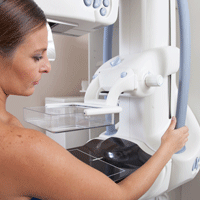
Monica Saini, MD, a radiologist in Santa Fe, New Mexico, and JoAnn Pushkin, executive director of the nonprofit educational website DenseBreast-info.org, engaged ObGyn attendees on “Breast density: Why it matters and what to do” at the American College of Obstetricians and Gynecologists (ACOG) 2017 Annual Clinical and Scientific Meeting (May 6–9, 2017) in San Diego, California. The program was sponsored by GE Healthcare.
DENSE BREASTS ARE A RISK FACTOR FOR CANCER
Breast density is the second largest risk factor for breast cancer after radiation treatment to the chest, so it is important to identify patients with dense breasts, according to Dr. Saini. The American College of Radiology’s Breast Imaging Reporting and Data System (BI-RADS) classifies breast density into 4 groups: 1) almost entirely fatty, 2) scattered fibroglandular densities, 3) heterogeneously dense, and 4) extremely dense. A woman whose mammograms show heterogeneously dense or extremely dense breasts is considered to have “dense breasts.”
Cancer is often difficult to identify with mammography in dense breasts because masses or lumps appear as white on a white (dense tissue) background; by contrast, a tumor in a nondense (fatty) breast would appear as white on a dark, fatty tissue background. Approximately one-third of cancers in dense breasts have a delayed diagnosis on mammography, and 70% of cancers occur in dense breasts, said Dr. Saini.
Having dense breasts is not an abnormal condition, however, and is actually common—about 40% of women aged 40 or older have dense breasts.
Supplement mammography with other screening modalities
While screening mammograms can save lives, mammography should not be viewed as a one-size-fits-all modality. Screening for breast cancer should be personalized, based on, among other factors, a woman’s personal and family history, age, genetic risk, lifestyle factors, and breast density.
Key point. Women with dense breasts should continue to have screening mammograms. In addition, mammography for these patients should be supplemented with other technologies, such as 3D mammography (digital tomosynthesis), handheld ultrasound, or automated breast ultrasound (ABUS). In women at higher risk (presence of BRCA1 or BRCA2 gene mutation, strong family history of breast cancer, or radiation treatment to the chest) magnetic resonance imaging (MRI) may be considered.
Data on adjunct screening modalities. Dr. Saini discussed the results of the ASTOUND trial, a prospective multicenter study that compared ultrasound and tomosynthesis for the detection of breast cancer in mammography-negative dense breasts.1 Among the 3,231 asymptomatic women included in the trial, 13 breast cancers were detected with tomosynthesis (incremental cancer detection rate [CDR], 4 per 1,000 screens; 95% confidence interval [CI], 1.8–6.2) and 23 were detected with ultrasound (incremental CDR, 7.1 per 1,000 screens; 95% CI, 4.4–10.0), P = .006. There were 107 false-positive results: 53 with tomosynthesis and 65 with ultrasound, a difference that was not statistically significant. The study authors noted that while ultrasound had better incremental breast cancer detection than tomosynthesis, and at a similar false-positive recall rate, tomosynthesis did detect more than half of the additional breast cancers in these women.1
Make screening easier for the patient
Dr. Saini noted that for women with dense breasts, performing mammography and adjunctive screening at the same visit is convenient for the patient. Physicians can also write prescriptions for follow-up based on density findings, for example, “3D mammography if available, if dense, order ultrasound.”
Read how to answer patient questions about breast density
ARE YOU READY TO ANSWER PATIENT QUESTIONS ABOUT BREAST DENSITY?
That is the question JoAnn Pushkin, executive director of DenseBreast-info.org, asked in her presentation. You should discuss with patients exactly what it means to have dense breasts, breast density as an independent risk factor for cancer, the breast imaging technologies available for screening (mammography, tomosynthesis, ultrasound, contrast-enhanced MRI), the risks and benefits of each screening modality, and surveillance intervals for women with dense breasts. Good communication with the patient’s radiology team assists in formulating an individualized screening strategy.
Patients may have concerns about the information provided—or not provided—in their state’s breast density notification letter after a mammogram. Currently, 31 states mandate some type of breast density notification, while 4 states have efforts for density reporting or education that do not require notification. The information given to patients and how they will be informed varies by state. Some states, for example, require that patients who have heterogeneously or extremely dense breasts be informed of this by letter, while other states require that all patients receive the same notification with information about dense breasts but does not tell them whether or not they have dense breasts.
A go-to resource for ObGyns and patients
The website of the nonprofit DenseBreast-Info.org (http://densebreast-info.org/), co-founded by Wendie Berg, MD, PhD, who serves as Chief Scientific Advisor to the organization and is Professor of Radiology at the University of Pittsburgh School of Medicine/Magee-Women’s Hospital of UPMC, provides an interactive US map that features state-by-state breast density reporting guidelines so you can stay up-to-date on notification legislation in your area.
Sections for patients offer comprehensive and clearly written information on categories of breast density, a patient risk checklist, screening test descriptions, frequently asked questions, educational videos, and a patient brochure in English and Spanish.
For health care providers, resources include:
- a screening decision support tool flowchart to help assess which patients need more screening
- a table summarizing the cancer detection rates for mammography alone and mammography plus another screening modality (tomosynthesis, ultrasound, MRI)
- a comparison of breast cancer screening guidelines from various medical societies, including the American College of Radiology/Society of Breast Imaging, the American Cancer Society, the American College of Obstetricians and Gynecologists, and the US Preventive Services Task Force.
A special section covers screening technology, and each page includes descriptions, benefits, and considerations for use. Photos of the equipment and images of breast scans with explanatory captions enhance understanding.
Screening for high-risk women
Ms. Pushkin noted that for high-risk patients with dense breasts, mammography plus MRI annually would be an appropriate option.
- Tagliafico AS, Calabrese M, Mariscotti G, et al. Adjunct screening with tomosynthesis or ultrasound in women with mammography-negative dense breasts: interim report of a prospective comparative trial [published online ahead of print March 9, 2015]. J Clin Oncol. doi:10.1200/JCO.2015.63.4147.
Monica Saini, MD, a radiologist in Santa Fe, New Mexico, and JoAnn Pushkin, executive director of the nonprofit educational website DenseBreast-info.org, engaged ObGyn attendees on “Breast density: Why it matters and what to do” at the American College of Obstetricians and Gynecologists (ACOG) 2017 Annual Clinical and Scientific Meeting (May 6–9, 2017) in San Diego, California. The program was sponsored by GE Healthcare.
DENSE BREASTS ARE A RISK FACTOR FOR CANCER
Breast density is the second largest risk factor for breast cancer after radiation treatment to the chest, so it is important to identify patients with dense breasts, according to Dr. Saini. The American College of Radiology’s Breast Imaging Reporting and Data System (BI-RADS) classifies breast density into 4 groups: 1) almost entirely fatty, 2) scattered fibroglandular densities, 3) heterogeneously dense, and 4) extremely dense. A woman whose mammograms show heterogeneously dense or extremely dense breasts is considered to have “dense breasts.”
Cancer is often difficult to identify with mammography in dense breasts because masses or lumps appear as white on a white (dense tissue) background; by contrast, a tumor in a nondense (fatty) breast would appear as white on a dark, fatty tissue background. Approximately one-third of cancers in dense breasts have a delayed diagnosis on mammography, and 70% of cancers occur in dense breasts, said Dr. Saini.
Having dense breasts is not an abnormal condition, however, and is actually common—about 40% of women aged 40 or older have dense breasts.
Supplement mammography with other screening modalities
While screening mammograms can save lives, mammography should not be viewed as a one-size-fits-all modality. Screening for breast cancer should be personalized, based on, among other factors, a woman’s personal and family history, age, genetic risk, lifestyle factors, and breast density.
Key point. Women with dense breasts should continue to have screening mammograms. In addition, mammography for these patients should be supplemented with other technologies, such as 3D mammography (digital tomosynthesis), handheld ultrasound, or automated breast ultrasound (ABUS). In women at higher risk (presence of BRCA1 or BRCA2 gene mutation, strong family history of breast cancer, or radiation treatment to the chest) magnetic resonance imaging (MRI) may be considered.
Data on adjunct screening modalities. Dr. Saini discussed the results of the ASTOUND trial, a prospective multicenter study that compared ultrasound and tomosynthesis for the detection of breast cancer in mammography-negative dense breasts.1 Among the 3,231 asymptomatic women included in the trial, 13 breast cancers were detected with tomosynthesis (incremental cancer detection rate [CDR], 4 per 1,000 screens; 95% confidence interval [CI], 1.8–6.2) and 23 were detected with ultrasound (incremental CDR, 7.1 per 1,000 screens; 95% CI, 4.4–10.0), P = .006. There were 107 false-positive results: 53 with tomosynthesis and 65 with ultrasound, a difference that was not statistically significant. The study authors noted that while ultrasound had better incremental breast cancer detection than tomosynthesis, and at a similar false-positive recall rate, tomosynthesis did detect more than half of the additional breast cancers in these women.1
Make screening easier for the patient
Dr. Saini noted that for women with dense breasts, performing mammography and adjunctive screening at the same visit is convenient for the patient. Physicians can also write prescriptions for follow-up based on density findings, for example, “3D mammography if available, if dense, order ultrasound.”
Read how to answer patient questions about breast density
ARE YOU READY TO ANSWER PATIENT QUESTIONS ABOUT BREAST DENSITY?
That is the question JoAnn Pushkin, executive director of DenseBreast-info.org, asked in her presentation. You should discuss with patients exactly what it means to have dense breasts, breast density as an independent risk factor for cancer, the breast imaging technologies available for screening (mammography, tomosynthesis, ultrasound, contrast-enhanced MRI), the risks and benefits of each screening modality, and surveillance intervals for women with dense breasts. Good communication with the patient’s radiology team assists in formulating an individualized screening strategy.
Patients may have concerns about the information provided—or not provided—in their state’s breast density notification letter after a mammogram. Currently, 31 states mandate some type of breast density notification, while 4 states have efforts for density reporting or education that do not require notification. The information given to patients and how they will be informed varies by state. Some states, for example, require that patients who have heterogeneously or extremely dense breasts be informed of this by letter, while other states require that all patients receive the same notification with information about dense breasts but does not tell them whether or not they have dense breasts.
A go-to resource for ObGyns and patients
The website of the nonprofit DenseBreast-Info.org (http://densebreast-info.org/), co-founded by Wendie Berg, MD, PhD, who serves as Chief Scientific Advisor to the organization and is Professor of Radiology at the University of Pittsburgh School of Medicine/Magee-Women’s Hospital of UPMC, provides an interactive US map that features state-by-state breast density reporting guidelines so you can stay up-to-date on notification legislation in your area.
Sections for patients offer comprehensive and clearly written information on categories of breast density, a patient risk checklist, screening test descriptions, frequently asked questions, educational videos, and a patient brochure in English and Spanish.
For health care providers, resources include:
- a screening decision support tool flowchart to help assess which patients need more screening
- a table summarizing the cancer detection rates for mammography alone and mammography plus another screening modality (tomosynthesis, ultrasound, MRI)
- a comparison of breast cancer screening guidelines from various medical societies, including the American College of Radiology/Society of Breast Imaging, the American Cancer Society, the American College of Obstetricians and Gynecologists, and the US Preventive Services Task Force.
A special section covers screening technology, and each page includes descriptions, benefits, and considerations for use. Photos of the equipment and images of breast scans with explanatory captions enhance understanding.
Screening for high-risk women
Ms. Pushkin noted that for high-risk patients with dense breasts, mammography plus MRI annually would be an appropriate option.
- Tagliafico AS, Calabrese M, Mariscotti G, et al. Adjunct screening with tomosynthesis or ultrasound in women with mammography-negative dense breasts: interim report of a prospective comparative trial [published online ahead of print March 9, 2015]. J Clin Oncol. doi:10.1200/JCO.2015.63.4147.
- Tagliafico AS, Calabrese M, Mariscotti G, et al. Adjunct screening with tomosynthesis or ultrasound in women with mammography-negative dense breasts: interim report of a prospective comparative trial [published online ahead of print March 9, 2015]. J Clin Oncol. doi:10.1200/JCO.2015.63.4147.
Los Angeles County encourages LARC use to decrease Zika cases
Los Angeles County, California, has been identified as one of 7 areas in the nation with the highest risk of local Zika transmission by the Centers for Disease Control and Prevention (CDC), advise Adriana Ramos and colleagues from Los Angeles County Department of Public Health (DPH), Maternal, Child & Adolescent Health Programs.1 One factor for this classification is the county’s high birth rate. According to Ramos at el the CDC recommends that, before a Zika outbreak occurs, health departments in areas with Aedes species mosquitos increase access to and use of effective contraception.1 Long-acting reversible contraceptives (LARCs), including the intrauterine device (IUD) and the implant, are proven most effective methods.1
In a poster presented at the American College of Obstetricians and Gynecologists (ACOG) Annual Clinical Meeting in San Diego, California, Ramos and colleagues summarized contraceptive use within LA County using data from the Los Angeles Mommy and Baby (LAMB) project, conducted by the Maternal, Child, and Adolescent Health (MCAH) Programs of the LA County DPH, which surveyed mothers who recently delivered a live baby about their preconception and perinatal experiences. In 2012, 6,893 mothers participated. In 2014, MCAH re-interviewed the 2012 LAMB respondents, excluding those with a subsequent pregnancy after the 2012 survey or who had not originally answered questions about family planning, leaving 3,175 respondents. Findings, weighted to the 2012 live-birth cohort, estimated the weighted population at 115,284 live births.1
The study defined contraception use by efficacy, identifying no contraception use, condoms, withdrawal, and the rhythm method as less effective; oral contraceptive pills and vaginal ring as moderately effective; and LARCs and sterilization as highly effective. Unintended births account for 47% of births in LAC and more than 59% of women report using less effective contraceptive methods.1
Results of the study
As a result of their study, MCAH researchers Adriana Ramos, Shin Chao, MD, MPH, and Diana E. Ramos, MD, MPH, conclude that educating providers to place LARC contraceptives and educating the public on the most effective contraceptive methods can decrease the neonatal Zika complication rates by preventing unplanned pregnancy. LAC is undertaking these activities to decrease the number of neonatal Zika cases.1
- Ramos A, Chao S, Ramos DE. Zika: Preconception & perinatal opportunities in Los Angeles County. Poster presented at: American College of Obstetricians and Gynecologists Annual Clinical and Scientific Meeting; May 6, 2017; San Diego, California.
Los Angeles County, California, has been identified as one of 7 areas in the nation with the highest risk of local Zika transmission by the Centers for Disease Control and Prevention (CDC), advise Adriana Ramos and colleagues from Los Angeles County Department of Public Health (DPH), Maternal, Child & Adolescent Health Programs.1 One factor for this classification is the county’s high birth rate. According to Ramos at el the CDC recommends that, before a Zika outbreak occurs, health departments in areas with Aedes species mosquitos increase access to and use of effective contraception.1 Long-acting reversible contraceptives (LARCs), including the intrauterine device (IUD) and the implant, are proven most effective methods.1
In a poster presented at the American College of Obstetricians and Gynecologists (ACOG) Annual Clinical Meeting in San Diego, California, Ramos and colleagues summarized contraceptive use within LA County using data from the Los Angeles Mommy and Baby (LAMB) project, conducted by the Maternal, Child, and Adolescent Health (MCAH) Programs of the LA County DPH, which surveyed mothers who recently delivered a live baby about their preconception and perinatal experiences. In 2012, 6,893 mothers participated. In 2014, MCAH re-interviewed the 2012 LAMB respondents, excluding those with a subsequent pregnancy after the 2012 survey or who had not originally answered questions about family planning, leaving 3,175 respondents. Findings, weighted to the 2012 live-birth cohort, estimated the weighted population at 115,284 live births.1
The study defined contraception use by efficacy, identifying no contraception use, condoms, withdrawal, and the rhythm method as less effective; oral contraceptive pills and vaginal ring as moderately effective; and LARCs and sterilization as highly effective. Unintended births account for 47% of births in LAC and more than 59% of women report using less effective contraceptive methods.1
Results of the study
As a result of their study, MCAH researchers Adriana Ramos, Shin Chao, MD, MPH, and Diana E. Ramos, MD, MPH, conclude that educating providers to place LARC contraceptives and educating the public on the most effective contraceptive methods can decrease the neonatal Zika complication rates by preventing unplanned pregnancy. LAC is undertaking these activities to decrease the number of neonatal Zika cases.1
- Ramos A, Chao S, Ramos DE. Zika: Preconception & perinatal opportunities in Los Angeles County. Poster presented at: American College of Obstetricians and Gynecologists Annual Clinical and Scientific Meeting; May 6, 2017; San Diego, California.
- Ramos A, Chao S, Ramos DE. Zika: Preconception & perinatal opportunities in Los Angeles County. Poster presented at: American College of Obstetricians and Gynecologists Annual Clinical and Scientific Meeting; May 6, 2017; San Diego, California.

Embolism major cause of stroke after open arch surgery in patients with carotid/intracranial stenosis
Embolization was the major cause of permanent stroke in patients with moderate or severe carotid or intracranial atherosclerosis who underwent elective open aortic arch surgery at a single institution, according to the results of a retrospective study.
Preoperative craniocervical and aortic screening may aid in modifying the operative strategy to reduce the incidence of stroke in these patients, according to a report published in the May issue of the Journal of Thoracic and Cardiovascular Surgery.
Preventing stroke in this patient population is an important consideration, because perioperative stroke is approximately 4 times more common in open aortic arch surgery (OAAS) than in coronary artery bypass grafting or valve surgery, according to Ken-ichi Imasaka, MD, and his colleagues at the National Hospital Organization Kyushu Medical Center, Fukuoka, Japan.
The study population comprised 200 consecutive patients undergoing elective OAAS at the institution between October 2008 and October 2015, including 34% women and with a mean patient age of 71 years (J Thorac Cardiovasc Surg. 2017;153:1045-53).
After preoperative screening, 21% of patients were diagnosed with carotid or intracranial artery disease (CIAD). None of these patients were diagnosed with impaired cerebral perfusion reserve on brain SPECT (single-photon emission computed tomography). A total of 92% of patients underwent ascending aorta or aortic arch replacement through a median sternotomy, while the remaining 8% underwent extended aortic arch replacement via L-incision (15 patients) or combined median sternotomy and left posterior lateral thoracotomy (1 patient). Among the patients, 16% underwent ascending aorta replacement; 8% had partial arch replacement; and the remaining 76% had total arch replacement.
Shaggy aorta was present in 19% of the patients, with 51% of these showing CIAD (P less than .0001). A total of 30% of the patients with shaggy aorta had the total arch replacement through an L-incision or combined median sternotomy and left posterior lateral thoracotomy, a significant difference (P less than .0001).

The overall in-hospital mortality rate was 3.5%. The overall incidence of permanent stroke and paraplegia or paraparesis was 4% (8 patients) and 2% (4 patients), respectively. Three (37.5%) of the 8 permanent stroke patients died during the postoperative hospital stay, compared with 2.1% of the 192 patients without stroke.
Univariate analysis indicated that previous cerebrovascular accident (P = .0002), shaggy aorta (P less than .0001), cardiopulmonary bypass time (P = .003), selective antegrade cerebral perfusion time (P = .004), operation time (P = .02), and extended aortic repair through L-incision or combined median sternotomy and left posterior lateral thoracotomy (P = .0002) were significant risk factors for neurologic morbidity.
“Preoperative intensive screening of carotid and intracranial artery disease is a useful step to identify patients at higher risk of hemodynamic ischemic stroke. Advanced systemic atherosclerosis may be a crucial determinant of perioperative stroke due to atherothrombotic embolization. Antiembolic measures during surgery are essential to prevent perioperative stroke,” the researchers concluded.
The authors reported that they had no disclosures.
During aortic arch surgery, the lack of blood supply as a result of emboli, rather than atherosclerosis itself, kills the cerebral neurons, according to Ourania Preventza, MD, and Joseph S. Coselli, MD, of the Baylor College of Medicine, Houston, in their invited commentary (J Thorac Cardiovasc Surg. 2017;153:1054-5).
Patients with carotid and intracranial disease should indeed have more intensive screening before undergoing major aortic surgery, they agreed, but pointed out that in the absence of carotid disease, large or complex aortic atheromas can be seen in the arch, indicating that, even though atherosclerosis is a systemic disease, using different sites of prediction can be uncertain.
This requires a broader approach to prevent stroke, including careful selection of the cannulation site in patients with diffuse and heavy arch atherosclerosis or currently ulcerated plaque, they added.
“To minimize postoperative neurologic morbidities after aortic arch surgery, an individually tailored perioperative approach should be in the armamentarium of cardiac surgeons,” Dr. Preventza and Dr. Coselli concluded.
Dr. Preventza consults for Medtronic and W. L. Gore & Associates. Dr. Coselli participates in clinical research trials conducted by GlaxoSmithKline, Edwards Lifesciences, and Bolton Medical, and consults for various companies.
Embolization was the major cause of permanent stroke in patients with moderate or severe carotid or intracranial atherosclerosis who underwent elective open aortic arch surgery at a single institution, according to the results of a retrospective study.
Preoperative craniocervical and aortic screening may aid in modifying the operative strategy to reduce the incidence of stroke in these patients, according to a report published in the May issue of the Journal of Thoracic and Cardiovascular Surgery.
Preventing stroke in this patient population is an important consideration, because perioperative stroke is approximately 4 times more common in open aortic arch surgery (OAAS) than in coronary artery bypass grafting or valve surgery, according to Ken-ichi Imasaka, MD, and his colleagues at the National Hospital Organization Kyushu Medical Center, Fukuoka, Japan.
The study population comprised 200 consecutive patients undergoing elective OAAS at the institution between October 2008 and October 2015, including 34% women and with a mean patient age of 71 years (J Thorac Cardiovasc Surg. 2017;153:1045-53).
After preoperative screening, 21% of patients were diagnosed with carotid or intracranial artery disease (CIAD). None of these patients were diagnosed with impaired cerebral perfusion reserve on brain SPECT (single-photon emission computed tomography). A total of 92% of patients underwent ascending aorta or aortic arch replacement through a median sternotomy, while the remaining 8% underwent extended aortic arch replacement via L-incision (15 patients) or combined median sternotomy and left posterior lateral thoracotomy (1 patient). Among the patients, 16% underwent ascending aorta replacement; 8% had partial arch replacement; and the remaining 76% had total arch replacement.
Shaggy aorta was present in 19% of the patients, with 51% of these showing CIAD (P less than .0001). A total of 30% of the patients with shaggy aorta had the total arch replacement through an L-incision or combined median sternotomy and left posterior lateral thoracotomy, a significant difference (P less than .0001).
The overall in-hospital mortality rate was 3.5%. The overall incidence of permanent stroke and paraplegia or paraparesis was 4% (8 patients) and 2% (4 patients), respectively. Three (37.5%) of the 8 permanent stroke patients died during the postoperative hospital stay, compared with 2.1% of the 192 patients without stroke.
Univariate analysis indicated that previous cerebrovascular accident (P = .0002), shaggy aorta (P less than .0001), cardiopulmonary bypass time (P = .003), selective antegrade cerebral perfusion time (P = .004), operation time (P = .02), and extended aortic repair through L-incision or combined median sternotomy and left posterior lateral thoracotomy (P = .0002) were significant risk factors for neurologic morbidity.
“Preoperative intensive screening of carotid and intracranial artery disease is a useful step to identify patients at higher risk of hemodynamic ischemic stroke. Advanced systemic atherosclerosis may be a crucial determinant of perioperative stroke due to atherothrombotic embolization. Antiembolic measures during surgery are essential to prevent perioperative stroke,” the researchers concluded.
The authors reported that they had no disclosures.
Key clinical point:
Major finding: Previous cerebrovascular accident and shaggy aorta were significant determinants of neurologic morbidity.
Data source: Retrospective study of 200 consecutive patients undergoing elective aortic arch surgery at a single institution.
Disclosures: The authors reported having no conflicts of interest.
Targeted treatment does not address RA patients’ mental health
BIRMINGHAM, ENGLAND – Improvements in physical health with response to treatment of rheumatoid arthritis (RA) don’t necessarily translate into improvements in patients’ mental health, based on results of a systematic review presented at the British Society for Rheumatology annual conference.
Reducing levels of joint pain and inflammation did not appreciably improve mental well-being, said Faith Matcham, PhD, of the Institute of Psychiatry, Psychology, and Neuroscience, King’s College London. The standardized mean difference between biologics and disease-modifying antirheumatic drugs (DMARDs) in improving the physical and mental components of the 36-item Short Form survey were a respective 0.47 and 0.25.

A pairwise meta-analysis was performed on 58 and 48 randomized controlled trials, respectively. More than 34,000 patients had been treated in these trials with 28 current or investigational RA therapies, and treatment outcomes were compared with an active, placebo, or usual-care arm. Studies were included if they had assessed any type of mood outcome.
Mental health had been measured in more than half of the RA treatment trials found on the original search, but just 40% of those trials reported mental health outcomes with enough information to be used in the meta-analyses.
Although studies that had compared biologics with placebo accounted for only four studies, none of those showed significant change in mental health outcomes with active therapy.
“Providing effective pharmacotherapy alone is going to be insufficient to produce meaningful improvement in mental health outcomes for the majority,” Dr. Matcham observed. She added that, even after treatment, mental health measures in RA patients remained lower than those in the general population. “It is essential to optimize both mental and physical health care outcomes,” she concluded, and an integrated approach needs to be taken.
Dr. Matcham reported having no financial disclosures.
BIRMINGHAM, ENGLAND – Improvements in physical health with response to treatment of rheumatoid arthritis (RA) don’t necessarily translate into improvements in patients’ mental health, based on results of a systematic review presented at the British Society for Rheumatology annual conference.
Reducing levels of joint pain and inflammation did not appreciably improve mental well-being, said Faith Matcham, PhD, of the Institute of Psychiatry, Psychology, and Neuroscience, King’s College London. The standardized mean difference between biologics and disease-modifying antirheumatic drugs (DMARDs) in improving the physical and mental components of the 36-item Short Form survey were a respective 0.47 and 0.25.
A pairwise meta-analysis was performed on 58 and 48 randomized controlled trials, respectively. More than 34,000 patients had been treated in these trials with 28 current or investigational RA therapies, and treatment outcomes were compared with an active, placebo, or usual-care arm. Studies were included if they had assessed any type of mood outcome.
Mental health had been measured in more than half of the RA treatment trials found on the original search, but just 40% of those trials reported mental health outcomes with enough information to be used in the meta-analyses.
Although studies that had compared biologics with placebo accounted for only four studies, none of those showed significant change in mental health outcomes with active therapy.
“Providing effective pharmacotherapy alone is going to be insufficient to produce meaningful improvement in mental health outcomes for the majority,” Dr. Matcham observed. She added that, even after treatment, mental health measures in RA patients remained lower than those in the general population. “It is essential to optimize both mental and physical health care outcomes,” she concluded, and an integrated approach needs to be taken.
Dr. Matcham reported having no financial disclosures.
AT RHEUMATOLOGY 2017
Key clinical point: A response to pharmacotherapy per se is not directly related to improved mental health outcomes in patients with rheumatoid arthritis.
Major finding: The standardized mean difference between biologics and DMARDs in improving the physical and mental components of the 36-item Short Form survey were 0.47 and 0.25, respectively.
Data source: A systematic review and meta-analysis of RA treatment trials involving 34,087 patients.
Disclosures: Dr. Matcham reported having no relevant financial disclosures.
CC-220 shows efficacy, safety concerns in phase II SLE trial
PORTLAND – The investigational agent CC-220 showed some efficacy but important safety signals in a 12-week, phase II, dose-escalation study of 42 patients with systemic lupus erythematosus (SLE).
In all, 14% of patients stopped treatment because of adverse effects, including five patients who were receiving CC-220 and one patient on placebo, Victoria Werth, MD, of the University of Pennsylvania, Philadelphia, said during a poster presentation at the annual meeting of the Society for Investigative Dermatology. Higher doses of CC-220 were associated with neutropenia, pneumonia, and dermatitis, she reported.

About 79% of patients completed the study, and none stopped because of lack of efficacy, Dr. Werth said. The most common treatment-emergent adverse effects were mild to moderate nausea and diarrhea. However, four patients experienced serious adverse events, including two patients in the placebo group and two patients who developed pneumonia at the highest (0.6 mg per day) CC-220 dose. In the two highest-dose groups, two patients also developed grade 3 neutropenia, one patient developed grade 1 neutropenia, and there were two cases each of dermatitis and urticaria.
CC-220 appeared to improve scores on the CLASI, SELENA-SENDAI, and PGA, Dr. Werth said. Average drops from baseline on the CLASI ranged from 0 (placebo group) to 7 points (0.3 mg every other day), and were 5 points at 0.3 mg daily, 4 points at alternating doses of 0.3 mg and 0.6 mg, and 6 points at the highest dose of 0.6 mg daily. Among patients whose baseline CLASI score was at least 10, mean decreases ranged between 0 (placebo) and 26 points (0.3 mg daily), and average decreases for the other groups were 9 points (0.3 mg every other day and 0.3 mg alternating with 0.6 mg) or 13 points (0.6 mg daily).
Compared with the placebo group, a greater proportion of CC-220 recipients also had at least a 4-point drop on SELENA-SENDAI and tended to improve more on the tender joint count and the swollen joint count, said Dr. Werth. The average drop in mean PGA score ranged between 0 (placebo group) and 0.9 (0.3 mg per day) and did not show a dose-response trend. The relatively small number of patients in this study and some variability in baseline SLE disease activity made it difficult to draw conclusions about dose-response relationships, Dr. Werth noted. However, there were signs of an overall dose-response trend when pooling mean changes in CLASI, tender and swollen joint counts, SELENA-SLEDAI, and PGA scores.
In terms of pharmacodynamics, doses of at least 0.3 mg CC-220 per day were associated with marked decreases in circulating plasmacytoid dendritic cells and B-cell subsets, which began as early as day 29, Dr. Werth said. Cereblon is part of the cullin-ring finger ligase-4 complex, she explained. Previous work has shown that CC-220 binds with high affinity to cereblon, inducing ubiquitination and increased breakdown of the transcription factors IKZF1 and IKZF3. The end result is an altered immune response.
This study includes an optional 2-year extension phase at the three highest doses of CC-220. Celgene also plans a phase IIb proof-of-concept study in a “broader SLE population,” Dr. Werth said.
Celgene funded the study. Dr. Werth disclosed grant or research support from Celgene, Janssen, Biogen, Roche, and Corbus Pharmaceuticals, and consulting or advisory fees from Celgene, Janssen, Genentech, and several other pharmaceutical companies.
PORTLAND – The investigational agent CC-220 showed some efficacy but important safety signals in a 12-week, phase II, dose-escalation study of 42 patients with systemic lupus erythematosus (SLE).
In all, 14% of patients stopped treatment because of adverse effects, including five patients who were receiving CC-220 and one patient on placebo, Victoria Werth, MD, of the University of Pennsylvania, Philadelphia, said during a poster presentation at the annual meeting of the Society for Investigative Dermatology. Higher doses of CC-220 were associated with neutropenia, pneumonia, and dermatitis, she reported.
About 79% of patients completed the study, and none stopped because of lack of efficacy, Dr. Werth said. The most common treatment-emergent adverse effects were mild to moderate nausea and diarrhea. However, four patients experienced serious adverse events, including two patients in the placebo group and two patients who developed pneumonia at the highest (0.6 mg per day) CC-220 dose. In the two highest-dose groups, two patients also developed grade 3 neutropenia, one patient developed grade 1 neutropenia, and there were two cases each of dermatitis and urticaria.
CC-220 appeared to improve scores on the CLASI, SELENA-SENDAI, and PGA, Dr. Werth said. Average drops from baseline on the CLASI ranged from 0 (placebo group) to 7 points (0.3 mg every other day), and were 5 points at 0.3 mg daily, 4 points at alternating doses of 0.3 mg and 0.6 mg, and 6 points at the highest dose of 0.6 mg daily. Among patients whose baseline CLASI score was at least 10, mean decreases ranged between 0 (placebo) and 26 points (0.3 mg daily), and average decreases for the other groups were 9 points (0.3 mg every other day and 0.3 mg alternating with 0.6 mg) or 13 points (0.6 mg daily).
Compared with the placebo group, a greater proportion of CC-220 recipients also had at least a 4-point drop on SELENA-SENDAI and tended to improve more on the tender joint count and the swollen joint count, said Dr. Werth. The average drop in mean PGA score ranged between 0 (placebo group) and 0.9 (0.3 mg per day) and did not show a dose-response trend. The relatively small number of patients in this study and some variability in baseline SLE disease activity made it difficult to draw conclusions about dose-response relationships, Dr. Werth noted. However, there were signs of an overall dose-response trend when pooling mean changes in CLASI, tender and swollen joint counts, SELENA-SLEDAI, and PGA scores.
In terms of pharmacodynamics, doses of at least 0.3 mg CC-220 per day were associated with marked decreases in circulating plasmacytoid dendritic cells and B-cell subsets, which began as early as day 29, Dr. Werth said. Cereblon is part of the cullin-ring finger ligase-4 complex, she explained. Previous work has shown that CC-220 binds with high affinity to cereblon, inducing ubiquitination and increased breakdown of the transcription factors IKZF1 and IKZF3. The end result is an altered immune response.
This study includes an optional 2-year extension phase at the three highest doses of CC-220. Celgene also plans a phase IIb proof-of-concept study in a “broader SLE population,” Dr. Werth said.
Celgene funded the study. Dr. Werth disclosed grant or research support from Celgene, Janssen, Biogen, Roche, and Corbus Pharmaceuticals, and consulting or advisory fees from Celgene, Janssen, Genentech, and several other pharmaceutical companies.
AT SID 2017
Key clinical point:
Major finding: Among patients whose baseline CLASI score was at least 10, mean decreases ranged between 0 (placebo), and 26 points (0.3 mg daily).
Data source: A 12-week, phase II, placebo-controlled dose-escalation study of 42 patients with systemic lupus erythematosus.
Disclosures: Celgene funded the study. Dr. Werth disclosed grant or research support from Celgene, Janssen, Biogen, Roche, and Corbus Pharmaceuticals, and consulting or advisory fees from Celgene, Janssen, Genentech, and several other pharmaceutical companies.
‘Rich pipeline’ of novel NASH treatments being studied
AMSTERDAM – There is a “very, very rich pipeline” of drugs being developed for the treatment of nonalcoholic steatohepatitis (NASH), Jean-François Dufour, MD, the head of hepatology and director of the University Clinic for Visceral Surgery and Medicine at the University of Berne (Switzerland) said at the International Liver Congress.
“We have many therapeutic options [under investigation],” Dr. Dufour noted at the Congress, which is sponsored by the European Association for the Study of the Liver (EASL). These include drugs that target metabolic homeostasis, insulin resistance, inflammation, oxidative stress, or fibrosis (Liver Int. 2017 May;37:634-47).
In fact, there is such a range of options that target different pathways, from fatty acid and bile acid synthesis to the early and late stages of fibrosis, that it is very likely that these drugs will be used in combination, Dr. Dufour observed as he gave an overview of the current trials that are underway in NASH.
There are currently five ongoing multicenter phase III trials being undertaken with four drugs. First, there is the REGENERATE trial with Intercept’s farnesoid X receptor obeticholic acid(Ocaliva). This is a placebo-controlled trial comparing two daily doses of obeticholic acid (10 and 25 mg) on top of the standard of care. The trial will recruit just over 2,000 patients with biopsy-proven stage 2-3 NASH fibrosis, and the primary endpoint is the resolution of NASH without fibrosis worsening or improvement in fibrosis without worsening of NASH at week 72.
Second there is the RESOLVE-IT trial with Genfit’s peroxisome proliferator-activated receptor alpha/delta agonist elafibranor. This randomized, double-blind trial hopes to recruit 2,000 patients with biopsy-proven NASH stage 1-3 fibrosis and will compare elafibranor 120 mg given once a day with placebo. The primary endpoint is the resolution of NASH without worsening of fibrosis at week 72.
Next, Tobira Therapeutics’ C-C chemokine receptor type 2 and 5 antagonist cenicriviroc is being studied in the AURORA trial. Again, recruiting around 2,000 patients is the target, but this time with stage 2-3 biopsy-proven NASH fibrosis. Cenicriviroc will be given daily at a dose of 150 mg and will be compared against placebo. The primary endpoint is the improvement of fibrosis by one or more stage with no worsening of steatohepatitis at 1 year.
Finally, there are the STELLA 3 and STELLA 4 trials with Gilead’s apoptosis signal-regulated kinase-1 inhibitor selonsertib. Target accrual in both studies is 800 patients with STELLA 3 recruiting patients with stage-3 NASH fibrosis and STELLA 4 recruiting those with compensated cirrhosis from NASH. Both trials will compared two daily doses of selonsertib (6 mg and 18 mg) versus placebo. The primary endpoints are the improvement of at least one or more fibrosis stage with no worsening of steatohepatitis at 48 weeks and event-free survival at week 240.
In addition, there are at least 20 phase 2b and 2a studies looking at a variety of other novel drugs with different therapeutic targets, Dr. Dufour said, and during separate presentations at the congress, results of several early trials with novel drugs being tested for NASH were given.
Eric J. Lawitz, MD, reported the promising results of a “proof of concept” open-label study in which the safety and efficacy of 12 weeks’ treatment with the oral acetyl-CoA carboxylase (ACC) inhibitor, GS-0976, was examined in 10 patients with a clinical diagnosis of nonalcoholic fatty liver disease (NAFLD).
“ACC catalyzes the rate-limiting step in de novo hepatic lipogenesis (DNL),” which is an underlying pathologic process in NASH, Dr. Lawitz, who is vice president of scientific and research development at the Texas Liver Institute, San Antonio, observed.
He reported that 12 weeks’ treatment with the ACC inhibitor GS-0976 suppressed DNL by 29%, compared with baseline (P = .022). There was also a 43% decrease in hepatic steatosis from baseline to 12 weeks (P = .006), as measured by the magnetic resonance imaging–proton-density fat fraction (MRI-PDFF), and a nonsignificant 9% reduction in liver stiffness measured using magnetic resonance elastography (MRE).
Two markers of fibrosis and cell death (TIMP-1 and CK18) were also improved, he said, noting that overall, the drug was well tolerated, bar a trend to an increase in triglycerides and reduction in high-density lipoprotein cholesterol that needs further follow up.
“There is a placebo-controlled phase II trial of GS-0976 in patients with NASH that is ongoing,” Dr. Lawitz said. Results of two phase II studies presented during the late-breaking abstracts session at the meeting showed similar promising results could be achieved with drugs mimicking the activity of different fibroblast growth factors.
“Fibroblast growth factor 21 (FGF21) is a nonmitogenic hormone produced in the liver that is an important regulator of energy metabolism,” said Arun J. Sanyal, MD, who presented the findings of a study with the FGF21 inhibitor BMS-986036.
”From a NASH perspective, it improves insulin sensitivity and by doing that, decreases lipogenesis, and it also has been shown to have some antifibrotic effects,” Dr. Sanyal of Virginia Commonwealth University in Richmond, added.
FGF21 has a short half-life, however, and BMS-986036 is a recombinant human analog of this hormone that could potentially allow it to be given up to once weekly.
The study involved 74 patients with stage 1-3 biopsy-proven NASH fibrosis and a hepatic fat fraction of 10% or greater measured by MRI-PDFF. Patients were randomized to treatment with BMS-986036 at subcutaneously administered doses of 10 mg given once daily or 20 mg given once weekly or to placebo for 16 weeks.
A significant reduction in the hepatic fat fraction was seen in patients treated with both the once-daily and once-weekly regimen of the active treatment relative to placebo, with absolute changes from baseline of –6.8% (P = .008) and –5.2% (P = .0004), respectively, and just –1.3% for placebo.
“Results suggest that BMS-986036 had beneficial effects on steatosis, liver injury, and fibrosis in NASH,” said Dr. Sanyal, who also noted that there were no deaths and no signal that there could be any safety concerns.
NGM282 is another recombinant human analog mimicking the action of an FGF, this time FGF19, and early data also suggest that it also reduces hepatic steatosis and key biomarkers of NASH. Dr. Stephen Harrison, MD, the medical director of Pinnacle Clinical Research in Live Oak, reported data on 82 patients with stage 1-3 NASH fibrosis who had been treated with NGM-282 3 mg or 6 mg subcutaneously once a day or placebo for 12 weeks.
“The primary endpoint [decrease in absolute liver fat content greater than or equal to 5%] was met in 79% of NGM-282-treated subjects, with over one-third of subjects achieving normalization of liver fat content with 12 weeks of therapy,” Dr. Harrison reported.
“There were significant and rapid reductions in multiple markers that are relevant to the resolution of NASH and improvement in fibrosis,” Dr. Harrison added.
One serious adverse event of acute pancreatitis occurred in a patient treated with FGF19, which was possibly thought to be treatment related, but otherwise adverse events were generally mild and included gastrointestinal effects such as diarrhea and nausea, and injection site reactions.
“These data strongly support the continued development of NGM282 in NASH,” Dr. Harrison said.
During his presentation at a symposium session on current and future approaches to NAFLD and NASH, Dr. Dufour was keen to point out that a combination of diet and exercise remains central to managing patients with NASH.
“We should not forget, that the first line of discussion with these patients should be about changing their lifestyles.” Improving diet and exercise is something that everybody can do, he said, it is widely available and inexpensive, associated with few side effects and can produce good results.
However, convincing some patients can be difficult and those with a low acceptance to lifestyle changes often prefer to take medication. That is likely to come at a cost, not just in terms of money but there are likely to be some side effects, and, of course, efficacy in NASH is yet to be proven in many cases, Dr. Dufour said.
Dr. Dufour disclosed he had been part of a number of advisory committees or received speaking and teaching fees from a host of pharmaceutical companies, many of whom have an interest in the development of treatments for NASH.
Gilead Sciences supported the study reported by Dr. Lawitz and he disclosed receiving research grants or other support from the company, as well as several other pharmaceutical companies.
The study presented by Dr. Sanyal was financed by Bristol-Myers Squibb and he disclosed research funding was provided to his institution. Dr. Sanyal also disclosed receiving research support and consulting fees from multiple other pharmaceutical companies.
Dr. Harrison acknowledged receiving research funding from and acting as a consultant to NGM Bio, who sponsored the study he presented. He also disclosed acting as an adviser or speaker, and receiving grants from other pharmaceutical companies in the past 12 months.
Key clinical point: Lots of new approaches to treating nonalcoholic steatohepatitis are being investigated, some with phase III trials underway.
Major finding: Hepatic steatosis was significantly reduced by the GS-0976, BMS-986036, and NGM282.
Data source: An expert review and three early-phase studies testing of the safety and efficacy of novel NASH treatments.
Disclosures: Dr. Dufour disclosed he had been part of a number of advisory committees or received speaking and teaching fees from a host of pharmaceutical companies, many of whom have an interested in the development of treatments for NASH. Gilead Sciences supported the study reported by Dr. Lawitz and he disclosed receiving research grants or other support from the company, as well as several other pharmaceutical companies. The study presented by Dr. Sanyal was financed by Bristol-Myers Squibb and he disclosed research funding was provided to his institution. Dr. Sanyal also disclosed receiving research support and consulting fees from multiple other pharmaceutical companies. Dr. Harrison acknowledged receiving research funding from and acting as a consultant to NGM Bio, who sponsored the study he presented. He also disclosed acting as an adviser or speaker and receiving grants from other pharmaceutical companies in the past 12 months.
Salvage chemo for NSCLC more effective after PD-1/PD-L1 inhibitors
GENEVA – Patients with stage IV non–small cell lung cancer (NSCLC) who have disease progression following treatment with an immune checkpoint inhibitor have a 30% better chance of achieving at least a partial response with salvage chemotherapy compared with patients who had received prior chemotherapy but not immunotherapy, according to Swiss and U.S. investigators.
In a study of 82 patients with advanced NSCLC, 18 of 67 patients (27%) who had progressed on an inhibitor of programmed death-1 (PD-1) or programmed death ligand-1 (PD-L1) had a partial response to combination chemotherapy, compared with just 1 of 15 (7%) of patients who had not received a checkpoint inhibitor, reported Sacha Rothschild, MD, PhD, of University Hospital Basel, Switzerland, and colleagues.
“The odds of achieving a partial response to salvage chemotherapy were significantly higher in patients with prior exposure to PD-1/PD-L1 inhibitors. This observed difference, however, warrants confirmation in larger cohorts. Ongoing investigations include the duration of response as well as evaluation of toxicity,” the researchers wrote in a scientific poster presented at the European Lung Cancer Conference.
Immune checkpoint inhibitors have been shown to be active in patients with late-stage NSCLC who have experienced disease progression following chemotherapy. To see whether salvage chemotherapy could offer any additional benefit to patients who had disease progression on immunotherapy, the investigators conducted a retrospective case-control study. They reviewed 355 patient records, and identified 82 patients – 46 men and 36 women – who met the study criteria.
Of this group, 67 patients had received a PD-1/PD-L1 checkpoint inhibitor, either nivolumab (Opdivo, 56 patients), pembrolizumab (Keytruda, 7), or atezolizumab (Tecentriq, 4). These patients were designated as cases. The remaining 15 patients, who had received prior chemotherapy or chemoradiotherapy only, were designated as controls.
Of all 82 patients, 63 (77%) had adenocarcinomas, 18 (22%) had squamous cell carcinomas, and 1 (1%) had a large-cell carcinoma.
Cases had a mean of 2.37 chemotherapy regimens prior to salvage chemotherapy, and controls had a mean of 1.93. Drugs used in the salvage regimens were docetaxel in 62% of patients, pemetrexed in 20%, gemcitabine in 12%, and paclitaxel in 6%.

The odds ratio for achieving a partial response was 0.30 (95% confidence interval, 0.18-0.50, P less than .0001).
In a multiple logistic regression model, neither age, sex, number of prior chemotherapy regimens, tumor histology, smoking status, nor type of salvage chemotherapy regimen were significantly associated with the likelihood of achieving a partial response.
In a poster discussion session, Simon Ekman, MD, of Karolinska Institute in Stockholm, Sweden, the invited discussant, said that the data from the study are convincing, and raise the question of what to do next.
“The question is how do we combine immunotherapy with chemotherapy? Should we use the immune therapy first, like in this trial, or vice versa, or should we combine concurrently” he said.
The strategy of chemotherapy first is supported by research showing that neoantigens crucial for the immune response are released during chemotherapy and radiotherapy, enabling immunotherapeutic agents to work better, he said.
The study was internally funded. Dr. Rothschild and Dr. Ekman reported no conflicts relevant to the research.
GENEVA – Patients with stage IV non–small cell lung cancer (NSCLC) who have disease progression following treatment with an immune checkpoint inhibitor have a 30% better chance of achieving at least a partial response with salvage chemotherapy compared with patients who had received prior chemotherapy but not immunotherapy, according to Swiss and U.S. investigators.
In a study of 82 patients with advanced NSCLC, 18 of 67 patients (27%) who had progressed on an inhibitor of programmed death-1 (PD-1) or programmed death ligand-1 (PD-L1) had a partial response to combination chemotherapy, compared with just 1 of 15 (7%) of patients who had not received a checkpoint inhibitor, reported Sacha Rothschild, MD, PhD, of University Hospital Basel, Switzerland, and colleagues.
“The odds of achieving a partial response to salvage chemotherapy were significantly higher in patients with prior exposure to PD-1/PD-L1 inhibitors. This observed difference, however, warrants confirmation in larger cohorts. Ongoing investigations include the duration of response as well as evaluation of toxicity,” the researchers wrote in a scientific poster presented at the European Lung Cancer Conference.
Immune checkpoint inhibitors have been shown to be active in patients with late-stage NSCLC who have experienced disease progression following chemotherapy. To see whether salvage chemotherapy could offer any additional benefit to patients who had disease progression on immunotherapy, the investigators conducted a retrospective case-control study. They reviewed 355 patient records, and identified 82 patients – 46 men and 36 women – who met the study criteria.
Of this group, 67 patients had received a PD-1/PD-L1 checkpoint inhibitor, either nivolumab (Opdivo, 56 patients), pembrolizumab (Keytruda, 7), or atezolizumab (Tecentriq, 4). These patients were designated as cases. The remaining 15 patients, who had received prior chemotherapy or chemoradiotherapy only, were designated as controls.
Of all 82 patients, 63 (77%) had adenocarcinomas, 18 (22%) had squamous cell carcinomas, and 1 (1%) had a large-cell carcinoma.
Cases had a mean of 2.37 chemotherapy regimens prior to salvage chemotherapy, and controls had a mean of 1.93. Drugs used in the salvage regimens were docetaxel in 62% of patients, pemetrexed in 20%, gemcitabine in 12%, and paclitaxel in 6%.
The odds ratio for achieving a partial response was 0.30 (95% confidence interval, 0.18-0.50, P less than .0001).
In a multiple logistic regression model, neither age, sex, number of prior chemotherapy regimens, tumor histology, smoking status, nor type of salvage chemotherapy regimen were significantly associated with the likelihood of achieving a partial response.
In a poster discussion session, Simon Ekman, MD, of Karolinska Institute in Stockholm, Sweden, the invited discussant, said that the data from the study are convincing, and raise the question of what to do next.
“The question is how do we combine immunotherapy with chemotherapy? Should we use the immune therapy first, like in this trial, or vice versa, or should we combine concurrently” he said.
The strategy of chemotherapy first is supported by research showing that neoantigens crucial for the immune response are released during chemotherapy and radiotherapy, enabling immunotherapeutic agents to work better, he said.
The study was internally funded. Dr. Rothschild and Dr. Ekman reported no conflicts relevant to the research.
FROM ELCC
Key clinical point: Salvage chemotherapy was more effective among patients who had disease progression following immunotherapy in the second line than among patients who received only chemotherapy.
Major finding: Among patients with disease progression while on a checkpoint inhibitor, 18 of 67 had a partial response to salvage chemotherapy, compared with just 1 of 15 controls.
Data source: Retrospective case-control study of 82 patients with advanced NSCLC.
Disclosures: The study was internally funded. Dr. Rothschild and Dr. Ekman reported no conflicts relevant to the research.