User login
Life-long risk reduction could cut late-life dementia by up to 35%
LONDON – As many as a third of dementia cases could be prevented worldwide if society could adopt a life course–focused approach of supporting brain health with mostly common-sense measures.
Improving childhood education, controlling blood pressure and cholesterol, keeping socially and intellectually active, exercising, and ceasing tobacco use are among the recommendations to reduce the incidence of dementia made by a worldwide panel of expert clinicians and researchers.
The findings are part of an exhaustive report commission by The Lancet and released at the Alzheimer’s Association International Conference. The report has concluded that nine lifestyle factors, most of which are modifiable from childhood though middle age, account for 35% of dementia that strikes elderly persons, Gillian Livingston, MD, said at the conference. The report was simultaneously published.
Being homozygous for the ApoE4 allele confers about an immutable 7% increased chance of developing Alzheimer’s disease. But two of the other factors identified in the Lancet report, low education in childhood and hearing loss at middle age, confer even higher individual risks of 8% and 9%. And when combined with other mid-life risks of hypertension and obesity, and late-life risks imposed by smoking, depression, inactivity, social isolation, and diabetes, these factors not only dwarf the potential impact of ApoE4, but offer a lifelong chance to forestall or even prevent dementia.
The findings, all gathered from an exhaustive literature review, bolster the notion that public health interventions could block the tsunami of dementia cases that threaten to overwhelm the world’s health care resources by 2050, Dr. Livingston said.
“While public health interventions won’t prevent or cure all potentially modifiable dementia, intervention for cardiovascular risk factors, mental health, and hearing may push back the onset in many people for years. Even if only some of this promise is realized, it could make a huge difference. We have, in fact, already seen that in some populations dementia is being delayed for years. If we could achieve an overall delay of onset by 5 years, we could cut the global prevalence by half.”
The Lancet commissioned the panel of global dementia experts to review the extant literature and construct a lifespan-focused risk model. In addition to examining risk and making recommendations to ameliorate it, the panel issued recommendations about treating cognition and psychiatric and behavioral problems; protecting dementia patients in both home and long-term care settings; supporting the family members who provide most of the care for dementia patients; and helping patients and families navigate end-of-life situations.
The literature review identified nine modifiable risk factors that account for 35% of dementia risk worldwide:
• Education in youth. Less education in childhood, which the commission identified as a lack of secondary schooling, increased the risk of dementia by 8%. Improving education at this age would remove this portion of the population attributable risk factor (PAF), Dr. Livingston said.
This finding represents an enormous opportunity for improvement: The decline in dementia incidence seen in some populations occurs mostly among the better-educated. “The mechanism of prevention here appears to be increasing brain resilience,” said Lon Schneider, MD, a member of the Lancet panel.
Tackling poor childhood education is a daunting task and requires commitment from both public and private sectors, the report noted, but its importance cannot be overstated.
• Hearing loss at mid-life. This emerged as the most powerful risk factor in the analysis, conferring an independent 9% increased risk of dementia, “a relatively new idea that has not been included in previous calculations of population attributable factors,” the commission wrote. The mechanism of increased risk isn’t clear, but may be a combination of neurodegeneration and social isolation imposed by being shut out of easy communication. It’s unclear whether hearing aids can mitigate the effects of hearing loss on dementia risk, the report noted.
• Hypertension, obesity, and diabetes. Respectively, these accounted for 2%, 1%, and 1% of the PAF. Obesity is linked to prediabetes and diabetes, which are in turn linked to insulin resistance, decreased brain amyloid clearance, high blood glucose, and inflammation – all risks for Alzheimer’s disease.
The report recommends that anyone aged 45-65 years who has hypertension should be actively treated for the disorder.
• Smoking. At 5%, smoking posted the third-highest PAF. The risk is probably mediated through smoking’s detrimental effects on cardiovascular health. But the report noted that tobacco smoke contains known neurotoxins as well.
Preventing the smoking/dementia connection is simple, Dr. Livingston said. “Simply stop smoking. If you’re smoking, just stop. Please.”
• Depression. Depression in late life imposed a 4% PAF. The evidence reviewed suggested that depression is not, in fact, linked to dementia when experienced at mid-life. But late-life depression may be a prodromal symptom of dementia and biologically linked to increased stress hormones, decreased neuronal growth factors, and decreased hippocampal volume. The commission noted animal models that suggest some antidepressants, including citalopram, decrease amyloid progression.
• Social isolation. Associated with a 2% PAF, social isolation may, like depression, be a prodromal symptom. But, the report said, there is growing evidence that it actually is an independent risk factor as well. It has been shown to also increase the risk of hypertension, cardiovascular disease, and depression, all dementia risk factors in their own right.
Longitudinal studies suggest that social activities and personal connections may prevent or delay dementia, although top-grade evidence is lacking. Still, maintaining a rich social network not only reduces the chance of isolation, but helps prevent depression as well.
• Physical inactivity. Sedentary lifestyle carried a 3% PAF for dementia. Older adults who maintain physical activity are more likely to remain cognitively intact. Physical exercise improves mood, reduces the risk of falls, and maintains normal physical function. The report cited a meta-analysis of 16 studies and almost 164,000 participants without dementia; it concluded that those in the highest level of activity had a 25% decreased risk of all-cause dementia and a 45% decreased risk of Alzheimer’s disease.
The strongest evidence for exercise’s benefit on cognition may be from the Finnish Geriatric Intervention Study to Prevent Cognitive Impairment and Disability (FINGER). Patients with a high risk of dementia who completed the lifestyle modification program of healthy diet cognitive training, vascular risk factor management, and aerobic exercise showed a slowing of cognitive decline and improvements in executive function and processing speed.
Becoming aware of the risk factors is one thing, the report said. Doing something about them is another. In general, the first step is to “be ambitious” about prevention.
“Prevention is always better than treatment,” Dr. Livingston said in an interview. “We need to start thinking about dementia not as something that simply happens outside our control, but as something that we can have an effect on.”
The Lancet commissioned the report. Dr. Livingston did not have any financial declarations but many of the other authors reported multiple relationships with pharmaceutical companies.
[email protected]
On Twitter @alz_gal
AT AAIC 2017
Amyloid PET imaging impacts diagnoses, management for those with MCI and dementia
LONDON – A study to prove the practical clinical utility of amyloid PET scanning appears to be doing just that: The knowledge of patients’ brain amyloid status changed clinical management for 68% of those who had the imaging done.
Interim results of the Imaging Dementia–Evidence for Amyloid Scanning (IDEAS) study far exceeded the 30% change rate that investigators hoped to see, Gil Rabinovici, MD, said at the Alzheimer’s Association International Conference. Not only did many patients receive a more suitable care plan, the scans actually changed diagnoses in three-quarters of those who tested positive, said Dr. Rabinovici of the University of California, San Francisco.

The U.S.-wide, open-label study is being conducted in Medicare beneficiaries who have been diagnosed with mild cognitive impairment (MCI) or dementia of uncertain cause. Its two goals are to show that amyloid PET imaging affects a patient’s care plan within 3 months of the scan and to show an impact on major medical outcomes 12 months later. In diagnostically uncertain cases, investigators theorized, amyloid PET imaging would lead to significant changes in patient management, which would then translate into improved medical outcomes.
At the meeting, Dr. Rabinovici discussed the preplanned interim analysis of 3-month care plan changes. After the scan was completed, physicians related the results to patients and recommended any appropriate management changes. At the 90-day post-scan visit, physicians documented how any changes had been implemented. Data on the 12-month outcomes have not been announced on any portion of the cohort.
The patients were a mean of 76 years old; 64% had a diagnosis of MCI and 36% a diagnosis of dementia. Alzheimer’s disease was the suspected etiology in 74% of the MCI group and in 80% of the dementia group. About one-third of MCI patients and two-thirds of dementia patients were taking Alzheimer’s medications at enrollment.
The scans were positive in 54% of the MCI patients and in 70% of dementia patients. In amyloid-positive patients, the rate of Alzheimer’s diagnosis increased from 78% to 85%. In amyloid-negative patients, the rate of Alzheimer’s diagnosis decreased from 73% to 14%.
More than two-thirds of patients in both groups experienced a change in management after the scans were read. Changes in Alzheimer’s drugs occurred in 48% of both groups, while changes in non-Alzheimer’s drugs occurred in 36% of the MCI group and 32% of the dementia group. In amyloid-positive patients, use of Alzheimer’s drugs increased from 51% to 84%. In amyloid-negative patients, the use of Alzheimer’s drugs decreased from 39% to 31%.
Patient counseling also changed in 24% of the MCI group and 16% of the dementia group.
Dr. Rabinovici was cautiously optimistic about the interim results. “The study has two aims, and Medicare has made it clear that we need to complete both before they will consider covering the scans. I hope they understand that these changes in management are incredibly important, but they also want to see that they result in improved outcomes. That will take a year from when we enroll our last patient.”
Although the study is designed to show change in clinical outcomes, it’s also changing something less tangible, but no less important: patients’ understanding of their situation.
“We may be looking at objective changes in management, but we are also seeing that patients really want to know what’s going on in their brain, what the cause of their cognitive impairment is. They don’t want to be told that it’s normal aging, because they know it isn’t. There is a real value to getting that diagnosis, even if it’s the bad news that there are amyloid plaques in the brain. It leads to a clear plan in terms of what drugs to use, the possibility of enrolling in clinical trials, and getting some clarity about what’s going on. A lot of times, the certainty is better than the uncertainty.”
As of June, IDEAS had recruited 12,484 patients and completed 11,712 amyloid PET scans. Dr. Rabinovici hopes the recruitment will be completed by late this year, or early 2018.
IDEAS is being funded by the Centers for Medicare & Medicaid Services, Avid Radiopharmaceuticals/Eli Lilly, GE Healthcare, Piramal Imaging, the Alzheimer’s Association, and the American College of Radiology. Dr. Rabinovici has received honoraria or consulting fees from Eisai, Genentech, Lundbeck, Merck, Putnam, and Roche.
[email protected]
On Twitter @alz_gal
LONDON – A study to prove the practical clinical utility of amyloid PET scanning appears to be doing just that: The knowledge of patients’ brain amyloid status changed clinical management for 68% of those who had the imaging done.
Interim results of the Imaging Dementia–Evidence for Amyloid Scanning (IDEAS) study far exceeded the 30% change rate that investigators hoped to see, Gil Rabinovici, MD, said at the Alzheimer’s Association International Conference. Not only did many patients receive a more suitable care plan, the scans actually changed diagnoses in three-quarters of those who tested positive, said Dr. Rabinovici of the University of California, San Francisco.
The U.S.-wide, open-label study is being conducted in Medicare beneficiaries who have been diagnosed with mild cognitive impairment (MCI) or dementia of uncertain cause. Its two goals are to show that amyloid PET imaging affects a patient’s care plan within 3 months of the scan and to show an impact on major medical outcomes 12 months later. In diagnostically uncertain cases, investigators theorized, amyloid PET imaging would lead to significant changes in patient management, which would then translate into improved medical outcomes.
At the meeting, Dr. Rabinovici discussed the preplanned interim analysis of 3-month care plan changes. After the scan was completed, physicians related the results to patients and recommended any appropriate management changes. At the 90-day post-scan visit, physicians documented how any changes had been implemented. Data on the 12-month outcomes have not been announced on any portion of the cohort.
The patients were a mean of 76 years old; 64% had a diagnosis of MCI and 36% a diagnosis of dementia. Alzheimer’s disease was the suspected etiology in 74% of the MCI group and in 80% of the dementia group. About one-third of MCI patients and two-thirds of dementia patients were taking Alzheimer’s medications at enrollment.
The scans were positive in 54% of the MCI patients and in 70% of dementia patients. In amyloid-positive patients, the rate of Alzheimer’s diagnosis increased from 78% to 85%. In amyloid-negative patients, the rate of Alzheimer’s diagnosis decreased from 73% to 14%.
More than two-thirds of patients in both groups experienced a change in management after the scans were read. Changes in Alzheimer’s drugs occurred in 48% of both groups, while changes in non-Alzheimer’s drugs occurred in 36% of the MCI group and 32% of the dementia group. In amyloid-positive patients, use of Alzheimer’s drugs increased from 51% to 84%. In amyloid-negative patients, the use of Alzheimer’s drugs decreased from 39% to 31%.
Patient counseling also changed in 24% of the MCI group and 16% of the dementia group.
Dr. Rabinovici was cautiously optimistic about the interim results. “The study has two aims, and Medicare has made it clear that we need to complete both before they will consider covering the scans. I hope they understand that these changes in management are incredibly important, but they also want to see that they result in improved outcomes. That will take a year from when we enroll our last patient.”
Although the study is designed to show change in clinical outcomes, it’s also changing something less tangible, but no less important: patients’ understanding of their situation.
“We may be looking at objective changes in management, but we are also seeing that patients really want to know what’s going on in their brain, what the cause of their cognitive impairment is. They don’t want to be told that it’s normal aging, because they know it isn’t. There is a real value to getting that diagnosis, even if it’s the bad news that there are amyloid plaques in the brain. It leads to a clear plan in terms of what drugs to use, the possibility of enrolling in clinical trials, and getting some clarity about what’s going on. A lot of times, the certainty is better than the uncertainty.”
As of June, IDEAS had recruited 12,484 patients and completed 11,712 amyloid PET scans. Dr. Rabinovici hopes the recruitment will be completed by late this year, or early 2018.
IDEAS is being funded by the Centers for Medicare & Medicaid Services, Avid Radiopharmaceuticals/Eli Lilly, GE Healthcare, Piramal Imaging, the Alzheimer’s Association, and the American College of Radiology. Dr. Rabinovici has received honoraria or consulting fees from Eisai, Genentech, Lundbeck, Merck, Putnam, and Roche.
[email protected]
On Twitter @alz_gal
AT AAIC 2017
Key clinical point:
Major finding: Amyloid status changed clinical management in 68% of patients diagnosed with mild cognitive impairment or dementia.
Data source: An interim analysis of 4,000 out of 18,500 planned for enrollment in the IDEAS study.
Disclosures: IDEAS is being funded by the Centers for Medicare & Medicaid Services, Avid Radiopharmaceuticals/Eli Lilly, GE Healthcare, Piramal Imaging, the Alzheimer’s Association, and the American College of Radiology. Dr. Rabinovici has received honoraria or consulting fees from Eisai, Genentech, Lundbeck, Merck, Putnam, and Roche.
Crohn’s & Colitis Congress registration open
Join the Crohn’s & Colitis Foundation and AGA for the inaugural Crohn’s & Colitis Congress™. Expand your knowledge, network with inflammatory bowel diseases (IBD) leaders across multiple disciplines and get inspired to improve patient care.
The Crohn’s & Colitis Congress, taking place Jan. 18-20, 2018 in Las Vegas, combines the strengths of the nation’s leading IBD patient organization, Crohn’s & Colitis Foundation, and the premier GI professional association, AGA. Together we are committed to convening the greatest minds in IBD to transform patient care and accelerate the pace of research.
The Crohn’s & Colitis Congress offers a bold multidisciplinary, clinically focused and forward-thinking program. The organizing committee and faculty represent key disciplines involved in the comprehensive care of IBD patients, as well as the foremost minds in research.
Sessions will emphasize case studies with multidisciplinary panel discussions covering:
- • Management of complicated IBD
- • Defining optimal treatment algorithms
- • Clinical and research challenges
Take a critical step toward finding cures and improving the lives of adults and children living with IBD. Learn more about the congress and register today by visiting www.crohnscolitiscongress.org.
Nomadic Mongolian skin care practices
In a large country of only 3 million people (where wild horses outnumber people), with an estimated 2 million who live in the capital and 1 million who live a traditional nomadic lifestyle, traditional skin care and beauty practices can still be found.
In the capital city of Ulaanbaatar, women practice many of the same beauty regimens as those of women in other parts of mainstream Asia, with access to department store beauty counters and shopping malls found in major cities throughout the world. With the influx of movies and media into Mongolia from South Korea in the late 1990s, South Korean beauty regimens and standards have weaved their way into the urban culture. However, in rural Mongolia, where a nomadic way of life still predominates, certain beauty and cultural practices remain intact without the influence of mainstream culture.

Homemade yogurt, a staple in rural Mongolia, is used on the face to help brighten the skin. In rural Mongolia, the yogurt is made and eaten fresh, thus lasts for 1-2 days if not refrigerated. The yogurt comes from cows and goats (rarely from other animals) that graze on open land without pesticides and are not fed hormones and grains. My personal diet doesn’t consist of much dairy, but I can say firsthand that in my opinion, it is delicious there. Yogurt is also applied to the skin to treat acne, but for acne the yogurt is fermented more than usual, so there is more acid to reduce the skin inflammation. (Lactic acid is typically what is found in fermented yogurt.)


Dr. Wesley and Dr. Talakoub are co-contributors to this column. Dr. Wesley practices dermatology in Beverly Hills, Calif. Dr. Talakoub is in private practice in McLean, Va. This month’s column is by Dr. Wesley. Write to them at [email protected]. They had no relevant disclosures.
Resource:
In a large country of only 3 million people (where wild horses outnumber people), with an estimated 2 million who live in the capital and 1 million who live a traditional nomadic lifestyle, traditional skin care and beauty practices can still be found.
In the capital city of Ulaanbaatar, women practice many of the same beauty regimens as those of women in other parts of mainstream Asia, with access to department store beauty counters and shopping malls found in major cities throughout the world. With the influx of movies and media into Mongolia from South Korea in the late 1990s, South Korean beauty regimens and standards have weaved their way into the urban culture. However, in rural Mongolia, where a nomadic way of life still predominates, certain beauty and cultural practices remain intact without the influence of mainstream culture.
Homemade yogurt, a staple in rural Mongolia, is used on the face to help brighten the skin. In rural Mongolia, the yogurt is made and eaten fresh, thus lasts for 1-2 days if not refrigerated. The yogurt comes from cows and goats (rarely from other animals) that graze on open land without pesticides and are not fed hormones and grains. My personal diet doesn’t consist of much dairy, but I can say firsthand that in my opinion, it is delicious there. Yogurt is also applied to the skin to treat acne, but for acne the yogurt is fermented more than usual, so there is more acid to reduce the skin inflammation. (Lactic acid is typically what is found in fermented yogurt.)
Dr. Wesley and Dr. Talakoub are co-contributors to this column. Dr. Wesley practices dermatology in Beverly Hills, Calif. Dr. Talakoub is in private practice in McLean, Va. This month’s column is by Dr. Wesley. Write to them at [email protected]. They had no relevant disclosures.
Resource:

New classification system for systemic lupus erythematosus moves forward
MADRID – A proposed classification scheme for systemic lupus erythematosus (SLE) relies on a combination of antinuclear antibody titer and the weighted scoring of signs and symptoms in seven clinical and three immunologic domains.
Anyone with an ANA titer of at least 1:80 on immunofluorescence and 84 points accumulated from the domains can be classified as having the disease, according to the proposed system. An international collaboration between the American College of Rheumatology and the European League Against Rheumatism, the system is the first classification scheme update since 2012, said Sindhu Johnson, MD, PhD, who discussed it during the European Congress of Rheumatology.

[polldaddy:9802068]
The proposed system is not intended to be a diagnostic tool, Dr. Johnson said in an interview. Rather, it’s meant to better stratify patients into research studies. “The prior criteria were missing patients. And, since classification criteria are used to decide whether patients can get into a clinical trial, we all felt that we were doing patients an injustice if the criteria were excluding some and denying them an opportunity to receive a novel therapy.”
There are currently two classification criteria in use: the 1982 American College of Rheumatology criteria and the Systemic Lupus International Collaborating Clinics Criteria (SLICC 2012). New understandings of SLE pathogenesis have rendered the 1982 ACR criteria outdated, according to a recently published paper (Arthritis Care Res. 2017 July 10. doi: 10.1002/acr.23317). While the SLICC 2012 criteria incorporated some of the new concepts and have increased sensitivity, compared with the 1982 ACR criteria, their specificity actually declined, partially because the document assigned equal weight to each of the clinical and immunologic criteria. The ACR/EULAR project takes a different tack. Criteria are weighted to reflect the clinical impact of different signs and symptoms, Dr. Johnson said.
“In clinical practice, if someone has class III/IV lupus nephritis, that’s a very different patient than someone who has leukopenia. As clinicians, we weight things differently, and so do these criteria, putting more weight on serious or internal organ manifestations of SLE.”
The ACR/EULAR criteria begin with a confirmed ANA titer of at least 1:80. “This has never been a requirement before, but the consensus now is that you need to have a positive ANA to be classified.”
Once that baseline is established, patients can be assessed in seven clinical domains and three immunologic domains. Each contains a subgroup of weighted signs and symptoms. These are ordered from those with least impact to those with most impact. Within each domain, only the highest-scoring criterion is counted toward the total score. When assessing, clinicians should not score any symptom if a cause other than SLE is more likely. The symptoms are not time-bound either, Dr. Johnson said. A symptom may have occurred only one time in the past, and that’s sufficient to earn a score. At least one clinical criterion must be present to be classified as SLE-positive.
The clinical domains
Constitutional: Fever (13 points). The only symptom in this domain, it’s never before been assessed in SLE criteria, Dr. Johnson said. “Our inclusion of fever is new, but our work in phase 1 of this project found that fever is a feature that can distinguish early lupus from mimickers. We want to identify patients as early in the disease course as possible so we can intervene, and fever appears to improve the ability to detect those patients.”
Cutaneous: Nonscarring alopecia (13), oral ulcers (14), subacute cutaneous or discoid lupus (29), and acute cutaneous lupus (38).
“Skin has long been recognized as an important part of lupus, but it got a lot of weight that some people felt was inappropriate. These criteria still include skin, but a patient can’t be classified on skin findings alone. There is concern that skin findings by themselves may not be lupus but something else, and some people even consider that cutaneous and systemic lupus are two different things.”
Arthritis: Synovitis in at least two joints (34).
Neurologic: Delirium (12), psychosis (20), and seizure (34).
Serositis: Pleural or pericardial effusion (34) and acute pericarditis (38).
Hematologic: Leukopenia (12), thrombocytopenia (26), and autoimmune hemolysis (28).
Renal: Proteinuria more than 0.5 g/24 hours (27), renal biopsy with class II or V lupus nephritis (55), and renal biopsy with class III or IV lupus nephritis (74).
The immunologic domains
Antiphospholipid antibodies: Anticardiolipin immunoglobulin G more than 40 GPL units, anti-beta2GP1 IgG more than 40 units, or lupus anticoagulant positive (13).
Complement proteins: Low C3 or low C4 (19) and low C3 and low C4 (27).
Highly specific antibodies: Anti-dsDNA antibody (38) and anti-Smith antibody (40).
Moving forward
Screening 10 domains with their attendant components may seem a bit clunky now, Dr. Johnson noted, but the final iteration should be more streamlined. Plus, she said, the system will be presented on a computer application that makes calculation much easier. “We’re aiming for feasibility and simplicity, but, at the same time, when you have a complex disease, you don’t want oversimplification. You may lose sensitivity and specificity.”
After further streamlining, Dr. Johnson said, the next step will be validating in a large retrospective patient cohort. “Right now, we are still collecting data for the validation cohort, which will be drawn from 36 centers. We’ll analyze sensitivity and specificity, comparing this system with the other two. We hope to present all these data at the ACR meeting in the fall.”
While research classification is the system’s raison d’être, it will undoubtedly influence diagnosis and clinical assessment as well, Dr. Johnson said. “ACR and EULAR are very clear that they only support the validation of classification criteria. The diagnosis of SLE is still within the hands of the physician. But, we know that classification criteria do inform our concept of the disease, so it’s likely these will shift the way we think about diagnosing lupus as well. We do hope to identify patients with earlier disease, so they have the opportunity to be involved in research” that may modify their disease course and, ultimately, prevent permanent damage and improve quality of life.
Dr. Johnson had no disclosures related to the development of the classification criteria.
[email protected]
On Twitter @alz_gal
This joint ACR/EULAR effort is very large, involving over 40 international SLE experts and centers, including many Systemic Lupus International Collaborating Clinics Criteria members. The goals are to develop new criteria that will be both sensitive and specific for SLE, which is a very heterogeneous and often elusive disease, using newer rigorous expert opinion–based and data-driven methodologies (as has been accomplished recently for rheumatoid arthritis, scleroderma, and gout).

This is a tall order! While genetic and cytokine-based biomarkers are being developed for the identification of SLE, unfortunately, we were not able incorporate them as they are not available for routine use at this time. The SLE classification criteria are used worldwide for inclusion in clinical trials and studies.
The work has been preceding in phases: phase 1, item generation; phase 2, item reduction and definition of criteria; phase 3, multicriteria decision analysis and threshold identification; and phase 4, validation. Phases 1-3 involved many iterative, group discussions, data collection and review, and novel “forced choice” methodologies for arriving at group consensus. In phase 4 (validation), the goal is to compare classification using these criteria with the existing ACR and SLICC criteria, as well as SLE expert physician diagnosis. We will see how they do after all this effort!
Karen H. Costenbader, MD, is the lupus program director at Brigham and Women’s Hospital, Boston. She is a member of the classification criteria steering committee and is the senior author on a paper describing the process to develop the draft classification criteria (Arthritis Care Res. 2017 July 10. doi: 10.1002/acr.23317).
This joint ACR/EULAR effort is very large, involving over 40 international SLE experts and centers, including many Systemic Lupus International Collaborating Clinics Criteria members. The goals are to develop new criteria that will be both sensitive and specific for SLE, which is a very heterogeneous and often elusive disease, using newer rigorous expert opinion–based and data-driven methodologies (as has been accomplished recently for rheumatoid arthritis, scleroderma, and gout).
This is a tall order! While genetic and cytokine-based biomarkers are being developed for the identification of SLE, unfortunately, we were not able incorporate them as they are not available for routine use at this time. The SLE classification criteria are used worldwide for inclusion in clinical trials and studies.
The work has been preceding in phases: phase 1, item generation; phase 2, item reduction and definition of criteria; phase 3, multicriteria decision analysis and threshold identification; and phase 4, validation. Phases 1-3 involved many iterative, group discussions, data collection and review, and novel “forced choice” methodologies for arriving at group consensus. In phase 4 (validation), the goal is to compare classification using these criteria with the existing ACR and SLICC criteria, as well as SLE expert physician diagnosis. We will see how they do after all this effort!
Karen H. Costenbader, MD, is the lupus program director at Brigham and Women’s Hospital, Boston. She is a member of the classification criteria steering committee and is the senior author on a paper describing the process to develop the draft classification criteria (Arthritis Care Res. 2017 July 10. doi: 10.1002/acr.23317).
MADRID – A proposed classification scheme for systemic lupus erythematosus (SLE) relies on a combination of antinuclear antibody titer and the weighted scoring of signs and symptoms in seven clinical and three immunologic domains.
Anyone with an ANA titer of at least 1:80 on immunofluorescence and 84 points accumulated from the domains can be classified as having the disease, according to the proposed system. An international collaboration between the American College of Rheumatology and the European League Against Rheumatism, the system is the first classification scheme update since 2012, said Sindhu Johnson, MD, PhD, who discussed it during the European Congress of Rheumatology.
[polldaddy:9802068]
The proposed system is not intended to be a diagnostic tool, Dr. Johnson said in an interview. Rather, it’s meant to better stratify patients into research studies. “The prior criteria were missing patients. And, since classification criteria are used to decide whether patients can get into a clinical trial, we all felt that we were doing patients an injustice if the criteria were excluding some and denying them an opportunity to receive a novel therapy.”
There are currently two classification criteria in use: the 1982 American College of Rheumatology criteria and the Systemic Lupus International Collaborating Clinics Criteria (SLICC 2012). New understandings of SLE pathogenesis have rendered the 1982 ACR criteria outdated, according to a recently published paper (Arthritis Care Res. 2017 July 10. doi: 10.1002/acr.23317). While the SLICC 2012 criteria incorporated some of the new concepts and have increased sensitivity, compared with the 1982 ACR criteria, their specificity actually declined, partially because the document assigned equal weight to each of the clinical and immunologic criteria. The ACR/EULAR project takes a different tack. Criteria are weighted to reflect the clinical impact of different signs and symptoms, Dr. Johnson said.
“In clinical practice, if someone has class III/IV lupus nephritis, that’s a very different patient than someone who has leukopenia. As clinicians, we weight things differently, and so do these criteria, putting more weight on serious or internal organ manifestations of SLE.”
The ACR/EULAR criteria begin with a confirmed ANA titer of at least 1:80. “This has never been a requirement before, but the consensus now is that you need to have a positive ANA to be classified.”
Once that baseline is established, patients can be assessed in seven clinical domains and three immunologic domains. Each contains a subgroup of weighted signs and symptoms. These are ordered from those with least impact to those with most impact. Within each domain, only the highest-scoring criterion is counted toward the total score. When assessing, clinicians should not score any symptom if a cause other than SLE is more likely. The symptoms are not time-bound either, Dr. Johnson said. A symptom may have occurred only one time in the past, and that’s sufficient to earn a score. At least one clinical criterion must be present to be classified as SLE-positive.
The clinical domains
Constitutional: Fever (13 points). The only symptom in this domain, it’s never before been assessed in SLE criteria, Dr. Johnson said. “Our inclusion of fever is new, but our work in phase 1 of this project found that fever is a feature that can distinguish early lupus from mimickers. We want to identify patients as early in the disease course as possible so we can intervene, and fever appears to improve the ability to detect those patients.”
Cutaneous: Nonscarring alopecia (13), oral ulcers (14), subacute cutaneous or discoid lupus (29), and acute cutaneous lupus (38).
“Skin has long been recognized as an important part of lupus, but it got a lot of weight that some people felt was inappropriate. These criteria still include skin, but a patient can’t be classified on skin findings alone. There is concern that skin findings by themselves may not be lupus but something else, and some people even consider that cutaneous and systemic lupus are two different things.”
Arthritis: Synovitis in at least two joints (34).
Neurologic: Delirium (12), psychosis (20), and seizure (34).
Serositis: Pleural or pericardial effusion (34) and acute pericarditis (38).
Hematologic: Leukopenia (12), thrombocytopenia (26), and autoimmune hemolysis (28).
Renal: Proteinuria more than 0.5 g/24 hours (27), renal biopsy with class II or V lupus nephritis (55), and renal biopsy with class III or IV lupus nephritis (74).
The immunologic domains
Antiphospholipid antibodies: Anticardiolipin immunoglobulin G more than 40 GPL units, anti-beta2GP1 IgG more than 40 units, or lupus anticoagulant positive (13).
Complement proteins: Low C3 or low C4 (19) and low C3 and low C4 (27).
Highly specific antibodies: Anti-dsDNA antibody (38) and anti-Smith antibody (40).
Moving forward
Screening 10 domains with their attendant components may seem a bit clunky now, Dr. Johnson noted, but the final iteration should be more streamlined. Plus, she said, the system will be presented on a computer application that makes calculation much easier. “We’re aiming for feasibility and simplicity, but, at the same time, when you have a complex disease, you don’t want oversimplification. You may lose sensitivity and specificity.”
After further streamlining, Dr. Johnson said, the next step will be validating in a large retrospective patient cohort. “Right now, we are still collecting data for the validation cohort, which will be drawn from 36 centers. We’ll analyze sensitivity and specificity, comparing this system with the other two. We hope to present all these data at the ACR meeting in the fall.”
While research classification is the system’s raison d’être, it will undoubtedly influence diagnosis and clinical assessment as well, Dr. Johnson said. “ACR and EULAR are very clear that they only support the validation of classification criteria. The diagnosis of SLE is still within the hands of the physician. But, we know that classification criteria do inform our concept of the disease, so it’s likely these will shift the way we think about diagnosing lupus as well. We do hope to identify patients with earlier disease, so they have the opportunity to be involved in research” that may modify their disease course and, ultimately, prevent permanent damage and improve quality of life.
Dr. Johnson had no disclosures related to the development of the classification criteria.
[email protected]
On Twitter @alz_gal
AT THE EULAR 2017 CONGRESS
Over 45% of American adults have type 2 diabetes mellitus or prediabetes
More than 114 million American adults have type 2 diabetes mellitus or prediabetes, according to estimates from the Centers for Disease Control and Prevention.
As of 2015, the combined prevalence of diabetes and prediabetes was 45.4% among adults in the United States: 11.5% (30.3 million) have diabetes and 33.9% have prediabetes, representing 84.1 million people who could develop type 2 diabetes within 5 years, the CDC said in the National Diabetes Statistics Report, 2017.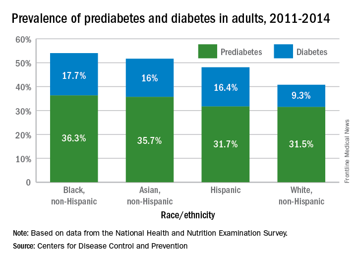
Lack of knowledge about having the disease was common: Almost a quarter (23.8%) of adults with diabetes didn’t know they had it, and the number jumped to 88% for those with prediabetes, based on data from the National Health and Nutrition Examination Survey, which included a blood test for fasting plasma glucose or hemoglobin A1c.
“More than a third of U.S. adults have prediabetes, and the majority don’t know it. Now, more than ever, we must step up our efforts to reduce the burden of this serious disease,” CDC Director Brenda Fitzgerald, MD, said in a statement.
“Diabetes and prediabetes remain serious threats for more than one-third of Americans – the statistics are staggering,” William T. Cefalu, MD, chief scientific, medical, and mission officer of the American Diabetes Association, said in a written statement.
“We must continue to innovate in scientific research and to translate findings to the clinical level to decrease the prevalence of diabetes. These data clearly confirm it is critical for us to continue to provide the education and support needed to improve health outcomes and decrease the daily burden of diabetes. We must reduce the incidence of diabetes and its enormous costs, including both the financial costs and the human toll of lost quality of life,” he said.
More than 114 million American adults have type 2 diabetes mellitus or prediabetes, according to estimates from the Centers for Disease Control and Prevention.
As of 2015, the combined prevalence of diabetes and prediabetes was 45.4% among adults in the United States: 11.5% (30.3 million) have diabetes and 33.9% have prediabetes, representing 84.1 million people who could develop type 2 diabetes within 5 years, the CDC said in the National Diabetes Statistics Report, 2017.
Lack of knowledge about having the disease was common: Almost a quarter (23.8%) of adults with diabetes didn’t know they had it, and the number jumped to 88% for those with prediabetes, based on data from the National Health and Nutrition Examination Survey, which included a blood test for fasting plasma glucose or hemoglobin A1c.
“More than a third of U.S. adults have prediabetes, and the majority don’t know it. Now, more than ever, we must step up our efforts to reduce the burden of this serious disease,” CDC Director Brenda Fitzgerald, MD, said in a statement.
GOP health reform dead for now
Senate Republicans are scrambling to come up with another plan now that at least four member of their caucus have said that they would vote against moving forward with debate on the Better Care Reconciliation Act.
Support for the bill, which included dramatic Medicaid cuts and stripped many coverage provisions of the Affordable Care Act, was lacking after revisions were announced on July 13. At that time, conservative Sen. Ran Paul (R-Ky.) and moderate Susan Collins (R-Maine) voiced their opposition for different ideological reasons. They were joined by Sen. Mike Lee (R-Utah) and Sen. Jerry Moran (R-Kan.), who also declined to support the bill. Senate GOP leadership, with a slim 52-48 majority, could only afford to lose two votes (Vice President Mike Pence would have been the tie-breaking vote).
No new timeline has been revealed for the next steps.
“The health reform debate is by no means over,” David Barbe, MD, president of the American Medical Association, said in a statement. “Congress must begin a collaborative process that produces a bipartisan solution. ... Near-term action is needed to stabilize the individual/nongroup health insurance marketplace. In the long term, stakeholders and policymakers need to address the unsustainable trends in health care costs while achieving meaningful, affordable coverage for all Americans.”
AGA believes that, at a minimum, any replacement package must ensure patient access to and coverage of specialty care, ensure patient access to and coverage of evidence-based preventive screenings without cost-sharing, ensure that insurers cannot discriminate because of a pre-existing condition, ensure that insurers cannot discriminate based on gender, allow parents to keep their children on their plans until age 26, and there should be no annual or lifetime caps.
Prenatal SSRI exposure’s effect on development
Unfortunately, the bottom line for most of these important questions is that we really don’t know as much as we probably should.
Just when we’ve read a convincing finding from a reputable journal that establishes a link between prenatal SSRI use and an untoward outcome, we see it disputed the next month. Why is this always happening, and why can’t we really know anything with certainty? Much of the confusion can be attributed to research methods and the obvious difficulty of using randomized, controlled trials to control for potential confounding factors. While statistical techniques have become increasingly sophisticated in addressing these confounding factors, they remain imperfect. For example, one of the most difficult challenges that remains is separating any effects of a medication from any effects caused by the condition it was designed to treat. Comparing women with the same underlying condition, some of whom are treated with a medication and some of whom are not, is a step forward, but there may be important reasons that one group decides to seek treatment and the other doesn’t. One clever research design that was employed to look at congenital anomalies in the offspring of women taking SSRIs accounted for siblings of these children who were born when their mother was not taking an SSRI. This study demonstrated that these women were more likely to have children with congenital malformations even when they weren’t taking the SSRIs.1 Other factors that render this literature difficult to interpret include small sample sizes when looking at specific SSRIs (many studies cluster them all), dose effects, timing (which trimester), duration of treatment, and method of recording compliance.
The potential link between SSRIs and autism has received a fair amount of attention lately, especially after a very well-designed study in 2016 suggested a significantly increased risk.10 However, as with many of the findings, this study was quickly disputed by other high-quality, well-powered research that found no increased risk after controlling for maternal illness.11,12
ADHD generally has not been found to be related to maternal SSRI use, although one study did find a link between ADHD and tricyclic antidepressants.12,13
In terms of other neurodevelopmental outcomes, there have been many negative studies examining IQ, nonverbal communication, as well as speech and motor skills.14,15,16 However, as with so many other outcomes, some other studies contradict these negative results. According to a recent, large cohort study, there may be some concern regarding SSRI exposure prenatally and an increase in speech disorders by age 14 years, as well as lower language competence at age 3 years.17,18 Likewise, mild motor abnormalities have been observed, with maternal depression severity as an independent but contributing factor.19
Several studies demonstrate a connection between prenatal SSRI exposure and childhood internalizing symptoms, such as depression and anxiety, independent of maternal depression.12,20 These findings must be balanced with our knowledge of the serious mental health conditions in offspring that are associated with untreated maternal illness, including both internalizing and externalizing disorders.21,22
How does one come to any firm conclusions to guide a primary care clinician’s practice and recommendations? Hopefully, the evidence will become clearer over time as we adopt more sophisticated designs and accumulate observations. A larger number of observations would allow us to decrease heterogeneity by studying subgroups according to type of SSRI and duration of exposure. Enhanced understanding of the role of genetic factors also may shed some light on individual variation as the serotonin transporter gene has been suggested as a potential moderator of sensitivity.23

Dr. Guth is an assistant professor in the department of psychiatry at the University of Vermont Medical Center and the University of Vermont Robert Larner College of Medicine, both in Burlington. She works with children and adolescents as well as women in the perinatal period. She has no relevant financial disclosures.
References
1. BMJ. 2015 Apr 17;350:h1798.
2. Can J Clin Pharmacol. 2009 Winter;16(1):e66-7.
3. J Matern Fetal Neonatal Med. 2008 Oct;21(10):745-51.
4. PLoS ONE. 2014 Nov; 9(11): e111327.
5. Pediatr Res. 2017 Jun 30. doi: 10.1038/pr.2017.156. [Epub ahead of print].
6. J Clin Psychiatry. 2017 May;78(5):605-11.
7. Acta Psychiatr Scand. 2010 Jun;121(6):471-9.
8. Am J Psychiatry. 2016 Feb 1;173(2):147-57.
9. J Perinatol. 2011 Sep;31(9):615-20.
10. JAMA Pediatr. 2016 Feb;170(2):117-24.
11. JAMA. 2017 Apr 18;317(15):1544-52.
12. J Am Acad Child Adolesc Psychiatry. 2016 May;55(5):359-66.
13. Paediatr Perinat Epidemiol. 2017 Jul;31(4):363-73.
14. Acta Obstet Gynecol Scand. 2015 May;94(5):501-7.
15. J Psychopharmacol. 2017 Mar;31(3):346-55.
16. CNS Drugs. 2005;19(7):623-33.
17. JAMA Psychiatry. 2016 Nov 1;73(11):1163-70.
18. BJOG. 2014. doi: 10.1111/1471-0528.12821.
19. BJOG. 2016 Nov;123(12):1908-17.
20. Pediatr Res. 2015 Aug;78(2):174-80.
21. Neuroscience. 2017 Feb 7;342:154-66.
22. Depress Anxiety. 2014 Jan;31(1):9-18.
23. Neuroscience. 2017 Feb 7;342:212-31.
Unfortunately, the bottom line for most of these important questions is that we really don’t know as much as we probably should.
Just when we’ve read a convincing finding from a reputable journal that establishes a link between prenatal SSRI use and an untoward outcome, we see it disputed the next month. Why is this always happening, and why can’t we really know anything with certainty? Much of the confusion can be attributed to research methods and the obvious difficulty of using randomized, controlled trials to control for potential confounding factors. While statistical techniques have become increasingly sophisticated in addressing these confounding factors, they remain imperfect. For example, one of the most difficult challenges that remains is separating any effects of a medication from any effects caused by the condition it was designed to treat. Comparing women with the same underlying condition, some of whom are treated with a medication and some of whom are not, is a step forward, but there may be important reasons that one group decides to seek treatment and the other doesn’t. One clever research design that was employed to look at congenital anomalies in the offspring of women taking SSRIs accounted for siblings of these children who were born when their mother was not taking an SSRI. This study demonstrated that these women were more likely to have children with congenital malformations even when they weren’t taking the SSRIs.1 Other factors that render this literature difficult to interpret include small sample sizes when looking at specific SSRIs (many studies cluster them all), dose effects, timing (which trimester), duration of treatment, and method of recording compliance.
The potential link between SSRIs and autism has received a fair amount of attention lately, especially after a very well-designed study in 2016 suggested a significantly increased risk.10 However, as with many of the findings, this study was quickly disputed by other high-quality, well-powered research that found no increased risk after controlling for maternal illness.11,12
ADHD generally has not been found to be related to maternal SSRI use, although one study did find a link between ADHD and tricyclic antidepressants.12,13
In terms of other neurodevelopmental outcomes, there have been many negative studies examining IQ, nonverbal communication, as well as speech and motor skills.14,15,16 However, as with so many other outcomes, some other studies contradict these negative results. According to a recent, large cohort study, there may be some concern regarding SSRI exposure prenatally and an increase in speech disorders by age 14 years, as well as lower language competence at age 3 years.17,18 Likewise, mild motor abnormalities have been observed, with maternal depression severity as an independent but contributing factor.19
Several studies demonstrate a connection between prenatal SSRI exposure and childhood internalizing symptoms, such as depression and anxiety, independent of maternal depression.12,20 These findings must be balanced with our knowledge of the serious mental health conditions in offspring that are associated with untreated maternal illness, including both internalizing and externalizing disorders.21,22
How does one come to any firm conclusions to guide a primary care clinician’s practice and recommendations? Hopefully, the evidence will become clearer over time as we adopt more sophisticated designs and accumulate observations. A larger number of observations would allow us to decrease heterogeneity by studying subgroups according to type of SSRI and duration of exposure. Enhanced understanding of the role of genetic factors also may shed some light on individual variation as the serotonin transporter gene has been suggested as a potential moderator of sensitivity.23
Dr. Guth is an assistant professor in the department of psychiatry at the University of Vermont Medical Center and the University of Vermont Robert Larner College of Medicine, both in Burlington. She works with children and adolescents as well as women in the perinatal period. She has no relevant financial disclosures.
References
1. BMJ. 2015 Apr 17;350:h1798.
2. Can J Clin Pharmacol. 2009 Winter;16(1):e66-7.
3. J Matern Fetal Neonatal Med. 2008 Oct;21(10):745-51.
4. PLoS ONE. 2014 Nov; 9(11): e111327.
5. Pediatr Res. 2017 Jun 30. doi: 10.1038/pr.2017.156. [Epub ahead of print].
6. J Clin Psychiatry. 2017 May;78(5):605-11.
7. Acta Psychiatr Scand. 2010 Jun;121(6):471-9.
8. Am J Psychiatry. 2016 Feb 1;173(2):147-57.
9. J Perinatol. 2011 Sep;31(9):615-20.
10. JAMA Pediatr. 2016 Feb;170(2):117-24.
11. JAMA. 2017 Apr 18;317(15):1544-52.
12. J Am Acad Child Adolesc Psychiatry. 2016 May;55(5):359-66.
13. Paediatr Perinat Epidemiol. 2017 Jul;31(4):363-73.
14. Acta Obstet Gynecol Scand. 2015 May;94(5):501-7.
15. J Psychopharmacol. 2017 Mar;31(3):346-55.
16. CNS Drugs. 2005;19(7):623-33.
17. JAMA Psychiatry. 2016 Nov 1;73(11):1163-70.
18. BJOG. 2014. doi: 10.1111/1471-0528.12821.
19. BJOG. 2016 Nov;123(12):1908-17.
20. Pediatr Res. 2015 Aug;78(2):174-80.
21. Neuroscience. 2017 Feb 7;342:154-66.
22. Depress Anxiety. 2014 Jan;31(1):9-18.
23. Neuroscience. 2017 Feb 7;342:212-31.
Corticosteroid therapy in Kawasaki disease
Clinical Question
What is the efficacy of corticosteroid therapy in Kawasaki disease?
Background
First described in 1967 in Japan, Kawasaki disease (KD), or mucocutaneous lymph node syndrome, is an acute systemic vasculitis of unclear etiology which primarily affects infants and children. Of significant clinical concern, 30%-50% of untreated patients develop acute coronary artery dilatation, and about one fourth progress to serious coronary artery abnormalities (CAA) such as aneurysm and ectasia.1,2 These patients have a higher risk of long-term complications, such as coronary artery thrombus, myocardial infarction, and sudden death.3 KD is typically treated with a combination of intravenous immunoglobulin (IVIG) and aspirin, which reduces the risk of CAA.1 However, more than 20% of cases are resistant to this conventional therapy and have higher risk of CAA than nonresistant patients.4 Corticosteroids have been suggested as therapy in KD, as the anti-inflammatory effect is useful for many other vasculitides, but studies to date have had conflicting results. This study was performed to comprehensively evaluate the effect of corticosteroids in KD as initial or rescue therapy (after failure to respond to IVIG).
Study design
Systematic review and meta-analysis.
Synopsis
The population of interest was children diagnosed with KD. The intervention of interest was treatment with adjunctive corticosteroids either as initial or rescue therapy. Comparisons were made between the corticosteroids group and the conventional therapy (IVIG) group. Outcome measurements included the incidence of CAA (primary outcome), duration until defervescence, and adverse events. IVIG resistance was defined as persistent or recurrent fever lasting (or relapsed within) 24-48 hours after the initial IVIG treatment.
A total of 681 articles were initially retrieved, and after exclusions, 16 comparative studies were enrolled for meta-analysis. In these studies, a total of 2,746 cases were involved, with 861 in the corticosteroid group and 1,885 in the IVIG group. Ten studies used corticosteroids as initial treatment (comparing this plus IVIG versus IVIG therapy alone), and six used steroids as rescue treatment after initial IVIG failure (comparing corticosteroids with additional IVIG therapy). Four studies enrolled patients with KD who were predicted to have high risk of IVIG resistance, based on published scoring systems. All patients in the studies received oral aspirin.

Overall, this meta-analysis found that adding corticosteroid therapy was associated with a relative risk reduction of 58% in CAA (odds ratio, 0.424; 95% confidence interval, 0.270-0.665; P less than .001). Further analysis showed that the longer the duration of illness prior to corticosteroids, the less of a treatment effect was noticed. The studies using steroids as initial adjunctive therapy had duration of illness 4.7 days prior to treatment, and showed an advantage, compared with IVIG alone, while studies using steroids as rescue therapy had a longer duration of illness prior to steroid therapy (7.2 days), and did not show significant benefit, compared with additional IVIG. In analyzing patients predicted to be at high risk of IVIG resistance at baseline, addition of corticosteroids with IVIG as initial therapy showed a significantly lower risk of CAA development (relative risk reduction of 76%) versus IVIG alone (OR, 0.240; 95% CI, 0.123-0.467; P less than .001). As a secondary outcome, the use of adjunctive corticosteroid therapy was associated with a quicker resolution of fever, compared with IVIG alone (0.66 days vs. 2.18 days). There was no significant difference in adverse events between the two groups.
Although this is the most comprehensive study of corticosteroids in KD, there are some limitations. High-risk patients were found to receive the greatest benefit, but the predictive ability of published scoring systems have not been optimized or generalized to all populations. Most of the studies included in this review were conducted in Japan, so it is uncertain if the results are applicable to other regions. Also, the selection of corticosteroids and treatment regimens were not consistent between studies.
Bottom line
This study suggests that corticosteroids combined with IVIG as initial therapy for KD showed a more protective effect against CAA, compared with conventional IVIG therapy, and the efficacy was more pronounced in the high-risk patient group.
Citation
Chen S, Dong Y, Kiuchi MG, et al. Coronary Artery Complication in Kawasaki Disease and the Importance of Early Intervention: A Systematic Review and Meta-analysis. JAMA Pediatr. 2016;170(12):1156-1163.
References
1. Newburger JW, Takahashi M, Gerber MA, et al. Committee on Rheumatic Fever, Endocarditis and Kawasaki Disease. Council on Cardiovascular Disease in the Young; American Heart Association; American Academy of Pediatrics: diagnosis, treatment, and long-term management of Kawasaki disease. Circulation. 2004;110:2747-71.
2. Daniels LB, Tjajadi MS, Walford HH, et al. Prevalence of Kawasaki disease in young adults with suspected myocardial ischemia. Circulation. 2012;125(20):2447-53.
3. Gordon JB, Kahn AM, Burns JC. When children with Kawasaki disease grow up: myocardial and vascular complications in adulthood. J Am Coll Cardiol. 2009;54(21):1911-20.
4. Tremoulet AH, Best BM, Song S, et al. Resistance to intravenous immunoglobulin in children with Kawasaki disease. J Pediatr. 2008;153(1):117-21.
Dr. Galloway is a pediatric hospitalist at Sanford Children’s Hospital in Sioux Falls, S.D., assistant professor of pediatrics at the University of South Dakota Sanford School of Medicine, and vice chief of the division of hospital pediatrics at USD SSOM and Sanford Children’s Hospital.
Clinical Question
What is the efficacy of corticosteroid therapy in Kawasaki disease?
Background
First described in 1967 in Japan, Kawasaki disease (KD), or mucocutaneous lymph node syndrome, is an acute systemic vasculitis of unclear etiology which primarily affects infants and children. Of significant clinical concern, 30%-50% of untreated patients develop acute coronary artery dilatation, and about one fourth progress to serious coronary artery abnormalities (CAA) such as aneurysm and ectasia.1,2 These patients have a higher risk of long-term complications, such as coronary artery thrombus, myocardial infarction, and sudden death.3 KD is typically treated with a combination of intravenous immunoglobulin (IVIG) and aspirin, which reduces the risk of CAA.1 However, more than 20% of cases are resistant to this conventional therapy and have higher risk of CAA than nonresistant patients.4 Corticosteroids have been suggested as therapy in KD, as the anti-inflammatory effect is useful for many other vasculitides, but studies to date have had conflicting results. This study was performed to comprehensively evaluate the effect of corticosteroids in KD as initial or rescue therapy (after failure to respond to IVIG).
Study design
Systematic review and meta-analysis.
Synopsis
The population of interest was children diagnosed with KD. The intervention of interest was treatment with adjunctive corticosteroids either as initial or rescue therapy. Comparisons were made between the corticosteroids group and the conventional therapy (IVIG) group. Outcome measurements included the incidence of CAA (primary outcome), duration until defervescence, and adverse events. IVIG resistance was defined as persistent or recurrent fever lasting (or relapsed within) 24-48 hours after the initial IVIG treatment.
A total of 681 articles were initially retrieved, and after exclusions, 16 comparative studies were enrolled for meta-analysis. In these studies, a total of 2,746 cases were involved, with 861 in the corticosteroid group and 1,885 in the IVIG group. Ten studies used corticosteroids as initial treatment (comparing this plus IVIG versus IVIG therapy alone), and six used steroids as rescue treatment after initial IVIG failure (comparing corticosteroids with additional IVIG therapy). Four studies enrolled patients with KD who were predicted to have high risk of IVIG resistance, based on published scoring systems. All patients in the studies received oral aspirin.
Overall, this meta-analysis found that adding corticosteroid therapy was associated with a relative risk reduction of 58% in CAA (odds ratio, 0.424; 95% confidence interval, 0.270-0.665; P less than .001). Further analysis showed that the longer the duration of illness prior to corticosteroids, the less of a treatment effect was noticed. The studies using steroids as initial adjunctive therapy had duration of illness 4.7 days prior to treatment, and showed an advantage, compared with IVIG alone, while studies using steroids as rescue therapy had a longer duration of illness prior to steroid therapy (7.2 days), and did not show significant benefit, compared with additional IVIG. In analyzing patients predicted to be at high risk of IVIG resistance at baseline, addition of corticosteroids with IVIG as initial therapy showed a significantly lower risk of CAA development (relative risk reduction of 76%) versus IVIG alone (OR, 0.240; 95% CI, 0.123-0.467; P less than .001). As a secondary outcome, the use of adjunctive corticosteroid therapy was associated with a quicker resolution of fever, compared with IVIG alone (0.66 days vs. 2.18 days). There was no significant difference in adverse events between the two groups.
Although this is the most comprehensive study of corticosteroids in KD, there are some limitations. High-risk patients were found to receive the greatest benefit, but the predictive ability of published scoring systems have not been optimized or generalized to all populations. Most of the studies included in this review were conducted in Japan, so it is uncertain if the results are applicable to other regions. Also, the selection of corticosteroids and treatment regimens were not consistent between studies.
Bottom line
This study suggests that corticosteroids combined with IVIG as initial therapy for KD showed a more protective effect against CAA, compared with conventional IVIG therapy, and the efficacy was more pronounced in the high-risk patient group.
Citation
Chen S, Dong Y, Kiuchi MG, et al. Coronary Artery Complication in Kawasaki Disease and the Importance of Early Intervention: A Systematic Review and Meta-analysis. JAMA Pediatr. 2016;170(12):1156-1163.
References
1. Newburger JW, Takahashi M, Gerber MA, et al. Committee on Rheumatic Fever, Endocarditis and Kawasaki Disease. Council on Cardiovascular Disease in the Young; American Heart Association; American Academy of Pediatrics: diagnosis, treatment, and long-term management of Kawasaki disease. Circulation. 2004;110:2747-71.
2. Daniels LB, Tjajadi MS, Walford HH, et al. Prevalence of Kawasaki disease in young adults with suspected myocardial ischemia. Circulation. 2012;125(20):2447-53.
3. Gordon JB, Kahn AM, Burns JC. When children with Kawasaki disease grow up: myocardial and vascular complications in adulthood. J Am Coll Cardiol. 2009;54(21):1911-20.
4. Tremoulet AH, Best BM, Song S, et al. Resistance to intravenous immunoglobulin in children with Kawasaki disease. J Pediatr. 2008;153(1):117-21.
Dr. Galloway is a pediatric hospitalist at Sanford Children’s Hospital in Sioux Falls, S.D., assistant professor of pediatrics at the University of South Dakota Sanford School of Medicine, and vice chief of the division of hospital pediatrics at USD SSOM and Sanford Children’s Hospital.
Pembrolizumab takes on r/r PMBCL
LUGANO, SWITZERLAND – Patients with relapsed/refractory primary mediastinal large B-cell lymphoma (PMBCL) have few good therapeutic options, but pembrolizumab (Keytruda) may be one of them, an interim analysis from a phase 2 trial suggests.
Six of 10 patients with relapsed/refractory PMBCL for whom autologous stem cell transplantation (ASCT) had failed had responses to salvage therapy with pembrolizumab, including three complete responses (CR) and three partial responses (PR), reported Pier Luigi Zinzani, MD, PhD, of the University of Bologna, Italy.
“In relapsed/refractory primary mediastinal large B-cell lymphoma including heavily pretreated patients in our series, pembrolizumab showed promising antitumor activity in these preliminary data from a phase 2 study, and a manageable safety profile,” Dr. Zinzani said at the International Conference on Malignant Lymphoma.

Although standard cytotoxic chemotherapy regimens are curative in approximately 80% of patients with PMBCL, those with relapsed/refractory disease have a poor prognosis and limited treatment options. In the United States, about 200 patients are diagnosed with relapsed/refractory PMBCL each year, Dr. Zinzani noted.
The rationale for using a programmed death-1 (PD-1) inhibitor such as pembrolizumab is that PMBCL tumors frequently harbor amplifications and/or translocations in the 9p24.1 locus that lead to overexpression of PD ligand 1 (PD-L1) or PD-L2. Pembrolizumab blocks interactions between PD-1 and its ligands.
In the Keynote-013 PMBCL cohort, the objective response rate (ORR) was 48%, including 24% CR.
Keynote 170 investigators are enrolling adults with relapsed/refractory PMBCL who are either ineligible for ASCT following at least two prior lines of therapy, or who had a relapse following a transplant.
Patients receive pembrolizumab 200 mg every 3 weeks. Those who achieve a complete response are allowed to discontinue after a minimum of 24 weeks on study, those with partial response or stable disease are treated for 35 cycles or until disease progression or intolerable toxicities, and those with disease progression are discontinued, but are allowed to remain on study until progression is confirmed.
Responses are assessed by PET and CT scans according to 2007 International Working Group criteria.
Among the 29 patients assessable for efficacy at the time of Dr. Zinzani’s presentations, the best response by blinded central review was 41% (12 patients: 4 CR and 8 PR). Three patients had stable disease, and eight had disease progression. Six patients died or discontinued participation prior to the first assessment.
In all, 16 of 22 patients had reductions in target lesions, and all but one of these patients had reductions greater than 50%.
After a median follow-up of 6.6 months, 10 patients had ongoing responses. The median time to response was 2.9 months. The median duration of response has not been reached.
Of the 10 patients for whom ASCT had failed, three had a CR, and three had a PR. Among 19 ASCT-ineligible patients, one had a CR and five had a PR.
The interim safety analysis included 49 patients. Of this group, only one discontinued treatment because of adverse events. There were 26 treatment-related adverse events, but no treatment-related deaths.
The most frequent adverse events were neutropenia, asthenia, hypothyroidism, and pyrexia.
The study is supported by Merck. Dr Zinzani disclosed serving on advisory boards for several companies.
LUGANO, SWITZERLAND – Patients with relapsed/refractory primary mediastinal large B-cell lymphoma (PMBCL) have few good therapeutic options, but pembrolizumab (Keytruda) may be one of them, an interim analysis from a phase 2 trial suggests.
Six of 10 patients with relapsed/refractory PMBCL for whom autologous stem cell transplantation (ASCT) had failed had responses to salvage therapy with pembrolizumab, including three complete responses (CR) and three partial responses (PR), reported Pier Luigi Zinzani, MD, PhD, of the University of Bologna, Italy.
“In relapsed/refractory primary mediastinal large B-cell lymphoma including heavily pretreated patients in our series, pembrolizumab showed promising antitumor activity in these preliminary data from a phase 2 study, and a manageable safety profile,” Dr. Zinzani said at the International Conference on Malignant Lymphoma.
Although standard cytotoxic chemotherapy regimens are curative in approximately 80% of patients with PMBCL, those with relapsed/refractory disease have a poor prognosis and limited treatment options. In the United States, about 200 patients are diagnosed with relapsed/refractory PMBCL each year, Dr. Zinzani noted.
The rationale for using a programmed death-1 (PD-1) inhibitor such as pembrolizumab is that PMBCL tumors frequently harbor amplifications and/or translocations in the 9p24.1 locus that lead to overexpression of PD ligand 1 (PD-L1) or PD-L2. Pembrolizumab blocks interactions between PD-1 and its ligands.
In the Keynote-013 PMBCL cohort, the objective response rate (ORR) was 48%, including 24% CR.
Keynote 170 investigators are enrolling adults with relapsed/refractory PMBCL who are either ineligible for ASCT following at least two prior lines of therapy, or who had a relapse following a transplant.
Patients receive pembrolizumab 200 mg every 3 weeks. Those who achieve a complete response are allowed to discontinue after a minimum of 24 weeks on study, those with partial response or stable disease are treated for 35 cycles or until disease progression or intolerable toxicities, and those with disease progression are discontinued, but are allowed to remain on study until progression is confirmed.
Responses are assessed by PET and CT scans according to 2007 International Working Group criteria.
Among the 29 patients assessable for efficacy at the time of Dr. Zinzani’s presentations, the best response by blinded central review was 41% (12 patients: 4 CR and 8 PR). Three patients had stable disease, and eight had disease progression. Six patients died or discontinued participation prior to the first assessment.
In all, 16 of 22 patients had reductions in target lesions, and all but one of these patients had reductions greater than 50%.
After a median follow-up of 6.6 months, 10 patients had ongoing responses. The median time to response was 2.9 months. The median duration of response has not been reached.
Of the 10 patients for whom ASCT had failed, three had a CR, and three had a PR. Among 19 ASCT-ineligible patients, one had a CR and five had a PR.
The interim safety analysis included 49 patients. Of this group, only one discontinued treatment because of adverse events. There were 26 treatment-related adverse events, but no treatment-related deaths.
The most frequent adverse events were neutropenia, asthenia, hypothyroidism, and pyrexia.
The study is supported by Merck. Dr Zinzani disclosed serving on advisory boards for several companies.
AT 14-ICML
Key clinical point: Poor-prognosis relapsed/refractory primary mediastinal large B-cell lymphoma may respond to PD-1 inhibition.
Major finding: The best response by blinded central review was 41% (12 patients: 4 complete and 8 partial responses).
Data source: Interim analysis from a phase 2 study with 29 patients evaluable for efficacy, 49 for safety.
Disclosures: Merck supports the study. Dr Zinzani disclosed serving on advisory boards for several companies.